Báo cáo y học: "Expression and function of inducible co-stimulator in patients with systemic lupus erythematosus: possible involvement in excessive interferon-γ and anti-double-stranded DNA antibody production" pot
Bạn đang xem bản rút gọn của tài liệu. Xem và tải ngay bản đầy đủ của tài liệu tại đây (750.53 KB, 14 trang )
Available online />
Research article
Open Access
Vol 8 No 3
Expression and function of inducible co-stimulator in patients
with systemic lupus erythematosus: possible involvement in
excessive interferon-γ and anti-double-stranded DNA antibody
production
Manabu Kawamoto1, Masayoshi Harigai1,2, Masako Hara1, Yasushi Kawaguchi1,
Katsunari Tezuka3, Michi Tanaka1, Tomoko Sugiura1, Yasuhiro Katsumata1, Chikako Fukasawa1,
Hisae Ichida1, Satomi Higami1 and Naoyuki Kamatani1
1Institute
of Rheumatology, Tokyo Women's Medical University, Tokyo, Japan
Research Center, Tokyo Medical and Dental University, Tokyo, Japan
3Central Pharmaceutical Research Institute, Japan Tobacco, Inc., Osaka, Japan
2Clinical
Corresponding author: Masayoshi Harigai,
Received: 9 Aug 2005 Revisions requested: 7 Sep 2005 Revisions received: 12 Jan 2006 Accepted: 21 Feb 2006 Published: 22 Mar 2006
Arthritis Research & Therapy 2006, 8:R62 (doi:10.1186/ar1928)
This article is online at: />© 2006 Kawamoto et al.; licensee BioMed Central Ltd.
This is an open access article distributed under the terms of the Creative Commons Attribution License ( />which permits unrestricted use, distribution, and reproduction in any medium, provided the original work is properly cited.
Abstract
Inducible co-stimulator (ICOS) is the third member of the CD28/
cytotoxic T-lymphocyte associated antigen-4 family and is
involved in the proliferation and activation of T cells. A detailed
functional analysis of ICOS on peripheral blood T cells from
patients with systemic lupus erythematosus (SLE) has not yet
been reported. In the present study we developed a fully human
anti-human ICOS mAb (JTA009) with high avidity and
investigated the immunopathological roles of ICOS in SLE.
JTA009 exhibited higher avidity for ICOS than a previously
reported mAb, namely SA12. Using JTA009, ICOS was
detected in a substantial proportion of unstimulated peripheral
blood T cells from both normal control individuals and patients
with SLE. In CD4+CD45RO+ T cells from peripheral blood, the
percentage of ICOS+ cells and mean fluorescence intensity with
JTA009 were significantly higher in active SLE than in inactive
SLE or in normal control individuals. JTA009 co-stimulated
peripheral blood T cells in the presence of suboptimal
concentrations of anti-CD3 mAb. Median values of
[3H]thymidine incorporation were higher in SLE T cells with
ICOS co-stimulation than in normal T cells, and the difference
between inactive SLE patients and normal control individuals
achieved statistical significance. ICOS co-stimulation
significantly increased the production of IFN-γ, IL-4 and IL-10 in
both SLE and normal T cells. IFN-γ in the culture supernatants of
both active and inactive SLE T cells with ICOS co-stimulation
was significantly higher than in normal control T cells. Finally,
SLE T cells with ICOS co-stimulation selectively and
significantly enhanced the production of IgG anti-doublestranded DNA antibodies by autologous B cells. These findings
suggest that ICOS is involved in abnormal T cell activation in
SLE, and that blockade of the interaction between ICOS and its
receptor may have therapeutic value in the treatment of this
intractable disease.
Introduction
ate tissue and organ damage [1]. Recent investigations suggest that collaboration between autoantibody-producing B
cells and antigen-specific T-helper (Th) cells is important to
the production of these pathogenic autoantibodies [2].
Systemic lupus erythematosus (SLE), a prototype autoimmune
disease, is characterized by activation of lymphocytes and the
presence of various types of autoantibodies in peripheral
blood. These autoantibodies are considered to form immune
complexes with their corresponding autoantigens and to medi-
B7RP-1 = B7-related protein-1; ds = double stranded; ELISA = enzyme-linked immunosorbent assay; FITC = fluorescein isothiocyanate; ICOS =
inducible costimulator; IFN = interferon; IL = interleukin; mAb = monoclonal antibody; KLH = keyhole limpet hemocyanin; MFI = mean fluorescence
intensity; PBL = peripheral blood lymphocyte; PBS = phosphate-buffered saline; PE = phycoerythrin; PerCP = peridinin chlorophyll protein; SD =
standard deviation; SLE = systemic lupus erythematosus; SLEDAI = Systemic Lupus Erythematosus Disease Activity Index; Th = T-helper (cell).
Page 1 of 14
(page number not for citation purposes)
Arthritis Research & Therapy
Vol 8 No 3
Kawamoto et al.
The fate of T cells, after they encounter specific antigens, is
modulated by co-stimulatory signals, which are required for
both lymphocyte activation and the development of adaptive
immunity (for review [3-6]). In general, activation of T cells
requires two signals: one from a T cell receptor and the other
from co-stimulatory molecules such as CD28 and tumour
necrosis factor family members [3,7]. The inducible co-stimulator (ICOS; also known as AILIM [activation-inducible lymphocyte immunomediatory molecule]) was identified in 1999
as a membrane glycoprotein that is expressed on the surface
of activated T cells and that shares several structural and functional similarities with CD28 [8-10]. Like CD28, ICOS has
potent co-stimulatory effects on proliferation of T cells and production of cytokines [8-12]. ICOS is also important for germinal centre formation, clonal expansion of T cells, antibody
production, and class switching in response to various antigens [13,14]. CD28 and cytotoxic T lymphocyte associated
antigen 4 use the MYPPPY motif in their extracellular domains
to bind to their ligands, namely B7.1 and B7.2. ICOS does not
possess this motif, and so B7.1 and B7.2 are not among its ligands [9]. Subsequently, it was shown that a B7-like molecule,
termed B7-related protein-1 (B7RP-1) (also referred to as B7H2, GL50 and LICOS), binds to ICOS [9,15-21]. B7RP-1
shares 20% identity with B7.1/B7.2 [9] and is constitutively
expressed on B cells and monocytes [13].
Accumulating evidence indicates that ICOS is involved in the
immunopathogenesis of animal models of various autoimmune
disorders, including SLE, rheumatoid arthritis, multiple sclerosis and asthma [21-28]. These data prompted us to investigate the possible role of ICOS in human SLE and its
importance as a therapeutic target. We found that ICOS was
over-expressed in peripheral blood CD4+ T cells from patients
with active SLE and that ICOS contributed not only to the
enhanced proliferation but also to the increased production of
IFN-γ in peripheral blood T cells from patients with SLE. ICOS
also augmented the ability of peripheral blood T cells from
patients with SLE to support the production of IgG anti-double
stranded (ds)DNA antibody by autologous peripheral blood B
cells. Thus, we examined the expression and function of ICOS
in peripheral blood T cells from patients with SLE. Our data
suggest that ICOS plays an important role in the immunopathogenesis of SLE and support the possibility that blockade of the interaction between ICOS and B7RP-1 may have
therapeutic value in treating this intractable autoimmune disorder.
Materials and methods
Patients
Twenty-two patients with active SLE (21 females and one
male), 17 patients with inactive SLE (16 females and one
male) and 24 normal control individuals (22 females and two
males) were included in the study. All SLE patients fulfilled the
SLE classification criteria proposed by the American College
of Rheumatology [29]. Disease activity in the SLE patients was
Page 2 of 14
(page number not for citation purposes)
evaluated using the Systemic Lupus Erythematosus Disease
Activity Index (SLEDAI) [30]. SLEDAI scores for the patients
with active SLE ranged from 6 to 22 (mean ± standard deviation [SD] 10.0 ± 6.2; median 10), whereas the scores for the
patients with inactive SLE ranged from 0 to 2 (mean ± SD 0.9
± 1.0; median 0). Sixteen of the 22 patients with active SLE
were examined before administration of corticosteroids and
immunosuppressants. Treatments for the remaining six
patients with active SLE were as follows: low-dose prednisolone (≤ 15 mg/day, median 9.5 mg/day; n = 4); 30 mg/day
prednisolone (n = 1); and 100 mg/day prednisolone and 250
mg/day cyclosporine A (n = 1). Sixteen of the 17 patients with
inactive SLE were treated with low-dose prednisolone
(median 10 mg/day); the remaining patients had been followed up without medication.
Peripheral blood samples were obtained with the informed
consent of all participating individuals. The Helsinki Declaration was adhered to throughout the study.
Generation of fully human anti-ICOS monoclonal
antibody (JTA009)
The generation and characterization of the Xeno-Mouse-G2
strains, engineered to produce fully human IgG2 antibodies,
were described by Mendez and coworkers [31]. Xeno-MouseG2 mice (aged 8–10 weeks) were immunized with a footpad
injection of the membrane fraction isolated from human ICOS
expressing CHO-K1 cells [32] in complete Freund's adjuvant.
Mice were boosted with the same amount of the fraction three
to four times before fusion. Popliteal lymph node and spleen
cells were fused with the murine myeloma cell line
P3X63Ag8.653 (CRL-1580; American Type Culture Collection, Manassas, VA, USA) using PEG1500. Hybridomas were
screened for their ability to bind to human ICOS expressed on
CHO-K1 or HPB-ALL cells [32]. One of the mAbs, JTA009,
exhibited high avidity for human ICOS and was used in the following experiments. The characteristics of JTA009 are
described below in the Results section. JMAb23, a classmatched control mAb for JTA009, was generated against keyhole limpet hemocyanin (KLH) in the same manner. All experiments were conducted following institutional guidelines for
the ethical treatment of animals.
Other antibodies
The anti-human ICOS mAb SA12 was generated and characterized as described previously [32]. Anti-CD3 mAb (clone
UCHT1) and anti-CD28 mAb (clone 28.2) were obtained from
Beckman Coulter Inc. (Fullerton, CA, USA). Anti-B7RP-1 mAb
was obtained from R&D Systems (Minneapolis, MN, USA).
Fluorescein isothiocyanate (FITC)-conjugated anti-CD3 mAb
was purchased from DAKO Japan (Tokyo, Japan). Phycoerythrin (PE)-conjugated anti-CD45RO mAb and PE-conjugated
control IgG were obtained from Nichirei (Tokyo, Japan). PEconjugated anti-CD25 mAb was obtained from eBioscience
(San Diego, CA, USA). PE-conjugated anti-CD69 mAb and
Available online />
peridinin chlorophyll protein (PerCP)-conjugated mAbs to
human CD3, CD4 and CD8 were purchased from BD Biosciences (San Jose, CA, USA). The F(ab')2 fraction of goat
anti-human IgG antibody was obtained from Biosource International Inc. (Camarillo, CA, USA). Peroxidase-conjugated
anti-human IgG was obtained from MBL (Nagoya, Japan).
Cell preparations
Peripheral blood lymphocytes (PBLs) were separated by centrifugation of heparinized blood over a Ficoll-Conray gradient.
B cells were isolated by positive selection from PBLs using
anti-CD19 MicroBeads (Miltenyi Biotech, Auburn, CA, USA),
in accordance with the manufacturer's instructions. T cells
were selected from CD19-depleted PBLs using the Pan T cell
Isolation Kit (Miltenyi Biotech) and anti-CD14 MicroBeads
(Miltenyi Biotech). The purities of B cells and T cells were in
excess of 97% and 95%, respectively, using flow cytometry.
Immunoprecipitation and Western blotting
Peripheral blood T cells from normal control individuals were
stimulated with anti-CD3 mAb (0.1 µg/ml) + anti-CD28 mAb
(2 µg/ml) for 72 hours. The surface of these cells was biotinylated using the ECL Protein Biotinylation Module (Amersham Bioscience Corp., Piscataway, NJ, USA) and lysates
were prepared with lysis buffer containing 25 mmol/l Tris-HCl
(at pH 7.5), 250 mmol/l NaCl, 5 mmol/l EDTA, 1% NP-40, protease inhibitor cocktail (Roche Diagnostics GmbH, Mannheim,
Germany) and 1 mmol/l phenylmethanesulfonyl fluoride.
JTA009 or JMAb23 were conjugated with Protein G-agarose
(Pierce Biotechnology Inc., Rockford, IL, USA) and incubated
with the cell lysate at 4°C overnight. After washing three times
with lysis buffer, the mAb-conjugated Protein G-agarose was
boiled for two minutes and the bound antigens were separated
using 12.5% SDS-PAGE gel and transferred to nitrocellulose
membrane (Bio-Rad Laboratories, Hercules, CA, USA). Transferred protein was visualized using streptavidin-peroxidase
(Amersham Bioscience Corp.) and SuperSignal West Pico
Chemiluminescent Substrate (Pierce Biotechnology Inc.).
Flow cytometry
Multicolour analysis was performed using flow cytometry.
Cells were washed three times in ice cold FCM buffer (phosphate-buffered saline [PBS] containing 0.1% bovine serum
albumin and 0.1% sodium azide) and incubated on ice for five
minutes with 10 µg purified human immunoglobulin (Cappel,
ICN, Aurora, OH, USA) and/or 10 µg purified mouse IgG
(Chemicon, Temecula, CA, USA) to block nonspecific IgG
binding. Cells were then incubated at 4°C with saturating
amounts of the fluorochrome (for instance, FITC, PE, or
PerCP) or biotin conjugated mAbs for 30 minutes. Cells were
washed twice in ice cold FCM buffer and incubated at 4°C
with streptavidin-FITC (DAKO Japan) for 30 minutes. After
incubation, cells were washed three times in ice cold FCM
buffer and fixed in PBS containing 1% paraformaldehyde. The
expression of cell surface markers was evaluated using an
EPICS® ALTRA (Beckman Coulter Inc.) cell sorter and
EXPO32™ analysis software (Beckman Coulter Inc.).
Stimulation of T cells
Peripheral blood T cells were stimulated either with anti-CD3
mAb (0.1 µg/ml) plus anti-CD28 mAb (2 µg/ml; CD28 costimulation), or with anti-CD3 mAb (0.1 µg/ml) plus JTA009 (8 µg/
ml; ICOS costimulation). Anti-CD3 mAb and JTA009 were
bound to flat-bottomed 96-well microtitre plates (IWAKI,
Tokyo, Japan) by incubating overnight at 4°C. Preliminary
experiments showed that anti-CD3 mAb alone at 0.1 µg/ml
induced modest proliferation of peripheral blood T cells under
the conditions described above (data not shown). In some
experiments, T cells were stimulated with anti-CD3 mAb plus
anti-ICOS mAb or anti-CD3 plus anti-CD28 mAb in the presence of various concentration of B7RP-1-Fc (R&D Systems;
165-B7). To determine proliferative response, T cells (2 × 10⁵
cells/well) were cultured for 72 hours with or without stimuli
and pulsed with [3H]thymidine (1 µCi/well; Amersham Bioscience Corp.) for the last 8 hours. The uptake of [3H]thymidine was measured using Matrix96 (Packard Instrument
Company, Meridian, CT, USA). To determine cytokine production, T cells (2 × 10⁵ cells/well) were cultured with or without
stimuli for 72 hours and culture supernatants were collected.
T/B cell co-culture
T cells and B cells, purified from the peripheral blood of
patients with active SLE with high serum levels of anti-dsDNA
antibody, were reconstituted at a 1:1 ratio (1 × 10⁵ T cells and
B cells/well), and were cultured in the presence of various
stimuli for seven days. Culture supernatants were collected
and stored at -80°C until assayed for anti-dsDNA antibody and
total IgG.
ELISA for cytokines, IgG anti-dsDNA antibody, total IgG
and anti-tetanus antibody
IL-2, IL-4, IL-10 and IFN-γ production in the culture supernatants was measured using ELISA kits, in accordance with the
manufacturer's protocol (IL-2 from R&D Systems, IL-4 and IL10 from Biosource International Inc., and IFN-γ from Amersham Bioscience Corp.). The sensitivities of these ELISA kits
were 1.60 pg/ml, 0.39 pg/ml, 0.78 pg/ml and 0.63 pg/ml for
IL-2, IL-4, IL-10 and IFN-γ, respectively. IgG anti-dsDNA antibody and total IgG in culture supernatants were determined as
described previously [33]. Anti-tetanus antibody was measured using ELISA kits from Virion/Serion (Würzburg, Germany), in accordance with the manufacturer's protocol.
ELISA for anti-ICOS mAbs
To compare the sensitivities of JTA009 and SA12, ELISA for
anti-ICOS mAbs was performed. Both antibodies and
JMAb23 were biotinylated using FluoReporter® Mini-biotin-XX
Protein Labeling Kit (Invitrogen Japan K.K., Tokyo, Japan), in
accordance with the manufacturer's instructions. Biotinylation
was confirmed by coating ELISA plates with serial dilutions of
Page 3 of 14
(page number not for citation purposes)
Arthritis Research & Therapy
Vol 8 No 3
Kawamoto et al.
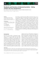
Figure 1
Characterization of JTA009, a novel anti-human ICOS mAb. JTA009, a fully human anti-human ICOS mAb, has greater avidity than SA12. (a) Avidity
ICOS mAb
of anti-human ICOS antibodies was evaluated by direct ELISA using ICOS-Fc (as described in Materials and method). JTA009 (open circles) exhibited stronger binding to ICOS-Fc than did SA12 (closed circle), a previously reported anti-human ICOS mAb. (b) Peripheral blood T cells from normal control individuals were stimulated with anti-CD3 mAb (0.1 µg/ml) plus anti-CD28 mAb (2 µg/ml) for 72 hours. These cells were biotinylated
and cell lysates were prepared. ICOS molecules in these lysates were immunoprecipitated, separated on SDS-PAGE gel, transferred to nitrocellulose membrane, and visualized using streptavidin-peroxidase and chemiluminescent substrate. A single band about 29 kDa was immunoprecipitated
with JTA009 but not with JMAb23, the control antibody. The thin lower band corresponded to the position of the front dye of the gel. Human ICOS
expressing (c) CHO-K1 and (d) its parental cell line CHO-K1 were stained with biotinylated JTA009 (thick line), biotinylated SA12 (broken line), or
biotinylated JMAb23 (human IgG2; thin line) and streptavidin-FITC, and then analyzed using flow cytometry. (e) Human ICOS expressing CHO-K1
cells were stained biotinylated SA12 (6.25 µg/test) and streptavidin-FITC in the presence of various amounts of nonbiotinylated JTA009 (thick line:
0 µg/test; thin line: 5 µg/test; thick broken line: 10 µg/test; thin broken line: 25 µg/test). JTA009 dose dependently decreased the binding of SA12
to the ICOS expressing CHO-K1 cells. FITC, fluorescein isothiocyanate; ICOS, inducible co-stimulator; mAb, monoclonal antibody.
the biotinylated mAbs and detecting them with streptavidinHRP (DAKO) and TMB+ substrate chromogen (DAKO). Both
antibodies were biotinylated at the same level. Then, various
amounts of ICOS-Fc (R&D Systems) were coated on the
ELISA plate at 4°C overnight. After blocking the wells with
PBS containing 0.01% Tween-20 (PBS-T) plus 1% casein, 50
µL of 0.3 µg/ml biotinylated anti-ICOS mAb (JTA-009 or
Page 4 of 14
(page number not for citation purposes)
SA12) or isotype-matched control antibody was added to the
wells and incubated at room temperature for 1 hour. After
washing away any unbound biotinylated antibody with PBS-T,
50 µl of 1/1000 diluted streptavidin-horseradish peroxidase
was added. After incubation at room temperature for 1 hour,
the plate was washed with PBS-T to remove unbound conjugate. TMB+ substrate chromogen was added to the wells.
Available online />
Table 1
Characterization of JTA009
JTA009 (%)
SA12 (%)
P
CD4+ICOS+/CD4+
29.2 ± 22.1
3.8 ± 2.4
0.0033
CD8+ICOS+/CD8+
11.6 ± 11.2
1.6 ± 1.0
0.0033
CD4+CD45RO+ICOS+/CD4+CD45RO+
37.3 ± 25.8
5.4 ± 4.0
0.0033
CD8+CD45RO+ICOS+/CD8+CD45RO+
17.1 ± 15.2
2.1 ± 1.5
0.0033
Peripheral blood T cells from 11 normal control individuals were multicolour stained and analyzed using flow cytometory. Values are expressed as
mean ± SD in 11 normal control individuals. Wilcoxon rank sum test was used for the comparison of data between JTA009 and SA12.
After stopping the colorization with 0.1 mol/l H2SO4 (Wako),
the optical density was measured at 450 nm using a spectrophotometer.
Statistical analysis
Values are expressed as mean ± SD, unless otherwise stated.
The differences between groups were evaluated using MannWhitney U test. Paired samples were analyzed using Wilcoxon's rank sum test. P < 0.05 was considered statistically
significant.
Results
Characterization of JTA009, a newly developed human
anti-ICOS mAb
We initially conducted experiments to characterize JTA009,
the newly developed human anti-human ICOS mAb (Figure 1).
Direct ELISA using a recombinant ICOS-Fc coated plate
clearly showed that JTA009 had greater avidity for the ICOS
molecule than did the previously reported anti-human ICOS
mAb SA12 (Figure 1a). We confirmed the specificity of
JTA009 by immunoprecipitation. JTA009 immunoprecipitated
a 29 kDa band (corresponding to the molecular weight of
human ICOS) on activated peripheral blood T cells, but the
control antibody JMAb23 did not (Figure 1b).
We then compared both anti-human ICOS mAbs using flow
cytometry. Both anti-ICOS mAbs bound to human ICOS
expressing CHO-K1 (CCL61) cells (Figure 1c) but not to control CHO-K1 cells (Figure 1d), indicating the specificity of
these two mAbs. Furthermore, binding of biotinylated SA12 to
ICOS expressing CHO-K1 cells was dose-dependently
replaced by nonbiotinylated JTA009 (Figure 1e). These data
strongly indicated that JTA009 was specific to human ICOS
and had greater avidity than SA12.
We also compared the binding profiles of SA12 and JTA009
to peripheral blood T cells from 11 normal control individuals.
Percentages of cells positive for JTA009 were 29.2 ± 22.1%
and 11.6 ± 11.2% (mean ± SD) for peripheral blood CD4+
and CD8+ T cells, respectively. These values were significantly
higher than those of SA12, which were 3.8 ± 2.4% for CD4+
T cells (P = 0.0033) and 1.6 ± 1.0% for CD8+ T cells (P =
0.0033; Table 1). We also performed multicolor staining and
analyzed the relationship between ICOS and CD45RO in
peripheral blood T cells. When JTA009 was used, percentages of ICOS+ cells on CD4+CD45RO+ and CD8+CD45RO+
normal peripheral blood T cells were 37.3 ± 25.8% and 17.1
± 15.2%, respectively, which were significantly higher than the
corresponding percentages using SA12 (P = 0.0033; Table
1). We compared mean fluorescence intensity (MFI) for ICOS
expression in CD45RO+ memory T cells and CD45- naïve T
cells using JTA009. MFI for ICOS expression in
CD4+CD45RO+ T cells and CD8+CD45RO+ T cells was significantly higher than that in CD4+CD45RO- T cells and
CD8+CD45RO- T cells, respectively (CD4+CD45RO+: 0.93
± 0.38; CD4+CD45RO-: 0.42 ± 0.19; CD8+CD45RO+: 0.42
± 0.25; CD8+CD45RO-: 0.19 ± 0.16; P = 0.0033 for CD4+
T cells and P = 0.0022 for CD8+ T cells). Thus, compared with
SA12, JTA009 possesses a stronger binding profile and is
more sensitive in detecting the expression of ICOS on human
T cells.
Augmented expression of ICOS on peripheral blood
CD4+ T cells from patients with active SLE
Peripheral blood T cells from SLE patients and normal control
individuals were analyzed for expression of ICOS using threecolor staining and flow cytometry. Because ICOS was predominantly expressed on CD45RO+ T cells in normal control
individuals as well as in patients with SLE (Table 1, Figure 2
and data not shown), we gated on either CD4+CD45RO+ or
CD8+CD45RO+ T cells and analyzed the expression of ICOS
on these subsets (Figure 2a–f). We determined the cutoff
points for positive staining so that the percentage of positive
cells with control antibody JMAb23 was less than 1%. The
percentage of CD4+CD45RO+ T cells expressing ICOS in
active SLE was significantly greater than the percentages in
inactive SLE and normal control individuals. Interestingly, percentages of both CD4+CD45RO+ and CD8+CD45RO+ T
cells expressing ICOS in inactive SLE were significantly lower
than those in active SLE and normal control (Figure 2c,d). The
MFIs of ICOS on both CD4+CD45RO+ and CD8+CD45RO+
T cells from patients with active SLE were significantly higher
Page 5 of 14
(page number not for citation purposes)
Arthritis Research & Therapy
Vol 8 No 3
Kawamoto et al.
Figure 2
Expression of ICOS on peripheral blood T cells from SLE patients and normal control individuals. Peripheral blood T cells were analyzed using threeT cells from SLE patients and normal control individuals
colour staining (anti-CD4-PerCP or anti-CD8-PerCP, anti-CD45RO-PE, and biotinylated JTA009 plus streptavidin-FITC) and flow cytometry for
ICOS expression. Representative patterns of ICOS expression on (a) CD4+CD45RO+ and (b) CD8+CD45RO+ peripheral blood T cells from a
patients with active SLE are shown. The background histograms (shown in black) were obtained by staining with anti-CD4-PerCP or anti-CD8PerCP, anti-CD45RO-PE, and biotinylated JMAb23 (control mAb) plus streptavidin-FITC. (c-f) Peripheral blood T cells from patients with active SLE
(n = 16), patients with inactive SLE (n = 16) and normal control individuals (n = 16) were analyzed using three-color staining and flow cytometry for
ICOS expression. Percentages of ICOS+ cells (panels c and d) and MFIs of ICOS+ cells (panels e and f) are shown. CD4+CD45RO+ (panels c and
e) and CD8+CD45RO+ (panels d and f) peripheral blood T cells were analyzed. Bars indicate median values of each group. Percentages (medians)
of CD4+CD45RO+ ICOS+ cells and CD8+CD45RO+ICOS+ cells, respectively, were as follows: active SLE, 71.3% and 33.2%; inactive SLE,
11.1% and 6.2%; and normal control individuals, 42.8% and 19.2%. The MFI (medians) of CD4+CD45RO+ ICOS+ cells and
CD8+CD45RO+ICOS+ cells, respectively, were as follows: active SLE, 1.80 and 1.25; inactive SLE, 0.45 and 0.40; and normal control individuals,
1.10 and 0.50. *P < 0.05, **P < 0.01, and ***P < 0.005, by Mann-Whitney U-test. FITC, fluorescein isothiocyanate; ICOS, inducible co-stimulator;
mAb, monoclonal antibody; MFI, mean fluorescence intensity; NC, normal control; PE, phycoerythrin; PerCP, peridinin chlorophyll protein; SLE, systemic lupus erythematosus.
Page 6 of 14
(page number not for citation purposes)
Available online />
Figure 3
Proliferative response of peripheral blood T cells to ICOS co-stimulation Peripheral blood T cells isolated from patients with active SLE (n = 14),
co-stimulation.
patients with inactive SLE (n = 16), and normal control individuals (n = 14) were cultured for 72 hours with or without stimulation and pulsed with
[3H]thymidine during the last 8 hours. (a) [3H]thymidine incorporation without stimulation. The median value of each group was as follows: active
SLE, 78.9 counts/min; inactive SLE, 15.9 counts/min; and normal control individuals, 9.9 counts/min. (b) Inhibition of ICOS co-stimulation by B7RP1. Peripheral blood T cells from normal control individuals were stimulated with either anti-CD3 mAb plus JTA009 or anti-CD3 mAb plus anti-CD28
mAb in the presence of various concentration of B7RP-1-Fc. Proliferation of peripheral blood T cells with ICOS co-stimulation, but not that with
CD28 co-stimulation, was dose dependently inhibited by the addition of B7RP-1-Fc to cell culture medium. (c) [3H]thymidine incorporation with
ICOS co-stimulation. The median values in each group for ICOS co-stimulation were as follows: active SLE, 8063 counts/min; inactive SLE, 6050
counts/min; and normal control individuals, 1481 counts/min. Bars indicate median values in each group. *P < 0.05, **P < 0.01, ***P < 0.005 by
Mann-Whitney U-test. B7RP, B7-related protein; ICOS, inducible co-stimulator; mAb, monoclonal antibody; NC, normal control; SLE, systemic
lupus erythematosus.
than those in inactive SLE patients and normal control individuals (Figure 2e,f). There was no significant correlation
between SLEDAI score and expression of ICOS in these
patients with SLE. We examined expression of ICOS in three
patients with active SLE before and after treatment with highdose prednisolone. In these three cases, percentages of
ICOS on both CD4+CD45RO+ and CD8+CD45RO+ T cells
drastically decreased (CD4+CD45RO+: 71.0 ± 11.7%
before treatment versus 13.4 ± 5.0% after treatment;
CD8+CD45RO+: 45.2 ± 12.9% before treatment versus 10.3
± 6.8% after treatment).
Proliferative response of peripheral blood T cells to
ICOS co-stimulation
We then investigated the effects of ICOS co-stimulation on
the proliferation of peripheral blood T cells. The [3H]thymidine
incorporation of unstimulated peripheral blood T cells from
active SLE patients was significantly greater than that for
Page 7 of 14
(page number not for citation purposes)
Arthritis Research & Therapy
Vol 8 No 3
Kawamoto et al.
Figure 4
Cytokine production by peripheral blood T cells from SLE patients after ICOS co-stimulation. Peripheral blood T cells were isolated from patients
co-stimulation
with active SLE (n = 14), patients with inactive SLE (n = 12) and normal control individuals (n = 12) and cultured with or without ICOS co-stimulation for 72 hours; the culture supernatants were collected and the production of IFN-γ, IL-4 and IL-10 were determined by ELISA. (a) Production of
IFN-γ without stimulation. (b) Production of IFN-γ with ICOS co-stimulation. (c) The production of IL-4 and IL-10 with or without ICOS co-stimulation.
*P < 0.05, **P < 0.01, ***P < 0.005 by Mann-Whitney U-test. #P < 0.05, ##P < 0.01, ###P < 0.005 by Wilcoxon rank sum test. ICOS, inducible
co-stimulator; NC, normal control; SLE, systemic lupus erythematosus.
patients with inactive SLE (P < 0.05) and normal control individuals(P < 0.005), indicating that peripheral blood T cells
from active SLE patients were already activated in vivo (Figure
3a). Peripheral blood T cells were stimulated with suboptimal
concentrations of anti-CD3 mAb (0.1 µg/ml) and optimal concentrations of anti-ICOS mAb or anti-CD28 mAb, as
described above under Materials and method. Anti-CD3 mAb
alone at this concentration induced modest proliferation of
peripheral blood T cells. CD28 co-stimulation was used as a
positive control. With the above experimental conditions,
ICOS co-stimulation as well as CD28 co-stimulation significantly increased [3H]thymidine incorporation for normal
peripheral blood T cells (n = 14; without stimulation: 15 ± 11
counts/minute; ICOS co-stimulation: 2244 ± 2160 counts/
minute; CD28 co-stimulation: 3101 ± 1900 counts/minute; P
< 0.001 for both co-stimulations versus without stimulation).
Proliferation of peripheral blood T cells with ICOS co-stimulation in normal control individuals, but not that with CD28 costimulation, was dose-dependently inhibited by the addition of
Page 8 of 14
(page number not for citation purposes)
B7RP-1-Fc, indicating the involvement of ICOS-B7RP-1 interaction in anti-CD3 mAb plus JTA009 stimulation (Figure 3b).
ICOS co-stimulation significantly increased the [3H]thymidine
incorporation of peripheral blood T cells in all three groups
(active SLE: P = 0.0012; inactive SLE: P = 0.0004; normal
control individuals: P = 0.001). The [3H]thymidine incorporation of peripheral blood T cells from inactive SLE patients after
ICOS co-stimulation was significantly higher than that for normal control individuals (P < 0.01; Figure 3c). Although the
median value of [3H]thymidine incorporation of peripheral
blood T cells from active SLE patients after ICOS co-stimulation was higher than those for inactive SLE patients and normal control individuals, the difference did not reach statistical
significance because of the presence of some patients with
active SLE who responded poorly to the co-stimulation (Figure
3c).
Because [3H]thymidine incorporation of T cells with ICOS costimulation was IL-2 dependent [11], we measured IL-2 in the
Available online />
Figure 5
Effects of dexamethasone on ICOS expression after T cell activation. (a) Peripheral blood T cells from patients with inactive SLE (n = 4) and normal
cell activation
control individuals (n = 5) were cultured with ICOS co-stimulation for 48 or 72 hours in the presence or absence of 10-6 mol/l dexamethasone and
were analyzed using three-colour staining (anti-CD3-PerCP, anti-CD45RO-PE, biotinylated JTA009 plus streptavidin-FITC) and flow cytometry for
ICOS expression. ICOS co-stimulation significantly induced ICOS expression on CD3+CD45RO+ T cells in both patients with inactive SLE and normal control individuals (dotted columns). Dexamethasone at 10-6 mol/l almost completely abrogated the induction of ICOS after ICOS co-stimulation
(hatched columns). The Y-axis showes percentages of ICOS+ cells among CD3+CD45RO+ cells. (b) Normal peripheral blood T cells (n = 4) were
cultured with ICOS co-stimulation for 48 or 72 hours in the presence or absence of 10-6 mol/l dexamethasone and were analyzed using two-color
staining (left panel, anti-CD3-FITC and anti-CD25-PE; right panel, anti-CD3-FITC and anti-CD69-PE) and flow cytometry. *P < 0.05 versus before
stimulation, by Wilcoxon rank sum test. #P < 0.05 versus without dexamethasone, by Wilcoxon rank sum test. DEXA, dexamethasone; FITC, fluorescein isothiocyanate; ICOS, inducible co-stimulator; NC, normal control; PE, phycoerythrin; PerCP, peridinin chlorophyll protein; SLE, systemic lupus
erythematosus.
culture supernatants of the above experiments at 72 hours
after ICOS co-stimulation. The mean levels of IL-2 production
by peripheral blood T cells were as follows: active SLE, 5.4 ±
5.5 pg/ml (n = 11); inactive SLE, 6.3 ± 4.6 pg/ml (n = 10); and
normal control individuals, 10.6 ± 10.8 pg/ml (n = 12).
Although these mean values for patients with SLE were lower
than that in normal control individuals, there was no statistical
difference between the groups. These data indicate that the
augmented proliferation of peripheral blood T cells from
patients with inactive SLE in response to ICOS co-stimulation
did not result from over-production of IL-2.
Enhanced IFN-γ production of peripheral blood T cells from
SLE patients with ICOS co-stimulation.
Page 9 of 14
(page number not for citation purposes)
Arthritis Research & Therapy
Vol 8 No 3
Kawamoto et al.
Previous reports revealed immunopathological roles of IFN-γ in
both human and murine lupus [34-40]. We therefore examined
the effects of ICOS co-stimulation on production of IFN-γ by
peripheral blood T cells. Peripheral blood T cells were cultured
with or without ICOS co-stimulation for 72 hours, and the production of IFN-γ in the culture supernatants was measured
using ELISA. Peripheral blood T cells from active SLE patients
spontaneously produced significantly larger amounts of IFN-γ
than did those from patients with inactive SLE and normal control individuals (median values: active SLE, 0.85 pg/ml; inactive SLE, <0.63 pg/ml [P < 0.05]; normal controls, <0.63 pg/
ml [P < 0.05]; Figure 4a). ICOS co-stimulation of peripheral
blood T cells significantly increased the production of IFN-γ in
all three groups (median values: active SLE, 612.8 pg/ml [P <
0.001]; inactive SLE, 1843.1 pg/ml [P < 0.005]; normal control individuals, 174.9 pg/ml [P < 0.05]). Peripheral blood T
cells from active and inactive SLE patients after ICOS co-stimulation produced significantly larger amounts of IFN-γ than did
those from normal control individuals (P < 0.05 for active SLE,
P < 0.005 for inactive SLE; Figure 4b). The enhanced production of IFN-γ in patients with SLE was also observed for CD28
co-stimulation, with a significant difference between patients
with inactive SLE and normal control individuals (median values: active SLE, 370.9 pg/ml; inactive SLE, 1292.6 pg/ml;
normal control individuals, 171.6 pg/ml; P < 0.01, patients
with inactive SLE versus normal control individuals). Because
ICOS has been shown to induce Th2-type cytokines, we
measured IL-4 and IL-10 in the same culture supernatants
[41,42]. ICOS co-stimulation of peripheral blood T cells significantly increased the production of both IL-4 and IL-10 in all
three groups. Peripheral blood T cells from patients with inactive SLE after ICOS co-stimulation produced significantly
larger amounts of IL-4 or IL-10 than did those from patients
with active SLE or normal control individuals (P < 0.01 for IL4, P < 0.05 for IL-10; Figure 4c)
Effects of dexamethasone on induction of ICOS in
peripheral blood T cells
Although the percentages of ICOS on both CD4+CD45RO+
and CD8+CD45RO+ T cells from more than half of the
patients with inactive SLE were relatively low (Figure 2c,d),
peripheral blood T cells from these patients with inactive SLE
exhibited significantly higher proliferative response (Figure 3)
and IFN-γ production (Figure 4) with ICOS co-stimulation than
did cells from normal control individuals. We therefore examined expression of ICOS on peripheral blood T cells after
ICOS co-stimulation in patients with inactive SLE and normal
control individuals. Because JTA009, an anti-ICOS mAb, was
bound to the microtitre plates during ICOS co-stimulation (as
described above, under Materials and method), it did not interfere with subsequent detection of ICOS molecule on stimulated T cells. ICOS co-stimulation of peripheral blood T cells
for 48 or 72 hours significantly enhanced expression of ICOS
on CD3+CD45RO+ T cells in both patients with inactive SLE
and normal control individuals (patients with inactive SLE:
Page 10 of 14
(page number not for citation purposes)
12.6 ± 3.9% before stimulation versus 27.5 ± 18.7% 48
hours after stimulation versus 63.5 ± 3.3 % 72 hours after
stimulation; normal control individuals: 33.6 ± 28.0% before
stimulation versus 53.2 ± 26.9% 48 hours after stimulation
versus 67.2 ± 29.3% 72 hours after stimulation; P < 0.05 for
both 48 and 72 hours compared with before stimulation in
each group).
We then examined effects of corticosteroid on induction of
ICOS after ICOS co-stimulation of peripheral blood T cells.
This is because all the patients except one with inactive SLE
were receiving maintenance doses of corticosteroid whereas
13 out of the 16 patients with active SLE considered in the
analysis of ICOS expression were examined before institution
of any treatments and the remaining three patients with active
disease were receiving 2.5, 15 and 30 mg/day prednisolone.
In this experiment, we used dexamethasone (Sigma-Aldrich,
St. Louis, MO, USA) instead of prednisolone. Dexamethasone
at 10-6 mol/l almost completely abrogated the induction of
ICOS 72 hours after ICOS co-stimulation in both patients with
inactive SLE and normal control individuals (Figure 5a).
Results with dexamethasone at higher concentrations were
essentially the same (data not shown). Inhibitory effects of dexamethasone on the induction of CD25 and CD69 with ICOS
co-stimulation were less prominent (Figure 5b), indicating that
ICOS is more sensitive to treatment with dexamethasone.
We also examined percentages of apoptotic cells with
Annexin-V staining (Annexin V-FITC Apoptosis Detection Kit;
BioVision, Mountain View, CA, USA). Treatment with dexamethasone at 10-6 mol/l did not increase the percentages of
Annexin-V positive T cells in gating of lymphocytes on flow
cytometry 48 and 72 hours after ICOS co-stimulation (with
and without dexamethasone, respectively: at 48 hours, 2.9 ±
1.0% and 1.7 ± 0.9%; at 72 hours, 0.7 ± 0.2% and 0.6 ±
0.3%). These data indicate that the relatively low expression of
ICOS on peripheral blood T cells from patients with inactive
SLE could be accounted for by treatment with maintenance
doses of corticosteroid. These data also suggest that ICOS
co-stimulation enhances the expression of ICOS on T cells
and amplifies their response to ICOS co-stimulation in both
patients with SLE and normal control individuals, and would
(at least in part) explain the discrepancy between the relatively
low expression of ICOS on peripheral blood T cells (Figure 2)
and augmented response to ICOS co-stimulation in inactive
SLE (Figures 3 and 4).
ICOS co-stimulated peripheral blood T cells from
patients with active SLE enhanced anti-dsDNA antibody
production by autologous B cells
Finally, we investigated the involvement of ICOS in pathogenic
autoantibody production in SLE. We purified peripheral blood
T cells and B cells from eight patients with active SLE with
high serum anti-dsDNA antibody levels and reconstituted
them at a ratio of 1:1 ratio. The reconstituted cells were cul-
Available online />
Figure 6
ICOS co-stimulated peripheral blood T cells in active SLE enhanced IgG anti-dsDNA antibody production by autologous B cells. Peripheral blood T
active SLE enhanced IgG anti-dsDNA antibody production by autologous B cells
cells and B cells were isolated from eight patients with active SLE, reconstituted at a 1:1 ratio and cultured in the presence of anti-CD3 mAb plus
JTA009 (ICOS co-stimulation), anti-CD3 mAb plus anti-CD28 mAb (CD28 co-stimulation), anti-CD3 mAb plus JMAb23 (anti-CD3), or without stimulation for 7 days. IgG anti-dsDNA antibody and total IgG were determined by ELISA. The mean ± SD production of IgG anti-dsDNA antibody and
total IgG, respectively, were as follows: anti-CD3 mAb plus JMAb23, 45.4 ± 64.4 U/ml and 274± 141 ng/ml; ICOS co-stimulation, 98.3 ± 118 U/ml
and 475 ± 297 ng/ml; CD28 co-stimulation, 46.1 ± 64.1 U/ml and 734 ± 694 ng/ml; and without stimuli, 22.0 ± 29.7 U/ml and 216 ± 180 ng/ml.
Co-stimulation indices for (a) IgG anti-dsDNA antibody and (b) total IgG were calculated as follows: the IgG anti-dsDNA antibody or total IgG production with co-stimulation/the IgG anti-dsDNA antibody or total IgG production with anti-CD3 mAb plus JMAb23. Differences between stimuli were
evaluated using Wilcoxon rank sum test. Ab, antibody; ds, double stranded; ICOS, inducible co-stimulator; SD, standard deviation; SLE, systemic
lupus erythematosus.
tured for seven days in the presence or absence of stimulation
with either anti-CD3 mAb plus JTA009 or anti-CD3 mAb plus
JMAb23 (as described above, under Materials and method).
Because ICOS and CD28 belong to the CD28 superfamily
and both of them provide positive co-stimulatory signal to T
cells, we also stimulated the reconstituted cells with anti-CD3
mAb (0.1 µg/ml) plus anti-CD28 mAb (2.0 µg/ml) for seven
days. The supernatants were collected and the concentrations
of IgG anti-dsDNA antibody and total IgG were measured
using ELISA. To evaluate the effects of co-stimulatory signals
on anti-dsDNA antibody or total IgG production, the results
were expressed as a co-stimulatory index, which was calculated as follows: (IgG anti-dsDNA antibody or total IgG production with co-stimulation)/(the IgG anti-dsDNA antibody or
total IgG production with anti-CD3 mAb plus JMAb23 stimulation).
The co-stimulatory index for IgG anti-dsDNA antibody with
ICOS co-stimulation was significantly higher than those with
anti-CD3 mAb plus JMAb23 stimulation or CD28 co-stimulation. There was no significant difference between the latter
two conditions (Figure 6a). Co-stimulatory index for total IgG
production with CD28 co-stimulation, but not with ICOS costimulation, was significantly higher than that with anti-CD3
mAb plus JMAb23 stimulation (Figure 6b). These data indicate
that ICOS co-stimulation selectively enhanced the production
of IgG anti-dsDNA antibody in this reconstitution experiment.
We also measured anti-tetanus antibodies in these culture
supernatants by ELISA, but almost all the results were under
the detection limit, except for some culture supernatants with
large amounts of total IgG (data not shown).
To examine whether direct contact between T and B cells is
required in the co-culture experiments, we separated T cells
and B cells using filter inserts. Within one well, B cells were
placed in the filter inserts whereas T cells were cultured under
the filter inserts with or without the same stimuli as described
above. In this culture system, T cells cannot stimulate B cells
via surface molecules, but would be able to stimulate B cells
via soluble factors secreted into the medium. The cells were
cultured for seven days and the supernatants were collected.
With or without stimulation, the separation of B cells from T
cells using the filter inserts drastically decreased the production of IgG anti-dsDNA antibody by the co-cultures (data not
shown). These data indicate that direct contact between T
cells and B cells is required to augment the IgG anti-dsDNA
antibody production of B cells by ICOS co-stimulated autologous T cells.
Discussion
In the present study we investigated the expression and function of ICOS in SLE. The major findings of this study are as follows. First, JTA009 – a newly developed fully human antihuman ICOS mAb – specifically binds to ICOS with high avidity. Second, expression of ICOS was detected on a substantial
proportion of peripheral blood T cells from normal control individuals. Third, expression of ICOS was augmented in peripheral blood CD4+CD45RO+ T cells from patients with active
Page 11 of 14
(page number not for citation purposes)
Arthritis Research & Therapy
Vol 8 No 3
Kawamoto et al.
SLE. Fourth, [3H]thymidine incorporation of peripheral blood T
cells from patients with inactive SLE after ICOS co-stimulation
was significantly higher than that for normal control individuals.
Fifth, production of IFN-γ in the culture supernatant of peripheral blood T cells from patients with active and inactive SLE
after ICOS co-stimulation was significantly increased compared with that in normal control individuals. Finally, induction
of IgG anti-dsDNA antibody production by peripheral blood B
cells by ICOS co-stimulated autologous T cells was relatively
selective.
The expression of ICOS in resting T cells has been reported
to be very low [9,32]. Sakamoto and coworkers [32] reported
that 1.54%, 2.0% and 8.0% of peripheral blood T cells
express ICOS in human, mouse and rat, respectively. In the
present study, however, using the high-avidity anti-human
ICOS mAb JTA009, we found that a substantial portion of
human peripheral blood T cells do express ICOS. In both SLE
patients and normal control individuals, ICOS was mainly
expressed in CD45RO+ T cells, which is consistent with the
fact that CD45RO+ T cells expressed ICOS more rapidly and
strongly when they were stimulated with superantigens and
human umbilical vein endothelial cells [43]. It has also been
reported that the activation of T cells with CD28 co-stimulation
or phorbol myristate acetate plus calcium ionophore strongly
induces the expression of ICOS [10,12,32,44]. The significantly increased percentage of ICOS+ cells and the significantly higher MFI with JTA009 in CD4+CD45RO+ T cells from
patients with active SLE therefore indicates that these T cells
are already activated in vivo (Figure 2c,e). This possibility
gains further support from the following results of the present
study: expression of ICOS on peripheral blood T cells from
patients with active SLE drastically decreased after treatment
with high-dose prednisolone; ICOS co-stimulation significantly enhanced expression of ICOS on peripheral blood T
cells from patients with inactive SLE and normal control individuals; and dexamethasone, a strong inhibitor of lymphocyte
activation, almost completely abrogated the induction of ICOS
with ICOS co-stimulation.
Recently, Hutloff and coworkers [45] also reported expression
of ICOS and B7RP-1 in peripheral blood lymphocytes from
patients with SLE using anti-ICOS mAb (F44) and anti-ICOSL
mAb (HIL-131). The mean percentages of ICOS+ cells for
both CD4+ and CD8+ T cells using F44 were less than 5%,
which were similar to the values obtained using SA12 but
apparently lower than the values obtained using JTA009
(Table 1). Thus JTA009 did provide novel findings regarding
the expression of ICOS on human peripheral blood T cells.
IFN-γ is a pivotal Th1 cytokine and has been involved in the
immunopathogenesis of both murine and human lupus [3440]. In mice, disruption of IFN-γ or IFN-γ receptor genes
resulted in greatly reduced autoantibody production and
organ destruction. Furthermore, treatment of MRL-Fas (lpr)
Page 12 of 14
(page number not for citation purposes)
mice with a plasmid encoding IFN-γ receptor-Fc fusion protein
significantly ameliorated disease manifestations [46]. In the
present study, we demonstrated that peripheral blood T cells
from patients with active SLE spontaneously produced significantly larger amounts of IFN-γ and that ICOS co-stimulation
induced significantly greater amounts of IFN-γ in peripheral
blood T cells from both active and inactive SLE patients compared with normal control individuals (Figure 4a,b). We also
observed significantly higher IFN-γ production by peripheral
blood T cells from patients with inactive SLE with anti-CD3
mAb plus anti-CD28 mAb stimulation compared with normal
control individuals. The excessive production of IFN-γ by
peripheral blood T cells in response to ICOS as well as CD28
co-stimulation may be relevant to the immunopathogenesis of
human SLE. ICOS co-stimulation also significantly increased
the production of both IL-4 and IL-10 in peripheral blood T
cells from the patients with SLE and normal control individuals,
which were compatible with previous reports [42].
ICOS gene knockout mice are defective in germinal centre formation, antibody production and class switching in response
to various antigens [13,47]. The ICOS-B7RP-1 interaction in
mice is involved in the initial clonal expansion of primary and
primed Th1 and Th2 cells in response to immunization and is
important for its ability to support the B cell response [14].
Treatment of lupus model mice with anti-ICOS mAb resulted
in reduced anti-dsDNA antibody in sera and renal pathology
[22]. Recently, a novel RING-type ubiquitin ligase family member, Roquin, has been identified as an autoimmune regulator.
Disrupted roquin in sanroque mice leads to over-expression of
ICOS and IL-21 in T cells, unrestrained formation of follicular
helper T cells, autoantibody production and lupus phenotype
[48]. These data suggest the possibility that the ICOS-B7RP1 interaction can also promote autoantibody production in
human SLE. Indeed, ICOS co-stimulated T cells, but not
CD28 co-stimulated T cells, from patients with active SLE
supported IgG anti-dsDNA antibody production (Figure 6a). In
contrast to IgG anti-dsDNA antibody production, total IgG
production did not increase significantly by ICOS co-stimulation, which suggests the relative selectivity of the co-stimulation for IgG anti-dsDNA antibody production (Figure 6b).
Conclusion
The data presented here indicate that ICOS co-stimulation is
involved in the immunopathogenesis of SLE via the stimulation
of proliferation of and cytokine production by T cells, and supporting IgG anti-dsDNA antibody production. Blockade of the
ICOS-B7RP-1 interaction may be a candidate novel strategy
for the treatment of this intractable autoimmune disease.
Competing interests
Katsunari Tezuka is an employee of Japan Tobacco, Inc. All
other authors declare that they have no competing interests.
Available online />
Authors' contributions
MK carried out fluorescence-activated cell sorting analysis
and ELISA for anti-dsDNA antibody, and prepared the manuscript. M Harigai conceived the study and contributed to the
preparation of the manuscript. M Hara contributed to the concept and interpretation of the study and separation of lymphocytes. Y Kawaguchi performed ELISA for human IgG. KT
developed antibodies to human ICOS. MT and TS participated
in ELISA for cytokines. Y Katsumata and SH carried out fluorescence-activated cell sorting analysis. CF and HI carried out
proliferation assays. NK made contributions to the design and
coordination of the study. All authors read and approved the
final manuscript.
17.
18.
19.
20.
21.
References
1.
2.
3.
4.
5.
6.
7.
8.
9.
10.
11.
12.
13.
14.
15.
16.
Hahn BH: An overview of the pathogenesis of systemic lupus
erythematosus. In Dubois' Lupus Erythematosus Edited by: Wallace DJ, Hahn BH. Baltimore, MA: Williams & Wilkins;
1997:69-76.
Rekvig OP, Nossent JC: Anti-double-stranded DNA antibodies,
nucleoses, and systemic lupus erythematosus: a time for new
paradigms? Arthritis Rheum 2003, 48:300-312.
McAdam AJ, Schweitzer AN, Sharpe AH: The role of B7 co-stimulation in activation and differentiation of CD4+ and CD8+ T
cells. Immunol Rev 1998, 165:231-247.
Watts TH, DeBendette MA: T cell co-stimulatory molecules
other than CD28. Curr Opin Immunol 1999, 11:286-293.
Carreno BM, Collins M: The B7 family of ligands and its receptors: new pathways for costimulation and inhibition of immune
responses. Annu Rev Immunol 2002, 20:29-53.
Sharpe AH, Freeman GJ: The B7-CD28 superfamily. Nat Rev
Immunol 2002, 2:116-126.
Croft M: Co-stimulatory members of the TNFR family: keys to
effective T-cell immunity? Nat Rev Immunol 2003, 3:609-620.
Hutloff A, Dittrich AM, Beier KC, Eljaschewitsch B, Kraft R, Anagnostopoulos I, Kroczek RA: ICOS is an inducible T-cell co-stimulator structurally and functionally related to CD28. Nature
1999, 397:263-266.
Yoshinaga SK, Whoriskey JS, Khare SD, Sarmiento U, Guo J,
Horan T, Shih G, Zhang M, Coccia MA, Kohno T, et al.: T-cell costimulation through B7RP-1 and ICOS.
Nature 1999,
402:827-832.
Tezuka K, Tsuji T, Hirano D, Tamatani T, Sakamaki K, Kobayashi Y,
Kamada M: Identification and characterization of rat AILIM/
ICOS, a novel T-cell costimulatory molecule, related to the
CD28/CTLA4 family. Biochem Biophys Res Commun 2000,
276:335-345.
Riley JL, Blair PJ, Musser JT, Abe R, Tezuka K, Tsuji T, June CH:
ICOS costimulation requires IL-2 and can be prevented by
CTLA-4 engagement. J Immunol 2001, 166:4943-4948.
McAdam AJ, Chang TT, Lumelsky AE, Greenfield EA, Boussiotis
VA, Duke-Cohan JS, Chernova T, Malenkovich N, Jabs C, Kuchroo
VK, et al.: Mouse inducible costimulatory molecule (ICOS)
expression is enhanced by CD28 costimulation and regulates
J Immunol 2000,
differentiation of CD4+ T cells.
165:5035-5040.
Yoshinaga SK, Zhang M, Pistillo J, Horan T, Khare SD, Miner K,
Sonnenberg M, Boone T, Brankow D, Dai T, et al.: Characterization of a new human B7-related protein: B7RP-1 is the ligand
to the co-stimulatory protein ICOS. Int Immunol 2000,
12:1439-1447.
Ling V, Wu PW, Finnerty HF, Bean KM, Spaulding V, Fouser LA,
Leonard JP, Hunter SE, Zollner R, Thomas JL, et al.: Cutting edge:
identification of GL50, a novel B7-like protein that functional
binds to ICOS receptor. J Immunol 2000, 164:1653-1657.
Wang S, Zhu G, Chapoval AI, Dong H, Tamada K, Ni J, Chen L:
Costimulation of T cells by B7-H2, a B7-like molecule that
binds ICOS. Blood 2000, 96:2808-2813.
Aicher A, Hayden-Ledbetter M, Brady WA, Pezzutto A, Richter G,
Magaletti D, Buckwalter S, Ledbetter JA, Clark EA: Characteriza-
22.
23.
24.
25.
26.
27.
28.
29.
30.
31.
32.
33.
34.
35.
tion of human inducible costimulator ligand expression and
function. J Immunol 2000, 164:4689-4696.
Ling V, Wu PW, Miyashiro JS, Marusic S, Finnerty HF, Collins M:
Differential expression of inducible costimulator-ligand splice
variants: lymphoid regulation of mouse GL50-B and human
GL50 molecules. J Immunol 2001, 166:7300-7308.
Guo J, Stolina M, Bready JV, Yin S, Horan T, Yoshinaga SK, Senaldi G: Stimulatory effects of B7-related protein-1 on cellular
and humoral immune responses in mice. J Immunol 2001,
166:5578-5584.
Dong C, Juedes AE, Temann UA, Shresta S, Allison JP, Ruddle
NH, Flavell RA: ICOS co-stimulatory receptor is essential for Tcell activation and function. Nature 2001, 409:97-101.
Tafuri A, Shahinian A, Bladt F, Yoshinaga SK, Jordana M, Wakeham A, Boucher LM, Bouchard D, Chan VS, Duncan G, et al.:
ICOS is essential for effective T-helper-cell responses. Nature
2001, 409:105-109.
Smith KM, Brewer JM, Webb P, Coyle AJ, Gutierrez-Ramos C,
Garside P: Inducible costimulatory molecule-B7-related protein 1 interactions are important for the clonal expansion and
B cell helper functions of naive, Th1, and Th2 T cells. J Immunol
2003, 170:2310-2315.
Iwai H, Abe M, Hirose S, Tsushima F, Tezuka K, Akiba H, Yagita H,
Okumura K, Kohsaka H, Miyasaka N, et al.: Involvement of inducible costimulator-B7 homologous protein costimulatory pathway in murine lupus nephritis.
J Immunol 2003,
171:2848-2854.
Iwai H, Kozono Y, Hirose S, Akiba H, Yagita H, Okumura K, Kohsaka H, Miyasaka N, Azuma M: Amelioration of collagen-induced
arthritis by blockade of inducible costimulator-B7 homologous protein costimulation. J Immunol 2002, 169:4332-4339.
Nurieva RI, Treuting P, Duong J, Flavell RA, Dong C: Inducible
costimulator is essential for collagen-induced arthritis. J Clin
Invest 2003, 111:701-706.
Rottman JB, Smith T, Tonra JR, Ganley K, Bloom T, Silva R, Pierce
B, Gutierrez-Ramos JC, Ozkaynak E, Coyle AJ: The costimulatory
molecule ICOS plays an important role in the immunopathogenesis of EAE. Nat Immunol 2001, 2:605-611.
Sporici RA, Beswick RL, von Allmen C, Rumbley CA, Hayden-Ledbetter M, Ledbetter JA, Perrin PJ: ICOS ligand costimulation is
required for T-cell encephalitogenicity. Clin Immunol 2001,
100:277-288.
Akbari O, Freeman GJ, Meyer EH, Greenfield EA, Chang TT,
Sharpe AH, Berry G, DeKruyff RH, Umetsu DT: Antigen-specific
regulatory T cells develop via the ICOS-ICOS-ligand pathway
and inhibit allergen-induced airway hyperreactivity. Nat Med
2002, 8:1024-1032.
Wiley RE, Goncharova S, Shea T, Johnson JR, Coyle AJ, Jordana
M: Evaluation of inducible costimulator/B7-related protein-1
as a therapeutic target in a murine model of allergic airway
inflammation. Am J Respir Cell Mol Biol 2003, 28:722-730.
Tan EM, Cohen AS, Fries JF, Masi AT, McShane DJ, Rothfield NF,
Schaller JG, Talal N, Winchester RJ: The 1982 revised criteria for
the classification of systemic lupus erythematosus. Arthritis
Rheum 1982, 25:1271-1277.
Bombardier C, Gladman DD, Urowitz MB, Caron D, Chang CH:
Derivation of the SLEDAI. A disease activity index for lupus
patients. The Committee on Prognosis Studies in SLE. Arthritis
Rheum 1992, 35:630-640.
Mendez MJ, Green LL, Corvalan JR, Jia XC, Maynard-Currie CE,
Yang XD, Gallo ML, Louie DM, Lee DV, Erickson KL, et al.: Functional transplant of megabase human immunoglobulin loci
recapitulates human antibody response in mice. Nat Genet
1997, 15:146-156.
Sakamoto S, Tezuka K, Tsuji T, Hori N, Tamatani T: AILIM/ICOS:
its expression and functional analysis with monoclonal antibodies. Hybrid Hybridomics 2001, 20:293-303.
Harigai M, Hara M, Fukasawa C, Nakazawa S, Kawaguchi Y,
Kamatani N, Kashiwazaki S: Responsiveness of peripheral
blood B cells to recombinant CD40 ligand in patients with systemic lupus erythematosus. Lupus 1999, 8:227-233.
Pollard KM, Hultman P, Kono DH: Using single-gene deletions
to identify checkpoints in the progression of systemic autoimmunity. Ann N Y Acad Sci 2003, 987:236-239.
Theofilopoulos AN, Koundouris S, Kono DH, Lawson BR: The role
of IFN-gamma in systemic lupus erythematosus: a challenge
Page 13 of 14
(page number not for citation purposes)
Arthritis Research & Therapy
36.
37.
38.
39.
40.
41.
42.
43.
44.
45.
46.
47.
48.
Vol 8 No 3
Kawamoto et al.
to the Th1/Th2 paradigm in autoimmunity. Arthritis Res 2001,
3:136-141.
Uhm WS, Na K, Song GW, Jung SS, Lee T, Park MH, Yoo DH:
Cytokine balance in kidney tissue from lupus nephritis
patients. Rheumatology (Oxford) 2003, 42:935-938.
Miyake K, Nakashima H, Akahoshi M, Inoue Y, Nagano S, Tanaka
Y, Masutani K, Hirakata H, Gondo H, Otsuka T, Harada M: Genetically determined interferon-gamma production influences the
histological phenotype of lupus nephritis. Rheumatology
(Oxford) 2002, 41:518-524.
Masutani K, Akahoshi M, Tsuruya K, Tokumoto M, Ninomiya T, Kohsaka T, Fukuda K, Kanai H, Nakashima H, Otsuka T, Hirakata M:
Predominance of Th1 immune response in diffuse proliferative lupus nephritis. Arthritis Rheum 2001, 44:2097-2106.
Akahoshi M, Nakashima H, Tanaka Y, Kohsaka T, Nagano S,
Ohgami E, Arinobu Y, Yamaoka K, Niiro H, Shinozaki M, et al.:
Th1/Th2 balance of peripheral T helper cells in systemic lupus
erythematosus. Arthritis Rheum 1999, 42:1644-1648.
Yokoyama H, Takabatake T, Takaeda M, Wada T, Naito T, Ikeda K,
Goshima S, Takasawa K, Tomosugi N, Kobayashi K, et al.: Up-regulated MHC-class II expression and gamma-IFN and soluble
IL-2R in lupus nephritis. Kidney Int 1992, 42:755-763.
Lohning M, Hutloff A, Kallinich T, Mages HW, Bonhagen K, Radbruch A, Hamelmann E, Kroczek RA: Expression of ICOS in vivo
defines CD4+ effector T cells with high inflammatory potential
and a strong bias for secretion of interleukin 10. J Exp Med
2003, 197:181-193.
Riley JL, Blair PJ, Musser JT, Abe R, Tezuka K, Tsuji T, June CH:
ICOS costimulation requires IL-2 and can be prevented by
CTLA-4 engagement. J Immunol 2001, 166:4943-4948.
Khayyamian S, Hutloff A, Buchner K, Grafe M, Henn V, Kroczek
RA, Mages HW: ICOS-ligand, expressed on human endothelial
cells, costimulates Th1 and Th2 cytokine secretion by memory
CD4+ T cells. Proc Natl Acad Sci USA 2002, 99:6198-6203.
Beier KC, Hutloff A, Dittrich AM, Heuck C, Rauch A, Buchner K,
Ludewig B, Ochs HD, Mages HW, Kroczek RA: Induction, binding specificity and function of human ICOS. Eur J Immunol
2000, 30:3707-3717.
Hutloff A, Buchner K, Reiter K, Baelde HJ, Odendahl M, Jacobi A,
Dorner T, Kroczek RA: Involvement of inducible costimulator in
the exaggerated memory B cell and plasma cell generation in
systemic lupus erythematosus.
Arthritis Rheum 2004,
50:3211-3220.
Lawson BR, Prud'homme GJ, Chang Y, Gardner HA, Kuan J, Kono
DH, Theofilopoulos AN: Treatment of murine lupus with cDNA
encoding IFN-gammaR/Fc. J Clin Invest 2000, 106:207-215.
Wong SC, Oh E, Ng CH, Lam KP: Impaired germinal center formation and recall T-cell-dependent immune responses in
mice lacking the costimulatory ligand B7-H2. Blood 2003,
102:1381-1388.
Vinuesa CG, Cook MC, Angelucci C, Athanasopoulos V, Rui L, Hill
KM, Yu D, Domaschenz H, Whittle B, Lambe T, et al.: A RING-type
ubiquitin ligase family member required to repress follicular
helper T cells and autoimmunity. Nature 2005, 435:452-458.
Page 14 of 14
(page number not for citation purposes)