Báo cáo khoa học: "Intra-arterial chemoradiation for T3-4 oral cavity cancer: Treatment outcomes in comparison to oropharyngeal and hypopharyngeal carcinoma" pot
Bạn đang xem bản rút gọn của tài liệu. Xem và tải ngay bản đầy đủ của tài liệu tại đây (243.64 KB, 6 trang )
BioMed Central
Page 1 of 6
(page number not for citation purposes)
World Journal of Surgical Oncology
Open Access
Research
Intra-arterial chemoradiation for T3-4 oral cavity cancer:
Treatment outcomes in comparison to oropharyngeal and
hypopharyngeal carcinoma
Ilana Doweck*
1
, K Thomas Robbins
2
, Sandeep Samant
3
and Francisco Vieira
3
Address:
1
Department of Otolaryngology- Head and Neck Surgery, Carmel Medical Center, and Bruce Rappaport Faculty of Medicine, Technion –
Israel Institute of Technology, Haifa, Israel,
2
Division of Otolaryngology-Head and Neck Surgery, Southern Illinois University School of Medicine,
Springfield, IL, USA and
3
Department of Otolaryngology-Head and Neck Surgery, University of Tennessee, College of Medicine, Memphis, TN,
USA
Email: Ilana Doweck* - ; K Thomas Robbins - ; Sandeep Samant - ;
Francisco Vieira -
* Corresponding author
Abstract
Background: Surgery followed by radiotherapy is the standard of care for resectable locally
advanced oral cavity squamous cell carcinoma (SCC). We report the treatment outcomes of
patients with T3-T4 SCC of the oral cavity treated with chemoradiation, an alternative approach.
Patients and methods: From a series of 240 patients with stage III-IV carcinoma of the upper
aerodigestive tract who were treated consecutively according to the RADPLAT protocol, a subset
analysis of 155 patients with T3-T4 SCC (Oral cavity SCC N = 22, oropharynx SCC N = 94 and
hypopharynx SCC N = 39), was performed. The goal was to test the hypothesis that oral cavity
SCC treated with chemoradiation has similar outcomes to the two comparison sites.
Results: With a median follow-up of 58 months, local disease control was 69% and the overall
survival was 37%. In comparison, local disease control for the oropharynx and hypopharynx groups
was 86% and 79% respectively. The overall survival rate for the oropharyngeal and hypopharyngeal
groups were 41% and 6% respectively
Conclusion: Patients with locally advanced oral cavity cancer treated with the chemoradiation
protocol RADPLAT have outcomes that are equal or better compared to patients with similar
disease involving the oropharynx and hypopharynx
Background
Chemoradiation has emerged as a viable option for
patients with advanced head and neck cancer. Through
meta-analyses and randomized trials, there is a growing
body of evidence to indicate improved overall survival
and organ preservation when compared to other treat-
ment modalities [1-3]. However, there remains a paucity
of data to determine whether there is a site-specific advan-
tage for patients who present with advanced disease
treated in this manner. Of particular interest are tumors
arising in the oral cavity, a site where clinicians often show
reluctance for treating patients with radiation, either
alone or combined with chemotherapy. In contrast to this
philosophy, we have followed the policy of offering
Published: 14 January 2008
World Journal of Surgical Oncology 2008, 6:2 doi:10.1186/1477-7819-6-2
Received: 22 August 2007
Accepted: 14 January 2008
This article is available from: />© 2008 Doweck et al; licensee BioMed Central Ltd.
This is an Open Access article distributed under the terms of the Creative Commons Attribution License ( />),
which permits unrestricted use, distribution, and reproduction in any medium, provided the original work is properly cited.
World Journal of Surgical Oncology 2008, 6:2 />Page 2 of 6
(page number not for citation purposes)
patients with T3-4 oral cavity cancer chemoradiation,
whether the disease is resectable or not [4]. Thus, over the
interval of 7 years during which patients were treated with
intra-arterial chemoradiation (RADPLAT), a substantial
number with oral cavity cancer were enrolled.
We hypothesized that there were no significant differences
in treatment outcomes based on site for patients receiving
RADPLAT for T3-4 carcinoma of the oral cavity, orophar-
ynx, and hypopharynx. The demonstration of equivalent
efficacy for patients with oral cavity cancer would support
the use of the RADPLAT protocol as an alternative to the
current standard of care for advanced resectable oral cavity
cancer: surgery and post-operative radiation therapy. The
non-surgical approach may have the advantage of preserv-
ing function that frequently is associated with procedures
like total and near-total glossectomy.
Patients and methods
240 patients with Stage III-IV carcinoma of the head and
neck were treated with the RADPLAT protocol at the Uni-
versity of Tennessee, Memphis, between 1993 and 2000.
The data of these patients was previously reported in ear-
lier studies, regarding analysis of distant metastasis [5]
and predictors of local failure [6]. Within this prospec-
tively collected database, we identified 155 patients with
T3-4 carcinoma of the oral cavity (22 patients), orophar-
ynx (94 patients) and hypopharynx (39 patients) to serve
as the subjects for this analysis, to test the hypothesis that
oral cavity carcinoma treated with RADPLAT has similar
outcome to oropharyngeal and hypopharyngeal carci-
noma. All patients in this subset analysis had advanced
local disease, and surgery will be extremely mutilating.
Nine of the patients (41%) with oral cavity carcinoma had
T3 whereas 13 patients had T4 lesions (59%). Three
patients (14%) had unresectable disease, and five patients
had bone invasion (23%). All patients were entered onto
an IRB-approved protocol and informed consent was
obtained from all patients. All patients had biopsy proven
squamous cell carcinoma. The demographics and tumor
staging for each of the sites are outlined in Table 1.
The RADPLAT protocol (4) included superselective, rapid,
intra-arterial infusions of high dose cisplatin (150 mg/
m2), which was delivered through a microcatheter. At the
same time, sodium thiosulfate was given intra-venously to
neutralize the systemic effects of cisplatin. The chemo-
therapy was delivered once each week over 3–4 consecu-
tive weeks. Concomitant radiation therapy (2 Gy/fraction
daily, 5 treatments/week over 7 weeks) was administered
beginning on day 1 of the treatment, to a total dose of 70
Gy.
Patients were followed every week during the treatment
protocol. Tumor response was determined during therapy
by physical examination, and restaging was performed 2
months after radiation by means of criteria based on phys-
ical examination, computed tomography or magnetic res-
onance studies, and examination under anesthesia with
biopsy of the tumor site. For patients with persistent lym-
phadenopathy, neck dissection was also performed at the
same time.
We evaluated the following treatment outcomes of
patients with oral cavity carcinoma and compared them
to those with oropharyngeal and hypopharyngeal carci-
noma:
1. Local failure, defined as histological evidence of carci-
noma at the local site within 6 months following the com-
pletion of treatment (persistent disease), or histological
evidence of carcinoma in the local site presenting after 6
months of follow-up (recurrent disease);
Table 1: Patient and tumor characteristics based on site of disease.
Parameter Oral Cavity Oropharynx Hypopharynx P value
Number 22 94 39
Median Age (years) 58.8 56.1 57.8 0.9
Gender 0.7
Male:Female 18:4 78:16 31:8
T classification 0.34
T3 9 (41%) 45 (48%) 23 (59%)
T4 13 (59%) 49 (52%) 16 (41%)
N classification 0.54
N0 6 (27%) 25 (27%) 8 (20%)
N1 3 (14%) 18 (19%) 7 (18%)
N2 12 (54%) 44 (47%) 17 (44%)
N3 1 (5%) 7 (7%) 7 (18%)
Stage 0.31
III 3 (14%) 25 (28%) 12 (31%)
IV 19 (86%) 69 (72%) 27 (69%)
World Journal of Surgical Oncology 2008, 6:2 />Page 3 of 6
(page number not for citation purposes)
2. Regional failure, defined as recurrence in the cervical
lymph nodes after completion of treatment;
3. Distant failure, defined as evidence of disease at distant
sites without local or regional failure; and
4. Overall survival.
Comparisons among the three sites were made for each of
the four treatment outcomes.
The statistical analysis was done using JMP 4 for Windows
(SAS Inc., NC.). Statistical analysis for all comparisons
was done using the Chi square method. Estimates of local
and regional disease control, and overall survival, at 5
years were done using the Kaplan-Meier method. The Log-
rank test was used to determine the significance of the dif-
ferences between the estimates for each subset. A Propor-
tional Hazard Model was used to identify the parameters
with the greatest effect on local control rate.
Results
Among the total group of 155 patients, 22 had oral cavity
cancer, 94 patients had oropharyngeal cancer, and 39
patients had hypopharyngeal cancer. The distribution of
patients based on age, gender, T classification, N Classifi-
cation, and stage, is shown in Table 1. There were no sig-
nificant differences noted for each of these parameters.
The mean age was 58 years (± 11 years, range 26–85.8
years). The median time for follow-up was 58 months
(range 12–96 months), 46 months for oral cavity cancer
patients, 58 months for patients with carcinoma of
oropharynx, and 66 months for patients with hypophar-
ynx primary. The differences are not significant (P = 0.12).
Acute toxicity
Mucositis was the most common grade III-IV toxicity
afflicting 49 patients (31%). This involved 8 patients
(36%) with oral cavity cancer, 34 patients (36%) with
oropharyngeal cancer, and 7 patients (18%) with
hypopharyngeal cancer. There were no significant differ-
ences between the groups. Grade III-IV hematologic toxic-
ity was observed in 17 patients (11%). There were no
significant differences among the groups. Neurologic tox-
icity was the third most common grade III-IV acute toxic-
ity, involving 8 patients (5%). Other categories of grade
III-IV toxicities were: gastrointestinal – 4 patients; cardiac
– 5 patients; circulatory – 1 patient; and otologic – 1
patient (Table 2)
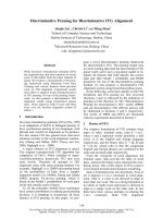
Local disease control
Based on the site of disease, the rate of local disease con-
trol for oral cavity was 17/22 (77%) compared to 83/94
(88%) for oropharynx and 33/39 (85%) for hypopharynx
(X
2
, p = 0.42). The estimates of local disease control at 5
years using the Kaplan Meier method were 69% for oral
cavity, 86% for oropharynx, and 79% for hypopharynx
(figure 1). There were no significant differences among
the 3 sites (Log-Rank test, p = 0.32). Using the Cox Pro-
portional Hazard Model to determine which factors influ-
enced the rate of local disease control, neither T
classification or disease site were found to be significant.
Regional disease control
Based on the site of disease, the rates of regional disease
control were: oral cavity – 21/22 (97.5%); oropharynx –
91/94 (96.8%); and hypopharynx – 38/39 (99%).
Distant metastases
Based on site of disease, the rates of disease failure initially
occurring at distant sites were: oral cavity – 9%; orophar-
ynx – 17%; hypopharynx – 36 % (p = 0.02).
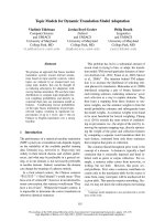
Survival
At the time of analysis, the proportion of patients who
remained alive according to site of disease was: oral cavity
– 50%; oropharynx – 47%; hypopharynx – 27% (X
2
, p =
0.026). The rates of overall survival at 5 years using Kap-
lan Meier projections based on site of disease were: oral
cavity – 37%; oropharynx – 41%; and hypopharynx – 6%
(figure 2)
Table 2: Distribution of Grade III-IV Toxicity based on Site of Disease
Toxicity grade III & IV Oral Cavity (n = 22) Oropharynx (n = 94) Hypopharynx (n = 39) Total (n = 155)
Mucositis 8 (36%) 34 (36%) 7 (18%) 49 (31%)
Hematologic 4 (18%) 11 (12%) 2 (5%) 17 (11%)
Neurologic 0 7 (7.5%) 1 (2.5%) 8 (5%)
Gastrointestinal 04 (4%)0 4 (2.5%)
Cardiac 05 (5%)0 4 (2.5%)
Circulatory 0 1 (1%) 0 1 (0.06%)
Ototoxicity 0 0 1 (2.5%) 1 (0.06%)
Total Events 12 62 11 85
No. of patients 11 (50%) 54 (57%) 10 (26%) 75 (48%)
10 patients had more than one episode of grade III-IV toxicity resulting in a total of 85 events among 75 patients.
World Journal of Surgical Oncology 2008, 6:2 />Page 4 of 6
(page number not for citation purposes)
Discussion
For patients who have resectable oral cavity disease, the
current standard of care for T3-4 squamous cell carcinoma
is surgery and postoperative radiation therapy. This treat-
ment preference is accepted by radiation oncologists as
well as surgeons and is most likely related to the biologic
behavior of oral cavity carcinoma as well as the lower tol-
erance of oral cavity tissue for radiation therapy. Biologi-
cally, it is generally accepted by clinicians that squamous
cell carcinoma of the oral cavity is more resistant to radia-
tion therapy [7,8]. In addition, when radiation therapy is
used as a primary modality, it is fraught with excessive tox-
icity, both acute and chronic. For acute problems, the
major issue is mucositis. For example, radiation of the
buccal mucosa often results in severe mucositis with ulcer-
ations. Also, the lips are problematic to include in the
radiation field because of the acute inflammation of the
mucosa. The dominant chronic toxicity of radiation treat-
ment to the oral cavity relates to osteoradionecrosis, many
of which become clinically manifested years later.
Although there were no recorded events in our series,
longer follow-up may subsequently document this event.
All patients in our series had dental evaluations prior to
therapy and were managed according to the condition of
the dentition, radiation fields, and patient compliance.
Thus, the potential for such toxicity has influenced clini-
cians to treat patients with oral cavity carcinomas with pri-
mary surgery, even in the current era when
chemoradiation is becoming the treatment of choice for
other organ sites such as the larynx, oropharynx, and
hypopharynx [2,3].
Although primary surgery for T3-4 oral cavity cancer
remains the standard of care, a major disadvantage for
patients undergoing this option is the associated func-
tional morbidity. In particular, dysphagia remains a major
challenge faced by patients undergoing surgery for oral
cavity cancer because excessive soft and bony tissue
removal is often necessary. Such procedures as total or
near-total glossectomy, resection of the supra-hyoid mus-
culature, and in some circumstances laryngectomy, have a
major impact on quality of life. There clearly is a need to
improve the treatment for T3-4 oral cavity cancer as this
relates to morbidity as well as efficacy.
Although a number of chemoradiation studies have
included patients with oral cavity tumors, the numbers of
patients entered into such trials have typically lagged
behind those with primary disease arising in other head
and neck sites. Furthermore, among the chemoradiation
trials reported, few have compared outcomes data specific
for the oral cavity and thus it is difficult to know whether
such protocols are effective for this site [9-15].
The 22 patients with T3-4 oral cavity carcinoma included
in our analysis involved some patients, who had unresect-
able disease. More recently, these patients have been des-
ignated as T4b according to the AJCC staging system
(2002) [16]. Patients in the oral cavity subset had a 69 %
rate of local disease control, a rate that was not signifi-
cantly different from the other 2 sites analyzed for com-
parison. Similarly, the projected overall survival rates at 5
years for patients with oral cavity tumors, was not signifi-
cantly different when compared to patients with oropha-
ryngeal tumors: 37% versus 41%. However, the 6%
overall survival rate at 5 years observed for the patients
with hypopharyngeal carcinoma was significantly less.
This difference can be explained by the higher rate of dis-
tant metastases (36%) for hypopharyngeal cancer, a well-
recognized characteristic of carcinomas arising in this site.
We have previously reported that carcinomas arising in
the hypopharynx treated with RADPLAT have the highest
Overall survival stratified by site of diseaseFigure 2
Overall survival stratified by site of disease.
Local-control rate stratified by site of diseaseFigure 1
Local-control rate stratified by site of disease.
World Journal of Surgical Oncology 2008, 6:2 />Page 5 of 6
(page number not for citation purposes)
risk of distant failure, and when combined with the pres-
ence of nodal disease involving multiple levels, this rate
approaches 60% [5].
A comparison of the results from our study with previ-
ously reported results of a treatment regimen consisting of
accelerated radiation therapy shows a striking difference
in the rate of local disease control. Fien et al., [7] treated
105 patients with oral tongue carcinoma using accelerated
radiotherapy and found the rate of local disease control to
be 45% for T3 disease and 0% for T4 disease. These results
led the authors to recommend surgery with post-operative
radiotherapy for advanced oral tongue carcinoma.
Mohr et al reported improved survival and loco-regional
control in patients with T2-T4 patients with oral cavity
and oropharyngeal carcinoma, treated with preoperative
radio-chemotherapy followed by radical surgery, com-
pared to patients with radical surgery alone. Patients with
radical surgery alone had 31% loco-regional recurrence
and 28% death, compared to 15.6% and 18.6%, respec-
tively, in the subset of patients who were treated with pre-
operative radio-chemotherapy [17]. Eckardt et al., found
that in a protocol includjng Taxol
®
and carboplatin given
concomitantly with radiotherapy for 40 Gy, followed by
surgery, 58% of the patients achieved a pathological com-
plete response, and the 3 year overall survival rate was
84% [18].
Our study is in agreement with Balm et al., who reported
treatment outcomes of 79 patients with unresectable car-
cinoma of the oral cavity, oropharynx, hypopharynx and
larynx. The study included 20 patients with oral cavity car-
cinoma, and there was no difference in the outcome of the
different sites regarding loco-regional control and survival
[19].
The main purpose of our analysis was to document and
compare the effects of the direct effects of the treatment
such as disease control, survival, and toxicity. Functional
outcomes such as swallowing and speech were not made
in the same systematic fashion. However, we have previ-
ously reported on function outcomes for selected compo-
nents of patients representing all sites treated with
RADPLAT [20,21]. In future studies, it will be important
to prospectively characterize such parameters specifically
for the oral cavity site and in particular, compare this to
patients with equivalent site-specific lesions who are
treated surgically.
Our findings support the hypothesis that patients with
oral cavity tumor are amendable to organ preservation
protocols in which concurrent chemotherapy is given. The
data indicates that there are no site-specific differences in
loco-regional control for upper aerodigestive tract carci-
nomas treated with targeted chemoradiation (RADPLAT).
Whether this feasibility is limited to protocols that
employ the intra-arterial approach remains to be seen.
The advantage of the intra-arterial technique is based on
the blood supply to the oral cavity. Tumors arising in this
site are amenable to selectively infusing the specific
branches of the external carotid artery, particularly the lin-
gual and facial arteries. Using contrast material and digital
subtraction imaging during the capillary phase of the infu-
sion, interventional radiologists are able to accurately
select the dominant blood supply to the tumor bed.
Conclusion
Patients with advanced oral cavity cancer treated with
RADPLAT respond favorably to RADPLAT, and possibly
other chemoradiation protocols. The effectiveness of the
therapy is comparable to the results using the same proto-
col for oropharyngeal and hypopharyngeal cancer. It is
likely that preservation of oral tissues such as the tongue
can be achieved in the majority of cases. Whether this
proves to preserve the function of the oral cavity such as
mastication, deglution, and articulation, remains to be
determined. Future trials of non-surgical treatment for
this disease site should incorporate prospective analysis of
such functions.
Competing interests
The author(s) declare that they have no competing inter-
ests.
Authors' contributions
ID: Contributed to concept and design, acquisition of
data, analysis and interpretation of data, drafting and
revising the manuscript KTR: contributed to concept and
design, analysis and interpretation of data and revising
the manuscript, SS: Helped in concept and design and
revising the manuscript; FV helped in acquisition of data
and revision of the manuscript. All authors read and
approved final manuscript.
Acknowledgements
Presented at the 6
th
International Conference on Head and Neck Cancer,
Washington, D.C. August 8, 2004.
References
1. Bourhis J, Amand C, Pignon JP, MACH-NC Collaborative Group:
Update of MACH-NC (meta-analysis of chemotherapy in
head & neck cancer) database focused on concomitant
chemotherapy [abstract 5505]. Proc ASCO 2004, 22(14S489
[ />Abstracts+%26+Virtual+MeetinAbstracts?&vmview=abst_detail_vie
w&confID=26&abstrac tID=3198].
2. Adelstein DJ, Li Y, Adams GL, Wagner H Jr, Kish JA, Ensley JF,
Schuller DE, Forastiere AA: An intergroup phase III comparison
of standard radiation therapy and two schedules of concur-
rent chemoradiotherapy in patients with unresectable squa-
mous cell head and neck cancer. J Clin Oncol 2003, 21:92-98.
3. Forastiere AA, Goepfert H, Maor M, Pajak TF, Weber R, Morrison
W, Glisson B, Trotti A, Ridge JA, Chao C, Peters G, Lee DJ, Leaf A,
Ensley J, Cooper J: Concurrent chemotherapy and radiother-
Publish with BioMed Central and every
scientist can read your work free of charge
"BioMed Central will be the most significant development for
disseminating the results of biomedical research in our lifetime."
Sir Paul Nurse, Cancer Research UK
Your research papers will be:
available free of charge to the entire biomedical community
peer reviewed and published immediately upon acceptance
cited in PubMed and archived on PubMed Central
yours — you keep the copyright
Submit your manuscript here:
/>BioMedcentral
World Journal of Surgical Oncology 2008, 6:2 />Page 6 of 6
(page number not for citation purposes)
apy for organ preservation in advanced laryngeal cancer. N
Engl J Med 349:2091-2098. 2003 Nov 27
4. Robbins KT, Kumar P, Wong FSH, Hartsell WF, Flick P, Palmer R,
Weir AB 3rd, Neill HB, Murry T, Ferguson R, Hanchett C, Vieira F,
Bush A, Howell SB: Targeted chemoradiation for advanced
head and neck cancer: analysis of 213 patients. Head Neck
2000, 22:687-693.
5. Doweck I, Robbins KT, Vieira F: Analysis of risk factors predic-
tive of distant failure after targeted chemoradiation for
advanced head and neck cancer. Arch Otolaryngol Head Neck Surg
2001, 127:1315-1318.
6. Robbins KT, Doweck I, Samant S, Vieira F, Kumar P: Factors pre-
dictive of local disease control after intra-arterial concomi-
tant chemoradiation (RADPLAT). Laryngoscope 2004,
114:411-417.
7. Fein DA, Mendenhall WM, Parsons JT, McCarty PJ, Stringer SP, Million
RR, Cassisi NJ: Carcinoma of the oral tongue: a comparison of
results and complications of treatment with radiotherapy
and/or surgery. Head Neck 1994, 16:358-365.
8. Palme CE, Gullane PJ, Gilbert RW: Current treatment options in
squamous cell carcinoma of the oral cavity. Surg Oncol Clin N
Am 2004, 13:47-70.
9. Koch WM, Lee DJ, Eisele DW, Miller D, Poole M, Cummings CW,
Forastiere A: Chemoradiotherapy for organ preservation in
oral and pharyngeal carcinoma. Arch Otolaryngol Head Neck Surg
1995, 121:974-980.
10. Taylor SG 4th, Murthy AK, Griem KL, Recine DC, Kiel K, Blendowski
C, Hurst PB, Showel JT, Hutchinson JC Jr, Campanella RS, Chen S,
Caldarelli DD: Concomitant cisplatin/5-FU infusion and radio-
therapy in advanced head and neck cancer: 8-year analysis of
results. Head Neck 1997, 19:684-691.
11. Chougule P, Wanebo H, Akerley W, McRae R, Nigri P, Leone L,
Safran H, Ready N, Koness RJ, Radie-Keane K, Cole B: Concurrent
paclitaxel, carboplatin, and radiotherapy in advanced head
and neck cancers: a phase II study – preliminary results.
Semin Oncol 1997, 24(6 Suppl 19):S19-57. S19–61
12. Lavertu P, Adelstein DJ, Saxton JP, Secic M, Eliachar I, Strome M, Larto
MA, Wood BG: Aggressive concurrent chemoradiotherapy
for squamous cell head and neck cancer: an 8-year single-
institution experience.
Arch Otolaryngol Head Neck Surg 1999,
125:142-148.
13. Pradier O, Eberlein K, Weiss E, Jackel MC, Hess CF: Radiotherapy
combined with simultaneous chemotherapy with mitomy-
cin-C and 5-fluorouracil for inoperable head and neck can-
cer. Br J Radiol 2001, 74:368-374.
14. Fornari G, Artusio E, Mairone L, Airoldi M, Bongioannini G, Amasio
E, Rosmino C, Gabriele P: Paclitaxel and carboplatin in neo-
adjuvant and concomitant chemoradiotherapy in locally
advanced head and neck squamous cell carcinoma. Tumori
2002, 88:489-494.
15. Airoldi M, Cattel L, Cortesina G, Giordano C, Pedani F, Recalenda V,
Danova M, Gabriele AM, Tagini V, Porta C, Bumma C: Docetaxel,
carboplatin and concomitant radiotherapy for unresectable
squamous cell carcinoma of the head and neck: pharmacok-
inetic and clinical data of a phase I-II study. Am J Clin Oncol
2004, 27:155-163.
16. Greene FL, Page DL, Fleming ID, Fritz A, Balch CM, Haller DG, Mor-
row M, (Eds.): AJCC Cancer Staging Manual. 6th edition.
Springer-Verlag, New-York; 2002.
17. Mohr C, Bohndorf W, Carstens J, Härle F, Hausamen JE, Hirche H,
Kimmig H, Kutzner J, Mühling J, Reuther J, et al.: Preoperative radi-
ochemotherapy and radical surgery in comparison with rad-
ical surgery alone: A prospective, multicentric, randomized
DOSAK study of advanced squamous cell carcinoma of the
oral cavity and the oropharynx (a 3-year follow-up). Int J Oral
Maxillofac Surg 1994, 23:140-148.
18. Eckardt A, Rades D, Rudat V, Hofele C, Dammer R, Dietl B, Wildfang
I, Karstens JH: Prospective phase II study of neoadjuvant radi-
ochemotherapy in advanced operable carcinoma of the
mouth cavity. 3-year outcome. Mund Kiefer Gesichtschir 2002,
6:117-121.
19. Balm AJ, Rasch CR, Schornagel JH, Hilgers FJ, Keus RB, Schultze-Kool
L, Ackerstaff AH, Busschers W, Tan IB: High-dose superselective
intra-arterial cisplatin and concomitant radiation (RAD-
PLAT) for advanced head and neck cancer. Head Neck 2004,
26:485-493.
20. Newman LA, Vieira F, Schwiezer V, Samant S, Murry T, Woodson G,
Kumar P, Robbins KT: Eating and weight changes following
chemoradiation therapy for advanced head and neck cancer.
Arch Otolaryngol Head Neck Surg 1998, 124:589-592.
21. Newman LA, Robbins KT, Logemann JA, Rademaker AW, Lazarus
CL, Hamner A, Tusant S, Huang CF: Swallowing and speech abil-
ity after treatment for head and neck cancer with targeted
intraarterial versus intravenous chemoradiation.
Head Neck
2002, 24:68-77.