Báo cáo khoa học: "Coxsackievirus and adenovirus receptor expression in human endometrial adenocarcinoma: possible clinical implications" doc
Bạn đang xem bản rút gọn của tài liệu. Xem và tải ngay bản đầy đủ của tài liệu tại đây (402.49 KB, 8 trang )
BioMed Central
Page 1 of 8
(page number not for citation purposes)
World Journal of Surgical Oncology
Open Access
Research
Coxsackievirus and adenovirus receptor expression in human
endometrial adenocarcinoma: possible clinical implications
Costas T Giaginis
1
, Apostolos C Zarros
1
, Maria A Papaefthymiou
1
,
Aikaterini E Papadopouli
1
, Ioannis K Sfiniadakis
2
and
Stamatios E Theocharis*
1
Address:
1
Department of Forensic Medicine and Toxicology, Medical School, University of Athens, Greece and
2
Department of Pathology, Naval
Hospital, Athens, Greece
Email: Costas T Giaginis - ; Apostolos C Zarros - ;
Maria A Papaefthymiou - ; Aikaterini E Papadopouli - ;
Ioannis K Sfiniadakis - ; Stamatios E Theocharis* -
* Corresponding author
Abstract
The coxsackievirus and adenovirus receptor (CAR) is a crucial receptor for the entry of both
coxsackie B viruses and adenoviruses into host cells. CAR expression on tumor cells was reported
to be associated with their sensitivity to adenoviral infection, while it was considered as a surrogate
marker for monitoring and/or predicting the outcome of adenovirus-mediated gene therapy. The
aim of the present study was to evaluate the clinical significance of CAR expression in endometrial
adenocarcinoma. CAR expression was assessed immunohistochemically in tumoral samples of 41
endometrial adenocarcinoma patients and was statistically analyzed in relation to various
clinicopathological parameters, tumor proliferative capacity and patient survival. CAR positivity was
noted in 23 out of 41 (56%) endometrial adenocarcinoma cases, while high CAR expression in 8
out of 23 (35%) positive ones. CAR intensity of immunostaining was classified as mild in 11 (48%),
moderate in 10 (43%) and intense in 2 (9%) out of the 23 positive cases. CAR positivity was
significantly associated with tumor histological grade (p = 0.036), as well differentiated tumors
more frequently demonstrating no CAR expression. CAR staining intensity was significantly
associated with tumor histological type (p = 0.016), as tumors possessing squamous elements
presented more frequently intense CAR immunostaining. High CAR expression showed a trend to
be correlated with increased tumor proliferative capacity (p = 0.057). Patients with tumors
presenting moderate or intense CAR staining intensity were characterized by longer survival times
than those with mild one; however, this difference did not reach statistical significance. These data
reveal, for the first time, the expression of CAR in clinical material obtained from patients with
endometrial adenocarcinoma in relation to important clinicopathological parameters for their
management. As CAR appears to modulate the proliferation and characteristics of cancer cells, its
expression could be considered of possible clinical importance for future (gene) therapy
applications.
Published: 17 June 2008
World Journal of Surgical Oncology 2008, 6:59 doi:10.1186/1477-7819-6-59
Received: 6 February 2008
Accepted: 17 June 2008
This article is available from: />© 2008 Giaginis et al; licensee BioMed Central Ltd.
This is an Open Access article distributed under the terms of the Creative Commons Attribution License ( />),
which permits unrestricted use, distribution, and reproduction in any medium, provided the original work is properly cited.
World Journal of Surgical Oncology 2008, 6:59 />Page 2 of 8
(page number not for citation purposes)
Background
The coxsackievirus and adenovirus receptor (CAR) is a 46-
kDa transmembrane protein, which functions as a pri-
mary receptor for both coxsackie B virus (CVB) and aden-
ovirus (Ad) [1]. This cell surface receptor plays a crucial
role in CVB and Ad entry into host cells [2]. CAR mediates
homotypic intercellular interactions, while in polarized
endothelial cells CAR is closely associated with the tight
junction, where it contributes to the barrier of paracellular
flow of solutes and macromolecules [3]. A strong correla-
tion of CAR levels with the viral sensitivity of several cell
types has been reported [4-6]. In fact, CAR has been
shown to be a docking site for Ad, thus acting as a key
receptor for the enhancement of the virus-to-host affinity
and the initiation of the virus internalization to the host
cell [7,8]. On cells lacking CAR, virus uptake takes place
with lower efficiency [7,9] due to the existence of a sec-
ondary pathway leading to the viral internalization [7,10].
The very promising use of Ad vectors in gene therapy,
since Ads are relatively safe, highly infectious, and capable
of delivering therapeutic genes to different cell types
[10,11], still faces a critical prerequisite, which is no other
than the identification of highly efficient and accurate sys-
tems for delivering the therapeutic genes into target cells
[12]. In this regard, CAR expression could be a surrogate
marker for monitoring and/or predicting the outcome of
gene therapy, while by increasing CAR levels, resistant
cells could become more sensitive to Ad infection [13].
However, only a limited number of studies concerning
CAR expression have been made on clinical tissue mate-
rial. In this aspect, Persson et al. presented an immunohis-
tochemical study in human normal brain and human
brain tumors, suggesting that neuroblastomas and medul-
loblastomas could be suitable for adenovirus-mediated
gene therapy [14,15]. Moreover, recent studies have sug-
gested a pathophysiological role for CAR in bladder can-
cer and glioma cells, rendering CAR as a membrane
receptor which conveys its signal into the nucleus and
results in cell proliferation suppression [16-18]. These
findings raise the question whether CAR expression could
be related to the tumor proliferative capacity or differenti-
ation amongst the different tumor cell types.
Endometrial adenocarcinoma is the most common malig-
nant tumor of the female tract and the fourth most com-
mon cancer in women following breast, colorectal and
lung cancer in the Western world [19]. A substantial
decrease in the incidence and mortality of endometrial
cancer seems unlikely in the next few years, as early detec-
tion and treatment modalities have not been proven to
possess a major impact on mortality [20]. Epigenetic
modification reagents, including DNA methyltransferase
and histone deacetylase inhibitors, when used alone or in
combination with conventional chemotherapy, seem to
be beneficial for endometrial cancer patients [21]. How-
ever, further research advancements are recommended to
bring about new strategies and technologies, which ulti-
mately improve the diagnosis and treatment of women
with endometrial cancer [21].
In the light of the above considerations, the present study
aimed to estimate the immunohistochemical CAR expres-
sion in tumoral specimens obtained from endometrial
adenocarcinoma patients. We also aimed to evaluate the
association of CAR expression and staining intensity with
various clinicopathological parameters, tumor prolifera-
tive capacity and patient survival.
Patients and Methods
Patients
Forty-one endometrial adenocarcinoma specimens
obtained from an equal number of patients who under-
went surgery due to endometrial cancer were included in
this study. None of the patients received any kind of anti-
cancer treatment prior to surgery. The mean age of the
patient cohort was 63.4 ± 9.6 years (median: 64 years,
range: 40–82 years). Tumors were typed according to the
presence or not of squamous elements. Three levels of dif-
ferentiation were used to classify grading as: well, moder-
ately and poorly differentiated. Tumors staging was
assessed according to the standards of the Federation
Internationationale de Gynecologistes et Obstetricianes
(FIGO) [22]. The patients were followed up, with the
length of the follow up varying from 22 to 94 months
(mean 65.51 ± 16.19 median 64 months). Twenty six
patients were followed up until death, while the remain-
ing 15 patients were remained disease free. All the exam-
ined clinicopathological parameters are reported in Tables
1, 2 and 3.
Immunohistochemistry
Immunostainings for CAR was performed on paraffin-
embedded tissue sections using a commercially available
rabbit anti-CAR monoclonal antibody (CAR H300, Santa
Cruz Biochemicals, Santa Cruz, CA, USA). Briefly, 4 μm
thick tissue sections were dewaxed in xylene and were
brought to water through graded alcohols. To remove the
endogenous peroxidase activity, sections were then
treated with freshly prepared 0.3% hydrogen peroxide in
methanol in the dark, for 30 minutes (min), at room tem-
perature. Non-specific antibody binding was then blocked
using Snipper, a specific blocking reagent for mouse pri-
mary antibodies (Sniper, Biocare Medical, Walnut, Creek,
CA, USA) for 5 min. The sections were then incubated for
1 hour (h), at room temperature, with the primary anti-
body, CAR, diluted 1:100, respectively, in phosphate buff-
ered saline (PBS). After washing three times with PBS,
sections were incubated at room temperature with bioti-
nylated linking reagent (Biocare Medical) for 10 min, fol-
World Journal of Surgical Oncology 2008, 6:59 />Page 3 of 8
(page number not for citation purposes)
lowed by incubation with peroxidase-conjugated
streptavidin label (Biocare Medical) for 10 min. The
resultant immune peroxidase activity was developed in
0.5% 3,3'-diaminobenzidine hydrochloride (DAB; Sigma,
Saint Louis, MO, USA) in PBS containing 0.03% hydrogen
peroxide for 3 min. Sections were counterstained with
Harris' hematoxylin and mounted in Entellan (Merck,
Darmstadt, Germany). An additional step of antigen
retrieval (citrate buffer at pH 6.1 and microwave heating)
was performed before incubation with the primary anti-
CAR antibody. Appropriate negative controls were per-
formed by omitting the primary antibody and/or substi-
Table 1: Associations between CAR positivity and clinicopathological characteristics of the 41 patients with endometrial
adenocarcinoma
Clinicopathological characteristics CAR positivity
Negative (%) Positive (%) P-value
Patients 18 (44) 23 (56)
Age (mean ± SD), years 64.7 ± 7.5 62.4 ± 11.0 0.623
< 63 8 (20) 12 (29)
≥63 10 (24) 11 (27)
pStage (FIGO) 0.675
I 16 (40) 18 (44)
II 1 (2) 3 (8)
III 0 (0) 1 (2)
IV 1 (2) 1 (2)
Histological type 0.684
Positive for squamous elements 3 (8) 5 (12)
Negative for squamous elements 15 (36) 18 (44)
Histological grade 0.036
Well differentiated 7 (17) 3 (8)
Moderately differentiated 7 (17) 18 (44)
Poorly differentiated 4 (10) 2 (4)
Ki-67 protein statement 0.829
Ki-67 below mean (<40%) 10 (24) 12 (29)
Ki-67 over mean (≥ 40%) 8 (20) 11 (27)
Table 2: Associations between CAR overexpression and clinicopathological characteristics of the 23 CAR positive endometrial
adenocarcinoma cases
Clinicopathological characteristics CAR expression
< 31% (%) ≥31% (%) P-value
Patients 15 (65) 8 (35)
Age (mean± SD), years 62.9 ± 12.1 61.5 ± 9.6 0.469
< 63 8 (35) 5 (22)
≥63 7 (30) 3 (13)
pStage (FIGO) 0.147
I 12 (52) 6 (27)
II 3 (13) 0 (0)
III 0 (0) 1 (4)
IV 0 (0) 1 (4)
Histological type 0.181
Positive for squamous elements 2 (9) 3 (13)
Negative for squamous elements 13 (56) 5 (22)
Histological grade 0.379
Well differentiated 3 (13) 0 (0)
Moderately differentiated 11 (48) 7 (31)
Poorly differentiated 1 (4) 1 (4)
Ki-67 protein statement 0.057
Ki-67 below mean (<40%) 10 (43) 2 (8)
Ki-67 over mean (≥ 40%) 5 (22) 6 (27)
World Journal of Surgical Oncology 2008, 6:59 />Page 4 of 8
(page number not for citation purposes)
tuting it with an irrelevant anti-serum. As positive control,
colon cancer tissue sections with known increased CAR
positivity were used [23]. The tumor proliferative capacity
was assessed immunohistochemically, using a mouse
anti-human Ki-67 antigen; IgG
1k
antibody (clone MIB-1,
Dakopatts) as previously described [24].
Evaluation of immunohistochemistry
The percentages of positively stained cells were obtained
by counting at least 1000 tumor cells in each case by two
independent observers (SET and IKS) blinded to the clin-
ical data with complete observer agreement. Specimens
were considered "positive" for CAR and Ki-67 when more
than 5% of the tumor cells were stained, while they were
characterized to present "high" CAR and Ki-67 expression
when the percentage of positively stained cells exceeded
the mean percentage value. The intensity of CAR immu-
nostaining was also estimated and graded on a three step
scale as: mild (+), moderate (++) and intense (+++). The
cellular pattern of distribution of CAR immunostaining
was characterized as membraneous and cytoplasmic. All
endometrial adenocarcinoma cases were Ki-67 positive,
presenting nuclear pattern of staining.
Statistical analysis
Chi-square tests were used to assess the association of CAR
positivity, overexpression and staining intensity with clin-
icopathological variables and tumor proliferative capac-
ity. Survival curves were constructed using the Kaplan-
Meier method and compared using the log-rank test. Cox
proportional hazard regression analysis was used to eval-
uate the effect of CAR positivity, level of expression (low
vs high level of CAR expression) and staining intensity as
prognostic factors on patient survival. A 2-tailed P < 0.05
was considered (statistically) significant. Statistical analy-
ses were performed using the software package SPSS for
Windows (version 11.0; SPSS Inc., Chicago, IL, USA).
Results
CAR positivity was noted in 23 out of 41 (56%) of the
examined endometrial adenocarcinoma cases (Table 1).
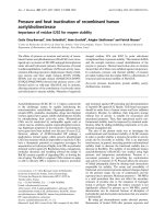
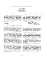
Representative CAR immunostaining is presented in Fig-
ure 1. The pattern of CAR distribution was both cytoplas-
mic and membraneous in all positive cases examined.
High CAR expression was noted in 8 out of 23 (35%) of
the positive cases (Table 2). The intensity of CAR immu-
nostaining was classified as mild in 11 (48%), moderate
in 10 (43%) and intense in 2 (9%) out of 23 positive cases
(Table 3).
CAR positivity was significantly associated with tumor
histological grade, as well differentiated tumors most fre-
quently presenting no CAR expression compared to mod-
erately and poorly differentiated ones. (P = 0.036, Table
1). High CAR expression showed a trend to be correlated
with increased proliferative capacity (P = 0.057, Table 2).
CAR staining intensity was significantly associated with
tumor histological type, as cases possessing squamous ele-
ments presented more frequently intense CAR immunos-
taining (P = 0.016, Table 3). CAR positivity, level of
Table 3: Associations between CAR staining intensity and clinicopathological characteristics of the 23 CAR positive endometrial
adenocarcinoma cases
Clinicopathological characteristics CAR intensity
Mild (%) Moderate (%) Intense (%) P-value
Patients 11 (48) 10 (43) 2 (9)
Age (mean ± SD), years 63.2 ± 11.5 63.6 ± 11.2 52.5 ± 0.7 0.359
< 63 6 (26) 5 (22) 2 (9)
≥63 5 (22) 5 (22) 0 (0)
pStage (FIGO) 0.395
I 7 (30) 9 (39) 2 (9)
II 3 (14) 0 (0) 0 (0)
III 0 (0) 1 (4) 0 (0)
IV 1 (4) 0 (0) 0 (0)
Histological type 0.016
Positive for squamous elements 1 (4) 2 (9) 2 (9)
Negative for squamous elements 10 (44) 8 (34) 0 (0)
Histological grade 0.881
Well differentiated 1 (4) 1 (4) 1 (4)
Moderately differentiated 9 (40) 7 (30) 1 (5)
Poorly differentiated 1 (4) 2 (9) 0 (0)
Ki-67 protein statement 0.555
Ki-67 below mean (< 40%) 7 (31) 4 (17) 1 (4)
Ki-67 over mean (≥ 40%) 4 (17) 6 (26) 1 (5)
World Journal of Surgical Oncology 2008, 6:59 />Page 5 of 8
(page number not for citation purposes)
expression and staining intensity were not significantly
associated with the other clinicopathological parameters
examined (Tables 1, 2 and 3).
The Kaplan-Meier product-limit method for overall anal-
ysis survival according to CAR positivity (positive vs nega-
tive CAR staining), level of expression (high vs low level of
CAR expression) and staining intensity (mild vs moderate
and intense CAR staining intensity) in patients with
endometrial adenocarcinoma did not reveal statistically
significant correlations (log-rank test, P = 0.799, P = 0.816
and P = 0.127, respectively) (data not shown). The sur-
vival of patients with tumors presenting moderate or
intense CAR staining intensity (mean survival rate 72.3 ±
11.0 months) was longer than those presenting mild
intensity (mean survival rate 55.6 ± 19.5 months); how-
ever the difference did not reach statistical significance in
univariate analysis (P = 0.127, data not shown).
Discussion
The expression of CAR in human tumors and tumor cell
lines has been subject of several studies [5,6,25-28] which
have detected this transmembrane protein in variable and
often low levels. The present study focused on the immu-
nohistochemical examination of CAR expression in
endometrial adenocarcinoma samples and revealed the
clinical significance of CAR in certain aspects of endome-
trial neoplasia, such as tumor differentiation, histological
type and proliferation. More to the point, CAR positivity
was noted in 56% of the examined cases. This incidence
of endometrial adenocarcinoma CAR positivity cannot be
considered among the highest ever found on tumor
malignancies, since Gu et al. have observed a 75% CAR
positivity on osteosarcoma samples; however, it is cer-
tainly higher than that of lung adenocarcinoma [29,30].
To this point, it should be noted the effectiveness of ade-
noviral gene therapy depends on the amount of CAR
expression on target cells. Thus, the current study rein-
forced the suitability for adenoviral gene therapy in the
case of endometrial adenocarcinoma.
Our study is the first report examining the clinical signifi-
cance of CAR expression in patients with endometrial ade-
nocarcinoma. We found that well differentiated tumors
more frequently presenting no CAR expression compared
to moderately and poorly differentiated ones. In this
aspect, Korn et al. also revealed a significant association
between CAR expression and tumor histological grade in
patients with gastrointestinal malignancies; however,
moderately to poorly differentiated tumors more fre-
quently demonstrating low or no CAR expression [31].
CAR expression was also reported to be increased with
increasing grade of tumor in breast cancer patients; how-
ever, this difference was not statistically significant [32].
Moreover, in the current study, high CAR expression
showed a trend to be correlated with increased prolifera-
tive capacity. In this context, CAR expression was reported
to modulate the proliferative capacity of cancer cells, in
vitro [13,16,18,33]. In fact, the presence of CAR was found
not only to facilitate viral uptake of adenovirus, but also
to inhibit cell growth in bladder cancer and malignant gli-
oma cells [13,16,18,33]. The latter is not in line with the
current findings and could be ascribed to the individual
characteristics among the different types of cancer. More-
over, our results were based on the in situ detection of CAR
protein by immunohistochemistry, while the potential
Intense immunostaining for CAR in tumor cells in representative endometrial adenocarcinoma cases (original magnification ×200)Figure 1
Intense immunostaining for CAR in tumor cells in representative endometrial adenocarcinoma cases (original magnification
×200). A. Negative for squamous elements. B. Positive for squamous elements.
World Journal of Surgical Oncology 2008, 6:59 />Page 6 of 8
(page number not for citation purposes)
tumor suppressor role of CAR was reported for cultured
cell lines or tumor cells injected into nude mice
[13,16,18,33]. We also found that CAR staining intensity
was significantly associated with the tumor histological
type. This result is in line with previous evidence where
higher rates of CAR expression were detected in lung squa-
mous cell carcinoma than in adenocarcinoma [30].
To our knowledge, there is limited data so far highlighting
to the prognostic value of CAR expression in cancer. In
this respect, our study is the first report examining the
clinical significance of CAR expression in the prognosis of
patients with endometrial adenocarcinoma. We did not
found any significant association of CAR positivity, level
of expression and staining intensity with patient survival.
It should be noted that the survival of patients with
tumors presenting moderate or intense CAR staining
(mean survival rate 72.3 ± 11.0 months) was longer than
those with mild (mean survival rate 55.6 ± 19.5 months);
however, this difference did not reach statistically signifi-
cance in univariate analysis (P = 0.127). In this context,
Martin et al. showed that elevated levels of CAR expres-
sion were significantly associated with poor overall sur-
vival in patients with breast cancer [32]. It has also been
shown that the soluble splice variants CAR 3/7 and CAR
4/7, but no the full-length hCAR were of independent
prognostic relevance for progression-free or overall sur-
vival of ovarian cancer patients [34].
In the last few years, the use of adenovirus vectors is gain-
ing increasingly interesting in order to advance new ther-
apeutic approaches against cancer. Thus, many tumor
samples have been examined for CAR expression, which
has generally been found to correlate with susceptibility
to transduction [5,18,28,35]. In several human malignan-
cies, including bladder and prostate carcinoma and gliob-
lastoma, CAR expression was downregulated during the
progression to malignancy [16,36,37]. In CAR-deficient
prostate and glioma tumor cell lines, expression of CAR
by transfection resulted in suppression of cell prolifera-
tion and decreased tumorogenicity [18,33]. CAR expres-
sion also inhibited cell proliferation and was associated
with modulations in the activity of the cell cycle regulators
p21-PIC and Rb in bladder cancer cells [16]. Importantly,
CAR dependent growth inhibition required the presence
of CAR-specific antibody which blocked homotypic adhe-
sion [16].
A strong correlation of CAR levels with the viral sensitivity
of any given cell has been reported [4-6]. Although the
normal cellular function of CAR is not known, some
researchers have suggested that CAR may serve as a cell-
cell adhesion molecule [38], while others have shown an
in vitro and in vivo tumor-suppressive role for CAR [33]. It
is thought that CAR can inhibit cancer growth by behav-
ing as a membrane receptor, which conveys its signal into
the nucleus, thus resulting in suppression of the prolifera-
tive mechanisms [13]. Moreover, reduced CAR expression
was shown to induce lung metastasis [39]. Overall, corre-
lating the CAR expression in all known tumor malignan-
cies with clinicopathological parameters cannot only
provide crucial information about its role in malignant
transformation, but it can also establish a better view for
future gene therapy approaches. Currently, Othman et al.
reported that endometriosis cells expressed higher levels
of CAR mRNA as compared with normal endometrial
cells [40]. In addition, it was shown that adenoviruses can
effectively transfect endometriosis cells in vitro. The dom-
inant negative mutants of Estrogen receptors (DN-ER)
delivered to endometriosis cells via an adenovirus
decreased cell proliferation, induced apoptosis and sup-
pressed cytokine production by these cells [40]. Such data
supported substantial evidence that adenovirus-mediated
delivery of DN-ER to endometriosis cells can be a poten-
tial therapeutic approach for endometriosis [40].
Conclusion
The data presented in this study revealed enhanced CAR
expression in endometrial adenocarcinoma specimens.
CAR protein expression was associated with important
clinicopathological parameters with respect to the diagno-
sis of patients with endometrial cancer. Although CAR
protein failed to predict patient survival, the current study
supports evidence for potential implication of CAR pro-
tein in endometrial carcinogenesis. The use of Ad vectors
in gene therapy needs an efficient and accurate system for
delivering the therapeutic gene into target cells [12]. In
this regard, CAR expression could be a surrogate marker
for monitoring and/or predicting the outcome of gene
therapy, while its increase might contribute to the upreg-
ulation of cellular sensitivity towards Ad infection [13]. It
is, however, without doubt that in order to understand the
physiological role of CAR in cellular function and prolif-
eration, a systematic approach towards the identification
of its natural ligand(s) should also be attempted.
Competing interests
The authors declare that they have no competing interests.
Authors' contributions
CTG participated in the design of the study, drafted the
paper and performed the statistical analysis, ACZ partici-
pated in the statistical analysis and drafted the paper, MAP
contributed to the immunostainings and clinical data col-
lection, AEP contributed to the immunostainings and
clinical data collection, IKS carried out the immunohisto-
chemistry data evaluation, SET designed the study, carried
out the immunohistochemistry data evaluation and cor-
rected the manuscript. All authors read and approved the
final manuscript.
World Journal of Surgical Oncology 2008, 6:59 />Page 7 of 8
(page number not for citation purposes)
References
1. Bowles KR, Gibson J, Wu J, Shaffer LG, Towbin JA, Bowles NE:
Genomic organization and chromosomal localization of the
human coxsackievirus B-adenovirus receptor gene. Hum
Genet 1999, 105:354-359.
2. Bergelson JM, Cunningham JA, Droguett G, Kurt-Jones EA, Krithivas
A, Hong JS, Horwitz MS, Crowell RL, Finberg RW: Isolation of a
common receptor for Coxsackie B viruses and adenoviruses
2 and 5. Science 1997, 275:1320-1323.
3. Coyne CB, Bergelson JM: CAR: A virus receptor within the tight
junction. Adv Drug Deliv Rev 2005, 57:869-82.
4. Kim JS, Lee SH, Cho YS, Choi JJ, Kim YH, Lee JH: Enhancement of
the adenoviral sensitivity on human ovarian cancer cells by
transient expression of coxsackievirus and adenovirus recep-
tor (CAR). Gynecol Oncol 2002, 85:260-265.
5. Hemmi S, Geertsen R, Mezzacasa A, Peter I, Dummer R: The pres-
ence of human coxsackievirus and adenovirus receptor is
associated with efficient adenovirus-mediated transgene
expression in human melanoma cell cultures. Hum Gene Ther
1998, 9:2363-2373.
6. Li Y, Pong RC, Bergelson JM, Hall MC, Sagalowsky AI, Tseng CP,
Wang Z, Hsieh JT: Loss of adenoviral receptor expression in
human bladder cancer cells: a potential impact on the effi-
cacy of gene therapy. Cancer Res 1999, 59:325-330.
7. Leon RP, Hedlund T, Meech SJ, Li S, Schaack J, Hunger SP, Duke RC,
DeGregori J: Adenoviral-mediated gene transfer in lym-
phocytes. Proc Natl Acad Sci USA 1998, 95:13159-13164.
8. Wang X, Bergelson JM: Coxsackievirus and adenovirus recep-
tor cytoplasmic and transmembrane domains are not essen-
tial for coxsackievirus and adenovirus infection. J Virol 1999,
73:2559-25562.
9. Bai M, Campisi L, Freimuth P: Vitronectin receptor antibodies
inhibit infection of HeLa and A549 cells by adenovirus type
12 but not by adenovirus type 2. J Virol 1994, 68:5925-5932.
10. Barnett BG, Crews CJ, Douglas JT: Targeted adenoviral vectors.
Biochim Biophys Acta 2002, 1575:1-14.
11. Zhang WW: Development and application of adenoviral vec-
tors for gene therapy of cancer. Cancer Gene Ther 1999,
6:113-138.
12. Hedley SJ, Chen J, Mountz JD, Li J, Curiel DT, Korokhov N, Kovesdi
I: Targeted and shielded adenovectors for cancer therapy.
Cancer Immunol Immunother 2006, 55:1412-1419.
13. Okegawa T, Li Y, Pong RC, Hsieh JT: Cell adhesion proteins as
tumor suppressors. J Urol 2002, 167:1836-1843.
14. Persson A, Fan X, Widegren B, Englund E: Cell type- and region-
dependent coxsackie adenovirus receptor expression in the
central nervous system. J Neurooncol 2006, 78:1-6.
15. Persson A, Fan X, Salford LG, Widegren B, Englund E: Neuroblast-
omas and medulloblastomas exhibit more coxsackie adeno-
virus receptor expression than gliomas and other brain
tumors. Neuropathology 2007, 27:233-236.
16. Okegawa T, Pong RC, Li Y, Bergelson JM, Sagalowsky AI, Hsieh JT:
The mechanism of the growth-inhibitory effect of coxsackie
and adenovirus receptor (CAR) on human bladder cancer: a
functional analysis of car protein structure. Cancer Res 2001,
61:6592-6600.
17. Huang KC, Altinoz M, Wosik K, Larochelle N, Koty Z, Zhu L, Holland
PC, Nalbantoglu J: Impact of the coxsackie and adenovirus
receptor (CAR) on glioma cell growth and invasion: require-
ment for the C-terminal domain. Int J Cancer 2005,
113:738-745.
18. Kim M, Sumerel LA, Belousova N, Lyons GR, Carey DE, Krasnykh V,
Douglas JT: The coxsackievirus and adenovirus receptor acts
as a tumour suppressor in malignant glioma cells. Br J Cancer
2003, 88:1411-1416.
19. Prat J, Gallardo A, Cuatrecasas M, Catasus L: Endometrial carci-
noma: pathology and genetics. Pathology 2007, 39:72-87.
20. Amant F, Moerman P, Neven P, Timmerman D, Limbergen EV, Ver-
gote I: Treatment modalities in endometrial cancer. Curr Opin
Oncol 2007, 19:479-485.
21. Zhou XC, Dowdy SC, Podratz KC, Jiang S-W: Epigenetics consid-
erations for endometrial cancer prevention, diagnosis and
treatment. Gynecol Oncol 2007, 107:143-153.
22. International Federation of Gynecology and Obstetrics: FIGO
stages: 1988 revision. Gynecol Oncol 1989, 25:
125-126.
23. Theocharis S, Papaefthymiou M, Giaginis C, Gatzidou E, Vgenopoulou
S, Sfiniadakis I, Kouraklis G: CAR expression in gastrointestinal
and pancreatic adenocarcinoma. Virchows Archiv 2007, 451:316.
24. Giaginis C, Davides D, Zarros A, Noussia O, Zizi-Serbetzoglou A,
Kouraklis G, Theocharis S: Clinical significance of tumor-associ-
ated antigen RCAS1 expression in human pancreatic ductal
adenocarcinoma. Dig Dis Sci 2008 in press.
25. Cripe TP, Dunphy EJ, Holub AD, Saini A, Vasi NH, Mahller YY, Collins
MH, Snyder JD, Krasnykh V, Curiel DT, Wickham TJ, DeGregori J,
Bergelson JM, Currier MA: Fiber knob modifications overcome
low, heterogeneous expression of the coxsackievirus-adeno-
virus receptor that limits adenovirus gene transfer and onc-
olysis for human rhabdomyosarcoma cells. Cancer Res 2001,
61:2953-2960.
26. Dmitriev I, Krasnykh V, Miller CR, Wang M, Kashentseva E, Mikheeva
G, Belousova N, Curiel DT: An adenovirus vector with geneti-
cally modified fibers demonstrates expanded tropism via uti-
lization of a coxsackievirus and adenovirus receptor-
independent cell entry mechanism. J Virol 1998, 72:9706-9713.
27. Fechner H, Wang X, Wang H, Jansen A, Pauschinger M, Scherubl H,
Bergelson JM, Schultheiss HP, Poller W: Trans-complementation
of vector replication versus Coxsackie-adenovirus-receptor
overexpression to improve transgene expression in poorly
permissive cancer cells. Gene Ther 2000, 7:1954-1968.
28. Miller CR, Buchsbaum DJ, Reynolds PN, Douglas JT, Gillespie GY,
Mayo MS, Raben D, Curiel DT: Differential susceptibility of pri-
mary and established human glioma cells to adenovirus
infection: targeting via the epidermal growth factor recep-
tor achieves fiber receptor-independent gene transfer. Can-
cer Res 1998, 58:5738-5748.
29. Gu W, Ogose A, Kawashima H, Ito M, Ito T, Matsuba A, Kitahara H,
Hotta T, Tokunaga K, Hatano H, Morita T, Urakawa S, Yoshizawa T,
Kawashima H, Kuwano R, Endo N: High-level expression of the
coxsackievirus and adenovirus receptor messenger RNA in
osteosarcoma, Ewing's sarcoma and benign neurogenic
tumors among musculoskeletal tumors. Clin Cancer Res 2004,
10:3831-3838.
30. Wang Y, Wang S, Bao Y, Ni C, Guan N, Zhao J, Salford LG, Widegren
B, Fan X: Coxsackievirus and adenovirus receptor expression
in non-malignant lung tissues and clinical cancers. J Mol Histol
2006, 37(3-4):
153-160.
31. Korn WM, Christian MM, Lacher MD, McMillian A, Rauen KA, War-
ren RS, Ferrell L: Expression of the coxsackievirus- and adeno-
virus receptor in gastrointestinal cancer correlates with
tumor differentiation. Cancer Gene Ther 2006, 13:792-797.
32. Martin TA, Watkins G, Jiang WG: The coxsackievirus adenovirus
receptor has elevated expression in human breast cancer.
Clin Exp Med 2005, 5:122-128.
33. Okegawa T, Li Y, Pong RC, Bergelson JM, Zhou J, Hsieh JT: The dual
impact of coxsackie and adenovirus receptor expression on
human prostate cancer gene therapy. Cancer Res 2000,
60:5031-5036.
34. Reimer D, Steppan I, Wiedemair A, Concin N, Hofstetter G, Marth
C, Müller-Holzner E, Zeimet AG: Soluble isoforms but not the
transmembrane form of coxsackie-adenovirus receptor are
of clinical relevance in epithelial ovarian cancer. Int J Cancer
2007, 120:2568-2575.
35. Qin M, Chen S, Yu T, Escuadro B, Sharma S, Batra RK: Coxsackiev-
irus adenovirus receptor expression predicts the efficiency
of adenoviral gene transfer into non-small cell lung cancer
xenografts. Clin Cancer Res 2003, 9:4992-4999.
36. Sachs MD, Rauen KA, Ramamurthy M, Dodson JL, De Marzo AM,
Putzi MJ, Schoenberg MP, Rodriguez R: Integrin alpha(v) and cox-
sackie adenovirus receptor expression in clinical bladder
cancer. Urology 2002, 60:531-536.
37. Fuxe J, Liu L, Malin S, Philipson L, Collins VP, Pettersson RF: Expres-
sion of the coxsackie and adenovirus receptor in human
astrocytic tumors and xenografts. Int J Cancer 2003,
103:723-729.
38. Honda T, Saitoh H, Masuko M, Katagiri-Abe T, Tominaga K, Kozakai
I, Kobayashi K, Kumanishi T, Watanabe YG, Odani S, Kuwano R: The
coxsackievirus-adenovirus receptor protein as a cell adhe-
sion molecule in the developing mouse brain. Brain Res Mol
Brain Res 2000, 77:19-28.
39. Yamashita M, Ino A, Kawabata K, Sakurai F, Mizuguchi H: Expression
of coxsackie and adenovirus receptor reduces the lung met-
Publish with BioMed Central and every
scientist can read your work free of charge
"BioMed Central will be the most significant development for
disseminating the results of biomedical research in our lifetime."
Sir Paul Nurse, Cancer Research UK
Your research papers will be:
available free of charge to the entire biomedical community
peer reviewed and published immediately upon acceptance
cited in PubMed and archived on PubMed Central
yours — you keep the copyright
Submit your manuscript here:
/>BioMedcentral
World Journal of Surgical Oncology 2008, 6:59 />Page 8 of 8
(page number not for citation purposes)
astatic potential of murine tumor cells. Int J Cancer 2007,
121:1690-1696.
40. Othman E-ER, Salama S, Ismail A, Al-Hendy A: Toward gene ther-
apy of endometriosis: adenovirus-mediated delivery of dom-
inant negative estrogen receptor genes inhibits cell
proliferation, reduces cytokine production, and induces
apoptosis of endometriotic cells. Fertil Steril 2007, 88:462-471.