Báo cáo khoa học: "Alveolar soft part sarcoma: clinicopathological findings in a series of 11 cases" pdf
Bạn đang xem bản rút gọn của tài liệu. Xem và tải ngay bản đầy đủ của tài liệu tại đây (1.25 MB, 7 trang )
BioMed Central
Page 1 of 7
(page number not for citation purposes)
World Journal of Surgical Oncology
Open Access
Research
Alveolar soft part sarcoma: clinicopathological findings in a series of
11 cases
Adrien Daigeler*
1
, Cornelius Kuhnen
2
, Joerg Hauser
1
, Ole Goertz
1
,
Daniel Tilkorn
1
, Lars Steinstraesser
1
, Hans-Ulrich Steinau
1
and
Marcus Lehnhardt
1
Address:
1
Department of Plastic Surgery, Burn Center, Hand surgery, Sarcoma Reference Center, BG-University Hospital Bergmannsheil, Ruhr
University Bochum, Buerkle-de-la-Camp-Platz 1, 44789 Bochum, Germany and
2
Institute for Pathology, BG-University Hospital Bergmannsheil,
Ruhr-University Bochum, Bürkle-de-la-Camp-Platz 1, 44789 Bochum, Germany
Email: Adrien Daigeler* - ; Cornelius Kuhnen - ; Joerg Hauser - ;
Ole Goertz - ; Daniel Tilkorn - ; Lars Steinstraesser - ; Hans-
Ulrich Steinau - ; Marcus Lehnhardt -
* Corresponding author
Abstract
Background: Alveolar sarcoma of the soft parts (ASPS) represents a very rare entity of soft tissue
sarcoma with special features such as young peak age incidence and frequent metastasis to the
brain. The aim of this study was a clinicopathological analysis with special reference to treatment
and outcome.
Methods: From the database of the BG-University Hospital Bergmannsheil, 1597 soft tissue
sarcoma (STS) cases were reviewed and 11 consecutive patients with ASPS were isolated. Data was
acquired from patients' charts and contact to patients, their relatives or general practitioners, with
special reference to treatment and clinical course. The average follow up time from the time of the
definite operation for the primary tumor was 6.5 years. Kaplan-Meier method was used to calculate
survival.
Results: Patients with localized disease who received complete resection and adjuvant radiation
and who did not develop recurrence or metastatic disease within 2 years after surgery had a
positive outcome. The size of the tumor, its localization, and the time of untreated growth before
treatment did not influence the long-term results. All patients who developed recurrent disease
also suffered from distant metastasis, reflecting the aggressive biology of the tumor. All patients
with distant metastasis had the lungs and the brain affected.
Conclusion: Due to the limited number of patients with ASPS, prospective studies would have to
span decades to gather a significant collective of patients; therefore, it is not possible to comment
meaningfully on a possible benefit of neoadjuvant or adjuvant therapy.
We recommend wide surgical excision and, in the absence of data telling otherwise, adjuvant
radiation. In cases with recurrent disease or metastasis, the prognosis is bad and further treatment
will be restricted to palliation in most cases.
Published: 1 July 2008
World Journal of Surgical Oncology 2008, 6:71 doi:10.1186/1477-7819-6-71
Received: 25 March 2008
Accepted: 1 July 2008
This article is available from: />© 2008 Daigeler et al; licensee BioMed Central Ltd.
This is an Open Access article distributed under the terms of the Creative Commons Attribution License ( />),
which permits unrestricted use, distribution, and reproduction in any medium, provided the original work is properly cited.
World Journal of Surgical Oncology 2008, 6:71 />Page 2 of 7
(page number not for citation purposes)
Background
Alveolar soft part sarcoma (ASPS) is a very rare type soft
tissue sarcoma (STS), with several unusual features, such
as a very young peak age incidence and frequent meta-
static spread to the brain [1]. Accounting for less than 1%
of STS, it presents at almost every part of the body with a
predominance of the trunk and the proximal extremities
[2-5] and usually affects patients younger than 40 years
[5]. The name "alveolar" was derived from its pseudo-
alveolar appearance with clustered polygonal cells lacking
central cohesion [4]. Recent cytogenetic studies revealed
chromosome rearrangements at t(X;17)(p11;q25) result-
ing in the ASPL-TFE3 fusion gene, but the origin of ASPS
still remains unclear, in fact, it seems that a normal cellu-
lar counterpart for this sarcoma does not exist [6-9]. Due
to its rarity, its unusual clinical course and an indolent
progression of disease diagnosis and treatment has been
proven to be a challenge for the pathologist and the sur-
geon as well. We reviewed our single center experience
with ASPS over a period of 16 years with special reference
to the clinical course and outcome and assessed our find-
ings against the background of the existing literature.
Methods
From 1991 to 2007, 11 out of 1597 patients that were
treated at our institution for STS were diagnosed with
alveolar sarcoma of the soft parts (ASPS). Data for this
case series were acquired retrospectively from the sarcoma
database of BG-University Hospital Bergmannsheil. Addi-
tional information regarding the clinical course and out-
come was collected from the patients' charts, and phone
calls to the patients, their relatives and their general prac-
titioners. Follow-up data were available for all patients
and consisted of clinical examination, chest X-ray or com-
puted tomography, abdominal ultrasound and CT or MRI
of the tumor site and the brain in three cases. Local recur-
rence was defined as tumor occurrence after treatment at
a site of previous operation. Metastasis was diagnosed
when the tumor occurred at any other site. Summary sta-
tistics were obtained using the Kaplan-Meier method for
calculating survival. Because of the low number of
patients we refrained from further statistical analysis.
Four patients were female, seven were male, and the aver-
age age at time of diagnosis was 32 years (range: 19–49).
The follow-up time from the time of the definite opera-
tion for the primary tumor was 78 months/6.5 years (5–
156 months).
Histopathological examination
In all cases the diagnosis of ASPS was confirmed by a
review of the pathology slides by experienced soft tissue
pathologists of our institution. In two cases (patient 5 and
9), tissue specimens were sent in for second opinion to
another experienced soft tissue pathologist. In both cases,
the primary diagnosis (ASPS) of our institution was con-
firmed.
Results
With 11 cases of alveolar sarcoma of the soft parts out of
1597 patients with soft tissue sarcoma, ASPS accounted
for 0.7% of STS in our data base. Ten patients could
remember the period of time the tumor was growing
before definite diagnoses was made. This time ranged
from one month (patient 11) to 20 years (patient 5). A
correlation between the duration of untreated tumor
growth and outcome could not be detected.
The site most often affected by the tumor was the thigh (n
= 4) followed by the lower leg (n = 2) and the thoracic
wall (n = 2), the upper arm, the forearm, and the foot in
one case each. All tumors were located intramuscular or
subfascial with an average of 6.8 cm in largest diameter
(range: 2.9–13.5 cm). All patients with an unfavorable
outcome had tumors below the average size (table 1).
At time of primary diagnosis no metastatic disease was
detected in any patient. A definite tumor grading accord-
ing to accepted grading classifications (Coindre classifica-
tion) was not applied due to the difficulty of using grading
as prognostic factor in ASPS. In one case, however, the
tumor was designated a poorly differentiated variant of
ASPS (patient 5).
Two patients (patient 1 and 5) had received neoadjuvant
therapy (chemotherapy with etoposide, vincristine, adri-
amycin, ifosfamide and isolated limb perfusion with mel-
phalan and TNF-alpha) prior to surgery because of a large
tumor mass adjacent to crucial structures, leaving 70%
and 30% of the tumor mass viable in the resection speci-
men. These two patients were alive with no evidence of
disease at follow-up. Incisional biopsy was performed in
six cases; fine needle biopsy in one case. Four patients
were primarily resected with microscopically positive
margins at other institutions and referred to BG-Univer-
sity Hospital Bergmannsheil for curative surgery after his-
tological diagnosis. In all but one case (patient 8), who
died of disseminated disease subsequently, free surgical
margins were achieved by definite surgery.
All but three patients received adjuvant radiation therapy
of the primary tumor site with a dose between 60 and
70Gy. One of these (patient 8) who additionally was not
completely resected in the definite operation died of his
disease (table 2).
Three patients (patients 2, 3, 8) developed metastases in
the lung and the brain (n = 3), the liver (n = 2) and the soft
tissue (n = 1). Two of those (3 and 8) developed subse-
quent recurrent disease 29 and 11 months after surgery,
World Journal of Surgical Oncology 2008, 6:71 />Page 3 of 7
(page number not for citation purposes)
which was treated with chemotherapy (patient 3) in one
case and lower leg amputation (patient 8) in the other
case. Two patients were operated on for their metastases.
One patient had the soft tissue metastases (R0) and 21
pulmonary metastases (R2) resected (patient 2); the other
one (patient 3) was operated on for his brain metastasis
(R1). All intracranial metastases were also treated with
adjuvant radiation (30Gy), as well as the one soft tissue
metastasis (60Gy). In addition, all patients with meta-
static disease received several chemotherapeutics, but
unfortunately they all died from disseminated disease
after 48, 79, and 97 months. The progression free interval
in these patients was 7, 9, and 12 months, respectively
(table 3). All other patients were alive with no evidence of
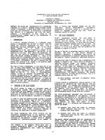
disease. The 2 year survival was calculated at 88%, the 5
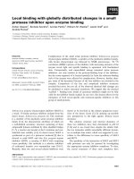
year survival was calculated 58% (figure 1).
Histopathology
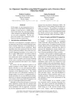
The firm, well vascularized tumors (figure 2) depicted a
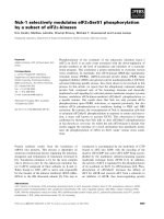
characteristic alveolar (or pseudo-alveolar) growth pat-
tern (figure 3); the tumor cells being epitheliod and polyg-
onal with eosinophilic cytoplasm, vesicular nuclei and
prominent nucleoli. Rhomboid crystalline inclusions
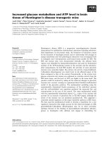
could be detected cytoplasmatically. A vascular invasion
as a typical finding in alveolar soft part sarcoma was evi-
dent in 5 of 11 tumors (figure 4).
In general, most of the tumors showed no or only faint
coagulation (tumor) necrosis. Mitotic activity was low (up
to 3 mitoses in 10 high power fields), except one case
(patient 7) which was characterized by 16 mitoses in 10
high power fields, including atypical ones.
Two patients (patient 1, 5) had been treated systemically
before tumor resection; these tumors showed regression
ranging from 30–40% vital tumor tissue (patient 5: regres-
sion grade IV according to Salzer-Kuntschik) to more than
70% vital tumor (patient 1: regression grade V according
to Salzer-Kuntschik).
Crystalline inclusions could be detected in 5 of 11 alveo-
lar soft part sarcomas. Using immunohistochemistry, var-
iable immunohistochemical reactions could be observed
with reactivity for S-100 in 2 of 5 examined tumors, focal
reactivity for desmin in 4 of 6 tumors, reactivity for actin
in 1 of 7 tumors, and weak reactivity for NSE in 1 exam-
ined ASPS specimen. No reactivity could be obtained in
any tumor for cytokeratins, HMB 45, myogenin, CD 31,
CD 34, factor VIII, and synaptophysin.
Discussion
Histopathology
The tumor harbors a specific chromosomal translocation
at der(17)t(X;17)(p11;q25), often with a loss of the chro-
mosomal region 17q25 (2,5). This translocation results in
a fusion of TFE-3-gene (coding for a transcription factor)
on chromosome Xp11 and the ASPL (RCC17)-gene of
chromosome 17q25. The resulting ASPL-TFE3-oncopro-
tein causes activation of aberrant transcription [3]. A
strong positive immunoreaction against TFE3 (nuclear
staining) is characteristic for alveolar soft part sarcoma
[1]. Other chromosomal abnormalities like trisomy 7,
monosomy 8 and monosomy 18 have also been
described [6].
As differential diagnosis especially, metastasis of renal cell
carcinoma has to be considered: this possibility can be
Table 1: Summarized tumor data.
Patient Localisation Size in cm TNM classification Comments Status
1 left thigh, intramuscular 10 × 8 × 4 ypT2 N0 M0 70% vital tumor in resection specimen: none
responder
alive, NED
2 right thigh, intramuscular 5.5 × 4 pT2 N0 M0 2 tumor free lymph nodes in primary
specimen, lymphangiosarcomatis
DOD
3 right upper arm, intramuscular 5.6 × 4.7 × 3.3 pT2 N0 M0 - DOD
4 left thigh, intramuscular 8 × 6,5 × 5 pT2 N0 M0 3 tumor free lymph nodes in primary
specimen, second expert opinion by Dr.
Mentzel (Friedrichshafen, Germany) and
Prof. Fletcher (Boston, USA)
alive, NED
5 right lower leg, intramuscular 13,5 × 9,5 × 8,3 ypT2b N0 M0 - alive, NED
6 right dorsum, intramuscular 3,5 × 2,9 × 1.9 pT1 N0 M0 - alive, NED
7 left thigh, intramuscular 10.5 × 7.3 × 5.3 pT2 N0 M0 - alive, NED
8 left heel, subfascial 4 × 3 × 2.5 pT1 N0 M0 - DOD
9 right lower leg, intramuscular 3.5 × 2.7 × 1.7 pT1b N0 M0 Inguinal dissection because of suspicious
lymph nodes, ruling out lymph node
metastasis, second expert opinion by Prof.
Katenkamp, Jena (Germany)
alive, NED
10 left thorax, subscapular, intramuscular 7.8 × 2.5 × 1.3 pT2b N0 M0 - alive, NED
11 right forearm, intramuscular 2.9 × 2.4 × 2 pT1b N0 M0 - alive, NED
World Journal of Surgical Oncology 2008, 6:71 />Page 4 of 7
(page number not for citation purposes)
excluded by history and further clinical and radiological
examination, whereby metastasic renal cell carcinoma
usually is positive for cytokeratin and vimentine in immu-
nohistochemistry. Other differential diagnoses for the his-
topathologist include paraganglioma, adrenal cortical
carcinoma, hepatocellular carcinoma, alveolar rhab-
domyosarcoma, malignant melanoma and granular cell
tumor. Paraganglioma may show alveolar structures as
well, but, in contrast, is positive by immunohistochemis-
try for chromogranin and synaptophysin (neuroendo-
Table 2: Summarized patient's and treatment data
Patient Age/Sex Untreated
tumor
growth
Initial
procedure
Neodjuvant
treatment for
primary
Definite
procedure
Ajuvant
radiation to
primary
Local and
recurrence
treatment
Metastasis and
treatment
Status
1 30/F 3 months incisional
biopsy
etoposide,
vincristine,
adriamycin,
ifosfamide
R0
resection
68 Gy - - alive, NED
2 40/M 2 years fine needle
biopsy
R0
resection
60 Gy - right lower leg: R0
resection +
radiation 60 Gy lung:
R2 resection of 21
metastases brain:
radiation 2 × 30Gy
DOD
3 21/M 3 years incisional
biopsy
R0
resection
60 Gy +/
adriamycin,
ifosfamide
lung: adriamycin,
ifosfamide brain:
radiation 30 Gy
liver: -
DOD
4 26/F 70 years incisional
biopsy
R0
resection
no - - alive, NED
5 30/M 20 years incisional
biopsy
ILP: Melphalan
+ TNF-alpha
R0
resection
60 Gy - - alive, NED
619/F3 years R1
resection
R0
resection
no - - alive, NED
7 30/M 2 years incisional
biopsy
R0
resection
65 Gy - - alive, NED
8 48/M n/a R1
resection
R1
resection
no +/R0
resection
lung: epirubicin,
ifosfamide brain: R1
resection liver: 5-
FU, cisplatin
DOD
9 24/M 6 months R1
resection
R0
resection
66 Gy - no alive, NED
10 49/M 6 months R1
resection
R0
resection
66 Gy - no alive, NED
11 34/F 1 month incisional
biopsy
R0
resection
70,4 Gy - no alive, NED
(F: female, M: male, R0-resection: complete resection with free margins, R1-resection: resection with microscopically positive margins, R2-
resection: tumor masses remaining in situ, ILP: isolated limb perfusion, NED: no evidence of disease, DOD: died of disease)
Table 3: Time elapsed: Time is calculated from primary diagnosis.
Patient Time to metastasis
(months)
Time to local recurrence
(months)
Progression free survival
(months)
Follow-up (months) Time to death (months)
1 - - 156 156 n/a
2 9 - 9 48 48
312 29 12 79 79
4- - 43 43 n/a
5- - 99 99 n/a
6- - 77 77 n/a
7 - - 125 125 n/a
87 11 7 97 97
9 - - 108 108 n/a
10 - - 25 25 n/a
11 - - 5 5 n/a
World Journal of Surgical Oncology 2008, 6:71 />Page 5 of 7
(page number not for citation purposes)
Overall survival after primary diagnosis of ASPSFigure 1
Overall survival after primary diagnosis of ASPS. The tick marks indicate the last follow-up.
Macroscopic appearance of an alveolar soft part sarcoma, showing a quite solid tumor mass located within the soft tis-suesFigure 2
Macroscopic appearance of an alveolar soft part sarcoma,
showing a quite solid tumor mass located within the soft tis-
sues. Necrosis is not a striking macroscopic appearance of
this sarcoma.
Histologic appearance of an alveolar soft part sarcoma: tumor growth characterized by a central loss of cohesion in cell lobules, depicting a pseudoalveolar archictectureFigure 3
Histologic appearance of an alveolar soft part sarcoma:
tumor growth characterized by a central loss of cohesion in
cell lobules, depicting a pseudoalveolar archictecture. Round
tumor cell lobules, delineated by fibrovascular septa, contain-
ing round to polygonally shaped tumor cells with eosinophilic
cytoplasm, vesicular nuclei and prominent nucleoli.
World Journal of Surgical Oncology 2008, 6:71 />Page 6 of 7
(page number not for citation purposes)
crine markers) and lacks crystalline cytoplasmatic
inclusions. Metastases of adrenal cortical carcinoma and
hepatocellular carcinoma may be excluded by means of
immunohistochemistry (melan-A-cross reactivity in adre-
nal cortical carcinoma, HepPar in hepatocellular carci-
noma) [1]. Alveolar rhabdomyosarcoma exhibits a
skeletal muscle differentiation which can be proven by
immunohistochemical stains for skeletal-muscle specific
markers (myogenin, Myo D1). Malignant melanomas
(especially as metastasis) have a growth pattern reminis-
cent of alveolar structures, but are positive for melanocytic
markers like S-100 and HMB 45. A granular cell tumor
does not include crystalline bundles and, in contrast to
ASPS, is positive for S-100 protein.
Due to the heterogeneity of the immunohistochemical
features and the small number of patients, we could not
detect a true correlation of histopathological and clinical
behavior in our series.
As in other studies with 0.7% of the 1597 soft tissue sar-
comas, ASPS represented a very small subgroup. In con-
trast to previous reports [1,4,5,10], all of our patients were
free of lymphatic or detectable hematogenic metastasis at
the time of diagnosis and were treated in curative intent.
In accordance with the literature, the lesions had been
growing to large tumor masses in most of our patients
without causing specific symptoms for an extended time
and were mostly located in the deep tissues of the thigh
[3-5]. The fact that all tumors were located within or close
to muscle may support the thesis that the origin of ASPS
is myogenic [11,12], although, due to recent genetic find-
ings, a myogenic line of differentiation seems unlikely [7].
Interestingly and contradictory to other reports [1,3,13]
the size of the primary at time of diagnosis does not seem
to affect the outcome because all three patients who died
from their disease had primaries below the average tumor
size in our series.
In this study, the age related gender ratio with a male pre-
ponderance in older patients -previously noted by Portera
[5] and Ordonez [14]) – was also observed. What is more,
all patients with an unfavorable outcome were male.
Complete resection of the primary, as well as the recurrent
tumor manifestation, seems to be essential for local con-
trol [3-5], but cannot prevent distant metastasis in case of
early spreading as seen in patient 2 and 3. Recurrent dis-
ease – that can be prevented by sufficient primary treat-
ment in about 90% of the cases [1,3,5] – itself seems to be
a negative prognostic factor, probably indicating an
aggressive tumor biology because all patients with recur-
rent disease also had distant metastases. Interestingly the
metastases occurred before the local recurrence. In these
cases, the preferred sequence of manifestation was lung,
brain and liver. No isolated brain metastases were
observed, whereas there are case reports describing those
[15-17] and the large series of 102 patients by Lieberman
et. al. mentioned four such cases [4]. As previously
reported, lung and brain are the most common sites of
metastases [1,16,18] which is why X-ray or CT scans of the
lung should be included in the follow-up examinations
for ASPS [19]. Considering metastases to the brain almost
exclusively occur synchronous or subsequent to pulmo-
nary metastasis, the value of routine intracranial imaging
is doubtful and we recommend it only in cases with neu-
rologic symptoms. For the same reason, the same
approach is recommended for abdominal MRI or CT
scans for liver metastases (with the exception of ultra-
sound) that are performed at routine follow-ups. Due to
the limited number of patients with ASPS, prospective
studies would have to span decades to gather a significant
collective of patients; therefore, it is not possible to com-
ment meaningfully on a possible benefit of neoadjuvant
or adjuvant therapy. In the absence of other data, how-
ever, it seems to be justified to recommend postoperative
radiation to potentially increase local control rates as in
other soft tissue sarcomas. The theory that radiation as
single therapy is beneficial for metastatic disease of the
brain must be questioned since it seems to be able to slow
down growth at best, but failed to induce significant
tumor shrinkage in our patients. In accordance with pre-
vious medical studies that described limited effects of
radiation and chemotherapy on metastatic disease,
patients who received such treatment for metastatic dis-
ease did not respond in our series[1,3-5,20]. The only
patient who received neoadjuvant chemotherapy for the
primary also did not respond to it, what was documented
by more than 70% of the tumor remaining viable in the
Poorly differentiated case of alveolar soft part sarcoma with overall lobular structure of tumor cell arrangementFigure 4
Poorly differentiated case of alveolar soft part sarcoma with
overall lobular structure of tumor cell arrangement.
World Journal of Surgical Oncology 2008, 6:71 />Page 7 of 7
(page number not for citation purposes)
resection specimen. Only one patient who was treated
with ILP with TNF-alpha and melphalan responded to
that treatment, with a necrosis of 70% of the tumor.
The long term outcome of patients with localized ASPS
with a 5-year disease free survival rate of 71% [5], 67%
[21], and 60% [4], respectively, and a 5-year actuarial sur-
vival rate of 88% [5] is considered relatively favorable.
From the time of diagnosis of metastatic disease, only
about 10% of the patients survive longer than 5 years
[4,5]. The clinical courses observed on our series agree
with these findings. Although the progression free interval
from primary diagnosis to the development of local recur-
rence or metastasis in three patients was short (7, 9, 12
months), patients lived with metastatic disease but had an
acceptable quality of life for a considerable time
(39,54,90 months). This prolonged survival with meta-
static disease has previously been observed by other
authors as well [4,5]. If this long survival time is due to the
multimodal therapy or in spite of it cannot be determined
thus far.
Conclusion
In case of acceptable patients' condition and justifiable
morbidity, our approach to recurrent disease or resectable
solitary metastases continues to be surgical excision fol-
lowed by radiation with the aim to improve overall qual-
ity of life. Chemotherapy is only recommended in
selected cases with disseminated disease. Isolated limb
perfusion may represent an additional therapeutic option
in order to prepare better resection conditions and
improve local control in extremity ASPS.
Competing interests
The authors declare that they have no competing interests.
Authors' contributions
AD conceptualized the study, gathered the data and wrote
the manuscript. CK performed the histopathological eval-
uation and interpretation of the data. JH analyzed and
interpreted the data. OG acquired and weighed the data.
DT drafted and revised the manuscript. LS reviewed the
literature and analyzed the data. HS conceptualized and
supervised the process. He gave final approval for publica-
tion. ML initiated the study and drafted the manuscript.
All authors read and approved the final manuscript.
Acknowledgements
Written informed consent was taken from all patients for publication of
their case records
We thank Dr. Mentzel, Friedrichshafen (Germany), Professor Katenkamp,
Jena (Germany) and Professor Fletcher, Boston (USA) for their expert sec-
ond opinion and Amanda Daigeler for her formal English revision of the
manuscript.
References
1. Ogose A, Morita T, Hotta T, Kobayashi H, Otsuka H, Hirata Y, Yosh-
ida S: Brain metastases in musculoskeletal sarcomas. Jpn J Clin
Oncol 1999, 29(5):245-247.
2. Lieberman PH, Foote FW Jr., Stewart FW, Berg JW: Alveolar soft-
part sarcoma. Jama 1966, 198(10):1047-1051.
3. Casanova M, Ferrari A, Bisogno G, Cecchetto G, Basso E, De Ber-
nardi B, Indolfi P, Fossati Bellani F, Carli M: Alveolar soft part sar-
coma in children and adolescents: A report from the Soft-
Tissue Sarcoma Italian Cooperative Group. Ann Oncol 2000,
11(11):1445-1449.
4. Lieberman PH, Brennan MF, Kimmel M, Erlandson RA, Garin-Chesa
P, Flehinger BY: Alveolar soft-part sarcoma. A clinico-patho-
logic study of half a century. Cancer 1989, 63(1):1-13.
5. Portera CA Jr., Ho V, Patel SR, Hunt KK, Feig BW, Respondek PM,
Yasko AW, Benjamin RS, Pollock RE, Pisters PW: Alveolar soft part
sarcoma: clinical course and patterns of metastasis in 70
patients treated at a single institution. Cancer 2001,
91(3):585-591.
6. Joyama S, Ueda T, Shimizu K, Kudawara I, Mano M, Funai H, Take-
mura K, Yoshikawa H: Chromosome rearrangement at 17q25
and xp11.2 in alveolar soft-part sarcoma: A case report and
review of the literature. Cancer 1999, 86(7):1246-1250.
7. Folpe AL, Deyrup AT: Alveolar soft-part sarcoma: a review and
update. J Clin Pathol 2006, 59(11):1127-1132.
8. Gomez JA, Amin MB, Ro JY, Linden MD, Lee MW, Zarbo RJ: Immu-
nohistochemical profile of myogenin and MyoD1 does not
support skeletal muscle lineage in alveolar soft part sar-
coma. Arch Pathol Lab Med 1999, 123(6):503-507.
9. Ladanyi M, Lui MY, Antonescu CR, Krause-Boehm A, Meindl A,
Argani P, Healey JH, Ueda T, Yoshikawa H, Meloni-Ehrig A, Sorensen
PH, Mertens F, Mandahl N, van den Berghe H, Sciot R, Cin PD, Bridge
J: The der(17)t(X;17)(p11;q25) of human alveolar soft part
sarcoma fuses the TFE3 transcription factor gene to ASPL,
a novel gene at 17q25. Oncogene 2001, 20(1):48-57.
10. Anderson ME, Hornicek FJ, Gebhardt MC, Raskin KA, Mankin HJ:
Alveolar soft part sarcoma: a rare and enigmatic entity. Clin
Orthop Relat Res 2005, 438:144-148.
11. Denk H, Krepler R, Artlieb U, Gabbiani G, Rungger-Brandle E, Leon-
cini P, Franke WW: Proteins of intermediate filaments. An
immunohistochemical and biochemical approach to the
classification of soft tissue tumors. Am J Pathol 1983,
110(2):193-208.
12. Mukai M, Torikata C, Iri H, Mikata A, Hanaoka H, Kato K, Kageyama
K: Histogenesis of alveolar soft part sarcoma. An immuno-
histochemical and biochemical study. Am J Surg Pathol 1986,
10(3):212-218.
13. Evans HL: Alveolar soft-part sarcoma. A study of 13 typical
examples and one with a histologically atypical component.
Cancer 1985, 55(4):912-917.
14. Ordonez NG: Alveolar soft part sarcoma: a review and
update. Adv Anat Pathol 1999, 6(3):125-139.
15. Bindal RK, Sawaya RE, Leavens ME, Taylor SH, Guinee VF: Sarcoma
metastatic to the brain: results of surgical treatment. Neuro-
surgery 1994, 35(2):185-90; discussion 190-1.
16. Salvati M, Cervoni L, Caruso R, Gagliardi FM, Delfini R: Sarcoma
metastatic to the brain: a series of 15 cases. Surg Neurol 1998,
49(4):441-444.
17. Wang CH, Lee N, Lee LS: Successful treatment for solitary
brain metastasis from alveolar soft part sarcoma. J Neurooncol
1995, 25(2):161-166.
18. Lewis AJ: Sarcoma metastatic to the brain. Cancer 1988,
61(3):593-601.
19. Pollack R, Brennan M, Lawrence W Jr.: Society of Surgical Oncol-
ogy practice guidelines. Soft-tissue sarcoma surgical practice
guidelines. Oncology (Williston Park) 1997, 11(9):1327-1332.
20. Pappo AS, Parham DM, Cain A, Luo X, Bowman LC, Furman WL, Rao
BN, Pratt CB: Alveolar soft part sarcoma in children and ado-
lescents: clinical features and outcome of 11 patients. Med
Pediatr Oncol 1996,
26(2):81-84.
21. Auerbach HE, Brooks JJ: Alveolar soft part sarcoma. A clinico-
pathologic and immunohistochemical study. Cancer 1987,
60(1):66-73.