Báo cáo khoa học: "Solitary fibrous tumor of the pleura presenting with syncope episodes when coughing" docx
Bạn đang xem bản rút gọn của tài liệu. Xem và tải ngay bản đầy đủ của tài liệu tại đây (2.88 MB, 5 trang )
BioMed Central
Page 1 of 5
(page number not for citation purposes)
World Journal of Surgical Oncology
Open Access
Case report
Solitary fibrous tumor of the pleura presenting with syncope
episodes when coughing
Luigi Santambrogio
1
, Mario Nosotti
1
, Alessandro Palleschi
1
, Lorenzo Rosso
1
,
Davide Tosi
1
, Matilde De Simone
2
, Michele M Ciulla
3
, Marco Maggioni
4
and
Ugo Cioffi*
2
Address:
1
Department of Surgery, Thoracic Unit, Fondazione Ospedale Maggiore Policlinico, Mangiagalli e Regina Elena, IRCCS, Milan, Italy,
2
Department of Surgery, Fondazione Ospedale Maggiore Policlinico, Mangiagalli e Regina Elena, IRCCS, Milan, Italy,
3
Istituto di Medicina
Cardiovascolare, Centro di Fisiologia Clinica e Ipertensione, University of Milan, Fondazione Ospedale Maggiore Policlinico, Mangiagalli e Regina
Elena, IRCCS, Milan, Italy and
4
A.O. San Paolo, U.O. Anatomia Patologica, Milan, Italy
Email: Luigi Santambrogio - ; Mario Nosotti - ;
Alessandro Palleschi - ; Lorenzo Rosso - ;
Davide Tosi - ; Matilde De Simone - ; Michele M Ciulla - ;
Marco Maggioni - ; Ugo Cioffi* -
* Corresponding author
Abstract
Background: Solitary fibrous tumor of the pleura is a rarely encountered clinical entity which may
have different clinical pictures. Although the majority of these neoplasms have a benign course, the
malignant form has also been reported.
Case presentation: We herein describe a case of 72 year-old man with head, facial, and thoracic
traumas caused by neurally-mediated situational syncope when coughing. The diagnostic work-up
including chest x-ray, CT and PET, revealed a large solitary mass of the left hemithorax. Radical
surgical resection of the mass was performed through a left lateral thoracotomy and completed
with a wedge resection of the lingula. Hystological examination of the surgical specimen showed an
encapsulated mass measuring 12 × 11.5 × 6 cm consistent with a solitary fibrous tumor of the
pleura. It's surgical removal definitively resolved the neurologic manifestations. The patient had no
postoperative complications. At two years follow-up the patient is free from recurrence and
without clinical manifestations.
Conclusion: In our case its resection definitively resolved the episodes of situational syncope due,
in our opinion, to the large thoracic mass compressing the phrenic nerve
Background
First described by Klemperer and Rabin [1], the solitary
fibrous tumor of the pleura (SFTP) is a localized benign
neoplasm arising from the submesothelial mesenchymal
layer [2] even if malignant forms have also been described
[3]. With about 800 cases reported in the world literature,
this rare entity contrast with the primary diffuse pleural
mesothelioma that have an incidence of 3000 new cases
every year in the USA [4]. In over half of patients the
tumor is asymptomatic, but if symptoms occur then chest
pain, cough and dyspnea are the most common com-
plaints. Complete en bloc surgical resection is the treat-
Published: 19 August 2008
World Journal of Surgical Oncology 2008, 6:86 doi:10.1186/1477-7819-6-86
Received: 11 April 2008
Accepted: 19 August 2008
This article is available from: />© 2008 Santambrogio et al; licensee BioMed Central Ltd.
This is an Open Access article distributed under the terms of the Creative Commons Attribution License ( />),
which permits unrestricted use, distribution, and reproduction in any medium, provided the original work is properly cited.
World Journal of Surgical Oncology 2008, 6:86 />Page 2 of 5
(page number not for citation purposes)
ment of choice for these neoplasms offering a cure in all
patients with benign form even if tumor recurrence may
occur also in tumors with benign histological features
[4,5]. We describe an unreported case of SFTP, to our
knowledge, manifesting with syncope episodes when
coughing.
Case presentation
A 72 year-old man was admitted to the hospital for head
injury, facial and left hemithorax contusions. The patient
referred to had fainted after coughing; the same symp-
toms had occurred six and three months earlier. Divertic-
ulosis of the colon was the only disease reported by the
patient in his medical history. He denied smoking, drug
or alcohol abuse. Physical examination showed dullness
to percussion and decreased breath sound in the affected
hemithorax. The neurological examination was negative.
Blood pressure was 170/80 mmHg, heart rate was 90
beats/minute and rhythmic. Laboratory findings, arterial
gas analysis, electrocardiogram, and brain computed tom-
ography were negative.
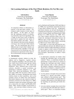
A chest x-ray revealed fractures of three left ribs plus a large
medium-basal opacity on the left hemithorax (Figure 1a).
Computed tomography (CT) of the thorax confirmed the
presence of a well-delineated, homogeneous, solid mass
of 11 × 8 cm, extending for about 10 cm on the vertical
axis. The mass presented a mild enhancement after con-
trast injection and calcifications in the basal part. It was
close to the chest wall, adjacent to the left pulmonary
artery, pulmonary artery trunk, and left ventricle with no
signs of infiltration (Figure 1b). Bronchoscopy showed an
insignificant bleeding from the upper left bronchus. Posi-
tron emission tomography (PET) revealed a mild positiv-
ity of the lesion (Figure 2). Echocardiogram, Holter ECG
monitoring, and carotid Doppler ultrasonography were
negative. With suspected diagnosis of SFTP, the patient
underwent surgery. Through a left lateral thoracotomy,
the neoplasm was carefully isolated, and its origin from
the visceral pleura of the pulmonary lingula segment
became evident. The adhesions with the phrenic nerve
were cut preserving the nerve integrity. The mass excision
was performed with clear surgical margins and completed
with a wedge resection of the lingula. The postoperative
course was uneventful, a good re-expansion of the left
lung was obtained, and the patient was discharged on the
fifth postoperative day.
Pathological examination showed a 12 × 11, 5 × 6 cm
encapsulated tumor mass (Figure 3a), whitish in color,
with whorled appearance and calcification on cut section
(Fig. 3b). Microscopic examination showed fibroblast-
Left panel: chest x-ray showing a large opacity on the left sideFigure 1
Left panel: chest x-ray showing a large opacity on the left side. Right panel: the CT scan of the chest showing a solid mass of 11
× 8 cm in the left hemi thorax, with vertical extension of 10 cm, mild enhancement after contrast medium infusion and some
calcifications in the basal part (asterisk). The lesion is in close relation with chest wall, left pulmonary artery (LPA) and pulmo-
nary trunk (PT), without signs of local infiltration.
World Journal of Surgical Oncology 2008, 6:86 />Page 3 of 5
(page number not for citation purposes)
like structures within the collagen (Figure 4). The diagno-
sis of benign SFTP was confirmed by mmunohistochemi-
cal analysis (CD34+, BCL-2+, SMA-, S100-).
After two years of follow-up the patient is in good clinical
condition without recurrence of disease or clinical symp-
toms.
Discussion
It is well known that solitary fibrous tumors of the pleura
are incidentally discovered during chest x-ray examina-
tion because these neoplasms often have a silent clinical
course for several years [4,6]. It has been described in all
ages, but the peak of incidence is in the sixth and seventh
decades of life [1]. The larger the tumor, the more likely it
is that there will be symptoms [2,4]. Systemic symptoms
such as weight loss, nocturnal sweating, chills, weakness,
digital clubbing, hypertrophic osteoarthropathy, and
hypoglycemia have also been reported [6,7]. Hyper-
trophic osteoarthropathy (Pierre Marie-Bamberg syn-
drome) [6-8], is related to the abnormal production of
hyaluronic acid by tumor cells and affect up to 20% of
patients. In less than 5% of cases, SFTP can secrete insulin-
like growth factor II which causes refractory hypoglycemia
[1,7]. Sometimes, large tumors might present an unusual
onset, such as the case of Shaker and et al, [8] in which a
woman with leg edema and dyspnea caused by a large
SFTP compressing the right atrium and the inferior vena
cava is described. In our case, the large tumor presented
with episodes of situational syncope when coughing. Sit-
uational syncope is a neurally-mediated syncope related
to a reflex response that, when triggered, determines
vasodilatation and bradycardia. Neurally-mediated syn-
cope is usually classified as vasovagal (common faint), or
situational [9]. Suggestive for vasovagal syncope are a long
history of syncope, a youthful age, a sudden and unpleas-
ant sight, pain or sound, prolonged standing in hot and/
Moderate activity of the mass on Positron Emission Tomog-raphy studyFigure 2
Moderate activity of the mass on Positron Emission Tomog-
raphy study.
Left panel: surgical specimen with detail of the wedge resection of the lingulaFigure 3
Left panel: surgical specimen with detail of the wedge resection of the lingula. Right panel: solitary fibrous tumor of the pleura,
whorled fibrous tissue is evident on the cut section.
World Journal of Surgical Oncology 2008, 6:86 />Page 4 of 5
(page number not for citation purposes)
or crowded places. It often associates with nausea and
vomiting. Situational syncope is diagnosed if syncope
occurs during or after urination, defecation, cough or
swallowing [9]. In our case the syncope occurred immedi-
ately after coughing, without nausea and vomiting; in
addition, the patient was old and reported a trauma. We,
therefore, hypothesize that coughing, due to the stimula-
tion of the phrenic nerve, resulted in a high intrathoracic
pressure producing an exaggerated Valsalva responce that
decreased venous return and, consequently, cardiac out-
put. At this regard it should be noticed that the accidental
phrenic nerve injury produces cough and dyspnea and
this evenience is well documented during right atrial cath-
eterization procedures [10]. All these details ruled out the
possibility of a common faint, and consequently a diag-
nosis of situational syncope when coughing was made.
The negative results of the cardiovascular tests associated
to the presence of a large thoracic mass convinced us to
consider the cough syncope related to the stimulation of
the phrenic nerve by the neoplasm. In fact, after surgical
removal of the tumor, the patient is free from syncope epi-
sodes confirming the direct implication of the solitary
fibrous tumor of the pleura in the neurologic manifesta-
tions.
Conclusion
Generally SFTP is a localized, benign tumor which may
have different clinical pictures. It is curable using a careful
and complete resection, provided that the surgical mar-
gins are free from neoplastic cells. In our case its resection
definitively resolved the episodes of situational syncope
due, in our opinion, to the large thoracic mass compress-
ing the phrenic nerve.
Consent
Written and informed consent was obtained from the
patient for publication of this case report and any accom-
panying images. A copy of the written consent is available
for review by the Editor-in-Chief of this journal.
Competing interests
The authors declare that they have no competing interests.
Authors' contributions
LS conceived the idea, did supervision of manuscript
preparation and proof reading initiated treatment, did
surgical procedures and approved the final version of the
paper. MN, AP, LR, DT proof reading, initiated treatment,
did surgical procedures. UC, MDS wrote the manuscript
Tumor consists of elongated cells that display a storiform pattern of growth and abundant stromal collagen (hematoxylin & eosin stain; magnification 100 ×)Figure 4
Tumor consists of elongated cells that display a storiform pattern of growth and abundant stromal collagen (hematoxylin &
eosin stain; magnification 100 ×). At higher magnification, tumor cells appear of small size, spindle, with no cytologic atypia
(insert; magnification 400 ×).
Publish with BioMed Central and every
scientist can read your work free of charge
"BioMed Central will be the most significant development for
disseminating the results of biomedical research in our lifetime."
Sir Paul Nurse, Cancer Research UK
Your research papers will be:
available free of charge to the entire biomedical community
peer reviewed and published immediately upon acceptance
cited in PubMed and archived on PubMed Central
yours — you keep the copyright
Submit your manuscript here:
/>BioMedcentral
World Journal of Surgical Oncology 2008, 6:86 />Page 5 of 5
(page number not for citation purposes)
and carried out literature review; MMC contributed to
data management and preparing of the manuscript. All
authors read and approved the final manuscript.
References
1. de Perrott M, Fischer S, Brundler MA, Sekine Y, Keshavjee S: Soli-
tary fibrous tumor of the pleura. Ann Thorac Surg 2002,
74:285-293.
2. Robinson LA: Solitary fibrous tumor of the pleura. Cancer Con-
trol 2006, 13:264-269.
3. Zhang H, Lucas DR, Pass HI, Che M: Disseminated malignant sol-
itary fibrous tumor of the pleura. Pathol Int 2004, 54:111-115.
4. Magdeleinat P, Alifano M, Petino A, Le Rochais JP, Dulmet E, Galateau
F, Icard P, Regnard JF: Solitary fibrous tumors of the pleura:
clinical characteristics, surgical treatment and outcome. Eur
J Cadio-thorac Surg 2002, 21:1087-1093.
5. Carretta A, Bandiera A, Melloni G, Ciriaco P, Arrigoni G, Rizzo N,
Negri G, Zannini P: Solitary fibrous tumors of the pleura:
Immunohistochemical analysis and evaluation of prognostic
factors after surgical treatment. J Surg Oncol 2006, 94:40-44.
6. Cardillo G, Facciolo F, Cavazzana AO, Capece G, Gasparri R, Martelli
M: Localized (solitary) fibrous tumors of the pleura: an anal-
ysis of 55 patients. Ann Thorac Surg 2000, 70:1808-1812.
7. Cole FH Jr, Ellis RA, Goodman RC, Weber BC, Courington DP:
Benign fibrous pleural tumor with elevation of insulin-like
growth factor and hypoglycemia. South Med J 1990, 83:690-694.
8. Shaker W, Meatchi T, Dusser D, Riquet M: An unusual presenta-
tion of solitary fibrous tumour of the pleura: right atrium
and inferior vena cava compression. Eur J Cadio-thorac Surg 2002,
22:640-642.
9. Brignole M: Neurally-mediated syncope. It Heart J 2005,
6:248-255.
10. Sacher F, Monahan KH, Thomas SP, Davidson N, Adragao P, Sanders
P, Hocini M, Takahashi Y, Rotter M, Rostock T, Hsu LF, Clémenty J,
Haïssaguerre M, Ross DL, Packer DL, Jaïs P: Phrenic nerve injury
after atrial fibrillation catheter ablation: characterization
and outcome in a multicenter study. J Am Coll Cardiol 2006,
47(12):2498-503.