Báo cáo khoa học: "Acute liver failure due to primary angiosarcoma: A case report and review of literature" doc
Bạn đang xem bản rút gọn của tài liệu. Xem và tải ngay bản đầy đủ của tài liệu tại đây (774.18 KB, 5 trang )
BioMed Central
Page 1 of 5
(page number not for citation purposes)
World Journal of Surgical Oncology
Open Access
Case report
Acute liver failure due to primary angiosarcoma: A case report and
review of literature
Chandra S Bhati, Anand N Bhatt, Graham Starkey, Stefan G Hubscher and
Simon R Bramhall*
Address: Liver Unit, Queen Elizabeth Hospital, Birmingham, UK
Email: Chandra S Bhati - ; Anand N Bhatt - ; Graham Starkey - ;
Stefan G Hubscher - ; Simon R Bramhall* -
* Corresponding author
Abstract
Background: Hepatic angiosarcoma is a primary sarcoma of the liver, accounting for only 2% of
all primary hepatic malignancies. Acute liver failure is an extremely rare presentation of a primary
liver tumour.
Case presentation: We report a case of a seventy year-old man who presented with a very short
period of jaundice leading to fulminant hepatic failure (FHF). On further investigation he was found
to have primary angiosarcoma of liver.
Conclusion: The treatment outcomes for hepatic angiosarcoma are poor, we discuss the options
available and the need for prompt investigation and establishment of a diagnosis
Background
Hepatic malignancies include primary hepatocellular car-
cinoma, metastases and primary or metastatic sarcomas
[1]. Hepatic angiosarcoma is a primary sarcoma of the
liver which accounts for only 2% of all primary hepatic
malignancies [2-5]. Angiosarcoma is associated with envi-
ronmental or occupational exposure to carcinogens (tho-
rium dioxide, vinyl chloride, arsenic and radiation). There
is also an association with hemochromatosis and von
Recklinghausen disease [1,2,4]. In most cases of primary
hepatic angiosarcoma, no obvious risk factor can be iden-
tified.
The most common causes of fulminant hepatic failure
(FHF) are drug toxicity and sero-negative hepatitis [6];
rarer causes include Bud-Chiari syndrome and acute Wil-
son's disease. FHF can also develop very rarely as a conse-
quence of primary or metastatic liver tumour, this
generally occurs as a result of massive neoplastic infiltra-
tion of the hepatic sinusoids leading to secondary necrosis
of hepatocytes [7]. Rowbotham et al reported 4020 cases
of FHF, malignant infiltration accounted for only 0.44%
(18 cases) [8].
There have been a number of case series reporting FHF
secondary to infiltration of the liver by malignant cells [7-
15], haematological malignancies are the most common
[7-10]. Other infiltrative metastatic malignancies that
rarely cause FHF include adenocarcinoma, melanoma,
and anaplastic tumours [11-15]. Although hepatic dys-
function due to malignancy such as hepatocellular carci-
noma or metastatic infiltration is common, acute liver
failure in these cases is rare. We report a case of primary
angiosarcoma of the liver which presented with FHF.
Published: 30 September 2008
World Journal of Surgical Oncology 2008, 6:104 doi:10.1186/1477-7819-6-104
Received: 28 June 2008
Accepted: 30 September 2008
This article is available from: />© 2008 Bhati et al; licensee BioMed Central Ltd.
This is an Open Access article distributed under the terms of the Creative Commons Attribution License ( />),
which permits unrestricted use, distribution, and reproduction in any medium, provided the original work is properly cited.
World Journal of Surgical Oncology 2008, 6:104 />Page 2 of 5
(page number not for citation purposes)
Case presentation
A seventy year old Caucasian male, who had no signifi-
cant previous medical history, was admitted to a local
hospital with a history of sudden onset jaundice and
weight loss. There was no previous history of jaundice or
hepatitis. There was no significant history of alcohol in-
take or exposure to arsenic, vinyl chloride, or Thorotrast.
He never used any hepatotoxic or herbal medications and
his mother died of undiagnosed liver disease.
Upon examination the patient was jaundiced without
encephalopathy or focal neurological findings. He had
bilateral pedal oedema and hepatomegaly. The patient
did not have any other signs of liver failure. Liver function
tests at admission revealed a total bilirubin of 203 mmol/
dL (normal, 5–17 mmol/dL), aspartate aminotransferase
(AST) 52 IU/L (normal, 4–44 IU/L), alkaline phosphatase
170 IU/L (normal, 67–213 IU/L), albumin 2.0 g/dL, PT 22
seconds, APTT 51 seconds and platelets 113,000/cm
3
.
An urgent ultrasound scan demonstrated hepatomegaly
with significant liver paranchymal alteration. A subse-
quent contrast enhanced abdominal CT showed gross
replacement of liver with tumour tissue suggestive of a pri-
mary liver tumour (Figure 1). The patient was at this point
referred to our centre.
The patient's initial evaluation in our Unit showed further
derangement in the patients liver functions tests; total
bilirubin had risen to 401 mmol/dL, AST to 132 IU/L,
alkaline phosphatase to 370 IU/L and INR to 2.1. A local
review of his CT scan raised the possibility of angiosar-
coma. To confirm the diagnosis a transjugular biopsy was
arranged as the clotting abnormality had been resistant to
correction with fresh frozen plasma at the referring centre.
Before this could be carried out patient rapidly deterio-
rated after admission and became progressively encepha-
lopathic, consistent with FHF. He was treated
conservatively with dextrose and broad spectrum antibiot-
ics but deteriorated further and died two days after admis-
sion to the liver unit.
A post mortem liver biopsy was carried out confirming
initial suspicions that this was a primary angiosarcoma of
the liver. Microscopically, tumour was composed of
poorly cohesive cells, oval to spindle shaped with high
grade cytological atypia. The tumour had a sinusoidal
growth pattern surrounding clusters of hepatocytes form-
ing cholestatic rosettes (Figure 2a). Immunohistochemis-
try staining was strongly and diffusely positive for vascular
endothelial markers (CD31, CD34) (Figure 2b) and for
vimentin. Stains for the cytokeratins and hepatocyte spe-
cific antigen highlighted the presence of entrapped non
neoplastic hepatocyte and bile ducts. Staining for smooth
muscle actin appeared to be confined to areas of fibrotic
tissue.
Discussion
Angiosarcoma usually presents in late adulthood [2] with
abdominal discomfort, distension, weight loss, and
fatigue [4,16]. On examination, the patients may have
jaundice, hepatomegaly, and ascites [4,16,17]. Our
patient was admitted with similar symptoms. Fulminant
hepatic failure (FHF) is defined as liver disease that results
in encephalopathy within 28 days from the onset of jaun-
dice in a patient with no prior evidence of liver disease.
Presentation as FHF is rare, Table 1 shows published
reports of clinical presentation and treatment of angiosar-
coma in the current literature. In an adult FHF Study
Group; acetaminophen overdose (46%), drug toxicity
(11%) and hepatitis (10%) were found to be the most
common causes for liver failure [18]. There are case
reports where association of FHF with liver metastasis
from other malignancies have been reported [7-15].
The liver is commonly involved in metastatic disease, and
the degree of liver biochemistry derangement tends to
reflect the extent of parenchymal replacement with
tumour [19]. In this patient, liver function tests were only
slightly abnormal two weeks before development of FHF.
Although, alteration of liver function tests in these
patients is very common [20], liver failure is extremely
rare.
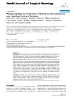
Abdominal CT scan showing complete replacement of liver parenchyma with liver tumourFigure 1
Abdominal CT scan showing complete replacement
of liver parenchyma with liver tumour.
World Journal of Surgical Oncology 2008, 6:104 />Page 3 of 5
(page number not for citation purposes)
CT scan is often diagnostic, demonstrating multiple
hypodense areas typical of angiosarcoma. Post contrast,
the lesions become partly or completely isodense com-
pared with normal hepatic tissue [1,21]. In our patient
liver parenchyma was completely replaced with tumour
tissue (Figure 1).
The mechanism of liver failure is multifactorial. Evidence
suggests a combination of hepatic ischaemia leading to
parenchymal infarction, vascular occlusion of portal vein
by tumour thrombi and nonocclusive infarction of liver
due to shock from secondary causes such as sepsis or car-
diac dysfunction plays an important role in these patients
[12,22]. In this patient, replacement of hepatocytes by
malignant cells, leading to secondary necrosis of hepato-
cytes played a significant role in development of liver fail-
ure.
Angiosarcoma has very limited treatment options, with-
out treatment the majority of patients die within 6
months of diagnosis [4]. Surgery has a limited role due to
the advanced stage at which these tumours present. Liver
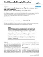
(A) Liver biopsy showing sinusoidal infiltration by pleomorphic spindle cells typical of hepatic angiosarcomaFigure 2
(A) Liver biopsy showing sinusoidal infiltration by pleomorphic spindle cells typical of hepatic angiosarcoma.
There is disruption of the normal trabecular architecture with hepatocyes forming glandular structures containing bile plugs
("cholestatic rosettes"). (B) Spindle cells are strongly immunoreactive for the vascular endothelial marker CD34. (A = Haema-
toxylin and eosin, B = immunoperoxidase).
World Journal of Surgical Oncology 2008, 6:104 />Page 4 of 5
(page number not for citation purposes)
transplantation is contraindicated, as patients who have
been transplanted incidentally have not shown any sur-
vival benefit. The data from European Liver Transplant
Registry on 17 patients who had undergone transplanta-
tion for angiosarcoma had a median survival of only 7
months [23]. Hepatic resection has been reported in
patients with limited disease but these results have also
been poor. There are very few published case reports with
good survival after liver resection (16 months [24] and 10
years [4]). The role of chemotherapy has been described
with very limited improvement in overall length of sur-
vival [25]. Treatment with new techniques like transcath-
eter arterial chemoembolization (TACE) techniques has
been described as a case report with very limited success
in overall survival improvement [26].
Conclusion
Our patient presented with mild hepatic failure that rap-
idly progressed to FHF. In the absence of a clear aetiology
for FHF primary liver tumour must be considered in the
differential diagnosis and a biopsy should be arranged to
reach definitive diagnosis.
Competing interests
The authors declare that they have no competing interests.
Authors' contributions
CSB – Contributions to case selection, analysis and draft-
ing the manuscript. ANB – Case analysis and initial draft-
ing of manuscript. GS – Contributions to conception,
arranging histopathology, revision of the manuscript.
SGH – Histopathology evaluation, further study of slides
and in depth analysis. SRB – Critical revision and final
approval of the version to be published. All authors read
and approved the final manuscript.
Consent
Written informed consent was obtained from the patient
for publication of this case report.
References
1. Buetow PC, Buck JL, Ros PR, Goodman ZD: Malignant vascular
tumours of the liver: radiologic-pathologic correlation. Radi-
ographics 1994, 14:153-166.
2. Mani H, Van Thiel DH: Mesenchymal tumors of the liver. Clin
Liver Dis 2001, 5(1):219-257.
3. Molina E, Hernandez A: Clinical manifestations of primary
hepatic angiosarcoma. Dig Dis Sci 2003, 48(4):677-682.
4. Timaran CH, Grandas OH, Bell JL: Hepatic angiosarcoma: long-
term survival after complete surgical removal. Am Surg 2000,
66(12):1153-1157.
5. Budd GT: Management of angiosarcoma. Current Oncology
Reports 2002, 4(6):515-519.
6. Lee W, Schiodt F: Fulminant hepatic failure. In Schiff's Diseases of
the Liver Volume 1. Edited by: Schiff E, Sorrell M, Maddrey W. Philadel-
phia: Lippincott-Raven; 1999:879-895.
7. Emile JF, Azoulay D, Gornet JM, Lopes G, Delvart V, Samuel D, Rey-
nes M, Bismuth H, Goldwasser F: Primary non-Hodgkin's lym-
phomas of the liver with nodular and diffuse infiltration
patterns have different prognoses. Ann Oncol 2001, 12:1005.
8. Rowbotham D, Wendon J, Williams R: Acute liver failure second-
ary to hepatic infiltration: a single centre experience of 18
cases. Gut 1998, 42(4):576-80.
9. Ghosh P, Fox IJ, Rader AM, Sorrell MF: Fulminant hepatic failure
as the initial manifestation of non-Hodgkin's lymphoma. Am
J Gastroenterol 1995, 90:2207-2209.
10. Zafrani ES, Leclercq B, Vernant JP, Pinaudeau Y, Chomette G,
Dhumeaux D: Massive blastic infiltration of the liver: a cause
of fulminant hepatic failure. Hepatology 1983, 3:428-432.
11. Alexopoulou A, Koskinas J, Deutsch M, Delladetsima J, Kountouras
D, Dourakis SP: Acute liver failure as the initial manifestation
of hepatic infiltration by a solid tumor: report of 5 cases and
review of the literature. Tumori 2006,
92(4):354-357.
12. Harrison HB, Middleton HM, Crosby JH 3rd, Crosby JH, Dasher MN
Jr: Fulminant hepatic failure: an unusual presentation of met-
astatic liver disease. Gastroenterology 1981, 80:820-825.
13. Rajvanshi P, Kowdley KV, Hirota WK, Meyers JB, Keeffe EB: Fulmi-
nant hepatic failure secondary to neoplastic infiltration of
the liver. J Clin Gastroenterol 2005, 39(4):339-43.
14. Athanasakis E, Mouloudi E, Prinianakis G, Kostaki M, Tzardi M, Geor-
gopoulos D: Metastatic liver disease and fulminant hepatic
failure: presentation of a case and review of the literature.
Eur J Gastroenterol Hepatol 2003, 15:1235-40.
15. Tanaka M, Watanabe S, Masaki T, Kurokohchi K, Kinekawa F, Inoue
H, Uchida N, Kuriyama S: Fulminant hepatic failure caused by
malignant melanoma of unknown primary origin. J Gastroen-
terol 2004, 39(8):804-6.
Table 1: Primary Angiosarcoma and fulminant liver failure and treatment
Case series No of patients FHF Treatment Median Survival
Monila et al [3] 5 No 1= R 6 mo
2 = C
2 = N
Forbes et al [16] 8 No 2 = OLTx <30 days (OLTx)
1.7 mo (N)
6 = N
Poggio et al [17] 3 No R N/A
Rademaker et al [21] 4 No N/A N/A
Vennarecci et al [25] 6 No 4 = C C = Max 8 mo
2 = OLTx Oltx = 10 mo
Husted et al [27] 6 N/A OLTx 5.7 mo
Wiitz et al [28] 5 No 3 = R 11 months (R)
2 = N
R = Resection, N = No treatment, C = Chemotherapy, N/A = not available
World Journal of Surgical Oncology 2008, 6:104 />Page 5 of 5
(page number not for citation purposes)
16. Forbes A, Portmann B, Johnson P, Williams R: Hepatic sarcomas
in adults: a review of 25 cases. Gut 1987, 28(6):668-74.
17. Poggio JL, Nagorney DM, Nascimento AG, Rowland C, Kay P, Young
RM, Donohue JH: Surgical treatment of adult primary hepatic
sarcoma. Br J Surg 2000, 87:1500-1505.
18. Lee WM, Squires RH Jr, Nyberg SL, Doo E, Hoofnagle JH: Acute
liver failure: Summary of a workshop. Hepatology 2008,
47(4):1401-15.
19. Jaffe B, Donegan W, Watson F, Spratt W: Factors influencing sur-
vival in patients with untreated hepatic metastases. Surg
Gynecol Obstet 1968, 127:1-11.
20. Roth A, Kolaric K, Dominis M: Histologic and cytologic liver
changes in 120 patients with malignant lymphomas. Tumori
1978, 64:45-53.
21. Rademaker J, Widjaja A, Galanski M: Hepatic hemangiosarcoma:
imaging findings and differential diagnosis. Eur Radiol 2000,
10(1):129-33.
22. Okuda K, Musha H, Kanno H, Igarashi M, Nakano M: Localized sub
massive liver cell necrosis as a terminal event of liver carci-
noma. Cancer 1976, 37:1965-1972.
23. Lerut J: Liver transplantation and vascular tumours, 7th World Congress of
the International Hepato-Pancreato-Biliary Association In:, Edinburgh UK
2006.
24. Arima-Iwasa S, Chijiiwa K, Makino I, Tanabe R, Ohuchida J, Kondo K:
A case of hepatic angiosarcoma surviving for more than 16
months after hepatic resection. Hepatogastroenterology 2007,
54(74):533-5.
25. Vennarecci G, Ismail T, Gunson B, McMaster P: Primary angiosar-
coma of the liver. Minerva Chir 1997, 52(10):1141-6.
26. Stambo GW, Guiney MJ: Hepatic angiosarcoma presenting as
an acute intraabdominal hemorrhage treated with transar-
terial chemoembolization. Sarcoma 2007:90169
27. Husted TL, Neff G, Thomas MJ, Gross TG, Woodle ES, Buell JF:
Liver
transplantation for primary or metastatic sarcoma to the
liver. Am J Transplant 2006, 6(2):392-7.
28. Weitz J, Klimstra DS, Cymes K, Jarnagin WR, D'Angelica M, La
Quaglia MP, Fong Y, Brennan MF, Blumgart LH, Dematteo RP: Man-
agement of primary liver sarcomas. Cancer 2007,
109(7):1391-6.