báo cáo khoa học: "Malignant fibrous histiocytoma originating from the mesorectum: a case report" ppt
Bạn đang xem bản rút gọn của tài liệu. Xem và tải ngay bản đầy đủ của tài liệu tại đây (1.36 MB, 5 trang )
CAS E REP O R T Open Access
Malignant fibrous histiocytoma originating from
the mesorectum: a case report
Yoshifumi Nakayama
*
, Noritaka Minagawa, Takayuki Torigoe, Koji Yamaguchi
Abstract
Background: Malignant fibrous histiocytoma (MFH) is a common sarcoma affecting soft tissues of the body,
especially of the extremities or trunk. Prognosis of the abdominal MFH is usually poor.
Case presentation: A 52-year-old female presented to our surgical outpatient clinic with a lower abdominal tumor
that had been gradually increasing in size. Clinical examination revealed a firm, irregularly surfaced, fixed, painless,
child-head-sized tumor located in her lower abdomen. Computed tomography (CT) and magnetic resonance
imaging (MRI) of the abdomen revealed a polycystic tumor at the lower abdomen which was 15 × 13 × 11 cm in
diameter and encased the colorectum to the left back side. A barium enema and a colonoscopy showed direct
invasion to the rectum. In 2001, the tumor had been excised along with a low anterior resection of the rectum
because of direct invasion. The origin of this tumor was the mesorectum. The weight of the excised tumor was
1,500 g, including 800 ml of a brown fluid. A histopath ological diagnosis revealed a common type of MFH, in
which mitotic figures are frequently seen.
Conclusion: This patient has survived without recurrence, for approximately 8 years since the completed tumor
resection. It is important to obtain a complete resection during the MFH treatment.
Background
Malignant fibrous histiocytoma (MFH) is a common
sarcoma affecting soft tissues of the body, especially of
the extremities or trunk [1-3]. The tumor cells are
derived from histiocytes capable of fibroblasti c transfor-
mation [4]. MFH is an aggressive tumor with a high
potential of demonstrating metastasis to other parts of
the body. The prognosis of patients with abdominal
MFH is usually poor [5].
Primary mesenteric MFH is a rare disease and few
cases have been reported in the English literature [6-10].
We herein report a surgical case of MFH that originated
from the mesorectum and affected the rectum.
Case Report
A 52-year-old female presented to our surgical outpati-
ent clinic with a lower abdominal tumor that had been
gradually increasing in size. She first noticed a fist sized,
painless tumor about four months ago. Clinical exami-
nation revealed a firm, irregularly surfaced, fixed,
painless, child-head-sized tumor in her lower abdomen.
Laboratory data showed that she had a white blood cell
count of 5500/mm
3
, hemoglobin of 8.3 g/dl, hematocrit
of 25.4%, platelets count of 429,000/mm
3
,normalelec-
trolytes, as well as normal blood urea nitrogen levels
and the liver function.
Computed tomography (CT) of the ab domen demon-
strated a large tumor in the lower abdomen which was
15 × 13 × 11 cm in diameter and encased the colorec-
tum to the left back side (Figure 1A). Magnet ic reso-
nance imaging (MRI) of the lower abdomen indicated a
polycystic tumor (Figure 1B). A barium enema revealed
that the tumor had encased the rect um toward left pos-
terior, and some parts of the upper rectum had irregular
mucosa (Figure 2A). A colonoscopy revealed an encase-
ment of the upper rectum and also round ulcers with a
white coating, whose histological examination deter-
mined granulation tissue (Figure 2B).
In 2001, this tumor was excised along with a low ante-
rior resection of the rectum for the complete resection
(Figure 3A, B). The mesorectum including the tumor
was well circumscribed from surrounding organs, with
the exception of the rectum. Therefore, the origin of
* Correspondence:
Department of Surgery 1, School of Medicine, University of Occupational
and Environmental Health, Japan
Nakayama et al. World Journal of Surgical Oncology 2011, 9:15
/>WORLD JOURNAL OF
SURGICAL ONCOLOGY
© 2011 Nakayama et al; licensee BioMed Central Ltd. This is an Open Access article distribute d under the terms of the Creative
Commons Attribution License (http://creativecom mons.org/licenses/by/2.0), which permits unr estricted use, distribution, and
reprodu ction in any medium, provided the original work is properly c ited.
this tumor was thought to be the mesorectum. The
uterus and the ovaries had no local lesions. The weight
of the excised tumor was 1,500 g. This tumor included
800 ml of a brown fluid, whose cytological examination
determined class I. A histopathological examination
revealed proliferation of pleomorphic cells in a storiform
pattern (Figure 4A). Mitoti c figures were also frequently
observed. Immunohistochemical analyses indicated that
many of the tumor cell s were positive for vimentin
(Figure 4B), while tumor cells were negative for cytoker-
atins, desmin, S-100 protein, actins, c-kit, and CD34.
These features are compatible with MFH of a common
type.
The postoperative course was u neventful, and she left
the hospital on the 15th postoperative day. A postopera-
tive follow-up CT revealed no recurrence of the MFH in
any organs since the resection was completed.
Discussion
Retroperioneal and mesenteric MFH have been reported
to occur in the rate from 5.7% to 16% of the total
MFHs cases [1,2,5,11]. Several reports have so far indi-
catedthatMFHderivedfromtheretroperitoneum
includes mesenteric MFH because it is difficult to deter-
minethetrueoriginofthehugetumor.Duetothis
condition, mesenteric MFH is very rarely observed
A
B
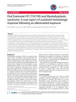
Figure 1 A) An abdominal CT revealed a large uneven tumor in the lower abdomen and encased the colorectum to the left back side
(arrows). B) An MRI of the lower abdomen revealed a polycystic tumor with a part of the thickened wall (arrows).
Nakayama et al. World Journal of Surgical Oncology 2011, 9:15
/>Page 2 of 5
[6-10]. In our case, the tumor originated from the
mesorectum because the huge tumor was limited to
within the capsule of the mesorectum.
MFH is an aggressive tumor with a high potential of
metastasis to other parts of the body. The rates of local
recurrence and distant metastasis are 44% and 42%,
respectively [1]. The most frequent metastatic site is the
lung (82%), followed by the lymph nodes (32%) [1].
Although the 5-year survival rate of all MFH patients that
underwent surgery is 67.2% [11], the 5-year survival rate
of patients with abdominal MFH is reportedly 14% [5]. On
the other hand, the 5-year survival rate of patients with
mesenteric MFH has not b een reported because the dis-
ease is extremely rare. The current treatment of choice for
A
B
B
Figure 2 A) A barium enema revealed an encasement of the sig moid colon and rectum toward the left back side. Some parts of the
upper rectum had irregular mucosa (arrows). B) A colonoscopy revealed a round ulcer with a white coating (arrows).
B
A
Figure 3 A) A laparotomy revealed a child-head-sized tumor that encased the other organs (arrows). B) An operative specimen revealed
a polycystic tumor with a part of the thickened wall.
Nakayama et al. World Journal of Surgical Oncology 2011, 9:15
/>Page 3 of 5
primary MFH is surgical resection which means wide exci-
sion of the tumor with an aim for a tumor-free margin.
Presently, the efficacy of adjuvant chemotherapy or radia-
tion has not been clear in the case of retroperitoneal and
visceral MFH. Therefore, this patient received neither
adjuvant chemotherapy nor radiation.
Mesenteric MFH initially grows without any signifi-
cant clinical symptoms, and finally produces clinical
symptoms and signs later when it compresses or invades
the adjacent organs [9]. In this case, she noticed the
lower abdominal tumor without any symptoms: never-
theless, the tumor size was 15 × 13 × 11 cm and the
tumor had directly invaded to the rectum. Therefore, a
complete tumor r esection combined with a low anterior
resection of the rectum was needed. A previous report
indicated that colonic lesions of retroperitoneal MFH
were detected by a barium enema and colonoscopic
examination before the laparotomy [12]. In the present
case, rectal ulcers were also identified by a barium
enema and colonoscopic examination before the laparot-
omy. Moreover, a histologic examination of the resected
specimen clearly demonstrat ed a submucosal invasion
by the MFH. It is very imp ortant that the requ irement
of the combined resection with other organs is decided
before the operation begins.
The efficacy of chemotherapy against advanc ed or
metastatic MFH is still unclear. On the other hand, sev-
eral phase studies which investigate the efficacy of che-
motherapy against advanced or metastatic soft tissue
sarcoma including MFH, have been reported [13-16].
These studies indicated that doxorubicin and ifosfami de
are the most active agents for the treatment of this clini-
calcondition[13-16].However,theresponserateof
these agents has been reported to range between 10%
and 36%, and the median overall survival of these agents
has been described to be between 10 and 12 months
[13-16]. T herefore, in order to obta in an improvement
in the survival i n the patients with soft tissue sarcoma
including MFH, further investigation of the new avail-
able agents is thus called for.
Conclusion
This patient has survived without recurrence for
approximately 8 years after the completed tumor resec-
tion. A complete resection is important to obtain a
favorable prognosis for MFH.
Consent
Written informed consent was obtained from the patient
for publication o f this case report and accompanying
images. A copy of the written consent is available for
review by the Editor-in-Chief of this journal.
Authors’ contributions
NY, MN, TT and YK contributed equally to this work; NY designed the
research; NY, MN and TT performed and analyzed the data; NY and YK wrote
the paper.
Competing interests
The authors declare that they have no competing interests.
Received: 29 July 2010 Accepted: 2 February 2011
Published: 2 February 2011
References
1. Weiss SW, Enzinger FM, Malignant fibrous histiocytoma: An analysis of 200
cases. Cancer 1978, 41:2250-2266.
2. Enjoji M, Hashimoto H, Tsuneyoshi M, Iwasaki H: Malignant fibrous
histiocytoma. A clinicopathological study of 130 cases. Acta Pathol Jpn
1980, 30:727-741.
3. Ros PR, Viamonte M Jr, Rywlin AM: Malignant fibrous histiocytoma:
Mesenchymal tumor of ubiquitous origin. Am J Roentgenol 1984,
142:753-759.
4. O’Brien JE, Stout AP: Malignant fibrous xanthoma. Cancer 1964,
17:1445-1455.
A
B
x400 x400
Figure 4 A) A histopathological examinati on rev ealed pleomorphic cells proliferating in a storiform pattern (H & E, ×400).B)Tumor
cells showing positive staining for vimentin (×400).
Nakayama et al. World Journal of Surgical Oncology 2011, 9:15
/>Page 4 of 5
5. Kearney MM, Soule EH, Ivins JC: Malignant fibrous histiocytoma:
A retrospective study of 167 cases. Cancer 1980, 45:167-178.
6. Bodner K, Bodner-Adler B, Mayerhofer S, Gruünberger W, Wierrani F,
Czerwenka K, Leodolter S, Mayerhofer K: Malignat fibrous histiocytoma
(MFH) of mesentery: A case report. Anticancer Res 2002, 22:169-170.
7. Hauser H, Beham A, Uranüs S, Frühwieth H, Lederer A, Klimpfinger M:
Malignat fibrous histiocytoma of the mesentery–a rare cause of
abdominal pain, Case report with a review of literature. Z Gastroenterol
1993, 31:735-738.
8. Basso M, Codaccipisanelli M, Bovino A, Vietri F, D’Ermo G, De Toma G:
Malignant fibrous histiocytoma of the mesentery: Report of two cases
and review of the literature. G Chir 2005, 26:43-46.
9. Shibuya T, Ishida H, Konno K, Komatsuda T, Hamashima Y, Sato M,
Masamune O: Sonographic findings of malignant fibrous histiocytoma of
mesentery-report of two cases. Eur J Ultrasound 1998, 8:207-212.
10. Higa T, Yamada M, Deguchi S, Tamaki S, Muto Y, Nakasone K, Kiyuna M,
Toda T: Malignant fibrous histiocytoma of the mesentery: A case report.
Ryukyu Med J 1996, 16:75-78.
11. Pezzi CM, Rawlings MS Jr, Esgro JJ, Pollock RE, Romsdahl MM: Prognostic
factors in 277 patients with malignant fibrous histiocytoma. Cancer 1992,
69:2098-2103.
12. Kobayashi S, Goto K, Shiraki S, Okayama Y, Ando H, Okumura F,
Nakamura S, Hattstori T, Joh T, Itoh M: Retroperitoneal malignant fibrous
histiocytoma causing variegated colonic lesion. Internal Medicine 1998,
37:376-380.
13. Lorigan P, Verweiji J, Papao Z, Rodenhuis S, Le Cesne A, Leahy MG,
Radford JA, Van Glabbeke MM, Kirkpatrick A, Hogendoorn PCW, Blay JY:
Phase III trial of two investigational schedules of ifosfamide compared
with standard-dose doxorubicin in advanced or metasttic soft tissue
sarcoma: A European organization for research and treatment of cancer
soft tissue and bone sarcoma group study. J Clin Oncol 2007,
25:3144-3150.
14. Maurel J, López-Pousa A, de Las Peñas R, Fra J, Martín J, Cruz J, Casado A,
Poveda A, Martínez-Trufero J, Balañá C, Gómez MA, Cubedo R, Gallego O,
Rubio-Viqueira B, Rubió J, Andrés R, Sevilla I, de la Cruz JJ, Del Muro XG,
Buesa JM: Efficacy of sequential high-dose doxorubicin and ifosfamide
compared with standard-dose doxorubicin in patients with advanced
soft tissue sarcoma: An open-label randomized phase II study of the
Spanish group for research on sarcoma. J Clin Oncol 2009, 27:1893-1898.
15. Karavasilis V, Seddon BM, Ashley S, AI-Muderis O, Fisher C, Judson I:
Significant clinical benefit of fierst-line palliative chemotherapy in
advanced soft-tissue sarcoma. Cancer 2008, 112:1585-1591.
16. Sleijfer S, Ouali M, Van Glabbeke M, Krarup-Hansen A, Rodenhuis S, Le
Cesne A, Hogendoorn PCW, Verweiji J, Blay JY: Prognostic and predictive
factors for outcome to first-line ifosfamide-containing chemotherapy for
adult patients with advanced soft tissue sarcomas. An exploratory,
retrospective analysis on large series from the european organization
for research and treatment of cancer-soft tissue and bone sarcoma
group (EORTG-STBSG). Eur J Cancer 2010, 46:72-83.
doi:10.1186/1477-7819-9-15
Cite this article as: Nakayama et al.: Malignant fibrous histiocytoma
originating from the mesorectum: a case report. World Journal of Surgical
Oncology 2011 9:15.
Submit your next manuscript to BioMed Central
and take full advantage of:
• Convenient online submission
• Thorough peer review
• No space constraints or color figure charges
• Immediate publication on acceptance
• Inclusion in PubMed, CAS, Scopus and Google Scholar
• Research which is freely available for redistribution
Submit your manuscript at
www.biomedcentral.com/submit
Nakayama et al. World Journal of Surgical Oncology 2011, 9:15
/>Page 5 of 5