Báo cáo khoa học: "Neoangiogenesis in early cervical cancer: Correlation between color Doppler findings and risk factors. A prospective observational study" pot
Bạn đang xem bản rút gọn của tài liệu. Xem và tải ngay bản đầy đủ của tài liệu tại đây (336.27 KB, 7 trang )
BioMed Central
Page 1 of 7
(page number not for citation purposes)
World Journal of Surgical Oncology
Open Access
Research
Neoangiogenesis in early cervical cancer: Correlation between
color Doppler findings and risk factors. A prospective observational
study
Matias Jurado
1
, Rosendo Galván
1
, Rafael Martinez-Monge
2
, Jesús Mazaira
1
and Juan Luis Alcazar*
1
Address:
1
Department of Gynecology, Clínica Universitaria de Navarra, School of Medicine, University of Navarra. Pamplona. Spain and
2
Department of Radiation Oncology, Clínica Universitaria de Navarra, School of Medicine, University of Navarra. Pamplona. Spain
Email: Matias Jurado - ; Rosendo Galván - ; Rafael Martinez-Monge - ;
Jesús Mazaira - ; Juan Luis Alcazar* -
* Corresponding author
Abstract
Background: The aim of the present article was to evaluate whether angiogenic parameters as
assessed by transvaginal color Doppler ultrasound (TVCD) may predict those prognostic factors
related to recurrence.
Methods: A total of 27 patients (mean age: 51.3 years, range: 29 to 85) with histologically proven
early stage invasive cervical cancer were evaluated by TVCD prior to surgery. Subjective
assessment of the amount of vessels within the tumor (scanty-moderate or abundant) and
pulsatility index (PI) were recorded. All patients underwent radical hysterectomy and pelvic lymph
node dissection. Postoperative treatment (RT or chemoradiotherapy) was given according to risk
factors (positive lymph nodes, parametrial and vaginal margin involvement, depth stromal invasion,
lymph-vascular space involvement)
Results: Tumors with "abundant" vascularization were significantly associated with pelvic lymph
node metastases, depth stromal invasion > 10 mm, lymph-vascular space involvement, tumor
diameter > 17.5 mm, and parametrial involvement. Postoperative treatment was significantly more
frequent in patients with "abundant" vascularization (OR: 20.8, 95% CIs: 2 to 211). The presence
of scanty-moderate vascularization with a PI < 0.82 or abundant vascularization with either PI >
0.82 or PI < 0.82 was associated with high-risk group in 94.4% of the cases (OR: 21.2, 95% CI: 1.9
to 236.0)
Conclusion: The results are consistent with a relationship between tumor angiogenesis and
prognostic factors for recurrence in early cervical cancer. "Abundant" vascularization and PI < 0.82
may be related to postoperative treatment due to risk factors.
Published: 25 November 2008
World Journal of Surgical Oncology 2008, 6:126 doi:10.1186/1477-7819-6-126
Received: 7 October 2008
Accepted: 25 November 2008
This article is available from: />© 2008 Jurado et al; licensee BioMed Central Ltd.
This is an Open Access article distributed under the terms of the Creative Commons Attribution License ( />),
which permits unrestricted use, distribution, and reproduction in any medium, provided the original work is properly cited.
World Journal of Surgical Oncology 2008, 6:126 />Page 2 of 7
(page number not for citation purposes)
Background
Angiogenesis has gained much attention in oncology in
recent years. It has been shown to be an essential event for
tumor growth and metastases [1]. Several studies have
demonstrated that tumor angiogenesis is an independent
prognostic factor in cervical cancer [2-4]. Therefore, the
assessment of this factor would seem to be important
when evaluating patients with this disease. However,
tumor angiogenesis can only be assessed on the surgical
specimen after surgery and therefore its prospective use, as
part of the treatment plan is difficult.
Transvaginal Color-Doppler Ultrasound (TVCD) allows
an in vivo non-invasive and prospective assessment of
tumor vascularization [5]. Some studies have shown that
color and Power-Doppler sonography can be used to
depict flow within arterioles and venules > 100 μm [6].
Furthermore, recent developments in this field have ena-
bled depiction of microvasculature (<7–10 μm)[7].
Treatment of early cervical cancer (FIGO stage Ia2, Ib1 and
II a <4 cm) is radical hysterectomy (RH) and pelvic lym-
phadenectomy (PLND). Radiotherapy is equally effective
with similar 5-year survival [8]. Some studies have found
that local recurrence in early cervical cancer surgically
treated is related to several prognostic factors such as
tumor size, lymph node (LN) metastases, parametrial or
vaginal margins involvement, depth of stromal invasion
(DSI) and lymph-vascular space invasion (LVSI). Accord-
ing to these data and based on different patterns of recur-
rence it has been proposed three different risk groups: low
(absence of any risk factor), intermediate (DSI ≥ 10 mm,
LVSI), and high risk (LN metastases, parametrial invasion,
or vaginal margin invasion) [9-14]. Prospective rand-
omized trials have shown a survival benefit after radiation
therapy for the intermediate risk group [15] as well as for
the high risk group after concomitant chemoradiation
[16]. Nonetheless, patients requiring adjuvant radiother-
apy after radical surgery have a higher long-term urologic
morbidity as well as intestinal and lymph-vascular com-
plications [17].
The aim of this prospective study is to evaluate whether
angiogenesis parameters as assessed by TVCD (amount of
intratumoral vessels and blood flow) may predict those
prognostic factors related to recurrence. A second objec-
tive is to study its ability to predict the need of postopera-
tive treatment.
Patients and methods
This is a prospective observational study. Clinical, sono-
graphic, and histopathologic data on 27 patients (mean
age: 51.3 years, ranging from 29 to 85 years) with histo-
logically proven invasive cervical cancer without evidence
of extra-uterine disease by CT scan or MRI, treated at our
institution were analyzed. Patients' characteristics are
shown in Table 1.
All patients underwent TVCD after diagnosis within one
week before surgery. Approval of Institutional Review
Board approval was obtained. TVCD data was not used for
clinical management decisions.
Transvaginal color Doppler sonography was performed in
all patients using a Toshiba SSA-370 A (Toshiba Medical
Systems, Tokyo, Japan), Sonoace 9900 (Kretztechnik,
Zipf, Austria) or Voluson 730 (GE, Milwaukee, USA)
machines equipped with real-time 5–7 MHz sector elec-
tronic array endovaginal probes with 5.0 MHz pulsed and
color Doppler capabilities.
After the endovaginal probe was gently inserted into the
vagina, the uterus and adnexal regions were scanned. Cer-
vical tumor size was estimated using electronic calipers on
the screen.
Table 1: Patients' characteristics
n%
FIGO Stage
Ia2 1 3.7
Ib1 25 92.6
IIa 1 3.7
Tumor size (cm)* 2.2 (1–3.9)
Histology
SCC 18 66.7
Non-SCC 9 33.3
Grade 1 11 40.7
Grade 2 13 48.1
Grade 3 3 11.1
Surgery
RH-II 22 81.5
RH-III 5 18.5
PLND** 14 (4–37)
+518.5
22 81.4
DSI (mm) < 10 8 29.6
DSI > 10 mm 19 70.4
LVSI+ 9 33.3
LVSI- 17 62.9
LVSI Unknown 1 3.7
Postop. Treat.
EPRT + Brachitherapy 11 40.7
Chemoradiation 7 25.9
No 9 33.3
*mean, range in parentheses ** median, range in parentheses
SCC = Squamous cell carcinoma. RH = radical hysterectomy. PLND =
Pelvic Lymph node dissection. DSI = Depth stromal invasion. LVSI =
Lymph-vascualr space invasion. EPRT = External pelvic radiation
World Journal of Surgical Oncology 2008, 6:126 />Page 3 of 7
(page number not for citation purposes)
After tumor size was estimated, color Doppler gate was
activated to identify intratumoral vessels. Color sensitivity
was set for slow velocities (1.5–10 cm/sec. PRF was set at
6.0 kHz). Color gain was set at maximum level and then
lowered until noise disappeared. As peripheral vessels
could not be reliably ascertained as neovascularized or
pre-existing vessels only central vessels were evaluated.
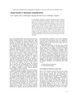
We arbitrarily considered as "central vessels" those located
at least at 5 mm far from the tumor's border. The amount
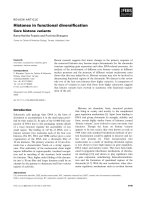
of vascularization was subjectively stated as scanty/mod-
erate (only few color spots seen) or abundant (multiple
color spots seen) (Figures 1 and 2). After a vessel was iden-
tified, pulsed Doppler volume sample was activated to
obtain the flow velocity waveform (FVW). Pulsatility
index (PI = [maximum peak systolic velocity- end diasto-
lic velocity]/mean velocity) was automatically calculated
for each vessel. We chose PI arbitrarily. The lowest PI
found was taken for analysis.
All sonographic examinations were performed by one of
the authors (JLA). Intra-observer coefficient of variation
(CV) for tumor size and PI were 5%, and 6%, respectively.
CV was calculated by performing two different measure-
ments at 10-minute interval in the first five patients
Following our institution's guidelines, surgical treatment
was a type II or III RH with PLND. Patients with two or
more intermediate risk factors received further treatment
with external pelvic radiation (EPRT) (45 Gy) and vaginal
high dose brachytherapy (HDB) (10 to 20 Gy). For
patients with at least one high risk factor the same radia-
tion regimen with concomitant weekly chemotherapy
with Taxol 50 mg/m
2
and Cisplatinum 40 mg/m
2
for a
total number of five courses was provided.
The Kolmogorov-Smirnov test was used to assess normal
distribution of continuous variables. One way analysis of
variance with Bonferroni post-hoc or Mann-Whitney tests
were used to compare RI and PI according to different
prognostic factors. The χ
2
with Pearson's correction was
used to compare categorical data. Receiver operating char-
acteristics (ROC) curves were plotted to determine the
best stromal invasion depth, tumor diameter and lowest
PI cutoff values to predict postoperative treatment. Odd
Ratios and positive likelihood ratios (LR+) were also
determined. Sensitivity, specificity, positive predictive
value (PPV) and negative predictive value (NPV) were also
calculated.
A p value ≤ 0.05 was considered statistically significant.
All statistical analyses were performed using the Statistical
Package SPSS 13.0.
Results
Prognostic factors prediction
ROC curves showed that the best cut-off values for tumor
diameter and DSI for predicting postoperative treatment
were 17.5 mm (AUC: 0.66, 95% CI: 0.41 to 0.91) and 10
mm (AUC: 0.78, 95% CI: 0.57 to 0.98), respectively.
The amount of vascularization was significantly associ-
ated with prognostic factors: Tumors with "abundant"
vascularization were significantly associated with pelvic
LN metastases, DSI > 10 mm, LVSI, tumor diameter > 17.5
mm, and parametrial involvement (Table 2). Lowest PI
were significantly lower in patients with DSI > 10 mm
(Table 3).
Transvaginal color Doppler ultrasound showing a cervical cancer with scanty vascularizationFigure 1
Transvaginal color Doppler ultrasound showing a cervical
cancer with scanty vascularization.
Transvaginal color Doppler ultrasound showing a cervical cancer with abundant vascularizationFigure 2
Transvaginal color Doppler ultrasound showing a cervical
cancer with abundant vascularization.
World Journal of Surgical Oncology 2008, 6:126 />Page 4 of 7
(page number not for citation purposes)
Further treatment prediction
Postoperative treatment (RT or chemoradiotherapy) was
significantly more frequent in patients with "abundant"
vascularization (OR: 20.8, 95% CI: 2 – 211). Thirteen out
of 18 patients who needed postoperative therapy had
abundant vascularization. Only one out of 9 patients who
did not need postoperative therapy had abundant vascu-
larization. Sensitivity, specificity, PPV and NPV for this
parameter were 72%, 89%, 93% and 61.5%, respectively.
Lowest PI was significantly lower in patients who needed
further treatment (0.79, 95% CI: 0.44 to 1.00) as com-
pared with those who did not (1.10, 95% CI: 0.86 to 1.36)
(p = 0.041)
ROC curves showed that the best cutoff value for PI was
0.82(AUC = 0.74, 95% CI: 0.56 to 0.93) (Figure 3).
Table 2: Amount of vascularization and prognostic factors
Parameter Scanty-Moderate (%) Abundant (%) p
PLN + 0 5 (43) 0.025
- 13 (100) 9 (57)
DSI <10 8 (100) 0 0.001
>10 5 (26) 14(74)
LVSI + 2 (15) 8 (85) 0.021
- 11 (69) 5(31)
T. size < 17.5 mm 7 (78) 2(22) 0.037
> 17.5 mm 6 (33) 12(67)
Parametrium + 0 6(42.9) 0.016
- 13(100) 8 (57.1)
Histology SCC 7 (53.8) 11 (78.6) 0.171
Non-SCC 6 (46.2) 3 (21.4)
SCC = Squamous cell carcinoma. PLN = Pelvic Lymph node. DSI = Depth stromal invasion. LVSI = Lymph-vascualr space invasion.
Table 3: Pulsatility index and prognostic factors
Lowest PI* P value
PLN 0.473
Negative 0.89 (0.68 – 1.10)
Positive 0.74 (0.52 – 0.95)
DSI 0.004
< 10 mm 1.20 (0.91 – 1.60)
> 10 mm 0.74 (0.55 – 0.92)
LVSI 0.073
Negative 1.00 (0.75 – 1.30)
Positive 0.68 (0.44 – 0.92)
Tumor size 0.158
< 17.5 mm 1.06 (0.68 – 1.40)
> 17.5 mm 0.80 (0.60 – 1.40)
Parametrium 0.171
Negative 0.95 (0.73 – 1.17)
Positive 0.67 (0.48 – 0.86)
Histology 0.406
SCC 0.84 (0.63 – 1.05)
Non-SCC 0.99 (0.61 – 1.37)
* Expressed as median, range in parentheses.
SCC = Squamous cell carcinoma. PLN = Pelvic Lymph node. DSI =
Depth stromal invasion. LVSI = Lymph-vascualr space invasion.
ROC curve for pulsatility indexFigure 3
ROC curve for pulsatility index. The best cut-off was 0.82.
World Journal of Surgical Oncology 2008, 6:126 />Page 5 of 7
(page number not for citation purposes)
Patients with PI < 0.82 needed more frequently postoper-
ative treatment (OR: 9.1, 95% CI: 1.4 to 59.6)
In order to develop a way to predict prospectively patients
that would be candidate for postoperative treatment, the
combination of the amount of vascularization and PI <
0.82 was evaluated according to prognostic factors. Two
main risk groups were established. The high-risk group
that was defined as having at least one of the following
prognostic factors: LVSI, DSI > 10 mm, tumor size > 17.5
mm, parametrial involvement or LN metastases. The low
risk group was defined as not having any of these factors.
The presence of scanty-moderate vascularization with a PI
< 0.82 or abundant vascularization with either PI > 0.82
or PI < 0.82 was associated with high-risk group in 94.4%
of the cases (OR: 21.2, 95% CI: 1.9 to 236.0) (Table 4).
LR+ for these three groups all together was 4.76
Discussion
Prognostic factors prediction
It is generally accepted that the rate of local recurrence for
early stage cervical cancer (FIGO Ib1 to II a < 4 cm) is sig-
nificantly lower than in advanced stages. The presence of
LN metastases has an overriding prognostic importance in
early stage cervical carcinoma with an overall survival
average of 90% if the pelvic nodes are negative and 65% if
pelvic nodes are positive. It is also important the number
of nodes involved, thus patients with one to three
involved nodes reported to have a 72% 5-year survival,
whereas the survival of patients with more than three
nodes involved averages only 40% [13,18]. Furthermore,
based on multivariate analysis, tumor size, LVSI, and
depth of cervical stromal invasion are independent pre-
dictors of lymph nodes metastases risk and, therefore, dis-
ease-free survival [9,13,19,20]. It has also been reported
that due to the presence itself of these prognostic factors
without pelvic lymph nodes involvement the rate of recur-
rence may increase from 2% to 31%, mainly locally, after
three years [15]. GOG prospective randomized trial [15]
has found a statistically significance decrease of local
recurrence after radiotherapy in this group of patients.
Other prospective randomized trials [16] have found a
benefit in overall survival and disease free survival with
postoperative concomitant chemoradiation over radia-
tion therapy alone in a higher risk group of patients with
early stage and with lymph node metastases, parametrial
or vaginal margin invasion due to its mixed recurrent pat-
tern.
Several publications [21-24] have pointed out the capabil-
ity of transvaginal color-Doppler to assess the intratu-
moral blood flow in cervical cancer. Velocimetric indexes
and color signals correlated with some prognostic factors.
Cheng et al [25] reported on a group of 35 patients with
stage Ib to II cervical cancer in whom they assessed tumor
angiogenesis by TVCD. They found that vascular index (VI
= number of colored pixels/number of total pixels) corre-
lated with prognostic factors. The higher the VI, the higher
the tumor stage, the deeper stromal invasion, the higher
the LVSI rate and the higher the pelvic LN metastases rate
was. Also interesting was this VI had a good correlation
with intratumoral microvessel density as assessed immu-
nohistochemically. The same group reported on a further
series of 60 patients with stage Ib to II a but using TVCD.
The presence of color signals was associated with a higher
probability of LN metastases and parametrial involve-
ment [26].
Hsu et al [27] reported their results on 141 patients with
early stage cervical cancer in who tumor angiogenesis was
assessed by 3-D Power-Doppler. They found that tumor
vascularization correlated with tumor volume.
Testa et al [28] also found a similar correlation between
tumor vascularization and its volume. In our study a sig-
nificant correlation between prognostic factors and tumor
vascularization was found, being the amount of vascular-
ization higher when tumor had deeper stromal invasion,
larger diameter, LVSI, parametrial involvement or LN
metastases. Vascular flow as assessed by velocimetric
indexes (the lowest PI) was correlated only with stromal
invasion higher than 10 mm. There was a trend for LVSI.
The lack of correlation with the rest of prognostic factors
could be due to the small number of patients in this series.
Postoperative treatment prediction
Cheng et al [26] in their above mentioned study per-
formed with TVCD reported results, found that the pres-
ence of color signals was associated with a higher
probability of LN metastases and parametrial invasion.
Although they did not made any specific statistical analy-
sis, they suggested that these findings could be helpful in
planning treatment for women with stage I–II a cervical
carcinoma.
To the best of our knowledge this is the first study regard-
ing the issue of tumor vascularization and its role to pre-
Table 4: Risk group according to amount of vascularization and
PI
Low Risk High Risk Total
Scanty Vascularization and PI > 0.82 5 (55.2%) 4 (44.8%) 9
Scanty vascularization and PI < 0.82
or
Abundant vascularization
1 (5.6%) 17 (94.4%) 18
Total 6 21 27
World Journal of Surgical Oncology 2008, 6:126 />Page 6 of 7
(page number not for citation purposes)
dict further treatment in early cervical cancer treated with
radical surgery. We have found that amount of vasculari-
zation and the lowest PI found within the tumor were
associated with the need for postoperative treatment due
to the presence of risk factors. Those with "abundant" vas-
cularization received more frequently adjuvant treatment
with radiation with or without simultaneous chemother-
apy, especially if PI was < 0.82. However, the clinical use
of PI as the unique parameter for predicting further treat-
ment may be questionable because the significant over-
lapping of individual values observed. This overlapping
could be explained by the fact of the small series herein
reported.
Another interesting question may be the use of 3D power
Doppler vascular indexes. To date the only study reported
did not find any relationship between 3D power Doppler
indexes and tumor features [28]. In our preliminary expe-
rience 3D power Doppler indexes were significantly
higher in locally advanced stage tumors as compared with
early stage cervical cancer [29]
Over the last ten years much attention has been paid to
morbidity after the combination of radical surgery and
pelvic radiotherapy. Some publications regarding this
issue [8,17] have found a significantly higher risk of post-
operative complications, specifically urologic and intesti-
nal. Therefore a judicious pretreatment selection of
patients with predictable risk factor for adjuvant therapy
would help to select patients who should not be sched-
uled for primary radical surgery. Whether TVCD and the
study of angiogenesis would help to avoid this morbidity
as a consequence of a more reasonable plan of treatment
based on prospectively predictable prognostic factors
needs further evaluation.
With angiogenic parameters, two main groups of risk for
adjuvant treatment could be defined. As patients with
intermediate risk factors are currently treated with radia-
tion alone [15] and with radiation and simultaneous
chemotherapy those with parametrial involvement or LN
metastases [16], it will be interesting to define this later
subset of patients in a larger series.
Conclusion
Our results are consistent with a relationship between
tumor angiogenesis and prognostic factors for recurrence
in early cervical cancer. "Abundant" vascularization and
the lowest PI are related to postoperative treatment due to
risk factors that can be easily and prospectively assessed by
TVCD and these findings encourage following with larger
series of study.
List of abbreviations
TVCD: Transvaginal Color Doppler; PI: Pulsatility index;
RT: Radiotherapy; FIGO: Federation International Gyne-
cology and Obstetrics; RH: Radical hysterectomy; PLND:
Pelvic lymph node dissection; LN: Lymph node; DSI:
Depth stromal invasion; LVSI: Lymph-vascular space inva-
sion; CT: Computed tomography; MRI: Magnetic reso-
nance imaging; EPRT: External pelvic radiation therapy;
HDB: High dose brachytherapy; GOG: Gynecologic
Oncology Group; OR: Odds ratio; CI: Confidence inter-
vals; ROC: Receiver Operator curves; AUC: Area under the
curve; NPV: Negative predictive value; PPV: Positive pre-
dictive value; LR: Likelihood ratio; CV: Coefficient of var-
iation.
Competing interests
The authors declare that they have no competing interests.
Authors' contributions
JLA was involved in study design, data collection, analysis,
patient recruitment and management. MJ was involved in
study design, data collection, analysis, patient recruitment
and management and preparation of the manuscript.
RMM was involved in patient recruitment and manage-
ment, helped in preparation of draft. RG was involved in
data analysis and interpretation of results. The final man-
uscript was approved by all authors.
Acknowledgements
The study was approved by Institutional review board. There was no fund-
ing source for this study. The corresponding author had full access to all
data of the study and has the final responsibility for data presented in the
study.
References
1. Carmeliet P, Jain RK: Angiogenesis in cancer and other dis-
eases. Nature 2000, 407:249-257.
2. Wiggins DL, Granai CO, Steinhoff MM, Calabresi P: Tumor angio-
genesis as a prognostic factor in cervical carcinoma. Gynecol
Oncol 1995, 56:353-356.
3. Schlenger K, Hockel M, Mitze M, Schäffer U, Weikel W, Knapstein
PG, Lambert A: Tumor vascularity – a novel prognostic factor
in advanced cervical carcinoma. Gynecol Oncol 1995, 59:57-66.
4. Tjalma W, Van Mark E, Weyler J, Dirix L, Van Daele A, Goovaerts G,
Albertyn G, van Dam P: Quantification and prognostic rele-
vance of angiogenic parameters in invasive cervical cancer.
Br J Cancer 1998, 78:170-174.
5. Cosgrove D: Angiogenesis imaging-ultrasound. Br J Radiol 2003,
76:43-S49.
6. Fleischer AC, Nierman KJ, Donnelly EF, Yankeelov TE, Canniff KM,
Hallahan DE, Rothenberg ME: Sonogrphic depiction of microves-
sel perfusion. J Ultrasound Med 2004, 23:1499-1506.
7. Foster FS, Burns PN, Simpson DH, Wilson SR, Christopher DA,
Goertz DE: Ultrasound of the visualization and quantification
of tumor microcirculation. Cancer Metastasis Rev 2000,
19:131-138.
8. Landoni F, Maneo A, Colombo A, Placa F, Milani R, Perego P, Favini
G, Ferri L, Mangioni C: Randomised study of radical surgery
versus radiotherapy for stage Ib-IIa cervical cancer. Lancet
1997, 350:535-540.
9. Delgado G, Bundy BN, Fowler WC, Stehman FB, Sevin B, Creasman
WT, Major F, DiSaia P, Zaino R: A prospective surgical patholog-
ical study of stage I squamous carcinoma of the cervix: a
Publish with BioMed Central and every
scientist can read your work free of charge
"BioMed Central will be the most significant development for
disseminating the results of biomedical research in our lifetime."
Sir Paul Nurse, Cancer Research UK
Your research papers will be:
available free of charge to the entire biomedical community
peer reviewed and published immediately upon acceptance
cited in PubMed and archived on PubMed Central
yours — you keep the copyright
Submit your manuscript here:
/>BioMedcentral
World Journal of Surgical Oncology 2008, 6:126 />Page 7 of 7
(page number not for citation purposes)
Gynecologic Oncology Group study. Gynecol Oncol 1989,
35:314-320.
10. Samlal RA, Velden J van der, Ten Kate FJ, Schilthuis MS, Hart AA,
Lammes FB: Surgical pathologic factors that predict recur-
rence in stage I b and II a cervical carcinoma patients with
negative pelvic lymph nodes. Cancer 1997, 80:1234-1240.
11. Singh N, Arif S: Histopathologic parameters of prognosis in
cervical cancer – a review. Int J Gynecol Cancer 2004, 14:741-750.
12. Inoue T, Okumura M: Prognostic significance of parametrial
extension in patients with cervical carcinoma stages I b, II a
and II b: A study of 628 cases treated by radical hysterec-
tolmy and lymphadenectomy with and without postopera-
tive radiation. Cancer 1984, 54:1714-1719.
13. Kamura T, Tsukamoto N, Tsuruchi N, Saito T, Matsuyama T, Aka-
zawa K, Nakano H: Multivariate analysis of the histopathologic
prognostic factors of cervical cancer in patients undergoing
radical hysterectomy. Cancer 1992, 69:181-186.
14. Estape RE, Angioli R, Madrigal M, Janicek M, Gomez C, Penalver M,
Averette H: Close vaginal margins as a prognostic factor after
radical hysterectomy. Gynecol Oncol 1998, 68:229-232.
15. Rotman M, Sedlis A, Piedmonte MR, Bundy B, Lentz SS, Muderspach
LI, Zaino RJ: A phase III randomized trial of postoperative pel-
vic irradiation in stage Ib cervical carcinoma with poor prog-
nostic features: follow-up of a Gynecologic Oncology group
study. Int J Radiat Oncology Biol Phys 2006, 65:169-176.
16. Peters WA 3rd, Liu PY, Barrett RJ 2nd, Stock RJ, Monk BJ, Berek JS,
Souhami L, Grigsby P, Gordon W Jr, Alberts DS: Concurrent
chemotherapy and pelvic radiation therapy compared with
radiation therapy alone as adjuvant therapy after radical sur-
gery in high-risk early-stage cancer of the cervix. J Clin Oncol
2000, 18:1606-1613.
17. Landoni F, Maneo A, Cormio G, Perego P, Milani R, Caruso O, Man-
gioni C: Class II versus class III radical hysterectomy in stage
Ib-IIa cervical cancer: a prospective randomized study. Gyne-
col Oncol 2001, 80:3-12.
18. Inoue T, Morita K: The prognostic significance of number of
positive nodes in cervical carcinoma stages Ib, IIa, and IIb.
Cancer 1990, 65:
1923-1927.
19. Delgado G, Bundy BN, Zaino R, Stehman FB, Sevin B, Creasman WT,
Major F, DiSaia P, Zaino R: A prospective surgical pathological
study of stage I squamous carcinoma of the cervix: a Gyne-
cologic Oncology Group Study. Gynecol Oncol 1989, 35:314-320.
20. Larsson G, Alm P, Gullberg B, Grundsell H: Prognostic factors in
early invasive carcinoma of the uterine cervix: a clinical, his-
topathologic, and statistical analysis of 343 cases. Am J Obstet
Gynecol 1983, 146:145-153.
21. Hsieh CY, Wu CC, Chen TM, Chen CA, Chen CL, Wang JF, Chang
CF, Hsieh FJ: Clinical significance of intratumoral blood flow in
cervical carcinoma assessed by color Doppler ultrasound.
Cancer 1995, 75:2518-2522.
22. Tepper R, Zalel Y, Altaras M, Ben-Baruch G, Beyth Y: Transvaginal
color Doppler ultrasound in the assessment of invasive cer-
vical carcinoma. Gynecol Oncol 1996, 60:26-29.
23. Alcazar JL, Jurado M: Transvaginal color Doppler for predicting
pathological response to preoperative chemoradiation in
locally advanced cervical carcinoma: a prliminary study.
Ultrasound Med Biol 1999, 25:1041-1045.
24. Wu YC, Yuan CC, Hung JH, Chao KC, Yen MS, Ng HT: Power Dop-
pler angiographic appearance and blood flow velocity wave-
forms in invasive cervical carcinoma. Gynecol Oncol 2000,
79:181-186.
25. Cheng WF, Lee CN, Chu JS, Chen CA, Chen TM, Shau WY, Hsieh
CY, Hsieh FJ: Vascularity index as a novel parameter for the in
vivo assessment of angiogenesis in patients with cervical car-
cinoma. Cancer 1999, 85(3):615-617.
26. Cheng WF, Wei LH, Su YN, Cheng SP, Chu JS, Lee CN: The possi-
ble use of color flow Doppler in planning treatment in early
invasive carcinoma of the cervix. Br J Obstet Gynaecol 1999,
106(11):1137-1342.
27. Hsu KF, SU JM, Huang SC, Cheng YM, Kang CY, Shen MR, Chang FM,
Chou CY: Three-dimensional power-Doppler imaging of
early-stage cervical cancer. Ultrasound Obstet Gynecol 2004,
24:664-671.
28. Testa AC, Ferrandina G, Distefano M, Fruscella E, Mansueto D, Basso
D, Salutari V, Scambia G:
Color Doppler velocimetry and three-
dimensional color power angiography of cervical carcinoma.
Ultrasound Obstet Gynecol 2004, 24:445-452.
29. Alcázar JL: Transvaginal color Doppler in the assessment of
cervical cancer. Cancer Ther 2005, 3:139-146.