Báo cáo y học: " Local treatment with the selective IκB kinase β inhibitor NEMO-binding domain peptide ameliorates synovial inflammation" pot
Bạn đang xem bản rút gọn của tài liệu. Xem và tải ngay bản đầy đủ của tài liệu tại đây (891.84 KB, 9 trang )
Open Access
Available online />Page 1 of 9
(page number not for citation purposes)
Vol 8 No 4
Research article
Local treatment with the selective IκB kinase β inhibitor
NEMO-binding domain peptide ameliorates synovial
inflammation
Sander W Tas
1
, Margriet J Vervoordeldonk
1
, Najat Hajji
1
, Michael J May
2
, Sankar Ghosh
3
and
Paul P Tak
1
1
Division of Clinical Immunology and Rheumatology F4-218, Academic Medical Center/University of Amsterdam, PO Box 22700, 1100 DE
Amsterdam, The Netherlands
2
School of Veterinary Medicine, Department of Animal Biology, University of Pennsylvania, 3800 Spruce Street, Philadelphia, PA 19104-6046, USA
3
Immunobiology Section, Yale University Medical School, 300 Cedar Street, New Haven, CT 06519, USA
Corresponding author: Paul P Tak,
Received: 22 Sep 2005 Revisions requested: 2 Nov 2005 Revisions received: 13 Mar 2006 Accepted: 18 Apr 2006 Published: 9 May 2006
Arthritis Research & Therapy 2006, 8:R86 (doi:10.1186/ar1958)
This article is online at: />© 2006 Tas et al.; licensee BioMed Central Ltd.
This is an open access article distributed under the terms of the Creative Commons Attribution License ( />),
which permits unrestricted use, distribution, and reproduction in any medium, provided the original work is properly cited.
Abstract
Nuclear factor (NF)-κB is a key regulator of synovial
inflammation. We investigated the effect of local NF-κB
inhibition in rat adjuvant arthritis (AA), using the specific IκB
kinase (IKK)-β blocking NF-κB essential modulator-binding
domain (NBD) peptide. The effects of the NBD peptide on
human fibroblast-like synoviocytes (FLS) and macrophages, as
well as rheumatoid arthritis (RA) whole-tissue biopsies, were
also evaluated. First, we investigated the effects of the NBD
peptide on RA FLS in vitro. Subsequently, NBD peptides were
administered intra-articularly into the right ankle joint of rats at
the onset of disease. The severity of arthritis was monitored over
time, rats were sacrificed on day 20, and tissue specimens were
collected for routine histology and x-rays of the ankle joints.
Human macrophages or RA synovial tissues were cultured ex
vivo in the presence or absence of NBD peptides, and cytokine
production was measured in the supernatant by enzyme-linked
immunosorbent assay. The NBD peptide blocked interleukin
(IL)-1-β-induced IκBα phosphorylation and IL-6 production in
RA FLS. Intra-articular injection of the NBD peptide led to
significantly reduced severity of arthritis (p < 0.0001) and
reduced radiological damage (p = 0.04). This was associated
with decreased synovial cellularity and reduced expression of
tumor necrosis factor (TNF)-α and IL-1-β in the synovium.
Incubation of human macrophages with NBD peptides resulted
in 50% inhibition of IL-1-β-induced TNF-α production in the
supernatant (p < 0.01). In addition, the NBD peptide decreased
TNF-α-induced IL-6 production by human RA synovial tissue
biopsies by approximately 42% (p < 0.01). Specific NF-κB
blockade using a small peptide inhibitor of IKK-β has anti-
inflammatory effects in AA and human RA synovial tissue as well
as in two important cell types in the pathogenesis of RA:
macrophages and FLS. These results indicate that IKK-β-
targeted NF-κB blockade using the NBD peptide could offer a
new approach for the local treatment of arthritis.
Introduction
Rheumatoid arthritis (RA) is a chronic inflammatory disease
predominantly affecting the joints [1]. Many different cell types
have been described as contributing to both the initiation
phase of the disease and the chronic perpetuation of synovial
inflammation. In rheumatoid synovium, the intimal lining layer
shows marked hyperplasia, mainly due to expansion of intimal
macrophages and fibroblast-like synoviocytes (FLS) [2]. Mac-
rophages appear to play a pivotal role in the pathogenesis of
RA because they are present in high numbers in RA synovial
tissue and clearly show signs of activation, including
AA = adjuvant arthritis; AUC = area under the curve; BSA = bovine serum albumin; DMEM = Dulbecco's modified Eagle's medium; ELISA = enzyme-
linked immunosorbent assay; FCS = fetal calf serum; FLS = fibroblast-like synoviocytes; HRP = horseradish peroxidase; i.a. = intra-articular; IκB =
inhibitor of κB; IKK = IκB kinase; mAb = monoclonal antibody; MOD = mean optical density; MUT = mutant nuclear factor-κB essential modulator
binding domain peptide; NBD = nuclear factor-κB essential modulator binding domain peptide; NEMO = nuclear factor-κB essential modulator; NF
= nuclear factor; PBS = phosphate-buffered saline; ph = phosphorylated; RA = rheumatoid arthritis; TBS = Tris-buffered saline; TNF = tumor necrosis
factor.
Arthritis Research & Therapy Vol 8 No 4 Tas et al.
Page 2 of 9
(page number not for citation purposes)
enhanced expression of cellular surface markers like major his-
tocompatibility complex class II molecules, pro-inflammatory
cytokines such as tumor necrosis factor-α (TNF-α) [3], chem-
okines, and matrix metalloproteinases [4]. Furthermore, there
is a highly significant positive correlation between scores for
local disease activity and macrophage numbers and the
expression of macrophage-derived cytokines in the synovium
[5]. In addition to macrophages, other cell types, like FLS, also
display altered biology. RA FLS are characterized by anchor-
age-independent growth and resistance to apoptosis due to
constitutive activation of multiple signaling cascades
(reviewed in [6,7]).
In many of the cells involved in synovial inflammation, altera-
tions are found in intracellular signaling cascades, leading to
unwanted interactions with other cells and resulting in pathol-
ogy [8]. Striking abnormalities are observed in the nuclear fac-
tor (NF)-κB signal transduction pathway [9]. Phosphorylation
of inhibitor of κB (IκBα) by the IκB kinase (IKK) complex is a
crucial step in NF-κB/Rel activation. The IKK complex contains
two catalytic subunits, named IKK-α and IKK-β, and a regula-
tory subunit termed NEMO (NF-κB essential modulator). NF-
κB activation in response to pro-inflammatory signals is
dependent mainly on IKK-β [10]. The subsequent polyubiquiti-
nation targets IκBα for degradation, releasing NF-κB dimers
from the NF-κB-IκBα complex, followed by translocation to the
nucleus and binding to κB enhancer elements of target genes
[11].
IKK is a key convergence site of many different stimuli that
induce NF-κB activation, such as pro-inflammatory cytokines
and ligation of Toll-like receptors, but triggering of highly spe-
cialized antigen receptors such as the T-cell receptor is also
dependent on this pathway [12]. Consequently, selective inhi-
bition of the IKK complex has emerged as a promising strategy
to block aberrant NF-κB activity in autoimmune and inflamma-
tory diseases as well as certain cancers [13].
NF-κB is highly activated in the synovial tissue of patients with
RA [14,15], with IKK-β being a key regulator of synovial inflam-
mation [16]. Various local or systemic approaches to specifi-
cally inhibit the activation of this transcription factor by
targeting the IKK complex have proven successful in the amel-
ioration of arthritis [16-19]. Obviously, NF-κB activity is also
required for normal physiology of cells or for clearing microbial
pathogens, raising toxicity concerns when this pathway is
blocked systemically in many different cell types at the same
time. Accordingly, for development of therapies blocking NF-
κB activity in RA, local intra-articular (i.a.) therapy appears
more attractive.
The present study was conducted to explore the effects of
specific inhibition of IKK-β-mediated NF-κB activation locally
in the inflamed joint, using the well-characterized NEMO-bind-
ing domain (NBD) peptide [20]. Our data indicate that local
IKK-β-targeted NF-κB blockade using a small peptide inhibitor
ameliorates synovial inflammation, both in an animal model of
arthritis and in human RA synovial tissue ex vivo, which opens
up a new approach for the local treatment of RA.
Figure 1
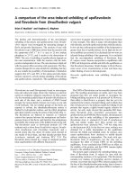
NBD peptide blocks interleukin (IL)-1-β-induced IκBα phosphorylation and IL-6 production in fibroblast-like synoviocytes (FLS)NBD peptide blocks interleukin (IL)-1-β-induced IκBα phosphorylation
and IL-6 production in fibroblast-like synoviocytes (FLS). (a) FLS were
pre-incubated with either NBD or mutant NBD (MUT) peptide at a con-
centration of 50 µM for 2 hours. Subsequently, cells were stimulated
with IL-1-β (2.5 ng/ml) for 30 minutes, extensively washed, and lysed in
sample buffer. Cell lysates were analyzed by Western blotting. One
representative blot out of three is shown. Densitometry includes all
three experiments, and data are expressed as mean ± standard error of
the mean (*p < 0.01). (b) NBD peptide blocks IL-1-β-induced IL-6 pro-
duction by FLS in vitro. FLS were pre-incubated with either NBD or
MUT peptide at a concentration of 50 µM for 2 hours. Subsequently,
cells were stimulated with IL-1-β (2.5 ng/ml). After 24 hours, superna-
tants were collected and IL-6 levels were measured by sandwich
enzyme-linked immunosorbent assay. Data are representative of three
independent experiments performed in triplicates (*p < 0.01).
Available online />Page 3 of 9
(page number not for citation purposes)
Materials and methods
Animals
Pathogen-free male Lewis rats (150–200 g; 8–10 weeks of
age at the start of the experiments) were obtained from Harlan
Sprague Dawley, Inc. (Horst, The Netherlands) and were main-
tained in our central animal facility. The Animal Care and Use
Committee of the University of Amsterdam, The Netherlands,
approved all experiments.
NBD peptides
Small-scale Fmoc (9-fluorenylmethoxycarbonyl) synthesis of
the peptides was carried out on a Rainin Symphony Instrument
(Rainin Instrument, LLC, Oakland, CA, USA) at the HHMI
Biopolymer-Keck Foundation Biotechnology Resource Labo-
ratory at Yale University (New Haven, CT, USA). Peptides
were characterized by matrix-assisted laser desorption ioniza-
tion mass spectrometry and analytical reverse-phase high-per-
formance liquid chromatography analysis. The peptides were
subsequently dissolved in dimethyl sulfoxide to a stock of 50
mM. The sequences of the wild-type and mutant (MUT) NBD
peptides have been described previously [20]. The NBD pep-
tide (3.7 kD) contains the region of IKK-β from T735 to E745
synthesized in tandem with a membrane permeabilization
sequence from the drosophila antennapedia homeodomain
protein. The MUT peptide (3.5 kD) is identical except that
W739 and W741 are replaced by alanines to render it biolog-
ically inactive.
Evaluation of NF-κB inhibition in FLS
Synovial biopsies were obtained by arthroscopy from different
seropositive RA patients with actively inflamed joints. Human
RA FLS were isolated from synovial tissue as described previ-
ously [21], grown in Dulbecco's modified Eagle's medium
(DMEM)/10% fetal calf serum (FCS), and used from passages
3 to 8. For stimulation experiments, FLS were seeded onto 24-
well dishes (Costar, now Corning Life Sciences, Acton, MA,
USA) at 1 × 10
4
per well. After serum-starving for 12 hours in
medium containing 0.5% FCS for synchronization, cells were
pre-incubated for 2 hours with NBD or MUT peptides (50 µM)
in medium containing 0.5% FCS and stimulated with IL-1-β
(2.5 ng/ml). After 30 minutes of stimulation, cells were washed
twice with ice-cold phosphate-buffered saline (PBS) to
remove all serum proteins and then lysed in 1× SDS-PAGE
sample buffer. Total intracellular protein was separated by
SDS-PAGE on a 10% gel, using Rainbow-colored protein
molecular weight markers (Amersham Biosciences, now GE
Healthcare, Little Chalfont, Buckinghamshire, UK) as a refer-
ence, and transferred onto a polyvinylidene difluoride mem-
brane (Bio-Rad Laboratories, Inc., Hercules, CA, USA). The
membrane was blocked in Tris-buffered saline (TBS)
containing 2% non-fat dry milk (Bio-Rad Laboratories, Inc.),
Na
3
VO
4
(2 mM), and 0.05% Tween 20 for 1 hour. Detection
of phosphorylated (ph) and unphosphorylated proteins was
performed by incubating the membranes with a primary anti-
body against the protein of interest overnight at 4°C. The
Figure 2
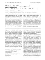
Intra-articular injection of the NBD peptide at the onset of disease ameliorates arthritisIntra-articular injection of the NBD peptide at the onset of disease
ameliorates arthritis. (a) Dose-finding experiments. To determine the
optimal dose of the NBD peptide for amelioration of arthritis, animals (n
= 6/group) were treated intra-articularly at the onset of arthritis (day 10)
and 2 days later (day 12) with either 50 or 150 µg of the peptides or
vehicle. Paw swelling was measured by water displacement plethys-
mometry until the animals were sacrificed at day 20. Data represent
mean ± standard error of the mean (SEM) paw swelling (p < 0.05). (b)
To evaluate the effect of the NBD peptide on clinical arthritis compared
with the mutant NBD (MUT) control peptide, animals (n = 15/group)
were treated intra-articularly at the onset of arthritis (day 10) and 2 days
later (day 12) with 150 µg of the peptides. Paw swelling was measured
by water displacement plethysmometry until the animals were sacri-
ficed at day 20. Data represent mean ± SEM paw swelling (p <
0.0001). (c) Results of the 'area under the curve' (AUC) calculation of
the experiments displayed in (b) (*p < 0.0001).
Arthritis Research & Therapy Vol 8 No 4 Tas et al.
Page 4 of 9
(page number not for citation purposes)
membranes were subsequently washed and incubated with
the appropriate horseradish peroxidase (HRP)-labeled sec-
ondary antibody (Bio-Rad Laboratories, Inc.) in TBS contain-
ing 2% non-fat dry milk, Na
3
VO
4
(2 mM), and 0.05% Tween 20
for 1 hour at room temperature, and after extensive washing
were assayed using the enhanced chemiluminescence detec-
tion system (GE Healthcare). Mouse monoclonal antibodies
(mAbs) to ph-IκBα and total IκBα were obtained from Cell
Signaling Technology, Inc. (Beverly, MA, USA). Densitometry
was performed using Quantity One software (Bio-Rad Labora-
tories, Inc.). To study the effects of NBD on cytokine produc-
tion, FLS were pre-incubated for 2 hours with NBD peptides
(50 µM) and stimulated for 24 hours with recombinant human
IL-1-β (2.5 ng/ml; R&D Systems, Minneapolis, MN, USA).
Supernatants were harvested, and the levels of IL-6 were
determined by sandwich enzyme-linked immunosorbent assay
(ELISA) as described previously [22].
AA
All rats were immunized at the base of the tail with 1 mg of
Mycobacterium tuberculosis H37RA (Difco, Detroit, MI, USA)
in 0.1 ml mineral oil on day 0 [16]. Clinical signs of arthritis
(that is, paw swelling) were usually observed by day 10 and
monitored during the course of disease by water displacement
plethysmometry. Paw swelling was expressed as delta paw
volume (that is, with paw volume before onset of arthritis sub-
tracted). For i.a. treatment, the right ankle joints were injected
at days 10 and 12 after immunization in animals (n = 6/group
[dose-finding]; n = 15/group [clinical study]) anesthetized
with isoflurane. The skin was prepared with ethanol, and NBD
or MUT peptides were injected in the indicated concentrations
anterolaterally into the right ankle joint in a total volume of 50
µl saline, using a 31-gauge needle on a glass syringe [23]. The
course of arthritis was monitored regularly until rats were sac-
rificed at day 20 by CO
2
inhalation and hind paws were col-
lected. X-rays of the ankle joints were made, and these plain
radiographs were scored for bone degradation, using a semi-
quantitative scoring system (demineralization [0-2+], ankle
and midfoot erosions [0-2+], calcaneal erosion [0-1+], heter-
otopic bone formation [0-1+]; maximum possible score = 6)
according to Boyle and colleagues [24].
Immunohistochemical analysis of synovial cytokine
expression
Hind paws were obtained from each rat, trimmed of skin, and
fixed in 4% paraformaldehyde. After 24 hours, paraformalde-
hyde was replaced by 70% ethanol and x-rays of the ankle
joints were made. Subsequently, the paws were decalcified for
4 weeks in decalcifying solution (15% EDTA [ethylenediami-
netetraacetic acid; pH 7.5]) on a rotator at 4°C, with decalci-
fying solution changed twice a week. After 1 week of
decalcification, the paws were longitudinally cut in half. After 4
weeks, the formalin-fixed paws were dehydrated in graded
alcohol and embedded in paraffin. Paraffin-embedded paws
were serially sectioned at a thickness of 4 µm. Sections were
subsequently deparaffinized in xylene and rehydrated in etha-
nol, followed by incubation with hydrogen peroxide 30% in
0.1% Na-azide-PBS to block endogenous peroxidase activity.
Antigen retrieval was obtained by boiling the sections in citrate
buffer (pH 6.0) for 10 minutes.
Cytokine expression was studied by staining the sections
overnight at 4°C with mAbs specific for TNF-α (10 µg/ml), IL-
6 (10 µg/ml), and IL-1-β (10 µg/ml) (all from R&D Systems,
Oxon, UK) in PBS/bovine serum albumin (BSA) 1%. Sections
were then washed extensively and incubated with secondary
HRP-conjugated swine anti-goat antibodies (Dako Denmark
A/S, Glostrup, Denmark) in PBS/BSA 1% + 10% N-hydroxy-
succinimide. Signal amplification was performed using bioti-
nylated tyramine (PerkinElmer Life and Analytical Sciences,
Boston, MA, USA) followed by streptavidine-HRP (Dako Den-
mark A/S) in PBS/BSA 1% as described previously [4].
Finally, peroxidase activity was detected with AEC (0.02% 3-
amino-9-ethylcarbazole; Vector Laboratories, Burlingame, CA,
USA) yielding red coloration. Sections were counterstained
with Mayer's haemalum solution (Merck, Darmstadt, Germany)
and mounted with Kaiser's glycerol gelatin (Merck) mounting
medium. For quantification of cytokine expression, the sec-
tions were blinded and analyzed in a random order by compu-
ter-assisted image analysis.
Digital image analysis
Six randomly selected fields within each section were chosen
for digitizing the amount of positive signal. These images were
acquired on an Olympus microscope (Olympus, Tokyo,
Japan), captured using a Charged Coupled Device video cam-
era (Sony, Tokyo, Japan), and digitized with a PV100 multime-
dia 16-bit color video digitizer card. In the resultant color
images, the area of positive staining and the mean optical den-
sity (MOD) were measured by a macro program as described
previously [25,26]. The MOD is proportional to the cellular
concentration of protein. The integrated optical density is
equal to the MOD multiplied by the area of positive staining.
Culture of normal macrophages and synovial biopsies
from patients with RA
For evaluating the effect of NBD on macrophage cytokine pro-
duction in vitro, monocytes were isolated from peripheral
blood of healthy controls as described previously [22] and
allowed to adhere to tissue culture plastic (24-well plates;
Corning Life Sciences) for 1 hour (1 × 10
6
cells; 1 ml). Subse-
quently, non-adherent cells were washed away and cells were
cultured for 8–9 days to obtain macrophages, with half of the
medium refreshed every 3 days [27]. Macrophages were pre-
incubated for 2 hours with NBD peptides (50 µM), and cells
were stimulated for 24 hours with rhIL-1-β (2.5 ng/ml; R&D
Systems). Supernatants were harvested, and the levels of
TNF-α were determined by sandwich ELISA as described pre-
viously [22].
Available online />Page 5 of 9
(page number not for citation purposes)
To evaluate the effects of NBD on human synovial tissue,
small-bore arthroscopy (2.7-mm arthroscope; Storz, Tuttlin-
gen, Germany) was performed under local anesthesia in three
patients with established, active seropositive RA [28]. The
obtained biopsies (± 5 mm
3
; 3 per well; mixed locations in the
joint to minimize sampling error) were cultured intact in
DMEM/10% FCS in a humidified 5% CO
2
atmosphere in the
presence or absence of NBD peptides (100 µM) and after 2-
hour pre-incubation stimulated with rhTNF-α (10 ng/ml; R&D
Systems). After 7 days, supernatants were collected and eval-
uated for the presence of IL-6 by sandwich ELISA as
described previously [22]. IL-6 levels were corrected for total
size of the biopsies by weighing the biopsies at day 7.
Statistical analysis
Treatment effects in the animal experiments were analyzed
using repeated measures analysis of variance, with treatment
and time as fixed factors and rat number as random factor. To
test whether treatment-induced amelioration of arthritis in time
was significant, the interaction-test treatment*time was
applied (SPSS 11.5.1 Statistics, SPSS Ltd., Surrey, UK),
resulting in the 'area under the curve' (AUC). Data from in vitro
and ex vivo experiments were analyzed for statistical signifi-
cance (GraphPad, InStat, version 2.02; GraphPad Software,
Inc., San Diego, CA, USA), using the Student's t test or Mann-
Whitney U test. A p value < 0.05 was taken as the level of
significance.
Results
NBD peptide blocks IκBα phosphorylation and IL-6
production in RA FLS
The NF-κB blocking effect of the NBD peptide has been
extensively characterized [17,18,20,29]. To evaluate the
effects of IKK-β inhibition in RA FLS, we analyzed the conse-
quences of pre-treatment with NBD on IL-1-β-induced IKK-
mediated phosphorylation of IκBα as readout for NF-κB acti-
Figure 3
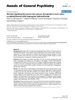
Intra-articular NBD treatment results in decreased pro-inflammatory cytokine expressionIntra-articular NBD treatment results in decreased pro-inflammatory cytokine expression. Shown are representative overview images of mutant NBD
(MUT)-treated ankle joints. Expression of different cytokines was evaluated by immunohistochemical staining of paraffin-embedded ankle joints.
Detailed images of synovial cytokine staining are provided (boxes in upper panels indicate synovial tissue location), followed by results from digital
image analysis (n = 10/group). Tumor necrosis factor (TNF)-α and interleukin (IL)-1-β expression was significantly reduced in the NBD-treated group
as compared with the MUT-treated group (*p = 0.05 and *p = 0.04, respectively). Data represent mean ± standard error of the mean.
Arthritis Research & Therapy Vol 8 No 4 Tas et al.
Page 6 of 9
(page number not for citation purposes)
vation in vitro. FLS were pre-treated for 2 hours with NBD or
the mutant control peptide and stimulated for 30 minutes with
IL-1-β. Cells were lysed and total intracellular protein was sep-
arated using SDS-PAGE. After immunoblotting, ph- and
unphosphorylated IκBα were detected using specific mAbs.
NBD pre-treatment resulted in reduced IL-1-β-induced IκBα
phosphorylation, whereas the mutant control peptide did not
affect IκBα phosphorylation in FLS. Using densitometry, we
found that the ph-IκBα/IκBα ratio was significantly reduced in
NBD compared with MUT-treated FLS (p < 0.01). NBD treat-
Figure 4
Nuclear factor-κB essential modulator binding domain (NBD) peptide treatment significantly reduces bone destructionNuclear factor-κB essential modulator binding domain (NBD) peptide
treatment significantly reduces bone destruction. (a) X-rays of the ankle
joints (n = 10/group) were made, and radiological damage was scored.
Representative pictures for NBD- and mutant NBD (MUT)-treated ankle
joints are shown. Large arrow indicates ankle demineralization. Small
arrow indicates midfoot demineralization and erosions. (b) NBD pep-
tide treatment significantly reduces bone destruction. Data represent
mean ± standard error of the mean (SEM) radiological scores (*p <
0.04). (c) No significant difference in bone destruction was observed in
the contralateral, non-injected paws. Data represent mean ± SEM radi-
ological scores.
Figure 5
NBD peptide blocks pro-inflammatory cytokine production by human macrophages in vitro and rheumatoid arthritis (RA) synovial biopsies ex vivoNBD peptide blocks pro-inflammatory cytokine production by human
macrophages in vitro and rheumatoid arthritis (RA) synovial biopsies ex
vivo. (a) NBD peptide blocks interleukin (IL)-1-β-induced tumor necro-
sis factor (TNF)-α production by human macrophages in vitro. Macro-
phages were pre-incubated with either NBD or mutant NBD (MUT)
peptide at a concentration of 50 µM for 2 hours. Subsequently, cells
were stimulated with IL-1-β (2.5 ng/ml). After 24 hours, supernatants
were collected, and TNF-α levels were measured by sandwich enzyme-
linked immunosorbent assay (ELISA). Data are representative of three
independent experiments performed in triplicates and are expressed as
mean ± standard error of the mean (SEM) (*p < 0.01). (b) NBD pep-
tide blocks TNF-α-induced IL-6 production of RA synovial biopsies ex
vivo. Synovial biopsies were cultured with TNF-α (10 ng/ml) in the
presence or absence of NBD peptides (100 µM). After 7 days, super-
natants were collected and IL-6 levels were measured by sandwich
ELISA and corrected for weight of the biopsy. Data are representative
of three independent experiments performed in triplicates and are
expressed as mean ± SEM (*p < 0.01).
Available online />Page 7 of 9
(page number not for citation purposes)
ment almost reduced the ph-IκBα/IκBα ratio to the level seen
in unstimulated cells (0.47 vs. 0.21 in unstimulated cells). The
mutant control peptide (MUT) did not affect IκBα phosphoryla-
tion (ph-IκBα/IκBα ratio 1.20 vs. 1.13 in stimulated control
cells) (Figure 1a). The low ph-IκBα/IκBα ratio after NBD treat-
ment of FLS was accompanied by a strong reduction in IL-1-
β-induced IL-6 secretion by these cells (1.4 ± 0.1 ng/ml vs.
8.5 ± 1.3 ng/ml in stimulated cells and 14.3 ± 4 ng/ml in MUT-
treated stimulated FLS, p < 0.01) (Figure 1b). Taken together,
these data demonstrate that the NBD peptide blocks IL-1-β-
induced IκBα phosphorylation in RA FLS, resulting in a less
inflammatory phenotype.
Intra-articular NBD treatment ameliorates AA in rats
Next, we investigated the therapeutic effects of this highly spe-
cific IKK-β inhibitor in established arthritis when administered
intra-articularly. In a dose-finding study (n = 6/group), we
found that two i.a. injections (on days 10 and 12) with a dose
of 150 µg NBD peptide significantly reduced arthritis severity
(P < 0.05), whereas a dose of 50 µg only marginally amelio-
rated arthritis (Figure 2a). Subsequently, we conducted a large
therapeutic study in which AA was induced in rats (n = 15/
group) on day 0. At the start of arthritis symptoms (day 10), the
animals received an i.a. injection with either the NBD peptide
or the MUT peptide (150 µg) into the right ankle joints. Two
days later, this procedure was repeated and the course of
arthritis was monitored by blinded observers until day 20 to
evaluate the effects of local IKK-β inhibition on paw swelling.
Intra-articular treatment with NBD resulted in significantly
reduced paw swelling (AUC 11.18 ± 1.14 vs. 15.40 ± 0.70,
NBD vs. MUT, respectively; p < 0.0001) (Figure 2b,c). Careful
evaluation of the internal organs of the animals (liver, kidney,
spleen, and so forth), conducted with a pathologist, did not
reveal any alterations compared with the control animals. Also,
no opportunistic infections occurred in the NBD-treated ani-
mals. Taken together, these findings do not suggest major sys-
temic effects. In short, we have demonstrated in two
independent AA experiments that two i.a. injections of the
NBD peptide significantly ameliorated arthritis severity.
Intra-articular NBD treatment results in reduced synovial
inflammation
Having shown the beneficial effect of the NBD peptide on the
severity of arthritis, we evaluated the effects of i.a. NBD treat-
ment on synovial cellularity in situ. Intra-articular injection of
the NBD peptide resulted in a significant decrease of inflam-
matory cells in the synovial tissue compared with MUT treat-
ment (239 ± 5 cells/mm
2
vs. 273 ± 7 cells/mm
2
, respectively;
p < 0.01). Histological evaluation of NBD-treated ankle joints
revealed less proliferation and invasive growth of the synovial
tissue (Figure 3). Next, we evaluated the effects of the NBD
peptide on synovial inflammation by performing immunohisto-
chemical stainings on sections from paraffin-embedded rat
ankle joints. Digital image analysis of comparable locations in
the synovial tissue showed a clear difference between NBD-
and MUT-treated animals in the expression of the pro-inflam-
matory cytokines TNF-α (4.06 × 10
2
± 2.40 × 10
1
versus 8.51
× 10
2
± 2.38 × 10
2
; p = 0.05) and IL-1-β (1.73 × 10
4
± 2.43
× 10
3
versus 2.66 × 10
4
± 5.30 × 10
3
; p = 0.04). IL-6
expression was not different between the two groups (1.70 ×
10
4
± 2.17 × 10
3
versus 1.87 × 10
4
± 2.10 × 10
3
) (Figure 3).
Intra-articular NBD treatment reduces bone destruction
We studied the effects of NBD treatment not only on synovial
inflammation, but also on bone destruction. Therefore, x-rays
of the ankle joints (Figure 4a) were made, and these plain radi-
ographs were scored for bone degradation, using a validated
scoring system [24]. Intra-articular NBD treatment significantly
reduced bone degradation of the injected ankle joints (p <
0.04) (Figure 4b) compared with MUT-treated or contralateral
joints. These findings show that local IKK-β inhibition in the
joint by i.a. injection of the small molecule NBD peptide not
only ameliorates arthritis, but concomitantly also reduces bone
destruction.
The NBD peptide inhibits pro-inflammatory cytokine
production in human macrophages and whole-tissue
synovial biopsies from patients with RA
To gain more knowledge on the therapeutic potential of the
NBD peptide in humans, we extended the experiments to
another pivotal cell type in the pathogenesis of RA, the macro-
phage. We found that NBD treatment of human macrophages
results in significantly reduced IL-1-β-induced TNF-α produc-
tion compared with MUT-treated macrophages (p < 0.01)
(Figure 5a). Finally, we conducted true translational research
in which we evaluated the effects of our highly specific IKK-β
inhibitor on human synovial tissue. Therefore, we collected
synovial biopsies from patients with RA by arthroscopy and
cultured the biopsies in the presence or absence of the NBD
peptide, followed by TNF-α stimulation. TNF-α was chosen for
stimulation of whole-tissue synovial biopsies because this
cytokine has been demonstrated to be pivotal in the pathogen-
esis of RA (reviewed in [30]). Supernatants were collected,
and IL-6 production was measured by ELISA. NBD treatment
resulted in a significant reduction of TNF-α-induced IL-6 pro-
duction compared with MUT treatment or TNF-α stimulation
alone (2.99 ± 0.01 versus 5.18 ± 0.61 or 6.40 ± 0.22; p <
0.01) (Figure 5b). In line with previous observations [17,18],
no effect of the NBD peptides on basal IL-6 production was
observed (data not shown), because the NBD peptide selec-
tively blocks the induction of NF-κB activity in response to pro-
inflammatory stimuli without affecting basal NF-κB activity
[20]. In conclusion, these experiments demonstrate the effec-
tiveness of the NBD peptide in human cells.
Discussion
In the present study, we show for the first time that i.a. admin-
istration of the highly specific IKK-β inhibitor NBD peptide sig-
nificantly reduces arthritis activity and bone destruction in vivo.
These results indicate that IKK-β-targeted NF-κB inhibition
Arthritis Research & Therapy Vol 8 No 4 Tas et al.
Page 8 of 9
(page number not for citation purposes)
using selective pharmacological inhibitors is beneficial in the
local treatment of established arthritis. Of note, only two i.a.
injections with the NBD peptide resulted in sustained reduc-
tion of the severity of arthritis in a therapeutic setting. Consist-
ent with these observations, synovial inflammation was
decreased as demonstrated by a decline in synovial cellularity
and reduced levels of the pro-inflammatory cytokines TNF-α
and IL-1-β. Importantly, i.a. NBD treatment concomitantly
resulted in reduced bone destruction, in agreement with the
effects shown after systemic treatment in murine collagen-
induced arthritis [18].
The biological effects of local NBD treatment are also consist-
ent with those observed using a gene therapy approach to tar-
get IKK-β locally in the joint [16]. Selective pharmacological
NF-κB inhibitors may reach the clinic faster because of possi-
ble safety and dose regulation issues that accompany gene
therapy. However, some of these issues might be resolved by
using vectors optimized for i.a. use (for example rAAV5 [31]
and disease-inducible promotors or other regulatable gene
expression systems [32]).
In RA, FLS and macrophages play important roles in the per-
petuation of synovial inflammation [5,6]. Our results indicate
that the NBD peptide may have great potential in humans as
well, because this NF-κB inhibitor efficiently blocked IL-1-β-
induced IκBα phosphorylation and IL-6 production in RA FLS,
as well as TNF-α production by human macrophages. One of
the important advantages of the NBD peptide, compared with
other IKK inhibitors, is that basal NF-κB activity remains unaf-
fected while NF-κB activation in response to pro-inflammatory
stimuli is effectively blocked [20]. Therefore, the beneficial role
of NF-κB in normal cellular functions is preserved, resulting in
less toxicity. Consequently, the effects of the NBD peptide on
pro-inflammatory cytokine production in vitro were not due to
increased apoptosis or necrosis (data not shown). In addition,
TNF-α-induced pro-inflammatory cytokine production in cul-
tured synovial biopsies from patients with RA was also signifi-
cantly reduced. In these synovial biopsies, the micro-
architecture of the synovium is preserved, allowing investiga-
tors to study the effects of the NBD peptide on synovial inflam-
mation in the complex, biologically relevant network of cells
that contribute to the inflammatory process rather than in indi-
vidually cultured cell types. Thus, this may serve as a model to
predict a possible therapeutic effect in human disease [33].
Many anti-inflammatory drugs used in the treatment of arthritis
target, at least in part, NF-κB. Of these drugs, glucocorticoids
like dexamethasone and prednisolone (although non-specific)
are considered the most powerful NF-κB inhibitors [34,35].
Intra-articular steroid injections are widely used to control local
inflammation. In addition to showing local side effects such as
reduced bone formation [36], recent work has shown
unwanted systemic effects due to absorption of steroids from
the i.a. space [37,38]. The most common side effect caused
by systemic absorption of i.a. steroids is suppression of pitui-
tary-adrenal axis function [38]. This suppression may last from
up to 2 weeks to even 6 months after i.a. injection and may ulti-
mately lead to adrenal failure [39]. Therefore, there is a clear
need for potent anti-inflammatory drugs for i.a. administration
without steroid action to prevent these unwanted side effects.
Pharmacological NF-κB inhibitors like the NBD peptide may
fulfill this need; because they are mainly peptide-based and
not steroid-based, they selectively block NF-κB activity in the
joint without causing these side effects. In addition, the NBD
peptide inhibits only pro-inflammatory IKK activity [20] and
may therefore be safer than other IKK inhibitors if absorbed
from the i.a. space and released systemically. However, exten-
sive pharmacological evaluation of this approach is required to
carefully monitor pharmacokinetics and pharmacodynamics,
as well as potential toxicity, of new pharmacological NF-κB
inhibitors like the NBD peptide before clinical trials with these
compounds may be initiated.
Conclusion
We have demonstrated that local small peptide-mediated NF-
κB inhibition not only ameliorated established arthritis and
reduced bone destruction in an animal model of RA, but also
prevented pro-inflammatory cytokine production by human RA
synovial biopsies. Our results suggest that i.a. treatment with
the NBD peptide may represent a novel therapeutic approach
in RA.
Competing interests
The authors declare that they have no competing interests.
Authors' contributions
SWT carried out Western blots, ELISAs, and animal studies,
evaluated radiological scores and immunohistochemical stain-
ings, and drafted the manuscript. MJV participated in the
design of the study, evaluated radiological scores, and helped
to draft the manuscript. NH assisted in the animal studies and
cell culture and performed immunohistochemical stainings
and digital image analysis. MJM and SG participated in the
design of the study and helped to draft the manuscript. PPT
conceived of the study, participated in its design and coordi-
nation, and helped to draft the manuscript. All authors read
and approved the final manuscript.
Acknowledgements
SWT was supported by the Dutch Arthritis Foundation (grant NR 01-1-
302) and a EULAR Young Investigator Award.
References
1. Firestein GS: Evolving concepts of rheumatoid arthritis. Nature
2003, 423:356-361.
2. Henderson B, Pettipher ER: The synovial lining cell: biology and
pathobiology. Semin Arthritis Rheum 1985, 15:1-32.
3. Chu CQ, Field M, Feldmann M, Maini RN: Localization of tumor
necrosis factor alpha in synovial tissues and at the cartilage-
pannus junction in patients with rheumatoid arthritis. Arthritis
Rheum 1991, 34:1125-1132.
Available online />Page 9 of 9
(page number not for citation purposes)
4. Smeets TJ, Barg EC, Kraan MC, Smith MD, Breedveld FC, Tak PP:
Analysis of the cell infiltrate and expression of proinflamma-
tory cytokines and matrix metalloproteinases in arthroscopic
synovialbiopsies: comparison with synovial samples from
patients with end stage, destructive rheumatoid arthritis. Ann
Rheum Dis 2003, 62:635-638.
5. Tak PP, Smeets TJ, Daha MR, Kluin PM, Meijers KA, Brand R,
Meinders AE, Breedveld FC: Analysis of the synovial cell infil-
trate in early rheumatoid synovial tissue in relation to local dis-
ease activity. Arthritis Rheum 1997, 40:217-225.
6. Pap T, Muller-Ladner U, Gay RE, Gay S: Fibroblast biology. Role
of synovial fibroblasts in the pathogenesis of rheumatoid
arthritis. Arthritis Res 2000, 2:361-367.
7. Yamanishi Y, Firestein GS: Pathogenesis of rheumatoid arthri-
tis: the role of synoviocytes. Rheum Dis Clin North Am 2001,
27:355-371.
8. Tas SW, Remans PH, Reedquist KA, Tak PP: Signal transduction
pathways and transcription factors as therapeutic targets in
inflammatory disease: towards innovative antirheumatic
therapy. Curr Pharm Des 2005, 11:581-611.
9. Tak PP, Firestein GS: NF-kappaB: a key role in inflammatory
diseases. J Clin Invest 2001, 107:7-11.
10. Karin M, Delhase M: The I kappa B kinase (IKK) and NF-kappa
B: key elements of proinflammatory signalling. Semin Immunol
2000, 12:85-98.
11. Li Q, Verma IM: NF-kappaB regulation in the immune system.
Nat Rev Immunol 2002, 2:725-734.
12. Ruland J, Mak TW: From antigen to activation: specific signal
transduction pathways linking antigen receptors to NF-kap-
paB. Semin Immunol 2003, 15:177-183.
13. Karin M, Yamamoto Y, Wang QM: The IKK NF-kappa B system:
a treasure trove for drug development. Nat Rev Drug Discov
2004, 3:17-26.
14. Handel ML, McMorrow LB, Gravallese EM: Nuclearfactor-kappa
B in rheumatoid synovium. Localization of p50 and p65. Arthri-
tis Rheum 1995, 38:1762-1770.
15. Marok R, Winyard PG, Coumbe A, Kus ML, Gaffney K, Blades S,
Mapp PI, Morris CJ, Blake DR, Kaltschmidt C, Baeuerle PA: Acti-
vation of the transcription factor nuclearfactor-kappaB in
human inflamed synovial tissue. Arthritis Rheum 1996,
39:583-591.
16. Tak PP, Gerlag DM, Aupperle KR, van de Geest DA, Overbeek M,
Bennett BL, Boyle DL, Manning AM, Firestein GS: Inhibitor of
nuclear factorkappaB kinase beta is a key regulator of syno-
vial inflammation. Arthritis Rheum 2001, 44:1897-1907.
17. di Meglio P, Ianaro A, Ghosh S: Amelioration of acute inflamma-
tion by systemic administration of a cell-permeable peptide
inhibitor of NF-kappaB activation. Arthritis Rheum 2005,
52:951-958.
18. Jimi E, Aoki K, Saito H, D'Acquisto F, May MJ, Nakamura I, Sudo T,
Kojima T, Okamato F, Fukushima H: Selective inhibition of NF-
kappa B blocks osteoclastogenesis and prevents inflamma-
tory bone destruction in vivo. Nat Med 2004, 10:617-624.
19. McIntyre KW, Shuster DJ, Gillooly KM, Dambach DM, Pattoli MA,
Lu P, Zhou XD, Qiu Y, Zusi FC, Burke UR: A highly selective
inhibitor of I kappa B kinase, BMS-345541, blocks both joint
inflammation and destruction in collagen-induced arthritis in
mice. Arthritis Rheum 2003, 48:2652-2659.
20. May MJ, D'Acquisto F, Madge LA, Glockner J, Pober JS, Ghosh S:
Selective inhibition of NF-kappaB activation by a peptide that
blocks the interaction of NEMO with the IkappaB kinase
complex. Science 2000, 289:1550-1554.
21. van Holten J, Reedquist K, Sattonet-Roche P, Smeets TJ, Plater-
Zyberk C, Vervoordeldonk MJ, Tak PP: Treatment with recom-
binant interferon-beta reduces inflammation and slows carti-
lage destruction in the collagen-induced arthritis model of
rheumatoid arthritis. Arthritis Res Ther 2004, 6:R239-R249.
22. Tas SW, de Jong EC, Hajji N, May MJ, Ghosh S, Vervoordeldonk
MJ, Tak PP: Selective inhibition of NF-kappaB in dendritic cells
by the NEMO-binding domain peptide blocks maturation and
prevents T cell proliferation and polarization. Eur J Immunol
2005, 35:1164-1174.
23. Nguyen KH, Boyle DL, McCormack JE, Chada S, Jolly DJ, Firestein
GS: Direct synovial gene transfer with retroviral vectors in rat
adjuvant arthritis. J Rheumatol 1998, 25:1118-1125.
24. Boyle DL, Moore J, Yang L, Sorkin LS, Firestein GS: Spinal ade-
nosine receptor activation inhibits inflammation and jointde-
struction in rat adjuvant-induced arthritis. Arthritis Rheum
2002, 46:3076-3082.
25. Kraan MC, Smith MD, Weedon H, Ahern MJ, Breedveld FC, Tak
PP: Measurement of cytokine and adhesion molecule expres-
sion insynovial tissue by digital image analysis. Ann Rheum
Dis 2001, 60:296-298.
26. Haringman JJ, Vinkenoog M, Gerlag DM, Smeets TJ, Zwinderman
AH, Tak PP: Reliability of computerized image analysis for the
evaluation of serial synovial biopsies in randomized controlled
trials in rheumatoid arthritis. Arthritis Res Ther 2005,
7:R862-R867.
27. Tas SW, Quartier P, Botto M, Fossati-Jimack L: Macrophages
from patients with SLE and rheumatoid arthritis have defective
adhesion in vitro, while only SLE macrophages have impaired
uptake of apoptotic cells. Ann Rheum Dis 2006, 65:216-221.
28. Kraan MC, Reece RJ, Smeets TJ, Veale DJ, Emery P, Tak PP: Com-
parison of synovial tissues from the knee joints and the small
joints of rheumatoid arthritis patients: implications for patho-
genesis and evaluation of treatment. Arthritis Rheum 2002,
46:2034-2038.
29. Dasgupta S, Jana M, Zhou Y, Fung YK, Ghosh S, Pahan K:
Antineuroinflammatory effect of NF-kappaB essentialmodi-
fier-binding domain peptides in the adoptive transfer model of
experimental allergic encephalomyelitis. J Immunol 2004,
173:1344-1354.
30. Feldmann M, Brennan FM, Williams RO, Woody JN, Maini RN: The
transfer of a laboratory based hypothesis to a clinically useful
therapy: the development of anti-TNF therapy of rheumatoid
arthritis. Best Pract Res Clin Rheumatol 2004, 18:59-80.
31. Adriaansen J, Tas SW, Klarenbeek PL, Bakker AC, Apparailly F,
Firestein GS, Jorgensen C, Vervoordeldonk MJ, Tak PP:
Enhanced gene transfer to arthritic joints using adeno-associ-
ated virus type 5: implications for intra-articular gene therapy.
Ann Rheum Dis 2005, 64:1677-84.
32. Goverdhana S, Puntel M, Xiong W, Zirger JM, Barcia C, Curtin JF,
Soffer EB, Mondkar S, King GD, Hu J: Regulatable gene expres-
sion systems for gene therapy applications: progress and
future challenges. Mol Ther 2005, 12:189-211.
33. Chabaud M, Miossec P: The combination of tumor necrosis fac-
tor alpha blockade with interleukin-1 and interleukin-17 block-
ade is more effective for controlling synovial inflammation and
bone resorptionin an ex vivo model. Arthritis Rheum 2001,
44:1293-1303.
34. Yamamoto Y, Gaynor RB: Therapeutic potential of inhibition of
the NF-kappaB pathway in the treatment of inflammation and
cancer. J Clin Invest 2001, 107:135-142.
35. De Bosscher K, Vanden Berghe W, Haegeman G: The interplay
between the glucocorticoid receptor and nuclear factor-kap-
paB or activator protein-1: molecular mechanisms for gene
repression. Endocr Rev 2003, 24:488-522.
36. Weitoft T, Larsson A, Saxne T, Ronnblom L: Changes of cartilage
and bone markers after intra-articular glucocorticoid therapy
with and without postinjection rest in rheumatoid arthritis
patients. Ann Rheum Dis 2005, 64:1750-1753.
37. Kumar S, Singh RJ, Reed AM, Lteif AN: Cushing's syndrome
after intra-articular and intradermal administration of triamci-
nolone acetonide in three pediatric patients. Pediatrics 2004,
113:1820-1824.
38. Mader R, Lavi I, Luboshitzky R: Evaluation of the pituitary-adre-
nal axis function following single intraarticularinjection of
methylprednisolone. Arthritis Rheum 2005, 52:924-928.
39. Wicki J, Droz M, Cirafici L, Vallotton MB: Acute adrenal crisis in
a patient treated with intraarticular steroid therapy. J
Rheumatol 2000, 27:510-511.