Báo cáo y học: "Inhibition of macropinocytosis blocks antigen presentation of type II collagen in vitro and in vivo in HLA-DR1 transgenic mice" docx
Bạn đang xem bản rút gọn của tài liệu. Xem và tải ngay bản đầy đủ của tài liệu tại đây (1.67 MB, 11 trang )
Open Access
Available online />Page 1 of 11
(page number not for citation purposes)
Vol 8 No 4
Research article
Inhibition of macropinocytosis blocks antigen presentation of
type II collagen in vitro and in vivo in HLA-DR1 transgenic mice
Alexei von Delwig
1
, Catharien MU Hilkens
1
, Daniel M Altmann
2
, Rikard Holmdahl
3
, John D Isaacs
1
,
Clifford V Harding
4
, Helen Robertson
5
, Norman McKie
1
and John H Robinson
1
1
Musculoskeletal Research Group, Clinical Medical Sciences, University of Newcastle upon Tyne, Framlington Place, Newcastle upon Tyne, UK
2
Human Disease Immunogenetics Group, Department of Infectious Diseases, Imperial College School of Medicine, Hammersmith Hospital, London,
UK
3
Department of Cell and Molecular Biology, Lund University, Lund, Sweden
4
Department of Pathology, Case Western Reserve University, Cleveland, OH, USA
5
BioImaging Facility, Clinical Laboratory Sciences, University of Newcastle upon Tyne, Framlington Place, Newcastle upon Tyne, UK
Corresponding author: Alexei von Delwig,
Received: 16 Feb 2006 Revisions requested: 27 Mar 2006 Revisions received: 13 Apr 2006 Accepted: 24 Apr 2006 Published: 16 May 2006
Arthritis Research & Therapy 2006, 8:R93 (doi:10.1186/ar1964)
This article is online at: />© 2006 von Delwig et al.; licensee BioMed Central Ltd.
This is an open access article distributed under the terms of the Creative Commons Attribution License ( />),
which permits unrestricted use, distribution, and reproduction in any medium, provided the original work is properly cited.
Abstract
Professional antigen-presenting cells, such as dendritic cells,
macrophages and B cells have been implicated in the
pathogenesis of rheumatoid arthritis, constituting a possible
target for antigen-specific immunotherapy. We addressed the
possibility of blocking antigen presentation of the type II
collagen (CII)-derived immunodominant arthritogenic epitope
CII
259–273
to specific CD4 T cells by inhibition of antigen uptake
in HLA-DR1-transgenic mice in vitro and in vivo. Electron
microscopy, confocal microscopy, subcellular fractionation and
antigen presentation assays were used to establish the
mechanisms of uptake, intracellular localization and antigen
presentation of CII by dendritic cells and macrophages. We
show that CII accumulated in membrane fractions of
intermediate density corresponding to late endosomes.
Treatment of dendritic cells and macrophages with cytochalasin
D or amiloride prevented the intracellular appearance of CII and
blocked antigen presentation of CII
259–273
to HLA-DR1-
restricted T cell hybridomas. The data suggest that CII was
taken up by dendritic cells and macrophages predominantly via
macropinocytosis. Administration of amiloride in vivo prevented
activation of CII-specific polyclonal T cells in the draining
popliteal lymph nodes. This study suggests that selective
targeting of CII internalization in professional antigen-presenting
cells prevents activation of autoimmune T cells, constituting a
novel therapeutic strategy for the immunotherapy of rheumatoid
arthritis.
Introduction
Professional antigen-presenting cells (APCs), such as den-
dritic cells (DCs), macrophages and B lymphocytes, play a piv-
otal role in the pathogenesis of autoimmune diseases in animal
models by presenting arthritogenic T cell epitopes to autoim-
mune T cells [1-3] Adoptive transfer of ex vivo cultured
autoantigen-specific DCs has been shown to induce a variety
of experimental autoimmune diseases, such as autoimmune
diabetes, experimental autoimmune encephalomyelitis and
erosive inflammatory arthritis [4-6]. DCs in situ are often sur-
rounded by a cluster of T cells [7] and are thought to internal-
ize autoantigens from the extracellular matrix and cartilage for
intracellular processing and presentation of arthritogenic
epitopes to specific CD4 T cells, as well as to induce activa-
tion of B lymphocytes in patients with rheumatoid arthritis (RA)
[8]. B lymphocytes have also been shown to be critical both as
antigen-presenting and antibody-secreting cells in the patho-
genesis of autoimmune arthritis [3,9-11] High efficiency of
antigen presentation of arthritogenic epitopes by macro-
phages has also been demonstrated [12,13].
Type II collagen (CII, α1(II)3), the most abundant fibrillar pro-
tein of articular cartilage [14], is considered an important
autoantigen involved in the pathogenesis of collagen-induced
APC = antigen-presenting cell; CII = type II collagen; DC = dendritic cell; DMA = 5-(N,N-dimethyl)amiloride; ELISA = enzyme-linked immunosorbent
assay; FBS = fetal bovine serum; LPS = lipopolysaccharide; mAb = monoclonal antibody; MHC = major histocompatibility complex; NF = nuclear
factor; RA = rheumatoid arthritis.
Arthritis Research & Therapy Vol 8 No 4 von Delwig et al.
Page 2 of 11
(page number not for citation purposes)
arthritis in mice and RA in humans [15,16] In addition, collagen
has been shown to deliver a direct maturation stimulus to DCs
[17], possibly via ligation of Toll-like receptor 4 (TLR4) or by
binding to cell surface integrins [18,19], suggesting that DCs
can present collagen T cell epitopes without additional inflam-
matory or danger signals. A direct co-stimulatory activity of col-
lagen has, however, not been demonstrated in vivo as there is
no evidence that collagen by itself displays adjuvant activity
[20]. No information is available on the mechanisms engaged
in CII uptake into professional APCs for presentation of arthri-
togenic epitopes to CD4 T cells.
Several mechanisms have been described to mediate internal-
ization of antigens into APCs, including phagocytosis, mac-
ropinocytosis, receptor-mediated endocytosis and caveolar
endocytosis. Phagocytosis follows the recognition of particles
≥0.25 μm by specific receptors and an F-actin microfilament-
dependent internalization into phagosomes [21]. Macropinoc-
ytosis does not require ligation of specific receptors and is
accompanied by membrane ruffling and F-actin-dependent
uptake into large macropinosomes of 0.15 to 5.0 μm [22].
Receptor-mediated endocytosis of smaller particles and mole-
cules engages clathrin-coated pits and F-actin recruitment at
endocytic sites [23], while clathrin-independent endocytosis
is dependent on intact caveolae and lipid rafts [24]. In contrast
to other internalization mechanisms, caveolar endocytosis
does not deliver antigens to lysosomes and, therefore, does
not appear to play a major role in antigen processing and pres-
entation [2,25].
In this report, we show that CII was taken up preferentially via
macropinocytosis into DCs and macrophages from HLA-DR1
transgenic mice for antigen presentation of both the glyco-
sylated and non-glycosylated forms of the arthritogenic CII
259–
273
epitope to CD4 T cells. Treatment of mice with an inhibitor
of macropinocytosis also prevented activation of CII
259–273
-
specific T cells in vivo.
Materials and methods
Antigens
Human CII purified from normal human cartilage was pur-
chased from MD BioSciences (Zürich, Switzerland). The glyc-
osylated peptide (GIAGF KGEQGPKGET; K = GalHyL
264
)
corresponding to epitope CII
259–273
GalHyL
264
was synthe-
sized using β-D-galactopyranosyl-5-hydroxy-L-lysine, as
described previously [26]. The non-glycosylated peptide
pCII
259–273
was purchased from GenScript Corp. (Piscata-
way, NJ, USA), and purity was confirmed by high-performance
liquid chromatography.
Animals
In all experiments described in this study we used previously
reported mice transgenic for HLA-DR1 on a major histocom-
patibility complex (MHC) class II-deficient background (desig-
nated C57BL/6J
0-0
HLA-DR1), which carried full-length
genomic constructs for HLA-DRA1*0101 and HLA-
DRB1*0101, developed by one of us (DMA) [27]. Experi-
ments described in this report have been performed under the
terms of Animals (Scientific Procedures) Act 1986, and
authorized by the Home Secretary, Home Office UK. The work
has been approved by the Ethical Review Committee of the
University of Newcastle upon Tyne.
Cells
Culture media ingredients and inhibitors were purchased from
Sigma Chemical Co. (Dorset, UK), unless stated otherwise.
Cells were grown in culture medium (RPMI 1640 medium con-
taining 3 mM L-glutamine, 50 μM 2-mercaptoethanol, 10%
FBS and 30 μg/ml gentamycin). T cell hybridomas HCII-9.1
(specific for the non-glycosylated peptide) and HCII-9.2 (spe-
cific for the glycosylated peptide) have been described previ-
ously [27]. Macrophages were grown from femoral bone
marrow cells in culture medium supplemented with 5% horse
serum, 1 mM sodium pyruvate, 10 mM HEPES and 7.5% of a
supernatant from the L929 cell line as a source of macrophage
colony stimulating factor (M-CSF), as described [27]. Macro-
phages were activated with 10 U/ml recombinant IFN-γ (R&D
Systems, Abingdon, UK) for 24 hours (purity approximately
95% based on CD11b expression).
Dendritic cells were grown from bone marrow progenitor cells
in the culture medium supplemented with 20 ng/ml recom-
binant mouse granulocyte-macrophage colony stimulating fac-
tor (GM-CSF; BioSource International, Nivelles, Belgium) for 5
days with culture medium changes on days 2 and 3. On day 5,
DCs were purified using CD11c-labeled magnetic
MicroBeads (Miltenyi Biotec, Bisley, Surrey, UK), according to
the manufacturer's instructions (purity approximately 92%
based on CD11c expression). Maturation was induced by
treatment of DCs with 0.2 μg/ml lipopolysaccharide (LPS;
purified by phenol extraction from Salmonella enterica, serovar
typhimurium, Sigma Chemical Co.) for 24 hours.
Antigen presentation assays
Adherent macrophages at 10
5
/well in 48 flat-well plates
(Corning Limited, Artington, Surrey, UK), or mature and imma-
ture DCs at 10
4
/well in flat-bottomed 96 well plates (Greiner
Bio-One Ltd, Stonehouse, Gloucestershire, UK) were pulsed
with a dilution series of CII or relevant synthetic peptides
(range 40.0 to 0.02 μg/ml) for 5 hours in the absence or pres-
ence of inhibitors of uptake (10.0 μM cytochalasin D, 5.0 μM
monodansylcadaverine, 1.0 mM amiloride, 0.2 mM 5-(N,N-
dimethyl) amiloride (DMA) or 0.4 μg/ml filipin) for 5 hours at
37°C [28,29]. Time and the optimal doses of APCs, antigens
and inhibitors were established in separate dose-response
experiments. Cells were fixed with 1.0% paraformaldehyde for
5 minutes, washed thoroughly to remove the fixative and T cell
hybridoma HCII-9.1 (specific for the non-glycosylated epitope)
and HCII-9.2 (specific for the glycosylated epitope) were
added (5 × 10
4
/well) and incubated for 24 hours at 37°C.
Available online />Page 3 of 11
(page number not for citation purposes)
Usage of synthetic peptides in all experiments controlled for
the non-specific toxic effect of metabolic inhibitors and the
responsiveness of T cell hybridomas. The interleukin-2 content
of hybridoma supernatants was measured by bioassay as the
proliferative response of the cytotoxic T cell line-2 (3 × 10
4
/
well; CTLL-2, ATCC, TIB 214, American Type Culture Collec-
tion, Rockville, MD, USA). Proliferation assays were performed
by incubating popliteal lymph node cells or spleen cells (2 ×
10
5
/well) with a dilution series of CII and synthetic peptides for
72 hours, as previously described [30].
Cells were incubated during the last 18 hours in the presence
of 14.8 kBq of [
3
H]thymidine (TRA310, specific activity 307
MBq/mg; Amersham International plc, Didcot, Oxfordshire,
UK), harvested on glass fiber membranes and radioactivity
was quantified using a direct Beta Counter (Matrix 9600,
Packard Instrument Company, Meridan, CT, USA).
Proliferation assays
For testing CII-specific T cell responses in draining lymph
nodes, mice were immunized in the footpad with 50 μg CII
emulsified 1:1 in TiterMax adjuvant in the absence or presence
of amiloride (150 μg/mouse [31]) and popliteal lymph nodes
were removed 7 days later. Cells (2 × 10
5
/well) were mixed
with a dilution series of CII, synthetic peptides or the polyclo-
nal T cell mitogen concanavalin A in round-bottomed 96 well
plates (Corning Limited) and incubated for 4 days at 37°C in
a humidified CO
2
incubator. Cells were incubated during the
last 18 hours in the presence of 14.8 kBq of [
3
H]thymidine,
harvested and radioactivity was measured, as described
above.
Subcellular fractionation
Macrophages (15 to 20 × 10
6
cells) were pulsed with 200 μg/
ml CII for 30 minutes and chased for different periods of time.
Macrophages were homogenized in buffer containing 0.25 M
sucrose, 10 mM HEPES, pH 7.4 in a Dounce tissue grinder
(Wheaton, Millville, NJ, USA) to obtain 80% to 85% cell lysis.
Subcellular fractionation of macrophages was performed by
density gradient centrifugation in 27% Percoll (Amersham plc,
Little Chalfont, Buckinghamshire, UK) using a Sorvall type A-
1256 fixed angle rotor (36,000 × g, 60 minutes, 4°C; Kendro
Laboratory Products plc, Bishop's Stortford, Hertfordshire,
UK), as described previously [27]. Six fractions of 1.5 ml were
collected manually numbered 1 to 6 from the top of the gradi-
ent. Percoll gradient fractions were each tested for β-hex-
osaminidase activity (marker for the presence of lysosomal
enzymes) and alkaline phosphodiesterase I activity (marker for
the presence of plasma membranes), as described [27,32]
The localization of markers of endosomal compartments and
CII within subcellular fractions was performed by ELISA, as
previously described [33]. Briefly, 50 μl of Percoll fractions
were dried in a constant flow cabinet in 96 well Microtiter
®
Immunoassays plates (Immulon
®
1, flat bottom, Dynex Tech-
nologies, Southampton, UK). Plates were blocked in phos-
phate-buffered saline containing 0.05% Tween 20, 10% FBS
and unlabeled anti-mouse mAb specific for FcγIIR and FcγIIIR
(1:200; clone 2.4G2, Fc Block
®
, PharMingen, Oxford, UK) for
1 hour at room temperature. Plates were washed and incu-
bated for 1 hour with goat anti-CII polyclonal antibody, goat
anti-Rab7 and Rab9 polyclonal antibody (1: 200; Santa Cruz
Biotechnology, Inc., Heidelberg, Germany). Normal goat
serum was used in control experiments. After washing, plates
were incubated for 1 hour with rabbit anti-goat IgG peroxidase
conjugate diluted 1:1000, washed and the reaction was devel-
oped with the liquid substrate system for ELISA 2,2'-azino-
bis(3-ethylbenzthiazoline-6-sulfonic acid. Absorbance was
measured at 405 nm.
Flow cytometry
Bone-marrow macrophages and DCs were incubated in the
absence or presence of inhibitors of uptake for five hours, and
the expression of HLA-DR, CD80, CD86 and CD40 mole-
cules was analyzed by flow cytometry, as described [28].
Briefly, cells were incubated for 30 minutes at 4°C in Hank's
balanced salt solution containing 2% FBS, 0.01 M HEPES
buffer with purified anti-mouse CD16/CD32 (Fc Block
®
, BD-
PharMingen) followed by incubation for 30 minutes at 4°C
with either of the following mAb fluorescent conjugates (BD-
PharMingen, Cowley, Oxford, UK): anti-HLA-DR FITC, anti-
CD40 FITC, anti-CD80 PE, anti-CD86 FITC, anti-CD11c
FITC, anti-CD11b FITC or isotype control, rat IgG2a PE plus
IgG2b FITC. Cells were analyzed with a FACScan
®
flow
cytometer (Becton Dickinson, Mount View, CA, USA), and
10,000 events were collected for each sample.
Electron microscopy
Bone-marrow macrophages and DCs were pulsed with 200
μg/ml CII in the absence or presence of inhibitors of uptake for
30 minutes. Transmission electron microscopy was performed
as described previously [28]. Briefly, cells were fixed in 2.5%
EM grade gluteraldehyde (TAAB Lab. Equipment, Aldermas-
ton, Berkshire, UK) diluted in 0.1 M phosphate buffer, pH 7.3,
washed in phosphate buffer and post-fixed with 1% osmium
tetroxide (Agar Scientific, Stansted, Essex, UK). Samples were
sequentially dehydrated through a graded acetone series,
impregnated with TAAB epoxy resin kit (TAAB Lab. Equip-
ment) and polymerized at 60°C for 24 hours. Blocks were thin
sectioned (80 nm), stained with uranyl acetate and lead citrate
(Leica UK Ltd, Milton Keynes, UK), and examined with a Philips
CM 100 (Compustage) Transmission Electron microscope
(Philips Electron Optics, Eindhoven, The Netherlands). Sec-
tions through several planes of more than 50 APCs were
examined for each treatment.
Confocal microscopy
Bone-marrow macrophages and DCs were pulsed with 200
μg/ml CII in the absence or presence of 1.0 mM amiloride for
30 minutes at 37°C. Cytospins were prepared by centrifuga-
tion of 2 × 10
4
cells in 200 μl in a Shandon Cytospin 3 cyto-
Arthritis Research & Therapy Vol 8 No 4 von Delwig et al.
Page 4 of 11
(page number not for citation purposes)
centrifuge (Thermo Electron Corp., Waltham, MA, USA). The
slides were air-dried at room temperature for 30 minutes, fixed
in acetone for 10 minutes at room temperature and permeabi-
lized in 0.1% Triton X-100 in PBS for 15 minutes at 4°C. After
washing (10 mM TRIS HCl pH 7.6, containing 150 mM NaCl,
TBS buffer) and blocking (normal rabbit serum 1:5 in TBS
buffer, 1 hour at room temperature) staining was performed
with goat anti-human CII polyclonal antibodies (1:100 in TBS
buffer, 4°C, 18 hours; Santa Cruz Biotechnology, Inc.). Slides
were washed and incubated with rabbit anti-goat IgG-FITC
(1:100, 2 hours, room temperature, in the dark). After washing,
slides were mounted in aqueous fluorescent mounting
medium (DAKO Cytomation, Carpenteria, CA, USA). Confo-
cal microscopy was performed at the BioImaging facility, Uni-
versity of Newcastle upon Tyne, using Leica TCS SP2 UV
laser scanning confocal microscope (Leica Microsystems
GmbH, Heidelberg, Germany) equipped with Time 63 oil
immersion 1.32 No Plan A Pro lens. Images were acquired
using the 488 excitation laser and emission was detected
between 500 and 560 nm. Images were collected using 0.5
μm Z-steps and these were projected using maximal projec-
tion and overlaid with single optimized transmitted light
images. In the control, cells were incubated in the absence of
CII, stained and imaged at the same gain and offset levels as
the positive cells and no fluorescence was observed.
Results
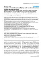
Mechanisms of CII uptake in macrophages and DCs
To study the mechanisms of uptake of CII, macrophages and
DCs from HLA-DR1-tg mice were incubated with CII for 30
minutes and visualized by transmission electron microscopy
(Figure 1a–d). CII fibrils of different size were seen inside mac-
rophages and DCs, showing that CII was internalized (Figure
1b,d). However, CII fibrils were rarely seen in the multiple sec-
tions examined, presumably because of the low probability of
the plane section coinciding with the longitudinal axis of the
CII fibrils.
Electron microscopy studies also revealed that cytochalasin
D, which prevents F-actin polymerization and hence inhibits
both phagocytosis and macropinocytosis [34], blocked the
appearance of CII inside both macrophages and DCs (Figure
2a). To distinguish between phagocytosis and macropinocyto-
sis, cells were treated with amiloride, which inhibits membrane
Na
+
/H
+
-ATPase, membrane ruffling and macropinocytosis
Figure 1
Electron micrographs of dendritic cells and macrophages pulsed with type II collagen (CII)Electron micrographs of dendritic cells and macrophages pulsed with
type II collagen (CII). (a,b) Macrophages and (c,d) dendritic cells were
incubated in the (a,c) absence and (b,d) presence of 200 μg/ml CII for
30 minutes and analyzed by transmission electron microscopy. The
arrows show fibrils of collagen aligned parallel to the plane of the sec-
tion. Magnification: (a) ×8,900; (b) ×6,610; (c) ×8,900; (d) ×21,000.
Bar = 1 μm. Sections through several planes of more than 50 cells
were examined for each treatment.
Figure 2
Electron micrographs of the effect of inhibitors of uptake on type II col-lagen (CII) internalization by macrophagesElectron micrographs of the effect of inhibitors of uptake on type II col-
lagen (CII) internalization by macrophages. Macrophages were pulsed
with 200 μg/ml CII for 30 minutes in the presence of (a) 10.0 μM cyto-
chalasin D, (b) 1.0 mM amiloride, (c) 5.0 μM monodansylcadaverine
(MDC) or (d) 0.4 μg/ml filipin and analyzed by electron microscopy.
Magnification: (a) ×6,610; (b) ×52,000; (c) ×21,000; (d) ×73,000. Bar
= (a,c) 1 μm or (b,d) 200 nm. Black arrows show fibrils of collagen
aligned parallel to the plane of the section; the white arrow shows an
unwinding collagen fibril inside the cell. Sections through several
planes of more than 50 cells were examined for each treatment.
Available online />Page 5 of 11
(page number not for citation purposes)
[35]. Internalization of CII was also undetectable in the pres-
ence of amiloride (Figure 2b), suggesting the involvement of
macropinocytosis rather than phagocytosis in the uptake of CII
[28]. In contrast, monodansylcadaverine, which inhibits forma-
tion of clathrin-coated pits and subsequent receptor-mediated
endocytosis [36], and filipin, which inhibits caveolae formation
[37], did not prevent CII uptake (Figure 2c,d). These data sug-
gest that CII was internalized by macrophages and DCs prima-
rily by macropinocytosis.
We confirmed the identity of the material internalized by mac-
rophages and DCs as CII by confocal microscopy using anti-
CII antibodies (Figure 3a,d). Interestingly, DCs displayed a rel-
atively stronger CII-specific fluorescence compared with mac-
rophages, which is consistent with the higher efficiency of
DCs as APCs compared with macrophages [38]. Amiloride
completely blocked the intracellular appearance of CII in both
Figure 3
Confocal micrographs of dendritic cells and macrophages pulsed with type II collagen (CII)Confocal micrographs of dendritic cells and macrophages pulsed with
type II collagen (CII). (a-c) Macrophages and (d-f) dendritic cells were
incubated in the (a,b,d,e) presence or (c,f) absence of 200 μg/ml CII
for 30 minutes, stained for CII expression and analyzed by confocal
microscopy. Magnification ×630, and bars denote (a) 6.63 μm, (b)
8.09 μm, (c) 5.0 μm, (d) 4.27 μm, (e) 4.64 μm and (f) 4.0 μm. More
than 50 cells were examined for each treatment.
Figure 4
Subcellular distribution of type II collagen (CII) in macrophagesSubcellular distribution of type II collagen (CII) in macrophages. (a)
Macrophages were subjected to subcellular fractionation and Percoll
fractions were analyzed for the expression of the plasma membrane-
associated enzyme alkaline phosphodiesterase I (open diamonds), the
lysosomal enzyme β-hexosaminidase (closed diamonds) and markers of
late endosomes Rab7 (closed circles) and Rab9 (open circles); 27%
Percoll alone is shown as fraction 0. Enzyme activity was measured as
absorbance at 405 nm. Goat serum was used as a negative control
(squares). (b) Macrophages were incubated in the absence (open cir-
cles) or presence of 200 μg/ml CII for 30 minutes and chased for 1
(open diamonds), 3 (closed diamonds), 5 (squares) and 24 h (closed
circles) followed by subcellular fractionation and CII-specific ELISA. (c-
d) Macrophages were pulse-chased with CII as above: (c) in the
absence (closed squares) or presence of cytochalasin D (closed cir-
cles), amiloride (open squares) and 5-(N,N-dimethyl)amiloride (DMA;
open circles); (d) in the presence of monodansylcadaverine (MDC;
open diamonds) and filipin (closed diamonds) in the doses shown in
the legend to Figure 1 or in the absence of CII and inhibitors (triangles).
Cells were subjected to subcellular fractionation followed by CII-spe-
cific ELISA. Absorbance was measured at 405 nm. One of two experi-
ments showing essentially the same results is shown. Error bars denote
standard deviation.
Arthritis Research & Therapy Vol 8 No 4 von Delwig et al.
Page 6 of 11
(page number not for citation purposes)
macrophages and DCs, leading to the accumulation of CII at
the cell surface (Figure 3b,e), which is in agreement with our
electron microscopy data. No unspecific fluorescence was
observed in control experiments in the absence of CII (Figure
3c,f).
Subcellular localization of CII after uptake
To establish the subcellular localization of CII after uptake,
macrophages were subjected to subcellular fractionation by
Percoll density gradient centrifugation, and subcellular frac-
tions were analyzed for markers characteristic of different sub-
cellular compartments. Alkaline phosphodiesterase I was
localized only to fraction 2, indicating enrichment for plasma
membranes [39], and the activity of the enzyme β-hexosamini-
dase was detected in dense membrane fraction 6 (Figure 4a),
indicating localization of lysosomes [40]. As Rab7 and Rab9
GTPases have been shown to be associated with late endo-
somes and MHC class II loading compartments [41,42], we
assayed Percoll fractions for Rab7 and Rab9 expression.
Membrane fractions 3 and 4 with intermediate density (Figure
4a) expressed Rab7 and Rab9, indicating the presence of late
endosomes including MHC class II loading compartments
[43].
Macrophages were pulsed with 200 μg/ml CII for 30 minutes
and chased for different periods of time. Following subcellular
fractionation, the distribution of intracellular CII was measured
by ELISA (Figure 4b). The intracellular level of CII peaked 3
hours after pulse and returned to the baseline after 24 hours.
After internalization, CII was detected in Percoll fractions 3
and 4 with intermediate density co-localizing with Rab7 and
Rab9 late endosomal markers. These pulse-chase experi-
ments showed that after uptake CII was present for about five
hours in membrane fractions corresponding to late endo-
somes, after which the level of intracellular CII dropped, prob-
ably due to terminal lysosomal transport and degradation.
We addressed the route of CII uptake into late endosomes in
pulse-chase experiments in the presence of inhibitors of
uptake. Pretreatment of macrophages with cytochalasin D or
amiloride reduced accumulation of CII in fractions 3 and 4
(Figure 4c). Monodansylcadaverine and filipin had no effect on
CII internalization (Figure 4d), consistent with data from elec-
tron microscopy (Figure 2c,d) and suggests internalization of
CII primarily by macropinocytosis.
Effect of uptake on activation of CII-specific T cells in
vitro
We studied whether prevention of CII uptake by DCs and
macrophages results in down-regulation of antigen presenta-
tion and inhibits activation of CII-specific T cells in in vitro anti-
gen presentation assays. Since T cells specific for the
glycosylated and non-glycosylated CII have been demon-
strated in peripheral blood of RA patients [44,45], T cell hybri-
domas HCII-9.2, specific for the glycosylated C
259–273
epitope, and HCII-9.1, specific for the non-glycosylated form
of the same epitope, were used in this study [27].
Macrophages were pulsed with CII or synthetic peptides in the
absence or presence of inhibitors for 5 hours, fixed and
assayed with T cell hybridomas HCII-9.2 and HCII-9.1. Macro-
phages were treated with cytochalasin D, which disrupts
actin-mediated uptake, and amiloride to block membrane Na
+
/
H
+
-ATPase, membrane ruffling and macropinocytosis. Both
inhibitors markedly reduced presentation of CII to both T cell
Figure 5
The effect of inhibitors of uptake on the intracellular processing of type II collagen (CII) by macrophagesThe effect of inhibitors of uptake on the intracellular processing of type
II collagen (CII) by macrophages. Macrophages from HLA-DR1-tg mice
were pulsed with a dilution series of (a,b) CII or (c,d) synthetic pep-
tides in the absence (closed squares) or presence of cytochalasin D
(triangles), amiloride (closed circles), 5-(N,N-dimethyl)amiloride (DMA;
diamonds), monodansylcadaverine (MDC; open circles) or filipin (open
squares) in the doses shown in the legend to Figure 1 for 5 hours. After
fixation, plates were assayed with the (a,c) T cell hybridoma HCII-9.2
specific for the glycosylated epitope CII
259–273
or (b,d) T cell hybridoma
HCII-9.1 specific for the non-glycosylated form of the same epitope. IL-
2 production by T cell hybridomas was assayed as proliferation of cyto-
toxic T cell line-2 (CTLL-2) cells in the presence of
3
H-thymidine, and
the results are presented as mean counts per minute (cpm) ± standard
deviation (SD). A representative of three experiments is shown and
error bars denote SD.
Available online />Page 7 of 11
(page number not for citation purposes)
hybridomas (Figure 5a,b). Monodansylcadaverine and filipin,
which interfere with clathrin-dependent and caveolin-depend-
ent endocytosis, respectively, had no major effect on CII pres-
entation (Figure 5a,b). We also confirmed the blocking effect
of amiloride by using and the membrane-permeable derivative
DMA (Figure 5a,b). Presentation of synthetic peptides was not
significantly affected by the inhibitors used (Figure 5c,d). Anti-
gen presentation by DCs was also inhibited by cytochalasin D,
amiloride or DMA, but not by monodansylcadaverine or filipin
(Figure 6a,b). Peptide presentation by DCs was not affected
by inhibitors of uptake (Figure 6c,d). Amiloride and cytochala-
sin D used in this study as inhibitors of uptake have been also
shown to inhibit activation of nuclear factor (NF)-κB and LPS-
mediated DC maturation [46,47] Therefore, in separate anti-
gen presentation experiments, immature DCs (not stimulated
with LPS) were tested with inhibitors of uptake, and similar
data were obtained (data not shown), suggesting that the
Figure 6
The effect of inhibitors of uptake on the intracellular processing of type II collagen (CII) by dendritic cellsThe effect of inhibitors of uptake on the intracellular processing of type
II collagen (CII) by dendritic cells. Dendritic cells from HLA-DR1-tg
mice were pulsed with a dilution series of (a,b) CII or (c,d) synthetic
peptides in the absence (closed squares) or presence of cytochalasin
D (triangles), amiloride (closed circles), 5-(N,N-dimethyl)amiloride
(DMA; diamonds), monodansylcadaverine (MDC; open circles) or filipin
(open squares) in the doses shown in the legend to Figure 1 for 5
hours. After fixation, plates were assayed with the (a,c) T cell hybridoma
HCII-9.2 specific for the glycosylated epitope CII
259–273
or (b,d) T cell
hybridoma HCII-9.1 specific for the non-glycosylated form of the same
epitope. Other details are as in the legend to Figure 4.
Figure 7
The effect of inhibitors of uptake on APC phenotypeThe effect of inhibitors of uptake on APC phenotype. (a) Dendritic cells
or (b) macrophages were pretreated for 5 hours with cytochalasin D
(open bars), monodansylcadaverine (MDC; ladder-hatched bars), 5-
(N,N-dimethyl)amiloride (DMA; hatched bars), amiloride (cross-hatched
bars) or filipin (back-hatched bars) in the doses shown in the legend to
Figure 1 before preparation for flow cytometry. Data for the expression
of HLA-DR1, CD40, CD80 and CD86 are shown as mean fluorescent
intensity. No significant differences were detected for all inhibitors com-
pared with untreated cells in three independent experiments by paired
two-tailed t test (P > 0.05).
Arthritis Research & Therapy Vol 8 No 4 von Delwig et al.
Page 8 of 11
(page number not for citation purposes)
effect of amiloride and cytochalasin D was independent of NF-
κB inhibition. Dose-response data obtained in the absence of
inhibitors presented in Figures 5 and 6 were also analyzed by
the four parameter logistic equation to measure the dose of CII
that causes 50% T cell hybridoma responses in antigen pres-
entation assays (Effective Dose50, ED50). According to our
calculations, DCs presented CII with about two-fold higher
efficiency compared with macrophages and there was no dif-
ference between the glycosylated and non-glycosylated
epitope presentation.
Mean fluorescence intensity analyzed by flow cytometry was
used as an indicator of the level of expression of MHC class II
and co-stimulatory molecules on the surface of macrophages
and DCs. The expression of HLA-DR1, or CD40, CD80 and
CD86 by macrophages and DCs was not significantly
affected by inhibitors of uptake (Figure 7a,b). Similarly, the pro-
portion of macrophages and DCs expressing these molecules
on the cell surface was not significantly affected by the inhibi-
tors used (data not shown). Therefore, the effect of inhibitors
of uptake on antigen presentation and T cell activation was
unlikely be due to expression of MHC class II or co-stimulatory
molecules. The level of expression of HLA-DR1, CD80, CD86
and CD40 was higher in DCs compared with macrophages,
which is consistent with the higher antigen presentation
capacity of DCs.
The effect of amiloride in vivo
Our data show that pretreatment of professional APCs with
amiloride prevents activation of CII-specific T cells in vitro. We
also confirmed the effect of amiloride on CII-specific T cell
responses in vivo. Mice were immunized with CII in adjuvant in
the absence or presence of 150 μg/mouse of amiloride fol-
lowed by assaying proliferation of popliteal lymph node cells 7
days later. The dose of amiloride was chosen based on the
previously published doses used for in vivo treatment for other
purposes [31]. T cell responses to concanavalin A were not
affected by amiloride treatment (Figure 8a). A reduction in the
CII-specific proliferative T cell responses in draining popliteal
lymph nodes from mice immunized in the presence of amilo-
ride was observed (Figure 8b), suggesting that CII uptake for
presentation to T cells could be prevented in vivo.
Discussion
We studied the mechanisms of uptake of CII by macrophages
and DCs for presentation to T cells specific for the arthri-
togenic epitope CII
259–273
. Electron microscopy and antigen
presentation to CII
259–273
-specific T and presentation cell
hybridomas demonstrated that uptake of CII by both types of
APCs depended on actin polymerisation (cytochalasin D-sen-
sitive) and membrane ruffling (amiloride-sensitive), suggesting
the principal route was macropinocytosis. Previous electron
microscopy studies showed that fibroblasts use an F-actin-
dependent mechanism for CII uptake, with no distinction
between phagocytosis and macropinocytosis [48]. Macro-
phages have also been shown to have vacuoles containing
collagen, suggesting their involvement in uptake and resorp-
tion of collagen [49]. However, no information was available on
the capacity of other cell types to take up CII, as well as on the
Figure 8
The effect of inhibitors of uptake on T cell proliferation in vivoThe effect of inhibitors of uptake on T cell proliferation in vivo. To test
the effect of amiloride on mitogenic and type II collagen (CII)-specific T
cell proliferation in vivo, groups of four mice were footpad immunized
with CII emulsified in TiterMax adjuvant in the absence (no inhibitor) or
presence of 150 μg/mouse amiloride (amiloride), and (a) mitogenic or
(b) CII-specific T cell responses of the popliteal lymph node cells were
assayed in triplicates 7 days later. Radioactivity incorporation was
quantified as counts per minute (cpm) and cpm of cells alone was
797.6 (95% confidence interval from 643.7 to 951.4; n = 35). To show
biological variation, mean data and error bars denoting 95% confi-
dence interval are presented.
Available online />Page 9 of 11
(page number not for citation purposes)
relevance of collagen uptake to antigen presentation and spe-
cific T cell activation. We extended the electron microscopy
studies with pulse-chase experiments and localization of CII by
subcellular fractionation and showed that after uptake, CII
accumulated in membrane fractions with intermediate density
corresponding to late endosomes. Moreover, blockade of
macropinocytosis prevented intracellular accumulation of CII
and resulted in profound blockade of antigen presentation to
T cells. The involvement of macropinocytosis in uptake of
autoantigens, such as CII, by both DCs and macrophages for
subsequent antigen processing and presentation to specific T
cells is a novel finding. Macropinocytosis has been previously
shown to deliver antigens for lysosomal processing and load-
ing of newly synthesized MHC class II molecules in DCs
[50,51] and macrophages [28]. This observation is in agree-
ment with our previous report that CII is processed in lyso-
somal compartments of macrophages for presentation by
newly synthesized MHC class II molecules [27].
Our model system used CD4 T cell hybridomas specific for
both the glycosylated and non-glycosylated arthritogenic
epitope CII
259–273
generated from HLA-DR1-transgenic mice
[27], which allowed us to test the effect of post-translational
modification on uptake and presentation of CII. No differential
effect of the inhibition of uptake on presentation of the glyco-
sylated and non-glycosylated CII
259–273
epitope was
observed. In a previous report we showed that glycosylated
and non-glycosylated forms of the same CII
259–273
epitope
were differentially processed in lysosomal compartments for
presentation to specific CD4 T cells [27]. Taken together, our
data indicate that following macropinocytosis CII is targeted to
lysosomes for antigen processing and presentation of both
glycosylated and non-glycosylated epitopes to T cells. This
conclusion is consistent with the presence of T cells specific
for both forms of the epitope in peripheral blood of RA patients
[44,45].
The importance of our finding that blockade of CII uptake pre-
vents activation of specific T cells in vitro was tested in vivo.
We administered amiloride in vivo and showed reduction in
the magnitude of CII-specific, but not polyclonal, T cell
responses in draining lymph nodes, suggesting that under
these experimental conditions amiloride did not directly affect
the T cell response, as has been reported in other experimen-
tal settings [52,53]. Our data suggest that amiloride caused
an immunosuppressive effect on T cell activation in vivo indi-
rectly via inhibition of uptake and antigen presentation, rather
than via a direct suppression of T cell proliferation [52,53]
Amiloride has also been shown to block soluble urokinase-
type plasminogen activator [54], a serine proteinase
expressed by macrophages and DCs (our unpublished obser-
vations), suggesting another mechanism underlying the effect
of this drug on antigen presentation.
The potential of immunotherapeutic protocols based on the
blockade of antigen presentation has been underscored in RA,
including targeting co-stimulatory or MHC class II molecules
[55,56] on APCs or T cell adhesion molecules on T cells [57],
which has prompted the search for new ways of down-regulat-
ing antigen presentation in vivo. The results of this study sug-
gest that interfering with antigen uptake could constitute a
novel effective target for blocking antigen presentation in DCs
and macrophages, as a way to prevent activation of specific
CII-specific T cells. The data obtained have implications for the
development of immunotherapeutic protocols for use in T cell-
mediated autoimmune diseases, such as RA.
Conclusion
This study shows that macropinocytosis was the predominant
mechanism of uptake of CII for antigen presentation by DCs
and macrophages. Treatment of both professional APC types
with amiloride, which prevents macropinocytosis, inhibited
intracellular accumulation of CII and antigen presentation of
the major arthritogenic T cell epitope in both glycosylated and
non-glycosylated forms. In addition, treatment of mice with
amiloride blocked the activation of collagen-specific T cells in
draining lymph nodes, constituting a novel therapeutic target
for the immunotherapy of RA.
Competing interests
The authors declare that they have no competing interests.
Authors' contributions
AvD was involved in study design, and was responsible for
data acquisition, analysis and interpretation as well as manu-
script preparation. CMUH, CVH, DMA, NM, HR, JDI and RH
contributed to study design and data analysis and interpreta-
tion. JHR was responsible for study design, data analysis and
interpretation, as well as manuscript preparation. All authors
read and approved the final manuscript.
Acknowledgements
We thank Jan Kihlberg, Umeå University, for synthesis of galactosylated
peptides, TE Cawston and Dr G McHaffie, University of Newcastle, for
discussions. We also thank T Booth, BioImaging Facilitiy, University of
Newcastle, for help with confocal microscopy. The work was supported
by grant MP/R0619 from the Arthritis Research Campaign, UK.
References
1. Bayry J, Thirion M, Delignat S, Misra N, Lacroix-Desmazes S,
Kazatchkine MD, Kaveri SV: Dendritic cells and autoimmunity.
Autoimmun Rev 2004, 3:183-187.
2. Holmdahl R, Tarkowski A, Jonsson R: Involvement of macro-
phages and dendritic cells in synovial inflammation of colla-
gen induced arthritis in DBA/1 mice and spontaneous arthritis
in MRL/lpr mice. Autoimmunity 1991, 8:271-280.
3. Panayi GS: B cells: a fundamental role in the pathogenesis of
rheumatoid arthritis? Rheumatology (Oxford) 2005, 44:3-7.
4. Ludewig B, Junt T, Hengartner H, Zinkernagel RM: Dendritic cells
in autoimmune diseases. Curr Opin Immunol 2001,
13:657-662.
5. Dittel BN, Visintin I, Merchant RM, Janeway CAJ: Presentation of
the self antigen myelin basic protein by dendritic cells leads to
Arthritis Research & Therapy Vol 8 No 4 von Delwig et al.
Page 10 of 11
(page number not for citation purposes)
experimental autoimmune encephalomyelitis. J Immunol
1999, 163:32-39.
6. Leung BP, Conacher M, Hunter D, McInnes IB, Liew FY, Brewer
JM: A novel dendritic cell-induced model of erosive inflamma-
tory arthritis: distinct roles for dendritic cells in T cell activation
and induction of local inflammation. J Immunol 2002,
169:7071-7077.
7. Sarkar S, Fox DA: Dendritic cells in rheumatoid arthritis. Front
Biosci 2005, 10:656-665.
8. Pettit AR, Thomas R: Dendritic cells: the driving force behind
autoimmunity in rheumatoid arthritis? Immunol Cell Biol 1999,
77:420-427.
9. Del Nagro CJ, Kolla RV, Rickert RC: A critical role for comple-
ment C3d and the B cell coreceptor (CD19/CD21) complex in
the initiation of inflammatory arthritis. J Immunol 2005,
175:5379-5389.
10. Holmdahl M, Vestberg M, Holmdahl R: Primed B cells present
type-II collagen to T cells. Scand J Immunol 2002, 55:382-389.
11. O'Neill SK, Shlomchik MJ, Glant TT, Cao Y, Doodes PD, Finnegan
A: Antigen-specific B cells are required as APCs and autoanti-
body-producing cells for induction of severe autoimmune
arthritis. J Immunol 2005, 174:3781-3788.
12. Holmdahl M, Grubb A, Holmdahl R: Cysteine proteases in Lang-
erhans cells limits presentation of cartilage derived type II col-
lagen for autoreactive T cells. Int Immunol 2004, 16:717-726.
13. Manoury-Schwartz B, Chiocchia G, Lotteau V, Fournier C: Selec-
tive increased presentation of type II collagen by leupeptin. Int
Immunol 1997, 9:581-589.
14. Cremer MA, Rosloniec EF, Kang AH: The cartilage collagens: a
review of their structure, organization, and role in the patho-
genesis of experimental arthritis in animals and in human
rheumatic disease. J Mol Med 1998, 76:275-288.
15. Brand DD, Kang AH, Rosloniec EF: The mouse model of colla-
gen-induced arthritis. In Autoimmunity. Methods and Protocols
Volume 102. Totowa, NJ: Humana Press; 2004:295-312. Edited
by Perl A
16. Kim WU, Cho ML, Yung YO, Min SY, Park SW, Min DJ, Joon JH,
Kim HY: Type II collagen autoimmunity in rheumatoid arthritis.
Am J Med Sci 2004, 327:202-211.
17. Lu L, Woo J, Rao AS, Li Y, Watkins SC, Qian S, Starzl TE, Deme-
tris AJ, Thomson AW: Propagation of dendritic cell progenitors
from normal mouse liver using granulocyte/macrophage col-
ony-stimulating factor and their maturational development in
the presence of type-1 collagen. J Exp Med 1994,
179:1823-1834.
18. Hao HN, Yang SY, Wooley PH: Direct interactions of collagen II
and Toll-like receptor depend upon the saccharide residue of
collagen II: a potential mechanism for collagen-induced arthri-
tis. Arthritis Rheum 2005, 52:S477.
19. Pribila JT, Itano AA, Mueller KL, Shimizu Y: The α1β1 and αEβ7
integrins define a subset of dendritic cells in peripheral lymph
nodes with unique adhesive and antigen uptake properties. J
Immunol 2004, 172:282-291.
20. Cremer MA, Stuart JM, Townes AS, Kang AH: Collagen-induced
polyarthritis in rats: a study of native type II collagen for adju-
vant activity. J Immunol 1980, 124:2912-2918.
21. Stuart LM, Ezekowitz RAB: Phagocytosis: elegant complexity.
Immunity 2005, 22:539-550.
22. Sansonetti PJ: Phagocytosis of bacterial pathogens: implica-
tions in the host response. Semin Immunol 2001, 13:381-390.
23. Kaksonen M, Toret CP, Drubin DG: A modular design for the
clathrin- and actin-mediated endocytosis machinery. Cell
2005, 123:305-320.
24. Nichols B: Caveosomes and endocytosis of lipid rafts. J Cell
Sci 2003, 116:4707-4714.
25. Bathori G, Cervenak L, Karadi I: Caveolae-an alternative endocy-
totic pathway for targeted drug delivery. Crit Rev Ther Drug
Carrier Syst 2004, 21:67-95.
26. Holm B, Baquer SM, Holm L, Holmdahl R, Kihlberg J: Role of the
galactosyl moiety of collagen glycopeptides for T-cell stimula-
tion in a model for rheumatoid arthritis.
Bioorg Med Chem
2003, 11:3981-3987.
27. von Delwig A, Altmann DM, Isaacs JD, Harding CV, Holmdahl R,
McKie N, Robinson JH: The impact of glycosylation on HLA-DR1
restricted T cell recognition of type II collagen in a mouse
model. Arthritis Rheum 2006, 54:482-491.
28. von Delwig A, Bailey E, Gibbs D, Robinson JH: The route of bac-
terial uptake by macrophages influences the repertoire of
epitopes presented to CD4 T cells. Eur J Immunol 2002,
32:3714-3719.
29. Werling D, Hope JC, Chaplin P, Collins RA, Taylor G, Howard CJ:
Involvement of caveolae in the uptake of respiratory syncytial
virus antigen by dendritic cells. J Leukoc Biol 1999, 66:50-58.
30. Delvig AA, Robinson JH, Wetzler LM: Testing meningococcal
vaccines for mitogenicity and superantigenicity. In Meningo-
coccal vaccines. Methods and protocols Volume 66. Totowa, NJ:
Humana Press; 2001:199-221. Edited by: Pollard AJ, Maiden
MCJ
31. Lyons JC, Ross BD, Song CW: Enhancement of hyperthermia
effect in vivo by amiloride and DIDS. Int J Radiat Oncol Biol
Phys 1993, 25:95-103.
32. von Delwig A, Ramachandra L, Harding CV, Robinson JH: Locali-
zation of peptide/MHC class II complexes in macrophages fol-
lowing antigen processing of viable Streptococcus pyogenes.
Eur J Immunol 2003, 33:2353-2360.
33. von Delwig A, Musson JA, Shim HK, Lee JJ, Walker N, Harding CV,
Williamson ED, Robinson JH: Distribution of productive antigen
processing activity for MHC class II presentation in macro-
phages. Scand J Immunol 2005, 62:243-250.
34. Grassme HU, Ireland RM, van Putten JPM: Gonococcal opacity
protein promotes bacterial entry-associated rearrangements
of the epithelial cell actin cytoskeleton. Infect Immun 1996,
64:1621-1630.
35. West MA, Bretscher MS, Watts C: Distinct endocytotic path-
ways in epidermal growth factor-stimulated human carcinoma
A431 cells. J Cell Biol 1989, 109:2731-2739.
36. Valentin-Weigand P, Benkel P, Rohde M, Chhatwal GS: Entry and
intracellular survival of group B Streptococci in J774 macro-
phages. Infect Immun 1996, 64:2467-2473.
37. Harris J, Werling D, Hope JC, Taylor G, Howard CJ: Caveolae and
caveolin in immune cells: distribution and functions. Trends
Immunol 2002, 23:158-164.
38. Tsark EC, Wang W, Teng YC, Arkfeld D, Dodge GR, Kovats S:
Differential MHC class II-mediated presentation of rheumatoid
arthritis autoantigens by human dendritic cells and macro-
phages. J Immunol 2002, 169:6625-6633.
39. Graham JM: Subcellular fractionation and isolation of
organelles. Isolation of lysosomes from tissues and cells by
differential and density gradient centrifugation. In Current Pro-
tocols in Cell Biology Edited by: Bonifacino JS, Dasso M, Hartford
JB, Lippincott-Schwartz J, Yamada KM. New York: John Wiley &
Sons, Inc; 2000:1-21.
40. Peters PJ, Neefjes JJ, Oorschot V, Ploegh HL, Geuze HJ: Segre-
gation of MHC class II molecules from MHC class I molecules
in the Golgi complex for transport to lysosomal compart-
ments. Nature 1991, 349:669-676.
41. Jordens I, Marsman M, Kuijl C, Neefjes JJ: Rab proteins, connect-
ing transport and vesicle fusion. Traffic 2005, 6:1070-1077.
42. Bertram EM, Hawley RG, Watts TH: Overexpression of rab7
enhances the kinetics of antigen processing and presentation
with MHC class II molecules in B cells. Int Immunol 2002,
14:309-318.
43. Zerial M, McBride H: Rab proteins as membrane organizers.
Nat Rev Mol Cell Biol 2001, 2:107-117.
44. Kim HY, Kim WU, Cho ML, Lee SK, Joun J, Kim SI, Yoo WH, Park
JH, Min JK, Lee SH, Park SH, Cho CS: Enhanced T cell prolifer-
ative response to type II collagen and synthetic peptide CII
(255–274) in patients with rheumatoid arthritis. Arthritis
Rheum 1999, 42:2085-2093.
45. Bäcklund J, Carlsen S, Hoger T, Holm B, Fugger L, Kihlberg J, Bur-
khardt H, Holmdahl R: Predominant selection of T cells specific
for the glycosylated collagen type II epitope (263–270) in
humanized transgenic mice and in rheumatoid arthritis. Proc
Natl Acad Sci USA 2002, 99:9960-9965.
46. Haddad JJ: Amiloride and the regulation of NF-κB: an unsung
crosstalk and missing link between fluid dynamics and oxida-
tive stress-related inflammation – controversy or pseudo-con-
troversy? Biochem Biophys Res Commun 2005, 327:373-381.
47. Cuschleri J, Gourlay D, Garcia I, Jelacic S, Maier RV: Endotoxin-
induced endothelial cell proinflammatory phenotypic differen-
tiation requires stress fiber polymerization.
Shock 2003,
19:433-439.
Available online />Page 11 of 11
(page number not for citation purposes)
48. Everts V, van der Zee E, Creemers LB, Beersten W: Phagocytosis
and intracellular digestion of collagen, its role in turnover and
remodelling. Histochem J 1996, 28:229-245.
49. Parakkal PF: Involvement of macrophages in collagen resorp-
tion. J Cell Biol 1969, 41:345-354.
50. Inaba K, Inaba M: Antigen recognition and presentation by den-
dritic cells. Int J Hematol 2005, 81:181-187.
51. Sallusto F, Cella M, Danieli C, Lanzavecchia A: Dendritic cells use
macropinocytosis and the mannose receptor to concentrate
macromolecules in the major histocompatibility complex
class II compartment: downregulation by cytokines and bacte-
rial products. J Exp Med 1995, 182:389-400.
52. Tang CM, Presser F, Morad M: Amiloride selectively blocks the
low threshold (T) calcium channel. Science 1988,
240:213-215.
53. Lai ZF, Chen YZ, Nishimura Y, Nishi K: An amiloride-sensitive
and voltage-dependent Na+ channel in an HLA-DR-restricted
human T cell clone. J Immunol 2000, 165:83-90.
54. Dyer KD, Linz-Mcgillem LA, Alliegro MA, Alliegro MC: Receptor-
bound uPA is reversibly protected from inhibition by low
molecular weight inhibitors. Cell Biol Int 2002, 26:327-335.
55. Liossis SN, Sfikakis PP: Costimulation blockade in the treat-
ment of rheumatic diseases. BioDrugs 2004, 18:95-102.
56. Falcioni F, Ito K, Vidovic D, Belunis C, Campbell R, Berthel SJ,
Bolin DR, Gillespie PB, Huby N, Olson GL, et al.: Peptidomimetic
compounds that inhibit antigen presentation by autoimmune
disease-associated class II major histocompatibility mole-
cules. Nat Biotechnol 1999, 17:562-567.
57. Zeyda M, Poglitsch M, Geyeregger R, Smolen JS, Zlabinger GJ,
Hörl WH, Waldhäusl W, Stulnig TM, Säemann MD: Disruption of
the interaction of T cells with antigen-presenting cells by the
active leflunomide metabolite teriflunomide: involvement of
impaired integrin activation and immunologic synapse forma-
tion. Arthritis Rheum 2005, 52:2730-2739.