Báo cáo y học: "Expression of bioactive bone morphogenetic proteins in the subacromial bursa of patients with chronic degeneration of the rotator cuff" pptx
Bạn đang xem bản rút gọn của tài liệu. Xem và tải ngay bản đầy đủ của tài liệu tại đây (1.49 MB, 9 trang )
Open Access
Available online />Page 1 of 9
(page number not for citation purposes)
Vol 8 No 4
Research article
Expression of bioactive bone morphogenetic proteins in the
subacromial bursa of patients with chronic degeneration of the
rotator cuff
Jana Neuwirth
1
, Renée AE Fuhrmann
1
, Amanda Veit
1
, Matthias Aurich
1
, Ilmars Stonâns
1
,
Tilo Trommer
1
, Peter Hortschansky
2
, Susanna Chubinskaya
3
and Juergen A Mollenhauer
1,3
1
Department of Orthopedics, University of Jena, Klosterlausnitzerstr. 81, D-07607 Eisenberg
2
Leibniz Institute for Natural Product Research and Infection Biology, Hans-Knöll Institute, Beutenbergstr. 11a, D-07745 Jena
3
Department of Biochemistry, Rush University Medical Center, 1653 W. Congress Parkway, Chicago, IL 60612, USA
Corresponding author: Juergen A Mollenhauer,
Received: 10 Oct 2005 Revisions requested: 27 Oct 2005 Revisions received: 7 Apr 2006 Accepted: 24 Apr 2006 Published: 23 May 2006
Arthritis Research & Therapy 2006, 8:R92 (doi:10.1186/ar1965)
This article is online at: />© 2006 Neuwirth et al.; licensee BioMed Central Ltd.
This is an open access article distributed under the terms of the Creative Commons Attribution License ( />),
which permits unrestricted use, distribution, and reproduction in any medium, provided the original work is properly cited.
Abstract
Degeneration of the rotator cuff is often associated with
inflammation of the subacromial bursa and focal mineralization
of the supraspinatus tendon. Portions of the supraspinatus
tendon distant from the insertion site could transform into
fibrous cartilage, causing rotator-cuff tears owing to mechanical
instability. Indirect evidence is presented to link this pathology to
ectopic production and secretion of bioactive bone
morphogenetic proteins (BMPs) from sites within the
subacromial bursa. Surgically removed specimens of
subacromial bursa tissue from patients with chronic tears of the
rotator cuff were analyzed by immunohistochemistry and reverse
transcription-PCR. Bioactive BMP was detected in bursa
extracts by a bioassay based on induction of alkaline
phosphatase in the osteogenic/myogenic cell line C2C12.
Topical and differential expression of BMP-2/4 and BMP-7
mRNA and protein was found in bursa tissue. The bioassay of
C2C12 cells revealed amounts of active BMP high enough to
induce osteogenic cell types, and blocking BMP with specific
antibodies or soluble BMP receptors Alk-3 and Alk-6 abolished
the inductive properties of the extract. Sufficient information was
gathered to explain how ectopic expression of BMP might
induce tissue transformation into ectopic bone/cartilage and,
therefore, promote structural degeneration of the rotator cuff.
Early surgical removal of the subacromial bursa might present an
option to interrupt disease progression.
Introduction
Alterations to the rotator cuff owing to chronic degenerative
and traumatic lesions are not only frequently diagnosed in the
elderly, but are also seen as a result of occupational or sports
activities. It is also widely accepted that cuff ruptures attrib-
uted to an accident mostly occur in patients suffering from
asymptomatic degenerative lesions of the tendon.
Chronic degeneration of the rotator cuff frequently presents as
an 'impingement syndrome', with painful restriction of joint
mobility. X-rays can reveal dense clouds within the tendon,
establishing the diagnosis of 'tendinosis calcarea' as an addi-
tional pathologic trait. Ultrasound or magnetic resonance
imaging usually demonstrates an enlarged subacromial bursa
adjacent to the rotator-cuff lesion. Surgical treatment focuses
on anatomic and functional reconstruction. Nevertheless, the
results are often poor. Even after debridement of the torn ten-
don, followed by suturing or osseous reattachment, the tissue
does not better: does not re-adjust to the functional and
mechanical demands. As a result, nearly two-thirds of surgi-
cally reconstructed rotator cuffs experience a re-rupture [1,2].
This clinical dilemma emphasizes that the underlying patho-
mechanisms involving the rotator-cuff tendon and subacromial
bursa are not yet understood.
AP = alkaline phosphatase; BMP = bone morphogenetic protein; ∆Ct = threshold cycle number for amplified cDNA; FGF = fibroblast growth factor;
GAPDH = glyceroaldehydephosphate dehydrogenase; IL = interleukin; PBS = phosphate-buffered saline; RT-PCR, reverse transcription-PCR;
TGF = tumor growth factor; TNF, tumor necrosis factor; VEGF = vascular endothelial growth factor.
Arthritis Research & Therapy Vol 8 No 4 Neuwirth et al.
Page 2 of 9
(page number not for citation purposes)
One of the pathologic hallmarks is inflammation, with conse-
quent hypertrophy of the subacromial bursa [3,4]. Hypertrophy
of the bursa is regarded as one of the causes of the chondroid
transformation of the supraspinatus tendon because the
expanded bursa tissue can act on the tendon and surrounding
tissues. Expansion of the bursa, however, does not explain cal-
cification of the tendon. Therefore, tendinosis calcarea is clas-
sified as a separate disease entity, although quite frequently
co-existing with an impingement syndrome attributable to rota-
tor-cuff tears [5-8].
Here, we present evidence that chronic degeneration of the
rotator cuff is associated with inflammation-related induction
of bone morphogenetic protein (BMP) activity in the joint. The
deposition and activation of significant quantities of BMP
could, in part, explain the observed rotator-cuff pathologies,
together with inflammation-induced proliferation of connective
tissue. BMP might induce differentiation of competent cell
types, such as mesenchymal precursors, tendon cells, and
other soft tissue cells, into osteochondral lineages, to form
ectopic populations of (fibro-) cartilage and mineralized tis-
sues. To support this hypothesis, experiments were carried out
to define the level of BMP and its activity within the affected
subacromial bursa.
Materials and methods
Tissue
Subacromial bursa was harvested from a total of 29 patients
(age range, 36–75 years; median age, 57 ± 9.8 years; normal
distribution (Kolmogorov-Smirnov) during surgical interven-
tions to address rotator-cuff tears, with patient consent and
institutional approval (approval #0981-10/02 from the Ethics
Committee of the Medical Faculty at the University of Jena,
Jena, Germany). Two patients were acute trauma cases who
received emergency shoulder surgery and served as donors of
'normal' bursa tissue. The tissue was collected in an oriented
fashion and immediately frozen using dry ice. Aliquots from
segments close to the acromion, the medial portion, the basal
portion, and close to the tendon (containing tendineus tissue)
were excised using a 2.5-mm tissue punch, placed into an
RNA extraction solution (TRIzol™ reagent; Life Technologies,
Invitrogen, Carlsbad, CA, USA), and stored at -80°C. The
deep-frozen tissue was also stored at -80°C before use.
Bone morphogenetic protein extraction
Deep-frozen tissue was placed into 4 M guanidinium/HCl
(approximately 1 ml/100 mg tissue), which was supplemented
with protease inhibitors (0.1 mM phenylmethanesulfonylfluo-
ride, 0.1 mM N-ethylmaleinimide, and 5 mM ethylendiamine-
tetraacetic acid). Extraction was performed overnight at 4°C.
The enrichment of BMP followed established procedures. The
extract was centrifuged at 10,000 rpm in a microvial centrifuge
and then exhaustively dialyzed against column buffer (0.05 M
Na-acetate and 30% isopropanol; pH 5.0). The clear solution
was placed on top of a small bed of MonoS beads (GE Health-
care, Munich, Germany) in a 10 ml syringe and allowed to flow
by gravity. The beads were washed with column buffer, and
the bound protein was stepwise eluted with 0.1 M, 0.5 M, and
1 M NaCl in column buffer. Typically, most of the BMP activity
was detected in the 0.5 M NaCl fraction. The eluates were
concentrated by lyophilization and dialyzed against PBS
before being applied to the cell culture.
Cell culture
The osteogenic mouse precursor cell line C2C12 (purchase
number CRL-1772) was purchased from American Type Cul-
ture Collection (ATCC) (LGC Promochem GmbH, Germany)
and routinely cultured at low density in Dulbecco's' modified
Eagle's medium supplemented with 10% fetal calf serum, with
serial passages by trypsination. The serum was pretested to
ensure that it contained as little as possible osteogenic activity
towards C2C12 [9]. For testing the extract, 1,000 cells/well
were placed into 96-well microplates and allowed to adhere
overnight. Medium was then exchanged with medium contain-
ing defined amounts of extract respectively recombinant
human BMP-2 [10] and BMP-7 (Stryker Biotech, Hopkinton,
MA, USA) standards, plus 10 µg/ml vitamin C and 20 nM vita-
min D (cholecalciferol, Sigma-Aldrich, Munich, Germany).
After 5 days of cultivation, the medium was removed and cells
were washed once with PBS before determination of alkaline
phosphatase (AP) activity, as a marker for osteogenic activa-
tion. To detect localized BMP activity in bursa tissue, frozen
sections (10 µm) were dried onto sterile glass slides and
10,000 C2C12 cells were seeded onto each slide. Following
5 days of incubation, the cells were fixed with 4% paraformal-
dehyde in PBS and submitted to AP detection, as described
below.
Alkaline phosphatase
AP activity in 96-well C2C12 cultures was detected and quan-
tified using an AP chemiluminescence detection kit (Roche
Diagnostics GmbH, Mannheim, Germany). As a calibration,
purified AP (activity 35,820 units/ml; EMD Biosciences, VWR
DEUTSCHLAND GMBH, Darmstadt, Germany) was serially
diluted and allowed to react with the kit in parallel wells without
cells. Detection of bioluminescence was performed in a FluorS
MultiImager (Bio-Rad Munich, Germany). Enzyme activity was
evaluated against a calibration curve in the activity range from
1791 down to 17.91 µU (regression coefficient, >0.9) with
purified bone AP. Significance (P) was calculated with a
paired t test, comparing quadruplicates from the control group
with each treatment group. For qualitative detection of AP
activity in histological sections, frozen sections or paraffin-
embedded sections (after deparaffination) were incubated
with an NBT/BCIP detection kit (Roche Diagnostics GmbH,
Mannheim, Germany), forming a dark blue precipitate. Levam-
isole (0.5 mM) was used, as directed by the manufacturer, to
differentiate bone AP from other isoenzymes.
Available online />Page 3 of 9
(page number not for citation purposes)
RNA and reverse transcription-PCR
Reverse transcription-PCR (RT-PCR) was applied to detect
mRNA of BMP, matrix proteins, and inflammatory cytokines
(primers and conditions; Table 1). The primers were optimized
for performance. Sequence specificity was tested by BLAST
searches [11]. Total RNA was isolated from the TRIzol™
extract according to the manufacturer's protocol and stored at
-80°C before use. Aliquots were submitted to reverse tran-
scription, amplified by PCR, analyzed in 0.8% agarose gels,
and normalized to glucosealdehyde phosphate dehydroge-
nase (GAPDH) and actin mRNA. The bands were visualized in
a FluorS MultiImager.
The real-time quantitative RT-PCR reactions were carried out,
using an iCycler 'iQ Real-Time PCR' instrument (Bio-Rad
Munich, Germany) and an 'IQ SYBR Green Supermix' (Bio-
Rad Munich, Germany), with primers that specifically amplified
the transcripts of the genes of interest. The primers used for
qRT-PCR were the same as those used for RT-PCR, with the
exception of GAPDH, type II collagen, and tumor growth factor
(TGF)-β mRNA-specific primer pairs. These were as follows
(upstream and downstream primers, and expected product
size (base pairs (bp))):for GAPDH, 5'-CATCACTGCCAC-
CCAGAAGA-3', 5'-CCTGCTTCACCACCTTCTTG-3', and
254 bp (NCBI: NM_000660); for type II collagen, 5'-
Table 1
Primers for RT-PCR
Gene Primer Sequence RT-PCR product
(base pairs)
Annealing
temperature
Cycles Reference
GAPDH up CCACCCATGGCAAATTCCATGGCA 600 60 25 Stratagene
down TCTAGACGGCAGGTCAGGTCCACC
Actin up TGAAGTCTGACGTGGACATC 254 60 25 [7]
down ACTCGTCATACTCCTGCTTG
COL1A2 up AGACCCAAGGACTATGAAGT 509 55 25 [NCBI: NM_000089]
down ACATCATTAGAGCCCTGTAG
COL2A1 up CATCTGGTTTGGAGAAACCATC 606 60 37 [NCBI: J00116]
down GCCCAGTTCAGGTCTCTTAG
COL3A1 up GATGGGGTCAAATGAAGGTGA 546 60 25 [NCBI: NM_000090]
down GCAGATGGGCTAGGATTCAAA
COL10A1 up CCTCTTGTTAGTGCCAACCAG 424 60 37 [NCBI: X98568]
down GAGCCACTAGGAATCCTGAG
Aggrecan up ACTTCCGCTGGTCAGATGGA 111 55 37 [8]
down TCTCGTGCCAGATCATCACC
TGF-β1 up CAGAAATACAGCAACAATTCCTGG 187 60 37 Stratagene
down TTGCAGTGTGTTATCCGTGCTGTC
BMP-7 up TTTGGGGCCAAGTTTTTCTG 410 60 37 [9]
down ACAGGAACTTCCGGGTCAAT
BMP-2 up GGGAAAACAACCCGGAGATT 503 60 37 [NCBI: NM_001200]
down TTAAGGCGTTTCCGCTGTTT
FGF-2 up TACAACTTCAAGCAGAAGAG 283 60 37 [10]
down CAGCTCTTAGCAGACATTGG
VEGF up AAGTGGTCCCAGGCTGCA 296 60 37 [NCBI: AY047581]
down ATCTCTCCTATGTGCTGGCC
IL-1 up AAGCAGCCATGGCAGAAGTA 482 60 37 [NCBI: M15840]
down GAACACCACTTGTTGCTCCA
TNF- up GAGTGACAAGCCTGTAGCCCATGTTGTAGCA 444 60 37 Clontech
down GCAATGATCCCAAAGTAGACCTGCCCAGACT
The sources for sequences are given in the last column, the numbers in brackets refer to the literature, and the other numbers indicate the codes
from sequences in PubMed. BMP, bone morphogenetic protein; COL, collagen type; FGF, fibroblast growth factor; GAPDH, glucosealdehyde
phosphate dehydrogenase; IL, interleukin; RT-PCR, reverse transcription-PCR; TNF, tumor necrosis factor; VEGF, vascular endothelial growth
factor.
Arthritis Research & Therapy Vol 8 No 4 Neuwirth et al.
Page 4 of 9
(page number not for citation purposes)
CAACACTGCCAACGTCCAGAT-3', 5'-CTGCTTCGTCCA-
GATAGGCAAT-3' [12], and 107 bp; for TGF-β, 5'-
CGGCAGCTGTACATTGACTT-3', 5'-AGCGCACGATCAT-
GTTGGAC-3', and 270 bp [NCBI: NM_002046]
An example of the standard curves obtained is given in Figure
1, including a calibration curve for an unknown sample. Each
sample was measured in duplicates, with a 2.3% average error
of measurement determined across all samples. Relative
gene-expression levels were calculated as 2(∆Ct), with
GAPDH used for normalization [12-15].
Immunohistochemistry
Frozen sections (5 µm) were obtained from the deep-frozen
bursae and stained by indirect immunofluorescence proce-
dures. Mouse monoclonal antibodies were chosen for BMP-2/
4 (final concentration, 2.5 µg/ml; MAB 355, R&D Systems,)
and for BMP-7 (final concentration, 40 µg/ml; MAB 3541,
R&D Systems, Wiesbaden.Nordenstadt, Germany). Parallel
sections were stained to obtain information on approximate
co-localization and BMP activity on application to C2C12 cells
(see above).
Statistical analysis and graphs
Statistical calculations were performed using SigmaStat 3.0
(SPSS GmbH, Munich, Germany). The graphs were made
with SigmaPlot 8.0 (SPSS).
Results
Immunohistology and histochemistry
The overall morphology of the inflamed bursa has been
described by others [6]. The top portion, close to the
acromion, looks similar to synovial tissue with lining cells. The
center comprises reticular connective tissue and fat tissue,
with vessel structures that seem not only to be arterioles and
venules, but also elements similar to interdigitations of the syn-
ovial lining. The bottom portion, close to the tendon, displays
the morphology of dense connective tissue, with transition to
a classical tendon-like structure. There were bursa tissues
from patients with various degrees of degeneration if amounts
of fat tissue and disorganized connective tissue were selected
as pathologic signs. But with the absence from the literature
of an established histological grading system for subacromial
bursa pathology, we refrained from discriminating grades or
stages. Collagen type I is present between the lobular ele-
ments in the supporting reticular tissue of the bursa and the
supraspinatus tendon fibers reaching into the basal portion of
the organ, in addition to the dense connective tissue between
the bursa and the tendon (Figure 2b). Although we detected
collagen type II mRNA in most specimens (see below), colla-
gen type II protein was detected only in some cases and only
with spotty distribution, preferentially towards or within the
tendon. Elements staining positive included reticular fibers,
dense connective tissue fibers proximal to the tendon, and the
tendon (Figure 2c). Safranin O staining, an indicator of large
deposits of proteoglycan, was negative (not shown). This is in
agreement with the absence of mRNA for aggrecan in this tis-
sue (see below).
Within these structures, there were significant deposits of
BMP-2 and BMP-7, as visualized by indirect immunofluores-
cence staining. The staining was concentrated in islets of high
cellularity (Figure 3a,b), vessel walls (Figure 3c,d), and seg-
mentally arranged lining cells (see below) rather than along
connective tissue fiber structures. The dense connective tis-
sue and tendon were mostly negative.
Bone AP, which could be inhibited by 5 mM levamisole to dif-
ferentiate it from leukocyte AP, was detected preferentially
within the bursa, not the tendon, and always close to vessel or
duct structures and the acinar elements in the acromial portion
(Figure 4).
Expression of mRNA for bone morphogenetic protein,
matrix proteins, and cytokines
Figures 5 and 6 show the results of expression profiles
obtained from areas of the bursa that are proximal to the
acromion, central, proximal to the tendon, and underlying the
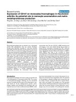
Figure 1
Example of a calibration experiment for the determination of the ∆∆Ct valuesExample of a calibration experiment for the determination of the ∆∆Ct
values. The figure shows the standard curve for glucosealdehyde phos-
phate dehydrogenase (GAPDH) based on the plasmid used for stand-
ardization. The regression curve has the function f(x) = -3.9x + 42.5.
The dashed lines indicate the 95% confidence intervals. Moreover, two
aliquots were prepared containing cDNA from a bursa sample, in addi-
tion to the plasmid standard. Upright triangle: one volume of bursa sam-
ple. Inverted triangle: two volumes of bursa sample. Note that the
linearity of measurement is undisturbed by the added samples and that
the detected quantities of GAPDH strictly correlate to the amounts
added.
Available online />Page 5 of 9
(page number not for citation purposes)
supraspinatus tendon segment. As expected, mRNA for colla-
gen types I and III are present in all samples. Collagen type II,
the major collagen type in chondrocytes, is significantly
expressed within the bursa and underlying tendineus tissue of
samples from almost all patients (Figures 5 and 6), possibly a
sign of the ongoing transformation of tendon to cartilage. The
ratio of collagen type II to GAPDH mRNA in diseased tissue is
less than or equal to 0.2, whereas in healthy tissue it is 0.02.
By contrast, freshly isolated adult articular chondrocytes can
have a collagen type II/GAPDH mRNA ratio of up to 100
(unpublished data). Interestingly, collagen type X expression
(Figures 5 and 6) within the bursa is higher than in the tendon
(ratios of collagen type X mRNA to GAPDH mRNA of less than
or equal to 0.3 and 0.04, respectively); such ratios in adult car-
tilage (Mollenhauer JA, unpublished data) are not different
from those found in the diseased bursa tissue. Collagen type
X is usually expressed in hypertrophic cartilage and is associ-
ated with mineralization processes; it is contained in mineraliz-
ing vesicles. In all of the samples tested there was no mRNA
for the large cartilage proteoglycan aggrecan. However, in
RNA prepared from articular cartilage, aggrecan mRNA was
strongly displayed using the same RT-PCR conditions (Figure
5).
mRNA for growth factors of the TGF-β superfamily was found
in all samples (some stratification was typical), with the follow-
ing order of magnitude: TGF-β (in a ratio to GAPDH of 0.3) >
BMP-7 (in a ratio to GAPDH of 0.03) > BMP-2 (in a ratio to
GAPDH of 0.001). BMP-2 did not show preferential subloca-
tion in its expression; however, BMP-7 mRNA in the acromial
portion of the diseased bursa was approximately two orders of
magnitude higher than in the healthy samples (in a ratio to
GAPDH of 0.09 compared with 0.0009, respectively), repre-
senting the largest difference in growth factor expression
between diseased and healthy tissues.
As examples of alternative tissue growth factors, fibroblast
growth factor (FGF)-2 and vascular endothelial growth factor
(VEGF) mRNA was also present and helps to explain the pro-
liferation of the bursa into granulation tissue, with loose con-
nective tissue and blood vessels amply present. However,
FGF-2 mRNA levels were low throughout the tissue – the ratio
of FGF-2 mRNA to GAPDH mRNA was about 10 times lower
than the ratio of BMP mRNA to GAPDH mRNA- and was not
preferentially localized within the bursa. Of the family of inflam-
matory cytokines, we tested for interleukin (IL)-1 and tumor
necrosis factor (TNFα. As in the present example, typically IL-
1β mRNA was not prominent, whereas TNF-α mRNA was
always expressed, in particular within the top portion of the
bursa. Figure 5b shows a representative cross-sectional
mRNA analysis from 14 patients.
Biogenic bone morphogenetic protein activity
We decided on a bioassay for differentiation rather than
directly determining quantities of specific BMPs. Although
measuring BMP concentrations is an exact method, no infor-
mation is given on the bioactivity, because molecules might
exist in an inactive pro-form, partially degraded or denatured,
or in mixtures that cancel out effects on differentiation. The
C2C12 cell line is an established detector of BMP type I
receptor-related BMP activity owing to its induction of AP [9].
Figure 2
Hematoxylin-eosin staining (a) and indirect immunofluorescence for collagen types I (b) and II (c) in comparable areasHematoxylin-eosin staining (a) and indirect immunofluorescence for collagen types I (b) and II (c) in comparable areas. The acromial portion of the
bursa is in the upper left-hand corner and the segment close to the rotator cuff is in the bottom right-hand corner, with some fat tissue in between.
The asterisk (*) marks dense connective tissue typical of the transition zone between bursa and tendon tissue.
Figure 3
Indirect immunofluorescence images of bone morphogenetic proteins (BMP)-2 (green) and BMP-7 (red) in two areas of inflamed bursaIndirect immunofluorescence images of bone morphogenetic proteins
(BMP)-2 (green) and BMP-7 (red) in two areas of inflamed bursa. (a)
and (b) are from cell-rich areas; (c) and (d) are microvessels. Objective
magnification: ×40.
Arthritis Research & Therapy Vol 8 No 4 Neuwirth et al.
Page 6 of 9
(page number not for citation purposes)
On the basis of this receptor constellation, BMP-2 and BMP-
4 should be initiators of osteogenic differentiation. However,
we also found, although to a lesser degree, that BMP-7 could
induce AP within these cells. In the extract, we found that the
BMP equivalent induced 380 ± 135 µU AP/100 mg of bursa
tissue, with the medium control displaying 175 ± 51 µU AP/
100 mg of bursa tissue (P = 0.0163). By contrast, under the
same conditions, 25 ng/ml recombinant BMP-2 induced 460
± 154 µU AP/100 mg of bursa tissue (P = 0.0021) and 50 ng/
ml of recombinant BMP-7 induced 430 ± 111 µU AP/100 mg
of bursa tissue (P = 0.0058). BMP activity could be com-
pletely abolished by incubation with neutralizing antibodies
against BMP-2 and BMP-7, in addition to incubation with the
soluble BMP receptors Alk-3 and Alk-6 (not shown).
Because the extraction process does not enable determina-
tion of the original site of expression within the bursa tissue,
we tested frozen sections of bursa tissue for their ability to
induce AP locally in C2C12 cells seeded onto a frozen sec-
tion. Surprisingly, the cells not only started to express AP, but
also expression was confined to the location where we
detected BMP-2 and BMP-7 in parallel sections (Figure 7). No
activation of AP expression took place distant from those sites.
Although the microwell assay worked well with the BMP
extract (BMP in solution), the result from the 'contact' experi-
ment indicates that BMP might also exert its effect strictly
locally, thus avoiding lateral expansion of the differentiation
signal.
Discussion
Degenerative alterations to the rotator cuff are generally
regarded as the consequence of mechanical abutment, unde-
fined 'rheumatoid' inflammation, or previous accident.
Although these causes could describe possible etiologies,
Figure 4
Alkaline phosphatase activity within the subacromial bursa varies from patient to patientAlkaline phosphatase activity within the subacromial bursa varies from
patient to patient. Some activity is located within the lining and underly-
ing 'lobules' (a) and (b), in addition to within the connective tissue of
the bursa (c) or the border between bursa and tendon tissue (d). In all
cases, treatment of the sections with 0.5 mM levamisole eradicated the
enzyme reaction, leaving behind only staining in granules within individ-
ual cells that appeared to be leukocytes (not shown).
Figure 5
A comparison of mRNA profiles for select genes from bursa tissue, ten-don and cartilageA comparison of mRNA profiles for select genes from bursa tissue, ten-
don and cartilage. (a) Gel-electrophoretic profile of reverse transcrip-
tion (RT)-PCR from RNA of a subacromial bursa and the underlying
torn supraspinatus tendon. Ethidium bromide-stained electrophoresis
gels are shown; the image grey scale has been inverted. The samples
have been normalized for total RNA extracted from the tissue, and
GAPDH and actin are presented as standards. Tissue aliquots were
sampled anatomically. The dark arrows indicate mRNA that have been
quantified by real-time PCR (Figure 4). An example from cultured
human articular chondrocytes has been added for comparison. Note
the absence of mRNA from the cartilage proteoglycan aggrecan in the
bursa specimens. (b) – Gel-electrophoretic RT-PCR profile of subacro-
mial bursa RNA from 14 patients. Note that the level of IL-1β mRNA is
weak or absent in most samples from the bursa; TNF-α is absent in
some bursa specimens. BMP, bone morphogenetic protein; COL, col-
lagen type; bFGF, basic fibroblast growth factor; GAPDH, glucoseal-
dehyde phosphate dehydrogenase; IL, interleukin; TGF, tumor growth
factor; TNF, tumor necrosis factor; VEGF, vascular endothelial growth
factor.
Available online />Page 7 of 9
(page number not for citation purposes)
they do not completely explain the cellular and molecular alter-
ations seen in and around the rotator cuff, such as chondro-
genic transformation and ectopic mineralization of the tendon
tissue. With the present set of data, an additional perspective
towards a causative explanation is given: chronic activation of
morphogenetic factors (BMP-2, BMP-7, TGF-β, VEGF, and
FGF) that might actively contribute to the rise of mechanically
incompetent and (because of the mineral deposits) chronically
irritating tissue components. The differential expression of
BMP-2 and BMP-7, with BMP-7 expressed in a decreasing
gradient from acromion to tendon, suggests a distinct contri-
bution of BMP-7 to the disease process. Additional expression
of inflammatory cytokines (IL-1β and TNF-α) might serve in
propagating local inflammation and tissue destruction.
In bursa samples from patients with ruptures of the rotator cuff,
expression of collagen types I and III was enhanced compared
with normal controls [16]. In addition, enhanced expression of
IL-1β, both secreted and cell-bound IL-1 receptor antagonists,
and VEGF have been demonstrated [4,17,18]. Participation of
the bursa in the disease progression of the rotator cuff has
also been shown in animal experiments with rabbits [19] and
chickens [20]. Specifically, chemical induction of a bursitis-
induced chondrogenic metaplasia of the supraspinatus inser-
tion site [21]. These reports are not only in line with our find-
ings, but also strongly suggest significant influence of
molecular events within the subacromial bursa on the fate of
the underlying supraspinatus tissue.
Unfortunately, there is no recent literature on the histology of
the normal bursa. Organ material from tumor-related and joint-
replacement surgery was available but always showed signs
of degeneration, depending on the primary disease, and was
thus unrepresentative of a normal situation. Nonetheless, the
presence of bioactive BMP in itself supports the hypothesis of
its role in induced chondrogenesis, irrespective of whether
and, if so, how much it is expressed in normal tissue.
The amounts of BMP we detected are quite significant.
Although direct estimates are hard to compare, the mid-ng
quantities/mg of bursa tissue represent an overwhelmingly
strong potential for morphogenetic signaling. In particular, the
in-situ transformation that was achieved by placing the detec-
tor cell line C2C12 onto tissue slices reveals the effectiveness
of the deposited BMP within the bursa. BMP detected within
the cell and extracellular matrix of blood vessels might have
been transported there through the bloodstream. However,
the microanatomic distribution of immunohistological signals
and the results from the PCR analysis strongly suggest local
production rather than introduction from outside the bursa.
Because we could block the differentiation signal by incubat-
ing the extracts with anti-BMP antibodies or soluble BMP
receptors, a dominant role of BMPs in activating tissue
transdifferentiation can be assumed, although a contribution of
other growth factors cannot be excluded.
Because of the normal function of the bursa, the BMP might
reach the tendon tissue through anatomic secretion pathways
from the glandular elements within the bursa. Unfortunately,
too little is reported in the literature about the secretary activity
of this normally rather inconspicuous layer of tissue under-
neath the acromion. It is obvious, however, that there was sig-
nificant cartilage differentiation, with RNA levels for cartilage
collagen types II and X at quite significant levels in parts of the
patients' tissue. Collagen to GAPDH ratios of >0.1, as
detected here, are usually found in normal articular, mineraliz-
ing, and osteoarthritic tissue [22,23]. In addition, studies per-
formed in the rat support our observation of long-term
preservation of cartilage-related gene expression in the
supraspinatus [24].
There is some information on the production and role of BMP
in adult soft tissue. BMP is produced by gingival and periodon-
tal fibroblasts [25], megakaryocytes and platelets [26], cells
Figure 6
Expression levels of cartilage-specific mRNA and growth factor mRNA types from seven diseased and two healthy bursa specimensExpression levels of cartilage-specific mRNA and growth factor mRNA types from seven diseased and two healthy bursa specimens. Each patient is
represented by four measurements (dots in the graph) across the bursa, which are derived from the acromial, medial, basal, and tendineous portions
of the organ. The box plots are made up of all the combined values. All data sets are statistically significant, with P < 0.05. ∆∆Ct, relative mRNA
expression levels; BMP, bone morphogenetic protein; bFGF, basic fibroblast growth factor; GAPDH, glucosealdehyde phosphate dehydrogenase;
TGF, tumor growth factor.
Arthritis Research & Therapy Vol 8 No 4 Neuwirth et al.
Page 8 of 9
(page number not for citation purposes)
supporting egg maturation [27,28], the kidney [29], and also
connective tissue tumor cells. More importantly, arthritic syno-
vial membranes have been shown to express BMP-2 and
BMP-6 and can influence cell turnover [30]. Early studies
showed that BMP induces tissue transdifferentiation of teno-
cytes into chondrocytes in vitro [31] and, more recently, stud-
ies showed BMP-induced transdifferentiation of kidney
fibroblasts into epithelial cells [32]. Our finding in itself is not
unexpected, retrospectively, but so far, to our knowledge, no
attempts have been made to directly link the 'shoulder syn-
drome' to ectopic overexpression of BMP. Our approach
using AP differentiation within the C2C12 cell line gave us a
tool to explore primary features of the BMP deposited within
the bursa. However, to describe the entire pathologic pathway
from soft tissue to mineral deposits and fiber cartilage, more
experiments are needed using chondrocytes and primary mes-
enchymal precursor cells in vitro or with exogenous deposits
of defined BMP in animals.
Conclusion
In the absence of pharmacologic strategies to counteract
untoward BMP activity, only surgical intervention is an option
for curative approaches to preventing eventually dramatic out-
comes, such as in cases of ossification of the rotator cuff [33].
Complete excision of the subacromial bursa either before
acute rupture or during restorative surgery of torn ligaments or
the tendon might represent the only option we currently have.
Competing interests
The authors declare that they have no competing interests.
Authors' contributions
RF, JN, AV, and IS contributed equally to the preparation of the
manuscript.
Acknowledgements
The excellent technical assistance of Christine Mollenhauer and Cordula
Mueller is gratefully acknowledged. This work was supported, in part, by
a grant from the Interdisciplinary Center for Medical Research at the Uni-
versity of Jena (# TP 2.7), a grant from the Deutsche Forschungsge-
meinschaft (AU 56/6-1), and both a research fellowship for MA and a
grant from the German Ministry of Science and Technology (0313177).
References
1. Cordasco FA, Backer M, Craig EV, Klein D, Warren RF: The par-
tial-thickness rotator cuff tear: is acromioplasty without repair
sufficient? Am J Sports Med 2002, 30:257-260.
2. Knudsen HB, Gelineck J, Sojbjerg JO, Olsen BS, Johannsen HV,
Sneppen O: Functional and magnetic resonance imaging eval-
uation after single-tendon rotator cuff reconstruction. J Shoul-
der Elbow Surg 1999, 8:242-246.
3. Suenaga N, Minami A, Fukuda K, Kaneda K: The correlation
between bursoscopic and histologic findings of the acromion
undersurface in patients with subacromial impingement
syndrome. Arthroscopy 2002, 18:16-20.
4. Yanagisawa K, Hamada K, Gotoh M, Tokunaga T, Oshika Y, Tomi-
sawa M, Lee YH, Handa A, Kijima H, Yamazaki H, et al.: Vascular
endothelial growth factor (VEGF) expression in the subacro-
mial bursa is increased in patients with impingement
syndrome. J Orthop Res 2001, 19:448-455.
5. Simon HW: Soft tissue disorders of the shoulder. Frozen
shoulder, calcific tendinitis, and bicipital tendinitis. Orthop Clin
North Am 1975, 6:521-539.
6. Uhthoff HK, Sano H: The rotator cuff, part I. Pathology of failure
of the rotator cuff tendon. Orthop Clin North Am 1997,
28:31-41.
7. Halverson PB: Crystal deposition disease of the shoulder
(including calcific tendonitis and Milwaukee shoulder
syndrome). Curr Rheumatol Rep 2003, 5:244-247.
8. Molloy ES, McCarthy GM: Hydroxyapatite deposition disease of
the joint. Curr Rheumatol Rep 2003, 5:215-221.
9. Katagiri T, Yamaguchi A, Komaki M, Abe E, Takahashi N, Ikeda T,
Rosen V, Wozney JM, Fujisawa SA, Suda T: Bone morphoge-
netic protein-2 converts the différentiation pathway of C2C12
myoblasts. J Cell Biol 1994, 127:1755-1766.
10. Gründer T, Gaissmaier C, Fritz J, Stoop R, Hortschansky P, Mollen-
hauer J, Aicher WK: Bone morphogenetic protein (BMP)-2
enhances the expression of type II collagen and aggrecan in
chondrocytes embedded in alginate beads. Osteoarthritis
Cartilage 2004, 12:559-567.
11. Altschul SF, Madden TL, Schäffer AA, Zhang J, Zhang Z, Miller W,
Lipman DJ: Gapped BLAST and PSI-BLAST: a new generation
of protein database search programs. Nucleic Acids Res 1997,
25:3389-3402.
12. Fan Z, Bau B, Yang H, Soeder S, Aigner T: Freshly isolated oste-
oarthritic chondrocytes are catabolically more active than nor-
mal chondrocytes, but less responsive to catabolic stimulation
with interleukin-1beta. Arthritis Rheum 2005, 52:136-143.
13. Melby PC, Darnell BJ, Tryon VV: Quantitative measurement of
human cytokine gene expression by polymerase chain
reaction. J Immunol Methods 1993, 159:235-244.
14. Livak KJ, Schmittgen TD: Analysis of relative gene expression
data using real-time quantitative PCR and the 2-(-Delta Delta
C(T)) method. Methods 2001, 25:402-408.
15. Ferreira ID, Rosario VE do, Cravo PV: Real-time quantitative with
SYBR Green I detection for estimating copy numbers of nine
drug resistance candidate genes in Plasmodium falciparum.
Malaria Journal 2006, 5:1.
Figure 7
BMP-2 and BMP-7 co-localization with AP in bursa tissue with and without C2C12 cellsBMP-2 and BMP-7 co-localization with AP in bursa tissue with and
without C2C12 cells. Indirect immunofluorescence staining of the topi-
cal distribution of (a) bone morphogenetic protein (BMP)-2 (green) and
(b) BMP-7 (red) and their local effects on C2C12 cells. The images
display objective magnifications (×5) of subsequent frozen sections.
(c) A section without cultured C2C12 cells stained directly for alkaline
phosphatase (AP). (d) A section seeded with C2C12 cells and cul-
tured for 5 days before staining for AP. Note the co-distribution of AP,
both the bursa-derived intrinsic enzyme and the C2C12-derived
enzyme, with the BMP deposits within the tissue.
Available online />Page 9 of 9
(page number not for citation purposes)
16. Tomonaga A, Hamada K, Gotoh M, Yamakawa H, Kobayashi K,
Fukuda H: Expression of procollagen alpha 1 type III mRNA in
rotator cuff tears. Tokai J Exp Clin Med 2000, 25:125-134.
17. Gotoh M, Hamada K, Yamakawa H, Yanagisawa K, Nakamura M,
Yamazaki H, Ueyama Y, Tamaoki N, Inoue A, Fukuda H: Inter-
leukin-1 induced subacromial synovitis and shoulder pain in
rotator cuff diseases. Rheumatology 2001, 40:995-1001.
18. Sakai H, Fujita K, Sakai Y, Mizuno K: Immunolocalization of
cytokines and growth factors in subacromial bursa of rotator
cuff patients. Kobe J Med Sci 2001, 47:25-34.
19. Uhthoff HK, Sano H, Trudel G, Ishii H: Early reactions after
replantation of the tendon of supraspinatus into bone. A study
in rabbits. J Bone Joint Surg Br 2000, 82:1072-1076.
20. Kobayashi K, Hamada K, Gotoh M, Handa A, Yamakawa H, Fukuda
H: Healing of full-thickness tears of avian supracoracoid ten-
dons: in situ hybridization of alpha1(I) and alpha1(III) procol-
lagen mRNA. J Orthop Res 2001, 19:862-868.
21. Tillander B, Franzén LE, Nilsson E, Norlin R: Carrageenan-
induced subacromial bursitis caused changes in the rat's rota-
tor cuff. J Orthop Res 2001, 19:441-447.
22. Fan Z, Bau B, Yang H, Soeder S, Aigner T: Freshly isolated oste-
oarthritic chondrocytes are catabolically more active than nor-
mal chondrocytes, but less responsive to catabolic stimulation
with Interleukin-1. Arthritis Rheum 2005, 52:136-143.
23. Gebauer M, Saas J, Haag J, Dietz U, Takigawa M, Bartnik E, Aigner
T: Repression of anti-proliferative factor Tob1 in osteoarthritic
cartilage. Arthritis Res Ther 2005, 7:R274-R284.
24. Yokota A, Gimbel JA, Williams GR, Soslowsky LJ: Supraspinatus
tendon composition remains altered long after tendon
detachment. J Shoulder Elbow Surg 2005, 14(1 Suppl
S):72S-78S.
25. Ivanovski S, Li H, Haase HR, Bartold PM: Expression of bone
associated macromolecules by gingival and periodontal liga-
ment fibroblasts. J Periodontal Res 2001, 36:131-141.
26. Sipe JB, Zhang J, Waits C, Skikne B, Garimella R, Anderson HC:
Localization of bone morphogenetic proteins (BMPs)-2, -4,
and -6 within megakaryocytes and platelets. Bone 2004,
35:1316-1322.
27. Pierre A, Pisselet C, Dupont J, Mandon-Pepin B, Monniaux D, Mon-
get P, Fabre S: Molecular basis of bone morphogenetic pro-
tein-4 inhibitory action on progesterone secretion by ovine
granulosa cells. J Mol Endocrinol 2004, 33:805-817.
28. Glister C, Kemp CF, Knight PG: Bone morphogenetic protein
(BMP) ligands and receptors in bovine ovarian follicle cells:
actions of BMP-4, -6 and -7 on granulosa cells and differential
modulation of Smad-1 phosphorylation by follistatin. Repro-
duction 2004, 127:239-254.
29. Ozkaynak E, Schnegelsberg PN, Oppermann H: Murine osteo-
genic protein (OP-1): high levels of mRNA in kidney. Biochem
Biophys Res Commun 1991, 179:116-123.
30. Lories RJ, Derese I, Ceuppens JL, Luyten FP: Bone morphoge-
netic proteins 2 and 6, expressed in arthritic synovium, are
regulated by proinflammatory cytokines and differentially
modulate fibroblast-like synoviocyte apoptosis. Arthritis
Rheum 2003, 48:2807-2818.
31. Sato K, Miura T, Iwata H: Cartilaginous transdifferentiation of rat
tenosynovial cells under the influence of bone morphogenetic
protein in tissue culture. Clin Orthop Relat Res 1988,
236:233-239.
32. Zeisberg M, Shah AA, Kalluri R: Bone morphogenic protein-7
induces mesenchymal to epithelial transition in adult renal
fibroblasts and facilitates regeneration of injured kidney. J
Biol Chem 2005, 280:8094-8100.
33. Matsumoto I, Ito Y, Tomo H, Nakao Y, Takaoka K: Case reports:
ossified mass of the rotator cuff tendon in the subacromial
bursa. Clin Orthop Relat Res 2005, 437:247-250.