Báo cáo y học: "β Scar wars: is TGFβ the phantom menace in scleroderma" doc
Bạn đang xem bản rút gọn của tài liệu. Xem và tải ngay bản đầy đủ của tài liệu tại đây (209.31 KB, 7 trang )
Page 1 of 7
(page number not for citation purposes)
Available online />Abstract
The autoimmune disease scleroderma (systemic sclerosis (SSc)) is
characterized by extensive tissue fibrosis, causing significant
morbidity. There is no therapy for the fibrosis observed in SSc;
indeed, the underlying cause of the scarring observed in this
disease is unknown. Transforming growth factor-β (TGFβ) has long
been hypothesized to be a major contributor to pathological fibrotic
diseases, including SSc. Recently, the signaling pathways through
which TGFβ activates a fibrotic program have been elucidated and,
as a consequence, several possible points for anti-fibrotic drug
intervention in SSc have emerged.
Introduction
During normal connective tissue repair, fibroblasts proliferate
and migrate into the wound, where they synthesize, adhere to
and contract extracellular matrix (ECM) proteins, resulting in
wound closure. It has been proposed that a failure to down-
regulate the normal tissue repair program causes the patho-
logical scarring characterizing fibrotic diseases [1,2]. Fibrotic
disease can affect individual organs, such as the kidney, liver,
pancreas or lung, or be systemic, affecting all organs [1-5]. In
its most severe forms, fibrosis results in organ failure and
death. The systemic autoimmune disease scleroderma
(systemic sclerosis (SSc)) possesses a significant fibrotic
component; indeed, pulmonary fibrosis is the cause of the high
mortality observed in SSc [6]. Identifying targets around which
to base selective, anti-fibrotic therapies is, therefore, essential.
The fundamental mechanism underlying the excessive
scarring observed in SSc is unknown. However, fibrosis is
generally considered to arise from a failure to down-regulate
the normal tissue repair program [2]. One of the major
cytokines induced during the tissue repair is transforming
growth factor (TGF)-β [7]. As TGFβ induces fibroblasts to
synthesize and contract ECM, this cytokine has long been
believed to be a central mediator in wound healing and
fibrotic responses, including SSc [5]; however, the exact
contribution of TGFβ to the fibrotic phenotype of SSc is
unclear. Furthermore, as TGFβ plays many roles in normal
physiology, including as a suppressor of the immune
response and epithelial proliferation, broadly targeting TGFβ
signaling for the treatment of disease is anticipated to be
problematic [8]. Thus, much interest exists, from both clinical
and pharmaceutical points of view, in identifying methods of
intervening within the TGFβ signaling cascade in such a
fashion that the pro-fibrotic aspects of TGFβ signaling are
blocked but other TGFβ-dependent processes are unaltered.
This review critically evaluates the evidence supporting the
notion that TGFβ is a critical mediator of fibrogenesis in SSc,
and assesses whether there are intervention points within the
TGFβ cascade that might be appropriate targets around
which to base selective anti-fibrotic therapies to treat SSc.
Transforming growth factor-
ββ
signaling
There are three TGFβ isoforms, TGFβ1, TGFβ2 and TGFβ3,
which are synthesized as latent precursors in complex with
latent TGFβ-binding proteins (for reviews, see [8-11]). When
these binding proteins are removed by proteolysis, TGFβ is
activated. The
‘
TGFβ activators’ include the proteases
plasmin, matrix metalloproteinase (MMP)-2 and MMP-9,
thrombospondin-1, and the integrin α
v
β
6
. Active TGFβ binds
to a heteromeric receptor complex, consisting of one TGFβ
type I and one TGFβ type II receptor. In the presence of
TGFβ ligand, the TGFβ receptor I kinase phosphorylates the
receptor-activated Smads (R-Smads), Smad2 and Smad3,
which are then able to bind the common mediator Smad,
Smad4, and translocate into the nucleus (Figure 1). The
Smad3-Smad4 pair binds promoters at the Smad consensus
sequence, CAGAC [12]. Smad2, on the other hand, is not
believed to bind DNA directly, but rather requires a nuclear
DNA-binding protein of the family Fast (Fast-1) to bind DNA
Review
Scar wars: is TGF
ββ
the phantom menace in scleroderma?
Andrew Leask
Division of Oral Biology and Department of Physiology and Pharmacology, CIHR Group in Skeletal Development and Remodeling, Division of Oral
Biology, Schulich School of Medicine and Dentistry, University of Western Ontario, Dental Sciences Building, London, ON N6A 5C1, Canada
Corresponding author: Andrew Leask,
Published: 9 June 2006 Arthritis Research & Therapy 2006, 8:213 (doi:10.1186/ar1976)
This article is online at />© 2006 BioMed Central Ltd
CTGF = connective tissue growth factor; ECM = extracellular matrix; EDA = extra domain A; ET = endothelin; ETA = endothelin receptor A; ETB =
endothelin receptor B; FAK = focal adhesion kinase; MMP = matrix metalloproteinase; SSc = systemic sclerosis; SMA = smooth muscle actin; TGF =
transforming growth factor.
Page 2 of 7
(page number not for citation purposes)
Arthritis Research & Therapy Vol 8 No 4 Leask
[13]. The ability of this complex to activate transcription
depends on the ability of Smads to recruit common
transcriptional cofactors, such as p300, and basal
transcription factors, which vary depending on the promoter
of interest. A third group of Smad proteins, the inhibitory
Smads Smad6 or Smad7, prevents R-Smad phosphorylation
and subsequent nuclear translocation of R-Smad-Smad4
heterocomplexes; it appears that Smad7 competes with
Smad2 and Smad3 for binding to the TGFβ type I receptor
[14]. TGFβ also induces Smad7 through a Smad3 and
Smad4-dependent mechanism, suggesting that TGFβ can
suppress its own action via the induction of Smad7 [15]
(Figure 1). Overall, the Smads mediate immediate-early
responses to TGFβ, and their activity is tightly controlled.
In addition to the Smads, TGFβ also causes the activation of
other signaling pathways, for example the mitogen activated
protein kinase cascades. These cascades are not required for
Smad activity per se, but are required for the activation of
basal transcription factors and potentially for the enhancement
of TGFβ responses [10,16-18] (Figure 2). Thus, trans-
criptional responses to TGFβ generally require the type I and
type II TGFβ receptors and the Smads (Figure 2). Specificity
of transcriptional responses to TGFβ rely, therefore, on
ancillary signaling pathways induced by TGFβ, and the basal
transcription factors recruited to the promoter (Figure 2).
The evidence for TGF
ββ
as a pro-fibrotic
cytokine in systemic sclerosis
Evidence supporting the contribution of TGFβ in fibrotic
responses has principally been derived using acute in vitro or
in vivo models. For example, treatment of fetal wounds with
TGFβ promotes wound closure and scarring [19,20]. In
addition, injection of TGFβ, either directly subcutaneously or
into metal chambers, results in enhanced deposition of ECM
[20-22]. Furthermore, incisional rat wounds treated with anti-
TGFβ antibodies or antisense oligonucleotides show a
marked reduction in ECM synthesis and scarring [23,24].
Although TGFβ1 deficient mice display markedly reduced
collagen deposition compared to control mice, such mice
also show a severe wasting syndrome accompanied by a
pronounced, generalized inflammatory response and tissue
necrosis, resulting in organ failure and death [25,26]. These
results are consistent with the fact that, as discussed above,
TGFβ is pleiotropic and that broad targeting of TGFβ in
humans is likely to have adverse side-effects. [8]. Indeed,
resistance to the antiproliferative effects of TGFβ is a hall-
mark of cancer cells [27].
Addition of TGFβ ligand to cells or mice causes only a transient
fibrotic response, which persists only as long as TGFβ ligand is
present [22,28]. TGFβ does promote persistent fibrotic
responses in vivo, but requires a cofactor, such as connective
tissue growth factor (CTGF, CCN2) [22]. This notion that other
factors are required to perpetuate fibrotic responses to TGFβ
is supported by observations using materials derived from SSc
patients. In dermal fibrotic lesions of scleroderma patients,
elevated TGFβ levels exist at the leading edge of the forming
scar tissue, but not within the established lesions [29]. In
addition, elevated serum TGFβ levels are found only in some
patients [30]. Finally, a recent report showed, paradoxically,
that TGFβ levels in patients with diffuse SSc showed lower
levels of active TGFβ in serum, relative to controls, and,
moreover, active TGFβ levels correlated inversely with the
severity of fibrosis [31]. These results may suggest, however,
that TGFβ might be preferentially sequestered, and consumed,
by the connective tissue in SSc [31]. The overexpression of
type I collagen by cultured SSc fibroblasts, which produce
neither elevated TGFβ levels nor increased latent TGFβ, is
nevertheless reduced by neutralizing TGFβ antibodies or
antisense RNA [32]. These results suggest that SSc
fibroblasts show an enhanced response to endogenous TGFβ
ligand. It is interesting to note, however, that an oral
presentation at the 2004 International Scleroderma Meeting at
Cambridge, UK, discussed a clinical safety trial using a
neutralizing anti-TGFβ antibody in SSc patients. This trial
showed that a neutralizing anti-TGFβ antibody showed neither
an anti-fibrotic ability nor a toxic side-effect. However, as only
one dose of antibody was used, it is difficult to evaluate from
this unpublished study whether targeting TGFβ ligand may be
an appropriate anti-fibrotic strategy in SSc.
A priori, the enhanced response to TGFβ in SSc fibroblasts
may arise through an elevation in TGFβ receptor levels.
Indeed, SSc fibroblasts possess increased levels of the
signaling TGFβ type I receptor, relative to the TGFβ type II
receptor [33]. As overexpression of TGFβ type I receptor in
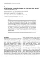
Figure 1
Transforming growth factor (TGF)β signaling generally occurs through
TGFβ type I and type II receptors and Smads. TGFβ binds to the TGFβ
type I and type II receptors. The type I receptor contains kinase activity,
and phosphorylates receptor-activated Smads, Smad2 and Smad3,
which dimerize with Smad4. The resultant complex migrates into the
nucleus to activate target gene expression. TGFβ induces the
inhibitory Smads, Smad6 and Smad7, which block TGFβ receptor type
I-dependent Smad 2/3 activation.
Page 3 of 7
(page number not for citation purposes)
normal fibroblasts increased basal collagen expression in a
dose-dependent manner, it is conceivable that the over-
expression of type I collagen observed in SSc fibroblasts may
arise because of this defect [32]. Supporting the idea that
TGFβ signaling through the TGFβ type I receptor contributes
to the pathogenesis of SSc, the over-expression of type I
collagen by SSc fibroblasts is blocked by a TGFβ type I
receptor antagonist [34]. Similarly, the enhanced ECM
contraction and adhesion observed in SSc fibroblasts
depends on TGFβ type I receptor activity [34,35]. However, it
should be pointed out that TGFβ type I receptor inhibition
also reduced basal collagen synthesis, adhesion and
contraction in normal and SSc fibroblasts, consistent with the
notion that the contribution of TGFβ and TGFβ signaling to
the phenotype of SSc fibroblasts may arise from an
exaggeration of processes operating in normal fibroblasts
[34,35]. Further complicating the issue, TGFβ type I receptor
inhibition had no significant effect on the overexpression of
CTGF or α-smooth muscle actin by SSc fibroblasts [34].
Collectively, these results suggest that signaling through the
TGFβ receptors is likely to contribute to some aspects of
SSc but not others. Finally, as discussed above, although
these data strongly suggest that the TGFβ ligand-type I
receptor axis contributes to the fibrotic phenotype in SSc,
broad targeting of the type I TGFβ receptors is likely to be
problematic in SSc as the type I receptor generally mediates
TGFβ signaling [10]. Indeed, animals genetically deficient in
TGFβ receptor type I die in utero and display severe vascular
defects [36]. That said, the above data suggest that
identifying a method of reducing the type I/type II receptor
ratio to that of normal fibroblasts might be a viable method for
selective anti-fibrotic drug intervention in SSc.
Smads in systemic sclerosis
In addition to implicating TGFβ ligand, acute in vitro and in
vivo models have strongly supported the role of Smad3 in
fibrogenesis. Following incisional wounding, animals lacking
Smad3 show accelerated wound healing, reduced granula-
tion tissue formation, increased epithelialization, and reduced
inflammation, possibly due to an impaired chemotactic
response [37]. Smad 3-deficient mice display resistance to
cutaneous fibrosis caused by radiation injury [38] or
bleomycin [39]. Experiments using microarrays and western
blot analyses have compared gene expression profiles of
fibroblasts taken from adult Smad3
–/–
and Smad3
+/+
mice,
and have shown that TGFβ was not able to induce
transcription in Smad3
–/–
fibroblasts, including the produc-
tion of matrix and proadhesive proteins such as collagen and
CTGF [40-42]. However, between four and six months of
age, Smad3 mutant mice become moribund, with chronic
inflammation and colorectal adenocarcinomas [43]. Smad 3
deficient mice also can develop degenerative joint disease
resembling human osteoarthritis, as characterized by
progressive loss of articular cartilage, decreased production
of proteoglycans, and abnormally increased number of type X
collagen-expressing chondrocytes in synovial joints [44].
These results strongly suggest that targeting Smad3 pharma-
cologically would be expected to have severe side-effects.
Within the context of SSc, leading edge SSc fibroblasts
show activation of Smad3; in some studies, this activation
has been shown to depend on TGFβ ligand, whereas other
studies have shown this may be ligand independent [45, 46].
Although both type I collagen and CTGF are induced by
TGFβ in a Smad-dependent fashion, the elevated activity of
Available online />Figure 2
Schematic diagram of general and gene-specific transforming growth factor (TGF)β signaling in fibroblasts. TGFβ binds to the TGFβ type I and
type II receptors, activates Smad3, which activates target gene expression by binding the sequence CAGA. This pathway regulates virtually every
TGFβ responsive gene in fibroblasts. Conversely, TGFβ can act with endothelin-1 (ET-1), connective tissue growth factor (CTGF) and extra domain
A-fibronectin (EDA-FN) via the endothelin receptor A and B (ETA/B) receptors, syndecan 4 and integrins to activate ERK and focal adhesion
kinase (FAK), which are required for target gene expression, in a promoter-specific fashion (for details see text).
the type I collagen promoter in SSc cells is dependent on the
Smad element; however, the over-expression of CTGF is not
[31,40]. Lesional SSc fibroblasts overexpress the TGFβ
ancillary receptor endoglin in a fashion correlating with
disease severity [47]. Endoglin, when overexpressed in
fibroblasts, suppresses the ability of TGFβ to induce Smad
activation [47]. These results suggest lesional SSc
fibroblasts overexpress endoglin to block further Smad-
dependent gene induction. In this regard, one report showed
decreased levels of the inhibitory Smad7 in scleroderma
fibroblasts [48], albeit in unaffected skin. Other studies
showed no difference, or even an increase in Smad7 levels in
SSc fibroblasts [40,45,49]. In one study, Smad7 was
constitutively present on the TGFβ receptors [49]. The result
of this defect is likely to be a bias away from Smad-
dependent signaling in lesional scleroderma fibroblasts.
Indeed, SSc fibroblasts are relatively non-responsive to
exogenously added TGFβ ligand [50].
Collectively, the above observations reveal the complex role
that TGFβ plays in mediating fibrogenesis, and seems to
suggest that targeting generic TGFβ pathways (TGFβ ligand,
receptors, Smads, p300) is likely to be problematic, not only
due to the pleiotropic nature of these molecules, but also due
to the complex possibly stage-specific ways that cells have of
suppressing these responses. Thus, given the pleiotropic
nature of TGFβ, it is preferable to target gene- or function-
specific, as opposed to general, pathways mediating TGFβ
signaling. Therefore, much recent interest has focused on
identifying mediators of TGFβ signaling affecting fibrosis-
specific, or fibrosis-selective, endpoints.
Gene-specific pathways contributing to
fibrogenesis in SSc
It is likely, then, that targeting the ability of TGFβ to induce
specific pathways or genes will be of benefit in generating
selective therapies in SSc. In fibroblasts, TGFβ transiently
activates the ras/MEK/ERK cascade, which is required for the
induction of CTGF expression [16-18]. Intriguingly, in both
mesangial cells and fibroblasts, TGFβ induction of a generic
Smad3-responsive promoter occurs in the presence of either
dominant negative ras or the mitogen activated protein kinase
inhibitor U0126, indicating that the absolute requirement for
the ras/MEK/ERK cascade in the induction of TGFβ-
responsive genes seems to be restricted in a promoter-
specific fashion [16-18]. The stable prostacyclin analog
Iloprost, which alleviates symptoms of fibrosis in vivo and
reduces CTGF expression and TGFβ-induced collagen
deposition, acts at least in part by antagonizing the
ras/MEK/ERK cascade via the elevation of cAMP [18].
Consistent with this notion, reduction in ERK reduces the
overexpression of type I collagen, and the enhanced adhesive
and contractile ability of SSc fibroblasts [35].
The ability of TGFβ to induce ERK is prevented in fibroblasts
genetically deficient for the proteoglycan syndecan 4, and
small interfering RNA recognizing syndecan 4 reduces the
elevated ERK activation seen in SSc fibroblasts [35].
Syndecan 4 knockout mice appear phenotypically normal, but
show reduced tissue repair responses, indicating that
syndecan 4 is selectively required for the tissue repair
program [51]. Thus, although broadly targeting MEK/ERK
inhibition would be expected to have severe side-effects due
to the involvement of this signaling pathway in many
processes, targeting syndecan 4 is likely to be of benefit in
selectively targeting fibrogenic responses (Figure 2).
Syndecan 4 is a receptor for fibronectin, and is a focal
adhesion component and is required for focal adhesion
kinase (FAK) phosphorylation [52]. Recent data have
suggested that FAK and the extra domain A (EDA) form of
fibronectin mediate the ability of TGFβ to induce α-smooth
muscle actin (α-SMA) [53,54]. Anti-EDA antibody or
recombinant EDA protein suppresses α-SMA induction, but
not that of other TGFβ-responsive genes [54]. Consistent
with this notion, elevated FAK phosphorylation is observed in
SSc fibroblasts [55]. Over-expression of a kinase-defective
FAK mutant reduced α-SMA expression in SSc fibroblasts
[55]. These results emphasize the notion that although the
generic TGFβ signaling pathways may not be suitable targets
for selective drug intervention in SSc, examining how
individual target genes are selectively induced is likely to
provide clues as to how to interfere with appropriate
pathways with drugs. Indeed, targeting adhesive signaling
might be of benefit in combating SSc (Figure 2).
Synergy between TGF
ββ
and other cytokines.
As mentioned above, in addition to signaling pathways
directly induced by TGFβ ligand, there is substantial evidence
for synergy between TGFβ and other extracellular ligands in
driving fibrogenic responses. These other ligands include
CTGF and endothelin (ET)-1.
Connective tissue growth factor (CTGF, CCN2)
CTGF, a member of the CCN family of proteins [56,57], is
induced by TGFβ in normal fibroblasts, but not in keratino-
cytes, through Smads, Ets-1, protein kinase C and ras/MEK/
ERK [16-18,40,58]. CTGF is constitutively expressed by
mesenchymal cells in development, and by kidney mesangial
cells and endothelial cells and is characteristically over-
expressed in fibrotic disease, including SSc, in a fashion
correlating with severity of fibrosis [59,60]. CTGF and TGFβ
act together to promote sustained fibrosis in rodents [22].
Consistent with this notion, CTGF-deficient embryonic
fibroblasts can respond to TGFβ through the Smad pathway,
but show impaired induction of adhesive signaling, as
visualized by the induction of FAK and Akt and the induction
of α-SMA and type I collagen [61]. These results are
consistent with the notion that CTGF executes its functions
through integrins [62-64], and with a previous hypothesis that
CTGF is a mediator of pro-fibrotic responses to TGFβ [65].
However, what is surprising is that the lack of responses
observed in CTGF-deficient embryonic fibroblasts were due
Arthritis Research & Therapy Vol 8 No 4 Leask
Page 4 of 7
(page number not for citation purposes)
to the absence of basal CTGF expression [61]. CTGF was
required for TGFβ to induce cell adhesion to fibronectin and
type I collagen [61]. These results suggest that CTGF acts
as a cofactor of TGFβ to induce adhesive signaling in cells
that are already activated and undergoing tissue remodeling
(e.g., embryonic and fibrotic fibroblasts), and also that
targeting basal CTGF expression, which is independent of
the TGFβ response element and is not blocked by inhibition
of the TGFβ type I receptor [33,38], might be of benefit in
SSc [66,67].
Endothelin-1
ET-1 is normally produced by endothelial cells, but is over-
expressed by SSc fibroblasts [68]. When added to fibro-
blasts, ET-1 independently induces a program of ECM
synthesis and contraction [68,69], and can act synergistically
with TGFβ [70,71]. Blockade of the endothelin receptors with
a dual endothelin receptor A and B (ETA/B) antagonist
significantly reduces α-SMA overexpression and ECM
contraction by SSc fibroblasts [68]. As addition of a TGFβ
receptor inhibitor to SSc fibroblasts does not affect α-SMA
expression but does impact ECM contraction [34,35], these
results suggest that ET-1 acts additively and cooperatively
with TGFβ. Significantly, and in contrast to TGFβ receptor
antagonism [34,35], ETA/B receptor blockade does not
block basal fibroblast activity [68]. ET-1 induces expression
of target genes through Akt and ras/MEK/ERK [68,69]. The
ET-1 response element in the CTGF promoter is distinct from
the TGFβ response element [16,17], and the ET-1 response
element, but not the TGFβ response element, is required for
the over-expression of CTGF in SSc [40]. Collectively, these
data support the notion that TGFβ and ET-1 act together to
promote fibrogenesis in SSc through differing yet
complementary pathways (Figure 1). The ETA/B receptor
antagonist bosentan is currently used clinically to treat
pulmonary hypertension and the formation of new digital
ulcers in SSc patients [72,73]. Thus ETA/B receptor
antagonism is well tolerated in patients, and is likely,
therefore, to be of clinical benefit in alleviating at least one
aspect of fibrosis in SSc, namely that of the persistently
activated fibroblast. A one-year, unpublished clinical trial in
which the efficacy of bosentan at alleviating pulmonary
fibrosis in SSc patients was tested was recently concluded,
and was negative. However, a non-fibrotic endpoint (a walk-
test) was used to evaluate the efficacy of bosentan on overall
lung function. Moreover, the length of the clinical trial may
have been too short to properly evaluate whether bosentan
has an anti-fibrotic effect. For final conclusions to be drawn
the published results are needed. Thus it remains unclear
whether bosentan may be effective in suppressing fibrosis in
patients.
Conclusion
TGFβ induces matrix synthesis in fibroblasts and fibrotic
responses in vivo and in vitro. The majority of the studies
conducted thus far has measured acute responses to TGFβ,
but suggest that TGFβ alone is insufficient for fibrogenesis.
Furthermore, genetic and pharmacological studies have
suggested that broad targeting of general TGFβ signaling
pathways, although perhaps of benefit in suppressing
aspects of the SSc phenotype, might be problematic for
treating SSc due to the pleiotropic nature of TGFβ. The past
several years have led to an appreciation that additional
pathways and receptors to the generic, universal TGFβ/TGFβ
type I and type II receptor/Smad axis are involved with fibro-
genic responses to TGFβ, including syndecan 4, EDA fibro-
nectin, ras/MEK/ERK, FAK, CTGF and ET-1 (Figure 2). By
manipulating these ancillary pathways, selective anti-fibrotic
effects might be achieved, for example, by identifying
inhibitors that block induction of fibrotic genes but leave other
pathways intact.
Competing interests
The author declares that they have no competing interests.
Acknowledgements
AL is supported by the Canadian Institute of Health Research and the
Canadian Foundation for Innovation and is an Arthritis Society (Sclero-
derma Society of Ontario) New Investigator.
References
1. Eckes B, Zigrino P, Kessler D, Holtkotter O, Shephard P, Mauch
C, Krieg T: Fibroblast-matrix interactions in wound healing
and fibrosis. Matrix Biol 2000, 19:325-332.
2. Gabbiani G: The myofibroblast in wound healing and fibro-
contractive diseases. J Pathol 2003, 200:500-503.
3. Bedossa P, Paradis V: Liver extracellular matrix in health and
disease. J Pathol 2003, 200:504-515.
4. Fogo AB: Mesangial matrix modulation and glomerulosclero-
sis. Exp Nephrol 1999, 7:147-159.
5. LeRoy EC, Trojanowska MI, Smith EA: Cytokines and human
fibrosis. Eur Cytokine Netw 1990, 1:215-219.
6. Steen VD, Medsger TA Jr: Severe organ involvement in sys-
temic sclerosis with diffuse scleroderma. Arthritis Rheum
2000, 43:2437-2444.
7. Kane CJ, Hebda PA, Mansbridge JN, Hanawalt PC: Direct evi-
dence for spatial and temporal regulation of transforming
growth factor beta 1 expression during cutaneous wound
healing. Cell Physiol 1991, 148:157-173.
8. McCartney-Francis NL, Frazier-Jessen M, Wahl SM: TGF-beta: a
balancing act. Int Rev Immunol 1998, 16:553-580.
9. Massague J: TGF-beta signal transduction. Annu Rev Biochem
1998, 67:753-791.
10. Leask A, Abraham DJ: TGF-beta signaling and the fibrotic
response. FASEB J 2004, 18:816-827.
11. Roberts AB: TGF-beta signaling from receptors to the
nucleus. Microbes Infect 1999, 1:1265-1273.
12. Zawel L, Dai JL, Buckhaults P, Zhou S, Kinzler KW, Vogelstein B,
Kern SE: Human Smad3 and Smad4 are sequence-specific
transcription activators. Mol Cell 1998, 1:611-617.
13. Liu B, Dou CL, Prabhu L, Lai E: FAST-2 is a mammalian
winged-helix protein which mediates transforming growth
factor beta signals. Mol Cell Biol 1999, 19:424-430.
14. Nakao A, Afrakhte M, Moren A, Nakayama T, Christian JL,
Heuchel R, Itoh S, Kawabata M, Heldin NE, Heldin CH, ten Dijke
P: Identification of Smad7, a TGFbeta-inducible antagonist of
TGF-beta signalling. Nature 1997, 389:631-635.
15. von Gersdorff G, Susztak K, Rezvani F, Bitzer M, Liang D, Bot-
tinger EP: Smad3 and Smad4 mediate transcriptional activa-
tion of the human Smad7 promoter by transforming growth
factor beta. J Biol Chem 2000, 275:11320-11326.
16. Chen Y, Blom IE, Sa S, Goldschmeding R, Abraham DJ, Leask A:
CTGF expression in mesangial cells: involvement of SMADs,
MAP kinase, and PKC. Kidney Int 2002, 62:1149-1159.
17. Leask A, Holmes A, Black CM, Abraham DJ: CTGF gene regula-
Available online />Page 5 of 7
(page number not for citation purposes)
tion: requirements for its induction by TGF
ββ
in fibroblasts. J
Biol Chem 2003, 278:13008-13015.
18. Stratton R, Rajkumar V, Ponticos M, Nichols B, Shiwen X, Black
CM, Abraham DJ, Leask A: Prostacyclin derivatives prevent the
fibrotic response to TGF-beta by inhibiting the Ras/MEK/ERK
pathway. FASEB J 2002, 16:1949-1951.
19. Mustoe TA, Pierce GF, Thomason A, Gramates P, Sporn MB,
Deuel TF: Accelerated healing of incisional wounds in rats
induced by transforming growth factor-beta. Science 1987,
237:1333-1336.
20. Lin RY, Sullivan KM, Argenta PA, Meuli M, Lorenz HP, Adzick NS:
Exogenous transforming growth factor-beta amplifies its own
expression and induces scar formation in a model of human
fetal skin repair. Ann Surg 1995, 222:146-154.
21. Duncan MR, Frazier KS, Abramson S, Williams S, Klapper H,
Huang X, Grotendorst GR: Connective tissue growth factor
mediates transforming growth factor beta-induced collagen
synthesis: down-regulation by cAMP. FASEB J 1999, 13:
1774-1786.
22. Mori T, Kawara S, Shinozaki M, Hayashi N, Kakinuma T, Igarashi
A, Takigawa M, Nakanishi T, Takehara K: Role and interaction of
connective tissue growth factor with transforming growth
factor-beta in persistent fibrosis: A mouse fibrosis model. J
Cell Physiol 1999, 181:153-159.
23. Shah M, Foreman DM, Ferguson MW: Neutralising antibody to
TGF-beta 1,2 reduces cutaneous scarring in adult rodents. J
Cell Sci 1994, 107:1137-1157.
24. Cordeiro MF, Mead A, Ali RR, Alexander RA, Murray S, Chen C,
York-Defalco C, Dean NM, Schultz GS, Khaw PT: Novel anti-
sense oligonucleotides targeting TGF-beta inhibit in vivo scar-
ring and improve surgical outcome Gene Ther 2003, 10:59-71.
25. Kulkarni AB, Karlsson S: Transforming growth factor-beta 1
knockout mice. A mutation in one cytokine gene causes a
dramatic inflammatory disease. Am J Pathol 1993, 143:3-9.
26. Bottinger EP, Letterio JJ, Roberts AB: Biology of TGF-beta in
knockout and transgenic mouse models. Kidney Int 1997, 51:
1355-1360.
27. Reiss M: TGF-beta and cancer. Microbes Infect 1999, 1:1327-
1347.
28. McWhirter A, Colosetti P, Rubin K, Miyazono K, Black C: Colla-
gen type I is not under autocrine control by transforming
growth factor-beta 1 in normal and scleroderma fibroblasts.
Lab Invest 1994, 71:885-894.
29. Querfeld C, Eckes B, Huerkamp C, Krieg T, Sollberg S: Expres-
sion of TGF-beta 1, -beta 2 and -beta 3 in localized and sys-
temic scleroderma. J Dermatol Sci 1999, 21:13-22.
30. Snowden N, Coupes B, Herrick A, Illingworth K, Jayson MI,
Brenchley PE: Plasma TGF beta in systemic sclerosis: a cross-
sectional study. Ann Rheum Dis 1994, 53:763-767.
31. Dziadzio M, Smith RE, Abraham DJ, Black CM, Denton CP: Cir-
culating levels of active transforming growth factor beta1 are
reduced in diffuse cutaneous systemic sclerosis and corre-
late inversely with the modified Rodnan skin score. Rheuma-
tology 2005, 44:1518-1524.
32. Ihn H, Yamane K, Kubo M, Tamaki K: Blockade of endogenous
transforming growth factor beta signaling prevents up-regu-
lated collagen synthesis in scleroderma fibroblasts: associa-
tion with increased expression of transforming growth factor
beta receptors. Arthritis Rheum 2001, 244:474-478.
33. Pannu J, Gore-Hyer E, Yamanaka M, Smith EA, Rubinchik S,
Dong JY, Jablonska S, Blaszczyk M, Trojanowska M: An
increased transforming growth factor beta receptor type I:
type II ratio contributes to elevated collagen protein synthesis
that is resistant to inhibition via a kinase-deficient transform-
ing growth factor beta receptor type II in scleroderma. Arthritis
Rheum 2004, 50:1566-1577.
34. Chen Y, Shi-wen X, Eastwood M, Black CM, Denton CP, Leask A,
Abraham DJ: ALK5 (TGF
ββ
receptor type I) signaling con-
tributes to the fibrotic phenotype of scleroderma fibroblasts.
Arthritis Rheum 2006, 54:1309-1316.
35. Chen Y, Shiwen X, van Beek J, Kennedy L, McLeod M, Renzoni
EA, Bou-Gharios G, Wilcox-Adelman S, Goetinck PF, Eastwood
M, Black CM, Abraham DJ, Leask A: Matrix contraction by
dermal fibroblasts requires TGFbeta/ALK5, heparan sulfate
containing proteoglycans and MEK/ERK: Insights into patho-
logical scarring in chronic fibrotic disease. Am J Pathol 2005,
167:1699-1711.
36. Larsson J, Goumans M, Sjöstrand LJ, van Rooijen MA, Ward D,
Levéen P, Xu X, ten Dijke P, Mummery CL, Karlsson S: Abnormal
angiogenesis but intact hematopoietic potential in TGF-
ββ
type
I receptor-deficient mice. EMBO J 2001, 20:1663-1673.
37. Ashcroft GS, Yang X, Glick AB, Weinstein M, Letterio JL, Mizel
DE, Anzano M, Greenwell-Wild T, Wahl SM, Deng C, Roberts
AB: Mice lacking Smad3 show accelerated wound healing
and an impaired local inflammatory response. Nat Cell Biol
1999, 1:260-266.
38. Flanders KC, Sullivan CD, Fujii M, Sowers A, Anzano MA, Arab-
shahi A, Major C, Deng C, Russo A, Mitchell JB, Roberts AB:
Mice lacking Smad3 are protected against cutaneous injury
induced by ionizing radiation. Am J Pathol 2002, 160:1057-
1068.
39. Lakos G, Takagawa S, Chen SJ, Ferreira AM, Han G, Masuda K,
Wang XJ, DiPietro LA, Varga J: Targeted disruption of TGF-
beta/Smad3 signaling modulates skin fibrosis in a mouse
model of scleroderma. Am J Pathol 2004, 165:203-217.
40. Holmes A, Abraham DJ, Sa S, Shiwen X, Black CM, Leask A:
CTGF and SMADs, maintenance of scleroderma phenotype is
independent of SMAD signaling. J Biol Chem 2001, 276:
10594-10601.
41. Yang YC, Piek E, Zavadil J, Liang D, Xie D, Heyer J, Pavlidis P,
Kucherlapati R, Roberts AB, Bottinger EP: Hierarchical model of
gene regulation by transforming growth factor beta. Proc Natl
Acad Sci USA 2003, 100:10269-10274.
42. Verrecchia F, Chu ML, Mauviel A: Identification of novel TGF-
beta/Smad gene targets in dermal fibroblasts using a com-
bined cDNA microarray/promoter transactivation approach. J
Biol Chem 2001, 276:17058-17062.
43. Yang X, Letterio JJ, Lechleider RJ, Chen L, Hayman R, Gu H,
Roberts AB, Deng C: Targeted disruption of SMAD3 results in
impaired mucosal immunity and diminished T cell respon-
siveness to TGF-beta. EMBO J 1999, 18:1280-1291.
44. Borton AJ, Frederick JP, Datto MB, Wang XF, Weinstein RS: The
loss of Smad3 results in a lower rate of bone formation and
osteopenia through dysregulation of osteoblast differentia-
tion and apoptosis. J Bone Miner Res 2001, 16:1754-1764.
45. Mori Y, Chen SJ, Varga J: Expression and regulation of intra-
cellular SMAD signaling in scleroderma skin fibroblasts.
Arthritis Rheum 2003, 48:1964-1978.
46. Mimura Y, Ihn H, Jinnin M, Asano Y, Yamane K, Tamaki K: Consti-
tutive thrombospondin-1 overexpression contributes to
autocrine transforming growth factor-beta signaling in cul-
tured scleroderma fibroblasts. Am J Pathol 2005, 166:1451-
1463.
47. Leask A, Abraham DJ, Finlay DR, Holmes A, Pennington D, Shi-
Wen X, Chen Y, Venstrom K, Dou X, Ponticos M, et al.: Dysregu-
lation of transforming growth factor beta signaling in
scleroderma: over-expression of endoglin in cutaneous scle-
roderma fibroblasts. Arthritis Rheum 2002, 46:1857-1865.
48. Dong C, Zhu S, Wang T, Yoon W, Li Z, Alvarez RJ, ten Dijke P,
White B, Wigley FM, Goldschmidt-Clermont PJ: Deficient
Smad7 expression: a putative molecular defect in sclero-
derma. Proc Natl Acad Sci USA 2002, 99:3908-3913.
49. Asano Y, Ihn H, Yamane K, Kubo M, Tamaki K: Impaired Smad7-
Smurf-mediated negative regulation of TGF-beta signaling in
scleroderma fibroblasts. J Clin Invest 2004, 113:253-264.
50. Kikuchi K, Hartl CW, Smith EA, LeRoy EC, Trojanowska M: Direct
demonstration of transcriptional activation of collagen gene
expression in systemic sclerosis fibroblasts: insensitivity to
TGF beta 1 stimulation. Biochem Biophys Res Commun 1992,
187:45-50.
51. Echtermeyer F, Streit M, Wilcox-Adelman S, Saoncella S, Denhez
F, Detmar M, Goetinck P: Delayed wound repair and impaired
angiogenesis in mice lacking syndecan-4. J Clin Invest 2001,
107:R9-R14.
52. Wilcox-Adelman SA, Denhez F, Goetinck PF: Syndecan-4 mod-
ulates focal adhesion kinase phosphorylation. J Biol Chem
2002, 277:32970-32977.
53. Thannickal VJ, Lee DY, White ES, Cui Z, Larios JM, Chacon R,
Horowitz JC, Day RM, Thomas PE: Myofibroblast differentiation
by transforming growth factor-beta1 is dependent on cell
adhesion and integrin signaling via focal adhesion kinase. J
Biol Chem 2003, 278:12384-12389.
54. Serini G, Bochaton-Piallat ML, Ropraz P, Geinoz A, Borsi L, Zardi
L, Gabbiani G: The fibronectin domain ED-A is crucial for
Arthritis Research & Therapy Vol 8 No 4 Leask
Page 6 of 7
(page number not for citation purposes)
myofibroblastic phenotype induction by transforming growth
factor-beta1. J Cell Biol 1998, 142:873-881.
55. Mimura Y, Ihn H, Jinnin M, Asano Y, Yamane K, Tamaki K: Consti-
tutive phosphorylation of focal adhesion kinase is involved in
the myofibroblast differentiation of scleroderma fibroblasts. J
Invest Dermatol 2005, 124:886-892.
56. Bork P: The modular architecture of a new family of growth
regulators related to connective tissue growth factor. FEBS
Lett 1993, 327:125-130.
57. Perbal B: CCN proteins: multifunctional signalling regulators.
Lancet 2004, 363:62-64.
58. Van Beek JP, Kennedy L, Rockel JS, Bernier SM, Leask A: The
induction of CCN2 by TGFbeta1 involves Ets-1. Arthritis Res
Ther 2006, 8:R36.
59. Dziadzio M, Usinger W, Leask A, Abraham D, Black CM, Denton
C, Stratton R: N-terminal connective tissue growth factor is a
marker of the fibrotic phenotype in scleroderma. QJM 2005,
98:485-492.
60. Roestenberg P, van Nieuwenhoven FA, Wieten L, Boer P,
Diekman T, Tiller AM, Wiersinga WM, Oliver N, Usinger W, Weitz
S, et al.: Connective tissue growth factor is increased in
plasma of type 1 diabetic patients with nephropathy. Diabetes
Care 2004, 27:1164-1170.
61. Shi-Wen X, Stanton L, Kennedy L, Pala D, Chen Y, Howat SL,
Renzoni EA, Carter DE, Bou-Gharios G, Stratton R, et al.: CCN2
is necessary for adhesive responses to TGFbeta 1 in embry-
onic fibroblasts. J Biol Chem 2006, 281:10715-10726.
62. Chen Y, Abraham DJ, Shi-Wen X, Pearson JD, Black CM, Lyons
KM, Leask A: CCN2 (connective tissue growth factor) pro-
motes fibroblast adhesion to fibronectin. Mol Biol Cell 2004,
15:5635-5646.
63. Babic AM, Chen CC, Lau LF: Fisp12/mouse connective tissue
growth factor mediates endothelial cell adhesion and migra-
tion through integrin alphavbeta3, promotes endothelial cell
survival, and induces angiogenesis in vivo. Mol Cell Biol 1999,
19:2958-2966.
64. Gao R, Brigstock DR: Connective tissue growth factor (CCN2)
induces adhesion of rat activated hepatic stellate cells by
binding of its C-terminal domain to integrin alpha(v)beta(3)
and heparan sulfate proteoglycan. J Biol Chem 2004, 279:
8848-8855.
65. Grotendorst GR: Connective tissue growth factor: a mediator
of TGF-beta action on fibroblasts. Cytokine Growth Factor Rev
1997, 8:171-179.
66. Leask A, Denton, CP and Abraham DJ: Insights into the molec-
ular mechanism of sustained fibrosis: The role of connective
tissue growth factor in scleroderma. J Invest Dermatol 2004,
122:1-6.
67. Takehara K: Hypothesis: pathogenesis of systemic sclerosis.
Rheumatology 2003, 30:755-759.
68. Shi-Wen X, Chen Y, Denton CP, Eastwood M, Renzoni EA, Bou-
Gharios G, Pearson JD, Dashwood M, du Bois RM, Black CM, et
al.: Endothelin-1 promotes myofibroblast induction through
the ETA receptor via a rac/phosphoinositide 3-kinase/Akt-
dependent pathway and is essential for the enhanced con-
tractile phenotype of fibrotic fibroblasts. Mol Biol Cell 2004,
15:2707-2719.
69. Shi-wen X, Howat SL, Renzoni EA, Holmes A, Pearson JD, Dash-
wood MR, Bou-Gharios G, Denton CP, du Bois RM, Black CM, et
al.: Endothelin-1 induces expression of matrix-associated
genes in lung fibroblasts through MEK/ERK. J Biol Chem
2004, 279:23098-23103.
70. Shephard P, Hinz B, Smola-Hess S, Meister JJ, Krieg T, Smola H:
Dissecting the roles of endothelin, TGF-beta and GM-CSF on
myofibroblast differentiation by keratinocytes. Thromb
Haemost 2004, 92:262-274.
71. Horstmeyer A, Licht C, Scherr G, Eckes B, Krieg T: Signalling
and regulation of collagen I synthesis by ET-1 and TGF-beta1.
FEBS J 2005, 272:6297-6309.
72. Denton CP, Black CM: Pulmonary hypertension in systemic
sclerosis. Rheum Dis Clin North Am 2003, 29:335-349, vii.
73. Korn JH, Mayes M, Matucci Cerinic M, Rainisio M, Pope J,
Hachulla E, Rich E, Carpentier P, Molitor J, Seibold JR, et al.:
Digital ulcers in systemic sclerosis: prevention by treatment
with bosentan, an oral endothelin receptor antagonist. Arthri-
tis Rheum 2004, 50:3985-3993.
Available online />Page 7 of 7
(page number not for citation purposes)