Báo cáo y học: "The spliceosomal autoantigen heterogeneous nuclear ribonucleoprotein A2 (hnRNP-A2) is a major T cell autoantigen in patients with systemic lupus erythematosus" potx
Bạn đang xem bản rút gọn của tài liệu. Xem và tải ngay bản đầy đủ của tài liệu tại đây (482.36 KB, 10 trang )
Open Access
Available online />Page 1 of 10
(page number not for citation purposes)
Vol 8 No 4
Research article
The spliceosomal autoantigen heterogeneous nuclear
ribonucleoprotein A2 (hnRNP-A2) is a major T cell autoantigen in
patients with systemic lupus erythematosus
Ruth Fritsch-Stork
1
, Daniela Müllegger
1,2
, Karl Skriner
1,3
, Beatrice Jahn-Schmid
4
,
Josef S Smolen
1,5
and Günter Steiner
1,2,5
1
Division of Rheumatology, Department of Internal Medicine III, Medical University of Vienna, Austria
2
Center of Molecular Medicine (CeMM) of the Austrian Academy of Sciences, Vienna, Austria
3
Charité University Medicine Berlin, Department of Rheumatology and Clinical Immunology, Humboldt University and Free University, Berlin, Germany
4
Institute of Pathophysiology, Medical University of Vienna, Austria
5
Ludwig Boltzmann Institute for Rheumatology and Balneology, Vienna, Austria
Corresponding author: Günter Steiner,
Received: 12 Apr 2006 Revisions requested: 19 May 2006 Revisions received: 8 Jun 2006 Accepted: 6 Jul 2006 Published: 19 Jul 2006
Arthritis Research & Therapy 2006, 8:R118 (doi:10.1186/ar2007)
This article is online at: />© 2006 Fritsch-Stork et al.; licensee BioMed Central Ltd.
This is an open access article distributed under the terms of the Creative Commons Attribution License ( />),
which permits unrestricted use, distribution, and reproduction in any medium, provided the original work is properly cited.
Abstract
A hallmark of systemic lupus erythematosus (SLE) is the
appearance of autoantibodies to nuclear antigens, including
autoantibodies directed to the heterogeneous nuclear
ribonucleoprotein A2 (hnRNP-A2), which occur in 20% to 30%
of SLE patients as well as in animal models of this disease. To
investigate the underlying cellular reactivity and to gain further
insight into the nature and potential pathogenic role of this
autoimmune response we characterized the T cell reactivity
against hnRNP-A2 in patients with SLE in comparison to healthy
controls. Cellular proliferation of peripheral blood T cells to
hnRNP-A2 was determined by [3H]thymidine incorporation and
T cell clones (TCCs) specific for hnRNP-A2 were grown by
limiting dilution cloning; IFNγ, IL-4 and IL-10 in culture
supernatants were measured by ELISA. Bioactivity of culture
supernatants was determined by incubation of anti-CD3/anti-
CD28 stimulated peripheral blood CD4+ T cells with
supernatants of TCCs. Stimulation assays performed with
peripheral blood mononuclear cells of 35 SLE patients and 21
healthy controls revealed pronounced proliferative responses in
66% of SLE patients and in 24% of the controls, which were
significantly higher in SLE patients (p < 0.00002). Furthermore,
hnRNP-A2 specific TCCs generated from SLE patients (n = 22)
contained a relatively high proportion of CD8+ clones and
mostly lacked CD28 expression, in contrast to TCCs derived
from healthy controls (n = 12). All CD4+ TCCs of patients and
all control TCCs secreted IFNγ and no IL-4. In contrast, CD8+
TCCs of patients secreted very little IFNγ, while production of IL-
10 did not significantly differ from other T cell subsets.
Interestingly, all CD8+ clones producing IL-10 in large excess
over IFNγ lacked expression of CD28. Functional assays
showed a stimulatory effect of the supernatants derived from
these CD8+CD28- hnRNP-A2 specific TCCs that was similar
to that of CD4+CD28+ clones. Taken together, the pronounced
peripheral T cell reactivity to hnRNP-A2 observed in the majority
of SLE patients and the distinct phenotype of patient-derived
CD8+ TCCs suggest a role for these T cells in the pathogenesis
of SLE.
Introduction
Systemic lupus erythematosus (SLE) is an autoimmune dis-
ease characterized by a wide spectrum of multi-organ manifes-
tations, and genetic, hormonal, environmental and
immunoregulatory factors are known to contribute to expres-
sion of the disease [1]. However, in spite of the considerable
accumulated knowledge, the detailed etiopathogenesis of
SLE still remains elusive. The presence of autoantibodies
(autoAbs) to nuclear antigens in virtually all SLE patients con-
stitutes the most characteristic serological feature of this dis-
autoAb = autoantibody; cpm = counts per minute; ds = double stranded; ECLAM = European Consensus Lupus Activity Measurement; ELISA =
enzyme-linked immunosorbent assay; FITC = fluorescein isothiocyanate; hnRNP = heterogeneous nuclear ribonucleoprotein; IFN = interferon; IL =
interleukin; mAb = monoclonal antibody; PBMC = peripheral blood mononuclear cell; RA = rheumatoid arthritis; SI = stimulation index; SEM = stand-
ard error of the mean; SLE = systemic lupus erythematosus; TCC = T cell clone.
Arthritis Research & Therapy Vol 8 No 4 Fritsch-Stork et al.
Page 2 of 10
(page number not for citation purposes)
Table 1
Demographic characteristics of systemic lupus erythematosus patients
Patient Sex Age (years) ECLAM score Medication
1 Female 25 1.5 HQ,P
2Female 250.50
3Female 522.50
4Female 241.5C,P
5Female 290HQ
6Female 4310
7 Female 29 0.5 HQ,P
8 Female 38 1.5 HQ,Aza,P
9 Female 34 0 MTX,P
10 Female 60 0 MTX,P
11 Female 52 1 P
12 Female 48 2 HQ,P
13 Female 43 0 0
14 Female 62 1 HQ,P
15 Male 33 0 HQ
16 Female 34 2 Aza,P
17 Female 37 3 0
18 Male 30 0 Ara,P
19 Female 45 4 0
20 Female 31 1 0
21 Female 24 6 Aza,P
22 Female 30 1 0
23 Female 29 1.5 MTX,HQ
24 Female 49 2 0
25 Female 28 2 HQ
26 Female 24 1.5 P
27 Male 45 1 HQ,P
28 Female 22 3.5 HQ,P
29 Female 26 0 HQ
30 Female 29 1 P
31 Female 24 2 0
32 Female 61 2 P
33 Female 43 5.5 MTX,P
34 Female 33 1 0
35 Male 44 1 C
0, none; Aza, azathioprin; C, cyclophosphamide; ECLAM, European Consensus Lupus Activity Measurement; HQ, hydroxychloroquine; MTX,
metothrexate; P, prednisolone.
Available online />Page 3 of 10
(page number not for citation purposes)
order. Among the numerous autoantigens described, the
nucleosome represents an important target structure of the
autoimmune attack, leading to the formation of a wide array of
autoAbs directed to single histones, histone-DNA complexes
and double-stranded (ds)DNA (reviewed in [2,3]). The search
for new serological markers has led to the identification of a
number of additional autoantigens, among them the Ro and La
proteins [4], as well as components of the spliceosome. This
large and highly dynamic complex contains many evolutionarily
conserved proteins, such as the U1–70 kD RNP or the Sm
proteins [5,6], which are targeted by approximately 30% of
SLE patients.
About 15 years ago, the heterogeneous nuclear ribonucleo-
protein (hnRNP-)A2, another component of the spliceosome,
was characterized as a novel target of autoAbs in systemic
autoimmune diseases [7]. Although initially described to occur
mainly in patients with rheumatoid arthritis (RA), autoAbs to
hnRNP-A2 (originally termed anti-RA33) were later detected
also in 20% to 30% of patients with SLE as well as in 40% of
patients with mixed connective tissue disease, usually in asso-
ciation with other anti-spliceosomal autoAbs [8,9]. The autoAb
response against hnRNP-A2 shows some differences in
epitope recognition among patients with RA, SLE and mixed
connective tissue disease [10]. Interestingly, autoAbs to
hnRNP-A2 have also been detected in several animal models
of RA and SLE [11,12]. HnRNP-A2 has a predominant nuclear
localization and exerts multiple functions, including regulation
of alternative splicing, transport of mRNA and regulation of
translation [13-17].
Most of the autoAbs in SLE patients have undergone immu-
noglobulin class switching and affinity maturation. This and the
association of HLA-DR subtypes with the presence of certain
autoAbs indicates an antigen-driven immune response,
emphasizing the role of T cells in SLE [18,19]. The cellular
aspect of the immune response to DNA in its various forms has
been extensively studied, including the characterization of T
cells inducing anti-dsDNA autoAb production by B cells
[20,21]. Interestingly, most of the T cell clones (TCCs) raised
from SLE patients were of the Th1 or Th0 subset [21,22].
However, there have been only few reports about the T cell
response to spliceosomal antigens, mainly focusing on the T
cell reactivity to small nuclear ribonucleoprotein antigens [23].
In a recent report by Greidinger and colleagues, the authors
characterized hnRNP-A2 specific CD4+ TCCs derived from
one mixed connective tissue disease patient and two SLE
patients, and attributed a possible pathogenic role to these T
cells in SLE [24]. However, the presence of autoreactive T
cells is not limited to patients, but has repeatedly been
observed also in healthy individuals [21,25,26]; thus, the
search for possible differences in autoantigen specific cellular
reactivity between patients and healthy controls might give
insight into the pathogenesis of the respective autoimmune
disease.
Recently, we were able to characterize the cellular response
against hnRNP-A2 in patients with RA [27]. We observed that
approximately half of the RA patients harbor T cells against
hnRNP-A2. In accordance with the perception of RA as an
inflammatory, Th1 type systemic autoimmune disease, all gen-
erated TCCs were of the Th1 subtype, as defined by their pre-
dominant IFNγ secretion.
To gain more insight into the role of autoantigen specific T
cells in SLE, we investigated spontaneous T cell responses to
hnRNP-A2 in patients with SLE and in healthy control subjects
and characterized TCCs specific for this antigen. The data
obtained suggest that hnRNP-A2 may constitute an important
T cell autoantigen in patients with SLE, indicating a potential
role for it in the pathogenesis of this disorder.
Materials and methods
Patients and controls
Peripheral blood from 35 patients with SLE (31 female, 4
male, mean age 36.7 ± 3.4 years, for demographic character-
istics see Table 1) classified according to the revised criteria
of the American College of Rheumatology [1] was drawn into
heparinized test tubes. Informed consent was obtained from
all patients. Most patients were treated with immunomodula-
tory drugs (n = 21) and/or low-dose glucocorticoids (n = 15).
Nine patients did not receive any medication. Disease activity
was determined by European Consensus Lupus Activity
Measurement (ECLAM) score [28]. While 87% of the patients
had moderately active disease (ECLAM score <3), 5 patients
had active SLE with an ECLAM score ≥3. The control popula-
tion consisted of 21 healthy individuals (10 female and 11
male, mean age 31.8 ± 1.7 years).
Antigens
Recombinant full-length hnRNP-A2 was used in all experi-
ments. The cDNA encoding the antigen [15] was cloned into
the pET-30 LIC vector (Novagen, Madison, WI, USA) and
expressed as His-tagged fusion protein as described [27].
Purification from bacterial lysates was achieved by Ni-NTA
affinity chromatography (Quiagen, Hilden, Germany) followed
by Polymyxin B Sepharose adsorption (BioRad, Hercules, CA,
USA) and anion exchange chromatography on DEAE Sepha-
rose (Pharmacia, Uppsala, Sweden) essentially as described
[27]. Endotoxin content was determined by the lympholytic
amoebocyte lysate assay (BioWhittaker, Verviers, Belgium).
Using this procedure, a more than 99% pure, endotoxin-free
preparation was obtained. The optimum concentration for pro-
liferation assays was found to be 0.35 µg/ml. Tetanus toxoid
as control antigen was obtained from Pasteur Merieux Con-
naught (Willowdale, Ontario, Canada) and used at a concen-
tration of 0.5 U/ml as previously described [29].
Dectection of antibodies and cytokines
AutoAbs to hnRNP-A2 were detected by immunoblotting as
described [8,10], employing the recombinant antigen and
Arthritis Research & Therapy Vol 8 No 4 Fritsch-Stork et al.
Page 4 of 10
(page number not for citation purposes)
additionally by ELISA (IMTEC, Berlin, Germany). Cytokines
were measured by ELISA (BioSource, Fleurus, Belgium) in
supernatants of TCCs after 24 hours incubation with 0.35 µg/
ml hnRNP-A2 or 0.5 U/ml tetanus toxoid as control antigen.
Detection limits were 9 pg/ml for IFNγ, 4 pg/ml for IL-4, and 8
pg/ml for IL-10.
T cell stimulation assays
Peripheral blood mononuclear cells (PBMCs) were isolated
from heparinized blood of SLE patients and controls by centrif-
ugation on Ficoll Hypaque (Pharmacia). After washing and
counting cells were either immediately used or frozen in RPMI
medium containing 10% dimethylsulfoxide and 20% fetal calf
serum. Cells were cultured in the presence of the antigens for
5 days at 37°C in triplicate in 96-well plates (Costar, Cam-
bridge, MA, USA) in a total volume of 200 µl (10
5
cells/well).
Culture medium consisted of Ultra Culture serum-free medium
(Biowhittaker, Wakersville, MD, USA) containing 2 mM
glutamine and 0.02 mM 2-mercaptoethanol supplemented
with 100 U/ml penicillin/streptomycin (Life Technologies,
Paisley, UK). Phytohemagglutinin (Life Technologies) and IL-2
(Roche Molecular Biochemicals, Mannheim, Germany) were
used as polyclonal stimuli. During the last 16 hours of culture
0.5 µCi per well [
3
H]TdR (Amersham-Pharmacia Biotech
Europe, Freiburg, Germany) was added and the incorporated
radioactivity was measured by scinitillation counting. Results
were expressed as stimulation index (SI) defined as the ratio
of mean counts per minute (cpm) obtained in cultures with
antigen to mean cpm obtained in cultures incubated in the
absence of antigen. An SI ≥3.0 and a ∆cpm >1,000 (mean
cpm obtained in cultures with antigen minus mean cpm
obtained in cultures incubated in the absence of antigen) was
regarded as a positive response.
Antigen-specific T cell lines and clones
Antigen specific T cell lines were obtained using a previously
established protocol [30]. In brief, 2 × 10
6
PBMCs were stim-
ulated with 0.35 µg/ml hnRNP-A2 for 5 days in 24-well flat-
bottomed culture plates. On the 5th day of culture, IL-2 was
added at 20 U/ml and the culture was continued for an addi-
tional 7 days. To generate TCCs, T cell lines were restimulated
with hnRNP-A2 and after 2 more days viable T cells were sep-
arated by Ficoll/Hypaque and seeded in limiting dilution (0.5
cells/well) in 96-wells plates. T cells were cultured in the pres-
ence of 1 × 10
5
irradiated (5,000 rad) allogeneic PBMCs as
'feeder cells', 0.5 µg/ml phytohemagglutinin and 20 U/ml IL-2
in medium containing 1% heat-inactivated human AB serum.
Growing microcultures were then expanded at weekly inter-
vals with fresh feeder cells in the presence of IL-2. The specif-
icity of TCCs was assessed by proliferation assays incubating
2 × 10
4
T cells with 0.35 µg/ml hnRNP-A2 in the presence of
10
5
autologous irradiated PBMCs. After 48 hours incubation
and pulsing with [
3
H]TdR for an additional 16 hours, cells were
harvested and the incorporated radioactivity was measured by
scintillation counting. Production of IL-4, IFNγ and IL-10 was
measured by ELISA in supernatants collected after 24 hours
of incubation as described [27].
For phenotyping, cloned T cells (0.5 to 1 × 10
5
) were washed
twice with ice-cold FACS buffer (phosphate-buffered saline,
5% fetal calf serum, 0.01% NaN
3
) and incubated for 30 min-
utes at 4°C with a fluorescein isothiocyanate (FITC) or phyco-
erythrin-conjugated monoclonal Ab (mAb) (BD Pharmingen,
San Diego, USA). Anti-T cell receptorα/β, anti-CD4 and anti-
CD28 mAbs were FITC-conjugated, and anti-T cell receptor γ/
δ and anti-CD8 mAbs were phycoerythrin-conjugated. Anti-
bodies of the appropriate IgG isotypes were used as negative
controls. Afterwards cells were washed again in FACS buffer
and analyzed with a FACScan flow cytometer (Becton Dickin-
son, Franklin Lakes, NJ, USA). The acquired data were ana-
lyzed using Flow-Jo software (Tree Star, Inc., Ashland, OR,
USA).
Stimulation assays with supernatants from T cell clones
To investigate a possible functional difference between the
CD4+CD28+ and CD8+CD28- TCCs, purified CD4+ T cells
from two SLE patients and two healthy controls (10
5
cells/
well) were stimulated with platebound anti-CD3 mAb (clone
OKT3, Janssen-Cilag, Saundertown, UK) and anti-CD28 mAb
(clone 15E8, Caltag, Burlingame, CA, USA) and incubated in
duplicates with either 50 µl supernatant derived from
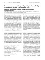
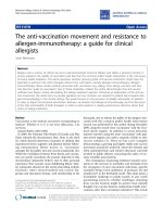
Figure 1
Proliferative responses of peripheral blood mononuclear cells (PBMCs) to heterogeneous nuclear ribonucleoprotein (hnRNP)-A2 in systemic lupus erythematosus (SLE) patients and healthy controlsProliferative responses of peripheral blood mononuclear cells (PBMCs)
to heterogeneous nuclear ribonucleoprotein (hnRNP)-A2 in systemic
lupus erythematosus (SLE) patients and healthy controls. Proliferation
was measured by [
3
H]thymidine incorporation in PBMCs of 35 SLE
patients and 21 controls in the presence or absence of hnRNP-A2. A
stimulation index ≥3.0 and a ∆cpm >1,000 cpm was considered a pos-
itive response (represented by the dashed line). The mean stimulation
index (SI) was 6.7 ± 2.3 for SLE patients, and 2.3 ± 0.2 for controls
(indicated by the solid bars). The difference between SLE patients and
controls was highly significant (p < 0.00002). HnRNP-A2 seropositive
SLE patients are indicated by crosses, and hnRNP-A2 seronegative
SLE patients by diamonds.
Available online />Page 5 of 10
(page number not for citation purposes)
CD4+CD28+ or CD8+CD28- TCCs, respectively. After 24
hours, cells were pulsed with 0.5 µCi per well [
3
H]TdR and
proliferation was measured after anadditional 16 hours. Incu-
bation with IL-10 alone (0.5 to 20 ng/ml; Insight Biotechnology
Ltd, Wembley, UK) was used as control.
Statistical analysis
Unless stated otherwise, SI are expressed as mean ± SEM of
the respective cohort analyzed. Student's t-test was used to
determine differences between controls and SLE patients. A p
value <0.05 was regarded significant. For assessment of cor-
relations the Pearson's correlation coefficient was used.
Results
Proliferative responses to hnRNP-A2 in SLE patients and
healthy controls
Cellular reactivity against hnRNP-A2 was investigated by
measuring proliferation of PBMCs obtained from 35 SLE
patients and 21 age and sex matched healthy controls. Signif-
icant responses (defined as SI ≥3, ∆cpm ≥1,000) were
observed in 66% of the SLE patients and 24% of the controls
(Figure 1). In SLE patients, the SI ranged from 0.5 to 18
(median SI = 4.4) with a mean SI of 6.7 ± 2.3, wheras the SI
was much lower in the control group, ranging from 0.7 to 4.1
(median SI = 2.0) with a mean SI of 2.3 ± 0.2 (p < 0.00002).
Moreover, a SI >4 was seen in 18 SLE patients (52%) but in
only one control subject (5%).
As seen in Figure 1, T cell responses of SLE patients did not
correlate with the presence of anti-hnRNP-A2 autoAbs, which
were detected in 20% of the SLE patients but not in healthy
controls, in accordance with previous observations [8,10].
There was also no correlation with anti-dsDNA antibodies or
disease activity as measured with the ECLAM score.
The response to tetanus toxoid as control antigen was meas-
ured in 21 of the SLE patients and 16 of the healthy controls.
Although stimulation elicited by this recall antigen was some-
what higher in SLE patients than in controls (6.2 ± 1.1 versus
5.0 ± 1.4), the difference was not significant. Of note, while in
controls the stimulation elicited by tetanus toxoid was consid-
erably higher than the one induced by hnRNP-A2, the
response of SLE patients to tetanus toxoid was slightly but sig-
nificantly lower than the response to hnRNP-A2 (Table 2).
Generation of hnRNP-A2 reactive T cell clones
T cell lines specific for hnRNP-A2 were established from the
peripheral blood of 8 SLE patients and 4 healthy controls.
Using the limiting dilution technique, between 1 and 6 antigen
specific TCCs per individual (34 TCCs in total) could be
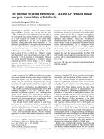
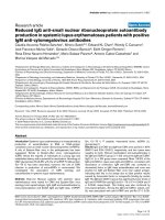
Figure 2
Phenotype, proliferation and cytokine production of heterogeneous nuclear ribonucleoprotein (hnRNP)-A2 specific T cell clones (TCCs) of systemic lupus erythematosus (SLE) patients and controlsPhenotype, proliferation and cytokine production of heterogeneous nuclear ribonucleoprotein (hnRNP)-A2 specific T cell clones (TCCs) of systemic
lupus erythematosus (SLE) patients and controls. TCCs were generated by limiting dilution and their specificity was assessed by measuring prolifer-
ation in response to hnRNP-A2. Cytokine production of TCCs was measured in the supernatants after 24 hours of stimulation with autologous anti-
gen presenting cells and hnRNP-A2. (a) Proliferation of CD4+ and CD8+ TCCs of SLE patients. (b) Proliferation and IFNγ production of TCCs of
SLE patients. (c) Proliferation of CD4+ and CD8+ TCCs of healthy controls (HCs). (d) Proliferation and IFNγ production of TCCs of healthy con-
trols. There was no correlation between proliferative capacity and cytokine secretion.
Arthritis Research & Therapy Vol 8 No 4 Fritsch-Stork et al.
Page 6 of 10
(page number not for citation purposes)
raised. Twenty-two TCCs were derived from SLE patients: 13
(59%) of them were CD4+/CD8- and thus belonged to the
helper T cell subset, while 9 were CD4-/CD8+. The TCCs
showed high variability in their degree of proliferation, with SIs
ranging from 3.0 to 26.0 and a mean SI of 6.2 ± 1.2. (Figure
2a). Analysis of the cytokine secretion pattern revealed pro-
nounced IFNγ production by 10 of the clones, while the
remaining 12 clones produced very little, if any, IFNγ. Cytokine
production did not correlate with the degree of proliferation as
even poorly proliferating TCCs secreted high amounts of IFNγ
and vice versa (Figure 2b), whereas IL-4 was not detectable at
all.
Proliferation of the 12 TCCs derived from control individuals
was comparable to patient-derived clones, with SIs ranging
from 3.6 to 37 and a mean SI of 9.0 ± 2.8 (Figure 2c). Pheno-
typing, on the other hand, revealed some differences. The
majority of TCCs (n = 8; 67%) were CD4+/CD8-, while 3
clones (25%) were CD4-/CD8+ and one TCC was CD4-/
CD8 In contrast to SLE TCCs, all clones secreted IFNγ,
though in highly varying amounts (Figure 2d). Whereas in SLE
patients IFNγ secretion was significantly higher in CD4+ than
in CD8+ TCCs (p < 0.003), no difference in cytokine produc-
tion was found in CD4+ and CD8+ TCCs from healthy con-
trols (Figure 3a). Additionally, IFNγ secretion of CD8+ SLE
TCCs was markedly lower than in CD8+ control TCCs (Figure
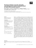
Figure 3
Cytokine production by heterogeneous nuclear ribonucleoprotein (hnRNP)-A2 specific T cell clones (TCCs) from systemic lupus ery-thematosus (SLE) patients and healthy controls (a) IFNγ productionCytokine production by heterogeneous nuclear ribonucleoprotein
(hnRNP)-A2 specific T cell clones (TCCs) from systemic lupus ery-
thematosus (SLE) patients and healthy controls (a) IFNγ production.
Whereas IFNγ production of CD4+ and CD8+ TCCs derived from
healthy controls was similar (mean ± SEM of 533 ± 148 for CD4+ ver-
sus 376 ± 312 for CD8+; p = not significant), IFNγ production by CD4
+ TCCs from SLE patients was significantly higher than IFNγ produc-
tion by patient derived CD8+ TCCs (500 ± 124 versus 40 ± 23 pg/ml;
p < 0.003). (b) Ratio of IL-10/IFNγ secretion in hnRNP-A2 specific
TCCs from SLE patients and controls. IL-10 secretion was measured in
supernatants of 11 TCCs from SLE patients and 5 TCCs from healthy
controls. IL-10 secretion largely exceeded IFNγ production in
CD8+CD28- TCCs, which were all derived from SLE patients.
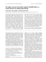
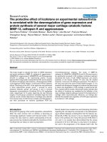
Figure 4
FACS analysis of four heterogeneous nuclear ribonucleoprotein (hnRNP)-A2 specific T cell clones (TCCs) derived from systemic lupus erythematosus (SLE) patientsFACS analysis of four heterogeneous nuclear ribonucleoprotein
(hnRNP)-A2 specific T cell clones (TCCs) derived from systemic lupus
erythematosus (SLE) patients. Left panels: clones were stained with
fluorescein isothiocyanate (FITC)- or phycoerythrin-conjugated mAbs
against CD4 or CD8, respectively. Right panels: clones were stained
with a FITC-conjugated anti-CD28 mAb and a phycoerythrin-conju-
gated control mAb. Top panel: FACS profile of a CD4+ CD28- TCC.
Second panel: FACS profile of a CD4+CD28+ TCC. Third panel:
FACS profile of a CD8+ CD28- TCC. Bottom panel: FACS profile of a
CD8+ CD28+ TCC.
Available online />Page 7 of 10
(page number not for citation purposes)
3a); however, due to the small number of CD8+ control clones
(n = 3), the difference between patient and control TCCs
(39.5 ± 29.4 pg/ml versus 376 ± 312 pg/ml) did not reach
statistical significance.
Expression of the costimulatory molecule CD28 was meas-
ured in six CD4+ and seven CD8+ TCCs from SLE patients
and in five TCCs from healthy subjects (Figure 4): while all
control TCCs examined (four CD4+ and one CD8+)
expressed CD28, only three SLE TCCs were CD28+ (two
CD4+ and one CD8+ TCC). Of note, all CD28+ clones and
also the four CD4+CD28- SLE TCCs produced IFNγ,
whereas this cytokine was barely detectable in the superna-
tants of the six CD8+CD28- TCCs; on the other hand, all
clones secreted comparable amounts of IL-10. Thus, IL-10
was produced in 6- to 1,000-fold excess over IFNγ by the
CD8+CD28- TCCs while all other clones examined (i.e.
CD8+CD28+, CD4+CD28+ and CD4+CD28-) secreted
comparable amounts of IFNγ and IL-10 (Figure 3b).
Stimulation assays with supernatants of T cell clones
As pronounced differences in the cytokine secretion pattern of
SLE patient derived CD4+CD28+ and CD8+CD28- TCCs
had been observed, we were interested in the functional prop-
erties of these two populations. To address this question,
CD4+ T cells (obtained from two SLE patients and two
healthy controls) were stimulated with anti-CD3 and anti-
CD28 mAbs and subsequently incubated with supernatants
from two CD8+CD28- and two CD4+CD28+ TCCs derived
from SLE patients (Figure 5). All supernatants caused a signif-
icant increase of the anti-CD3/anti-CD28 induced proliferative
response: supernatants from CD8+CD28- clones enhanced
proliferation by 452 ± 103% (p < 0.001), and CD4+CD28+
derived supernatants enhanced proliferation by 522 ± 161%
(p < 0.02). Since the CD8+CD28- clones produced IL-10 in
large excess over IFNγ, we examined the effect of IL-10 on pro-
liferation: a modest reduction of proliferation was reproducibly
seen, which, however, was not statistically significant (16.6 ±
6.6%, p = 0.07). Therefore, the pronounced stimulatory
capacity of these supernatants cannot be attributed to IL-10 or
IFNγ. Unfortunately, the TCCs were short-lived, limiting the
number of experiments, and additional studies will be required
to further investigate this issue.
Discussion
In previous investigations we characterized the humoral
response to hnRNP-A2 in SLE patients and some clinical
implications thereof [8,10,31,32]. In this study we examined
SLE patients and healthy controls for the presence of autore-
active T cells to hnRNP-A2 to better understand the role of this
Figure 5
Proliferative responses of CD4+ T cells upon incubation with supernatants from T cell clones (TCCs)Proliferative responses of CD4+ T cells upon incubation with supernatants from T cell clones (TCCs). CD4+ T cells of two healthy controls (HC-1,
HC-2) and two systemic lupus erythematosus (SLE) patients (SLE-1, SLE-2) were stimulated with platebound anti-CD3/anti-CD28 mAbs. Cells
were incubated for 24 hours with the supernatants of a CD4+CD28+ and a CD8+CD28
-
TCC derived from an SLE patient, and proliferation was
measured after an additional 16 hours. The increase in proliferation was statistically significant for all supernatants examined (indicated by a star).
Arthritis Research & Therapy Vol 8 No 4 Fritsch-Stork et al.
Page 8 of 10
(page number not for citation purposes)
autoimmune response in the pathogenesis of SLE. The data
obtained show the existence of a pronounced cellular
response to hnRNP-A2 in the majority of SLE patients, which
was far more vigorous than in healthy controls. In contrast, the
response to the control antigen tetanus toxoid was similar in
patients and controls, demonstrating the specificity of the
immune response towards hnRNP-A2. Moreover, the
response of SLE patients to hnRNP-A2 was of similar magni-
tude, or even slightly higher, than the response to tetanus tox-
oid, which is a recall antigen eliciting a pronounced response
in the majority of individuals tested.
The occurrence of autoreactive T cells in healthy individuals is
a common finding that has previously been reported for anti-
gens associated with SLE [21] as well as other autoimmune
diseases like pemphigus foliaceus [25] and multiple sclerosis
[26]. Thus, the presence of hnRNP-A2 auoreactive T cells in
healthy controls by itself was not surprising; a striking differ-
ence, however, was the significantly higher strength of the cel-
lular immune response observed in SLE patients, which may
be considered an indication for pathogenic involvement of
these autoreactive cells and/or a lack of counter-regulation in
SLE patients.
Interestingly, no correlation of cellular reactivity to hnRNP-A2
with the appearance of the respective autoAbs in SLE patients
could be observed. Thus, hnRNP-A2 appears to be a predom-
inant T cell antigen, while the generation of autoAbs might rep-
resent a bystander phenomenon occurring in a subgroup of
patients. However, recent data indicate that in SLE patients
the humoral response to hnRNP-A2 fluctuates considerably
and increases during flares. Therefore, the prevalence of these
autoAbs may be considerably higher than previously assumed
(unpublished observation).
Although the cellular reactivity to hnRNP-A2 appeared to be
primarily a Th1 response, we observed a relatively high per-
centage of CD8+ TCCs in SLE patients. Of particular interest,
most of these clones lacked CD28 expression and produced
neither IFNγ nor IL-4, but did produce IL-10. This may indicate
a special pathological role of this T cell subset because this
phenotype appeared to be restricted to SLE patient derived
TCCs. In previous reports, CD4+ T cells in SLE were shown
to be of predominantly Th1 or Th0 subtypes [21,24]. Further-
more, autoreactive CD4+ TCCs from SLE patients and
healthy controls showed similar cytokine production [22].
Whereas the relatively high proportion of hnRNP-A2 specific
CD8+ TCCs might mirror the common understanding of a
generally increased CD8:CD4 ratio in SLE patients [33-35],
there are only anecdotal reports about autoreactive CD8+
TCCs in SLE [6] and their role is not entirely clear and may be
quite complex indeed. Thus, on the one hand, the rather low
IFNγ secretion of patient derived CD8+ TCCs might be due to
inherent abnormalities of CD8+ (suppressor) T cell function in
SLE, as shown recently by Filaci and colleagues [36]. On the
other hand, these TCCs secreted considerable amounts of IL-
10 and, in contrast to the TCCs derived from healthy controls,
were CD28 negative. Recently, a new T suppressor popula-
tion was defined, which could be generated in vitro from
CD8+CD28- T cells [37]. This subset exerted its regulatory
and suppressive function in a cell contact independent man-
ner via secretion of IL-10, which is in line with previous reports
attributing a regulatory function to CD8+CD28- T cells [38-
40]. An increase of these cells in patients with SLE might thus
constitute an effort of counter-regulation within the disturbed
immune system of SLE patients.
Surprisingly, though, the supernatants of the hnRNP-A2 spe-
cific CD8+CD28- TCCs derived from SLE patients did not
inhibit proliferation of CD4+ T cells, but instead enhanced
anti-CD3/anti-CD28 induced stimulation, similar to superna-
tants derived from CD4+CD28+ TCCs. As IL-10 alone had an
antiproliferative effect, the increased content of IL-10 in the
supernatants of CD8+CD28- TCCs was obviously counter-
acted by other yet unknown stimulatory components of the
supernatant, which remain to be identified. Of note, the lack of
CD28 expression is also a hallmark of senescent T cells, which
are known to be increased in various autoimmune diseases
and chronic inflammatory conditions and may show a rather
aggressive phenotype [41]. Therefore, it is conceivable that
the CD8+CD28- TCC may be derived from the senescent T
cell pool of SLE patients; this also correlates better with our
data.
Table 2
Proliferative responses to hnRNP-A2 and the control antigen tetanus toxoid in SLE patients and healthy controls
Proliferative response to
hnRNP-A2 Tetanus toxoid P value
SLE patients (n = 21) 7.5 ± 1.2 6.2 ± 1.1 P < 0.05
Healthy controls (n = 16) 2.5 ± 0.6 5.0 ± 1.4 P < 0.002
P value P < 0.000002 NS
Data are given as mean stimulation index ± SEM. Student's paired t-test was used to calculate statistical significances. hnRNP, heterogeneous
nuclear ribonucleoprotein; NS, not significant; SLE, systemic lupus erythematosus.
Available online />Page 9 of 10
(page number not for citation purposes)
Elevated IL-10 serum levels have been reported in SLE
patients and correlated with clinical and serological disease
activity, especially anti-DNA antibody titres [42]. Several cell
types have been implicated in the production of IL-10 [43] and
augmented IL-10 secretion has been linked to autoAb produc-
tion in a model where PBMCs from patients with SLE were
transferred into mice with severe combined immunodieficiency
[44]. So, altogether, the CD8+CD28- T cell subpopulation
might enhance the inflammatory process in SLE patients by
stimulation of B cells via IL-10. Unfortunately, because of the
short life-span of the clones, additional functional assays could
not be performed and remain the subject of future investiga-
tions.
Finally, the question remains why hnRNP-A2 is such a pre-
ferred target of the T cell response in SLE, while autoAbs
occur less frequently and may represent an epiphenomenon of
ongoing T cell autoreactivity. On the other hand, autoAb titers
increase during disease flares (unpublished observation) and,
importantly, autoAbs to hnRNP-A2 are, together with anti-Sm
antibodies, among the earliest detectable autoAbs in MRL/lpr
mice, where they were found to precede anti-dsDNA autoAbs
[11]. HnRNP-A2 is a multifunctional protein that, in the
nucleus, partially colocalizes with spliceosomal complexes
[13,14], a preferred autoimmune target in SLE. Thus, the data
obtained in the course of this study further strengthen the view
of the spliceosome being one of the predominant autoimmune
target structures in SLE, playing a presumably pivotal role in
the pathogenesis of this disease, even though the reasons for
this are still far from being fully understood.
Conclusion
Our data reveal a pronounced T cell response against the
autoantigen hnRNP-A2 in the majority of SLE patients in con-
trast to a scarcer and lower response in healthy subjects.
Although this autoreactivity seemed to be mainly conferred by
CD4+ T cells showing a Th1 phenotype, a newly defined
hnRNP-A2 specific CD8+CD28- T cell-subset was observed
in SLE patients. This subpopulation showed a predominant IL-
10 secretion and a lack of IFNγ or IL-4 production. Our data
suggest involvement of both populations in the complex and
still incompletely understood pathogenesis of SLE, and war-
rant further investigations into the cellular aspects of this
autoimmune response.
Competing interests
They authors declare that they have no competing interests.
Authors' contributions
RF participated in the design of the study, worked with the T
cell cultures and drafted the manuscript. DM performed the T
cell cloning and T cell assays. KS prepared the recombinant
antigen. BJS participated in the design of the study. JS and
GS conceived of the study, participated in its design and coor-
dination and helped to write the manuscript. All authors read
and approved the final manuscript.
Acknowledgements
We thank Dr Alexander Ploner for his help in statistics and Elisabeth
Höfler for expert technical help with determination of autoantibodies and
Karolina von Dalwigk for performing functional assays with supernatat-
nts of TCCs. The work was supported by CeMM, the Center of Molec-
ular Medicine of the Austrian Academy of Sciences.
References
1. Tan EM, Cohen AS, Fries JF, Masi AT, McShane DJ, Rothfield NF,
Schaller JG, Talal N, Winchester RJ: The 1982 revised criteria for
the classification of systemic lupus erythematosus (SLE).
Arthritis Rheum 1982, 25:1271-1277.
2. Pisetsky DS: Anti-DNA and autoantibodies. Curr Opin Rheuma-
tol 2000, 12:364-368.
3. Schett G, Rubin RL, Steiner G, Hiesberger H, Muller S, Smolen J:
The lupus erythematosus cell phenomenon: comparative
analysis of antichromatin antibody specificity in lupus ery-
thematosus cell-positive and -negative sera. Arthritis Rheum
2000, 43:420-428.
4. Alspaugh MA, Tan EM: Antibodies to cellular antigens in
Sjogren's syndrome. J Clin Invest 1975, 55:1067-1073.
5. Reyes PA, Tan EM: DNA-binding property of Sm nuclear anti-
gen. J Exp Med 1977, 145:749-754.
6. Holyst MM, Hill DL, Hoch SO, Hoffmann RW: Analysis of T and
B cell responses against U small nuclear ribonucleoprotein
70-kD, B and D polypeptides among patients with systemic
lupus erythematosus and mixed connective tissue disease.
Arthritis Rheum 1997, 40:1493-1503.
7. Steiner G, Hartmuth , Skriner K, Maurer-Fogy I, Sinski A, Hassfeld
W, Barta A, Smolen JS: Purification and partial sequencing of
the nuclear autoantigen RA33 shows that it is indistinguisha-
ble from the A2 protein of the heterogeneous nuclear ribonu-
cleoprotein complex. J Clin Invest 1992, 90:1061-1066.
8. Hassfeld W, Steiner G, Studnicka-Benke A, Skriner K, Graninger
W, Fischer I, Smolen JS: Autoimmune response to the spliceo-
some: an immunologic link between rheumatoid arthritis,
mixed connective tissue disease, and systemic lupus ery-
thematosus. Arthritis Rheum 1995, 38:777-785.
9. Steiner G, Skriner K, Smolen JS: Autoantibodies to the A/B pro-
teins of the heterogeneous nuclear ribonucleoprotein com-
plex: novel tools for the diagnosis of rheumatic diseases. Int
Arch Allergy Immunol 1996, 111:314-319.
10. Skriner K, Sommergruber WH, Tremmel V, Fischer I, Barta A, Smo-
len JS, Steiner G: Anti-hnRNP-A2 autoantibodies are directed
to the RNA binding region of the A2 protein of the heterogene-
ous nuclear ribonucleoprotein complex: differential epitope
recognition in rheumatoid arthritis, systemic lupus erythema-
tosus, and mixed connective tissue disease. J Clin Invest
1997, 100:127-135.
11. Dumortier H, Monneaux F, Jahn-Schmid B, Briand JP, Skriner K,
Cohen PL, Smolen JS, Steiner G, Muller S: B and T cell
responses to the spliceosomal heterogeneous nuclear ribo-
nucleoproteins A2 and B1 in normal and lupus mice. J Immu-
nol 2000, 165:2297-2305.
12. Hayer S, Tohidast-Akrad M, Haralambous S, Jahn-Schmid B,
Skriner K, Trembleau S, Dumortier H, Pinol-Roma S, Redlich K,
Schett G, et al.: Aberrant expression of the autoantigen
hnRNP-A2/B1 (RA33) and spontaneous formation of rheuma-
toid arthritis associated anti-RA33 autoantibodies in TNFα
transgenic mice. J Immunol 2005, 175:8327-8336.
13. Weighardt F, Biamonti G, Riva S: The roles of heterogeneous
nuclear ribonucleoproteins (hnRNP) in RNA metabolism.
Bioessays 1996, 18:747-756.
14. Krecic AM, Swanson MS: HnRNP complexes: composition,
structure, and function. Curr Opin Cell Biol 1999, 11:363-371.
15. Mayeda A, Munroe SH, Caceres JF, Krainer AR: Function of con-
served domains of hnRNP A1 and other hnRNP A/B proteins.
EMBO J 1994, 13:5483-5495.
16. Carson JH, Kwon S, Barbarese E: RNA trafficking in myelinating
cells. Curr Opin Neurobiol 1998, 8:607-612.
Arthritis Research & Therapy Vol 8 No 4 Fritsch-Stork et al.
Page 10 of 10
(page number not for citation purposes)
17. Kamma H, Horiguchi H, Wan L, Matsui M, Fujiwara M, Fujimoto M,
Yazawa T, Dreyfuss G: Molecular characterization of the hnRNP
A2/B1 proteins: tissue-specific expression and novel iso-
forms. Exp Cell Res 1999, 246:399-411.
18. Smolen JS, Klippel JH, Penner E, Reichlin M, Steinberg AD,
Chused TM, Scherak O, Graninger W, Hartter E, Zielinski CC, et
al.: HLA-DR antigens in systemic lupus erythematosus: asso-
ciation with specificity of autoantibody reponses to nuclear
antigens. Ann Rheum Dis 1987, 46:457-462.
19. Stephens HA, McHugh NJ, Maddison PJ, Isenberg DA, Welsh KI,
Panayi GS: HLA class II restriction of autoantibody production
in patients with systemic lupus erythematosus. Immuno-
gengetics 1991, 33:276-280.
20. Mohan C, Adams S, Stanik V, Datta SK: Nuclesosome: a major
immunogen for pathogenic autoantibody-inducing T cells of
lupus. J Exp Med 1993, 177:1367-1381.
21. Voll RE, Roth EA, Girkontaite I, Fehr H, Herrmann M, Lorenz HM,
Kalden JR: Histone-specific Th0 and Th1 clones derived from
systemic lupus erythematosus patients induce double-
stranded DNA antibody production. Arthritis Rheum 1997,
40:2162-2172.
22. Converso M, Bertero MT, Vallario A, Caligaris-Cappio F: Analysis
of T-cell clones in systemic lupus erythematosus. Haemato-
logica 2000, 85:118-123.
23. Talken BL, Bailey CW, Reardon SL, Caldwell CW, Hoffman RW:
Structural analysis of TCRalpha and beta chains from human
T-Cell clones specific for small nuclear ribonucleoprotein
polypeptides Sm-D, Sm-B and U1–70 kDa: TCR complemen-
tarity determining region 3 usage appears highly conserved.
Scand J Immunol 2001, 54:204-210.
24. Greidinger EL, Gazitt T, Jaimes KF, Hoffman RW: Human T cell
clones specific for heterogeneous nuclear ribonucleoprotein
A2 autoantigen from connective tissue disease patients assist
in autoantibody production. Arthritis Rheum 2004,
50:2216-2222.
25. Gebhard KL, Veldman CM, Wassmuth R, Schultz E, Schuler G,
Hertl M: Ex vivo analysis of desmoglein 1-responsive T-helper
(Th) 1 and Th2 cells in patients with pemphigus foliaceus and
healthy individuals. Exp Dermatol 2005, 14:586-592.
26. Diaz-Villoslada P, Shih A, Shao L, Genain CP, Hauser SL: Autore-
activity to myelin antigens: myelin/oligodendrocyte glycopro-
tein is a prevalent autoantigen. J Neuroimmunol 1999,
99:36-43.
27. Fritsch R, Eselbock D, Skriner K, Jahn-Schmid B, Scheinecker C,
Bohle B, Tohidast-Akrad M, Hayer S, Neumuller J, Pinol-Roma S,
et al.: Characterization of autoreactive T cells to the autoanti-
gens heterogeneous nuclear ribonucleoprotein A2 (RA33) and
filaggrin in patients with rheumatoid arthritis. J Immunol 2002,
169:1068-1076.
28. Bencivelli W, Vitali C, Isenberg DA, Smolen JS, Snaith ML, Sciuto
M, Bombardieri S: Disease activity in systemic lupus erythema-
tosus: report of the Consensus Study Group of the European
Workshop for Rheumatology Research. III. Development of a
computerised clinical chart and its application to the compar-
ison of different indices of disease activity. The European Con-
sensus Study Group for Disease Activity in SLE. Clin Exp
Rheumatol 1992, 10:549-554.
29. Scheinecker C, Machold KP, Majdic O, Hocker P, Knapp W, Smo-
len JS: Initiation of the autologous mixed lymphocyte reaction
requires the expression of costimulatory molecules B7-1 and
B7-2 on human peripheral blood dendritic cells. J Immunol
1998, 161:3966-3973.
30. Fritsch R, Bohle B, Vollmann U, Wiedermann U, Jahn-Schmid B,
Krebitz M, Breiteneder H, Kraft D, Ebner C: Bet v 1, the major
birch pollen allergen, and Mal d 1, the major apple allergen,
cross-react at the level of allergen-specific T helper cells. J
Allergy Clin Immunol 1998, 102:679-686.
31. Isenberg DA, Steiner G, Smolen JS: Clinical utility and serologi-
cal connections of anti-RA33 antibodies in systemic lupus ery-
thematosus. J Rheumatol 1994, 21:1260-1263.
32. Richter-Cohen M, Steiner G, Smolen JS, Isenberg DA: Erosive
arthritis in systemic lupus erythematosus: analysis of a dis-
tinct clinical and serological subgroup. Br J Rheumatol 1998,
37:421-424.
33. Smolen JS, Chused TM, Leiserson WN, Reeves JP, Alling D, Stein-
berg AD: Heterogeneity of immunoregulatory T-cell subsets in
systemic lupus erythematosus. Am J Med 1982, 72:783-790.
34. Bakke AC, Kirkland PA, Kitridou RC, Quismorio FP Jr, Rea T,
Ehresmann GR, Horwitz AD: T lymphocyte subsets in systemic
lupus erythematosus. Arthritis Rheum 1983, 26:745-750.
35. Maeda N, Sekigawa I, Matsumoto M, Hashimoto H, Hirose S:
Relationship between CD4+/CD8+ T cell ratio and T cell acti-
vation in systemic lupus erythematosus. Scand J Rheumatol
1999, 28:166-170.
36. Filaci G, Bacilieri S, Fravega M, Monetti M, Contini P, Ghio M, Setti
M, Puppo F, Indiveri F: Impairment of CD8+ T suppressor cell
function in patients with active Systemic Lupus Erythmatosus.
J Immunol 2001, 166:6452-6457.
37. Filaci G, Fravega M, Negrini S, Procopio F, Fenoglio D, Rizzi M,
Brenci S, Contini P, Olive D, Ghio M, et al.: Nonantigen specific
CD8+ T suppressor lymphocytes originate from CD8+CD28- T
cells and inhibit both T-cell proliferation and CTL function.
Hum Immunol 2004, 65:142-156.
38. Liu Z, Tugulea S, Cortesini R, Suciu-Foca N: Specific suppres-
sion of T helper alloreactivity by allo-MHC class I restriced
CD8+CD28- T cells. Int Immunol 1998, 10:775-783.
39. Colovai AI, Liu Z, Ciubotariu R, Lederman S, Cortesini R, Suciu-
Foca N: Induction of xenoreactive CD4-T cell anergy by sup-
pressor CD8+CD28- Tcells. Transplantation 2000,
69:1304-1310.
40. Najafian N, Chitnis T, Salama AD, Zhu B, Benou C, Yuan X, Clark-
son MR, Sayegh MH, Khoury SJ: Regulatory functions of
CD8+CD28- T cells in an auoimmune disease mode. J Clin
Invest 2003, 112:1037-1048.
41. Vallejo AN: CD28 extinction in human T cells: altered functions
and the program of T-cell senescence. Immunol Rev 2005,
205:158-169.
42. Houssiau FA, Lefebvre C, Vanden Berghe M, Lambert M, Devoge-
laer JP, Renauld JC: Serum IL-10 titres in systemic lupus ery-
thematosus reflect disease activity. Lupus 1995, 4:393-395.
43. Llorente L, Richaud-Patin Y, Fior R, Alcocer-Varela J, Wijdenes J,
Fourrier BM, Galanaud P, Emilie D: In vivo production of inter-
leukin-10 by non-T cells in rheumatoid arthritis, Sjogren's syn-
drome, and systemic lupus erythematosus. Arthritis Rheum
1994, 37:1647-1656.
44. Llorente L, Zou W, Levy Y, Richaud-Patin Y, Wijdenes J, Alcocer-
Varela J, Morel-Fourrier B, Brouet JC, Alarcon-Segovia D,
Galanaud P, Emilie D: Role of interleukin-10 in the B lym-
phocyte hyperactivity and autoantibody production of human
systemic lupus erythematosus. J Exp Med 1995, 181:839-844.