Báo cáo y học: "Prostaglandin E2 synthesis in cartilage explants under compression: mPGES-1 is a mechanosensitive gene" docx
Bạn đang xem bản rút gọn của tài liệu. Xem và tải ngay bản đầy đủ của tài liệu tại đây (1.85 MB, 14 trang )
Open Access
Available online />Page 1 of 14
(page number not for citation purposes)
Vol 8 No 4
Research article
Prostaglandin E2 synthesis in cartilage explants under
compression: mPGES-1 is a mechanosensitive gene
Marjolaine Gosset
1
, Francis Berenbaum
1,2
, Arlette Levy
1
, Audrey Pigenet
1
, Sylvie Thirion
3
, Jean-
Louis Saffar
4
and Claire Jacques
1
1
UMR 7079 CNRS, Physiology and Physiopathology Laboratory, University Paris 6, quai St-Bernard, Paris, 75252 Cedex 5, France
2
Department of Rheumatology, UFR Pierre et Marie Curie, Saint-Antoine Hospital, 75012 Paris, France
3
CNE Neuroendocrine Cellular Interactions, UMR CNRS 6544, Mediterranean University, Faculty of Medecine, 13916 Marseille Cedex 20, France
4
Laboratory on Oro-facial Repair and Replannings EA 2496, University Paris Descartes, Faculty of Odontology, 92120 Montrouge, France
Corresponding author: Francis Berenbaum,
Received: 21 Feb 2006 Revisions requested: 6 Apr 2006 Revisions received: 5 Jul 2006 Accepted: 27 Jul 2006 Published: 27 Jul 2006
Arthritis Research & Therapy 2006, 8:R135 (doi:10.1186/ar2024)
This article is online at: />© 2006 Gosset et al.; licensee BioMed Central Ltd.
This is an open access article distributed under the terms of the Creative Commons Attribution License ( />),
which permits unrestricted use, distribution, and reproduction in any medium, provided the original work is properly cited.
Abstract
Knee osteoarthritis (OA) results, at least in part, from
overloading and inflammation leading to cartilage degradation.
Prostaglandin E2 (PGE
2
) is one of the main catabolic factors
involved in OA. Its synthesis is the result of cyclooxygenase
(COX) and prostaglandin E synthase (PGES) activities whereas
NAD+-dependent 15 hydroxy prostaglandin dehydrogenase
(15-PGDH) is the key enzyme implicated in the catabolism of
PGE
2
. For both COX and PGES, three isoforms have been
described: in cartilage, COX-1 and cytosolic PGES are
constitutively expressed whereas COX-2 and microsomal
PGES type 1 (mPGES-1) are inducible in an inflammatory
context. COX-3 (a variant of COX-1) and mPGES-2 have been
recently cloned but little is known about their expression and
regulation in cartilage, as is also the case for 15-PGDH. We
investigated the regulation of the genes encoding COX and
PGES isoforms during mechanical stress applied to cartilage
explants. Mouse cartilage explants were subjected to
compression (0.5 Hz, 1 MPa) for 2 to 24 hours. After
determination of the amount of PGE
2
released in the media
(enzyme immunoassay), mRNA and proteins were extracted
directly from the cartilage explants and analyzed by real-time RT-
PCR and western blotting respectively. Mechanical
compression of cartilage explants significantly increased PGE
2
production in a time-dependent manner. This was not due to the
synthesis of IL-1, since pretreatment with interleukin 1 receptor
antagonist (IL1-Ra) did not alter the PGE
2
synthesis.
Interestingly, COX-2 and mPGES-1 mRNA expression
significantly increased after 2 hours, in parallel with protein
expression, whereas COX-3 and mPGES-2 mRNA expression
was not modified. Moreover, we observed a delayed
overexpression of 15-PGDH just before the decline of PGE
2
synthesis after 18 hours, suggesting that PGE
2
synthesis could
be altered by the induction of 15-PGDH expression. We
conclude that, along with COX-2, dynamic compression
induces mPGES-1 mRNA and protein expression in cartilage
explants. Thus, the mechanosensitive mPGES-1 enzyme
represents a potential therapeutic target in osteoarthritis.
Introduction
Osteoarthritis (OA) is the leading cause of disability among
the elderly population [1]. Traumatic joint injury and joint over-
load are two major causes of cartilage degradation leading to
OA. Although the process of this disease is not yet fully under-
stood, it results from an imbalance in the loss of cartilage
caused by matrix degradation and the death of the unique cel-
lular population of cartilage, the chondrocytes. Joints are phys-
iologically exposed to mechanical stress, which triggers gene
expression and metabolic activity of chondrocytes in order to
turn over the extra cellular matrix and eventually adapt the tis-
15-PGDH = NAD+-dependent 15 hydroxy prostaglandin dehydrogenase; BSA = bovine serum albumin; C/EBP = CAAT enhancer binding protein
(C/EBP); COX = cyclooxygenase; cPGES = cytosolic PGES; CRE = cyclic AMP response element; CREB = cyclic AMP response element-binding
protein; ERK = extracellular signal regulated kinases; FGF = fibroblast growth factor; HPRT = hypoxanthine-guanine phosphoribosyltransferase; IL =
interleukin; IL1-Ra = inteleukin 1 receptor antagonist; JNK = c-jun-N-terminal kinase; LPS = lipopolysaccharides; MAPK = mitogen-associated protein
kinase; mPGES = microsomal PGES; NFkB = nuclear factor kappa-B; NO = nitric oxide; OA = osteoarthritis; PBS = phosphate-buffered saline;;
PGE
2
= prostaglandin E2; PGES = prostaglandin E synthase; RT-PCR = reverse transcription PCR; SEM = standard error of the mean; SSRE =
shear stress response element.
Arthritis Research & Therapy Vol 8 No 4 Gosset et al.
Page 2 of 14
(page number not for citation purposes)
sue to loading. The magnitude of the forces that are physiolog-
ically applied to cartilage is up to 20 MPa, according to the
type of articulation, movement and weight of the individual [2].
Moreover, pressure that is applied on joints comprises a com-
plex combination of strain, shear stress and compressive
forces, the latter seemingly being more prevalent in cartilage.
The duration of mechanical stress is less than 1 second and
leads to cartilage deformation of only 1% to 3% [3]. Many bio-
chemical changes are associated with cartilage degradation
and OA progression. These include an increased production
of matrix metalloproteinases, proinflammatory cytokines, proin-
flammatory lipid mediators, extracellular nucleotides, reactive
oxygen species and reactive nitrated oxygen species as nitric
oxide (NO). It is noteworthy that abnormal cartilage loading
may trigger the synthesis of all of these mediators [4-6]. Nota-
bly, Fermor and colleagues [6] described that intermittent
compression (0.5 Hz, 24 hours, 0.1 to 0.5 MPa) caused an
increase in NO production and inducible NO synthase activity
(P < 0.05). Different mechanoreceptors have been proven to
be at the surface of chondrocytes [7], but the integrin α5β1
could be the major link between extracellular mobilization and
intracellular events [8], which eventually promote the synthesis
of the various mediators described above. Recent studies
have focused on the intracellular events that promote these
syntheses under mechanical stress. Among them are the
extracellular signal regulated kinases 1/2 (ERK1/2), p38
mitogen-activated protein kinase (p38) and c-jun-N-terminal
kinase (JNK) [9], known for their involvement in many biologi-
cal events.
Prostaglandin E2 (PGE
2
) is one of the major catabolic media-
tors involved in cartilage degradation and chondrocyte apop-
tosis [10-12]. OA cartilage spontaneously releases more
PGE
2
than normal cartilage [13] and in knock-out mice for
EP4, a membrane receptor for PGE2, a decreased incidence
and severity of cartilage degradation in the collagen-induced
arthritis model is observable [14]. Several studies have exam-
ined the effects of physical forces on PGE
2
release. On the
one hand, cyclic tensile strain [15] and dynamic compression
applied on chondrocytes cultured in agarose for 48 hours [16]
inhibited the release of PGE
2.
On the other hand, fluid-induced
shear stress [17] as intermittent mechanical compression for
1 hour increased PGE
2
release in chondrocytes [6]. So,
depending on the type, the magnitude and duration of
mechanical stress, different molecular events, such as PGE
2
release, are triggered in chondrocytes.
PGE
2
is a prostanoid derived from arachidonic acid released
from membranes by phospholipase A
2
. Arachidonic acid is
metabolized by cyclooxygenase (COX) activity to form the
prostaglandin endeperoxyde H
2
. Three isoforms of COX
(COX-1, COX-2 and COX-3) have been cloned. Whereas
COX-1 is constitutively expressed in various cell types to
maintain homeostasis, COX-2 is inducible in an inflammatory
environment. COX-3 is a recently described derivative of
COX-1 that occurs as the result of conservation of the first
intron and is also called COX-1 V1. At this time, its expression
is described in both canine and human cortex and aorta, and
in the rodent heart, kidney and neuronal tissues [18]. Prostag-
landin endeperoxyde H
2
is subsequently metabolized by PGE
synthase (PGES) to form PGE
2
. Three types of PGES have
been cloned. The cytosolic form (cPGES) is ubiquitous and
non-inducible, whereas the microsomal PGES type 1
(mPGES-1) is involved in PGE
2
synthesis during inflammation.
mPGES-1-deficient mice exhibit a significant reduction in dis-
ease severity and cartilage degradation in the collagen-
induced arthritis model [19,20]. mPGES-1 belongs to the
MAPEG family (membrane associated proteins in eicosanoid
and glutathion metabolism) and is glutathion dependent. We
and others have recently shown that IL-1β upregulates
mPGES-1 expression in OA chondrocytes [21,22]. A third
form of PGES, called microsomal PGES type 2 (mPGES-2),
has recently been cloned. This glutathion-independent enzyme
is expressed in various cells and seems to be poorly regulated
by inflammation [23]; however, its expression and regulation
have not yet been elucidated in cartilage.
Our investigation sought to explore the activation of the arachi-
donic acid cascade. We hypothesized that mechanical com-
pression in certain conditions would lead to PGE
2
synthesis by
chondrocytes. Furthermore, we wanted to determine whether
genes encoding COX and PGES isoforms are mechanosensi-
tive or not.
Materials and methods
Materials
All of the reagents were purchased from Sigma-Aldrich (St
Quentin Fallavier, France), unless stated otherwise. Colla-
genase D and a Complete protease inhibitor mixture were from
Roche Diagnostics (Meylan, France). Antibodies used were:
anti-mouse COX-2 polyclonal antibody (Santa Cruz Biotech-
nology from Tebu, Le Perray-en-Yvelines, France); anti-mouse
COX-3 polyclonal antibody (Alpha Diagnostic International,
San Antonio, Texas, USA); anti-mouse COX-1 polyclonal anti-
body; anti-mouse mPGES-1 polyclonal antibody; anti-mouse
mPGES-2 polyclonal antibody; anti-mouse cPGES polyclonal
antibody (Cayman from SPI-BIO, Massy, France); and anti-
mouse β-actine monoclonal antibody. The ECL western-blot
analysis system was purchased from Amersham Pharmacia
Biotech (Orsay, France). The Immuno-Blot polyvinylidene dif-
luoride (PVDF) membranes for western-blotting and kaleido-
scope prestained standards were obtained from Bio-Rad (Ivry-
sur-Seine, France). Inteleukin 1 receptor antagonist (IL1-Ra)
was obtained from R&D Systems (Minneapolis, MN, USA).
Anti-goat fibronectin receptor (integrin α5β1) blocking poly-
clonal antibody (AB1950) was purchased from Euromedex for
Chemicon Inc. (Strasbourg, France) and rat anti-mouse β1
subunit of VLA1 integrins non-blocking monoclonal antibody
(VMA 1997), was purchased from AbCys SA for Chemicon
Inc. (Souffelweyersheim, France).
Available online />Page 3 of 14
(page number not for citation purposes)
Compression experiments
All of the experiments were performed according to the proto-
cols approved by the French/European ethics committee.
Compression was applied either on costal cartilage or on artic-
ular catilage. For each experiment, all of the rib cages and all
of the knees and the hips were harvested from 6-day-old new-
borns from one Swiss mouse litter according to the procedure
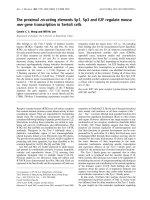
described in [24,25] (Figure 1).
For costal cartilage, explants were cleaned in PBS to eliminate
soft tissues and bone sternum parts were discarded. The cos-
tal cartilage was cut and divided into segments, which were
pooled. Each sample consisted of 50 mg of costal cartilage
explants. For articular cartilage, cartilage of two femoral heads
and two knees constitute one sample.
Immediately after the dissection, each sample was placed into
individual compression wells of Biopress culture plates (Flex-
ercell International, Hillsborough, NC, USA) in 1.5 ml of culture
medium (DMEM, containing penicillin-streptomycin 1% v/v,
glutamin 2% v/v, albumin 0.1% v/v and Hepes 30 mM) (Figure
1). All of the experiments were performed at 37°C, in air. The
compressive stress was applied to individual samples by the
Biopress system (Flexercell International) described by Fermor
and colleagues [26], whereas the control explants were kept
in unloaded conditions. At each time point (2 h, 4 h, 18 h and
24 h), we analyzed compressed and uncompressed explants
supplemented or not with effectors. Our results are expressed
as fold-induction in comparison to controls. After the applica-
tion of the mechanical regimen, supernatants and cartilage
explants were collected and stored immediately at -20°C and
-80°C, respectively.
Intermittent compression was applied using a sinusoidal wave-
form at 0.5 Hz (1 s on, 1 s off) for 30 minutes to 24 hours. Fer-
mor and colleagues [26] have established a calibration graph
for the Biopress system. This calibration establishes a linear
relationship between air pressure and the corresponding com-
pression force applied on a 5 mm diameter cartilage disk. This
calibration was calculated on a cross-sectional area of the
explant. In our model, the cartilage explants were disposed of
in order to form a 5 mm disk, which was composed of several
cartilage explants. We considered that the mechanical stress
applied is less uniform, but still is 1.0 MPa for an air pressure
of 30 kPa, according to the calibration from Fermor and col-
leagues [26].
Cell viability assay
Immediately after compression, cartilage was first incubated
with collagenase D solution (3 mg/ml) for 90 minutes at 37°C,
Figure 1
Mouse cartilage explants and Flexercell apparatus employed for mechanical stimulationMouse cartilage explants and Flexercell apparatus employed for mechanical stimulation. (a,b) Rib cages were harvested from one litter of 6-day-old
Swiss mice. (c) Costal cartilage was cleaned and cut into little segments. 50 mg of the costal cartilage pool were put into a Biopress culture plate
and 1.5 ml of media was added. (d) Each well was hermetically sealed with a specific cap. (e) The physiological compressive stress was applied by
the Flexercell Compression Plus system described by Fermor and colleagues [26] on mouse costal cartilage explants. Intermittent compression was
applied using a sinusoidal waveform at 0.5 Hz and 1.0 MPa of magnitude.
Arthritis Research & Therapy Vol 8 No 4 Gosset et al.
Page 4 of 14
(page number not for citation purposes)
and then incubated with collagenase D (0.5 mg/ml) overnight
at 37°C. The cell suspension obtained was mixed to disperse
any cell aggregates, producing a suspension of isolated cells.
Cells suspended in a culture medium were colored with
Trypan blue (0.04%) and counted in a hemocytometer. This
cell viability assay was carried out on one uncompressed and
two compressed explants, at 4 hours and 24 hours, and on
one explant immediately after the dissection, in two independ-
ent experiments.
PGE
2
and NO assays
Absolute concentrations of nitrite, a stable end-product of NO
metabolism, were determined in the media of the cartilage
explants using a spectrophotometric method based on the
Griess assay (Griess Reagent System, Promega, Charbon-
nières, France). Absorbance was measured at 550 nm and
nitrite concentration was determined by comparison with
standard solutions of sodium nitrite.
PGE
2
production was measured in the media by a high sensi-
tivity commercially available enzyme immunoassay kit (Cayman
Chemical, Ann Arbor, MI, USA), as previously described [27].
The cross-reactivity of the antibody with other prostanoids is
43% PGE
3
, 37.4% 8-iso PGE
2
, 18.7% PGE
1
, 1% PGF
1
α and
0.25% 8-iso PGF
2
α. The limit of detection was 9 pg/ml. PGE
2
concentration was analyzed at serial dilutions in duplicate and
was read against a standard curve.
RNA extraction, reverse transcription and quantitative
real-time PCR
Frozen cartilage explants (50 mg) were milled in 600 µL of RLT
buffer (from RNeasy Mini Kit, Qiagen GmbH, Hilden, Ger-
many) using a Mixer Mill MM 300 apparatus (Qiagen). Disrup-
tion was achieved through the beating and grinding effect of
beads on the cartilage samples as they were shaken together
in the grinding vessels. One steel ball (diameter 5 mm) was
added to each sample and they were mixed, at a cool temper-
ature, for two cycles of 2 minutes at 25 pulses/second. Then,
after removing the beads, the total RNA was extracted from
each sample using the RNeasy Kit (Qiagen) according to the
manufacturer's instructions. A proteinase K (Qiagen) digestion
step was performed after the lysis of cartilage explants and a
DNAse digestion step (RNAse free DNAse set, Qiagen) was
added. RNA concentration was then measured using a spec-
trophotometer. The migration in an agarose gel enabled quality
control.
Total RNA (1 µg) was reverse transcribed with Omniscript
(Qiagen) in a final volume of 20 µL containing 50 ng of oligos
dT. The enzyme was then inactivated by heating and the inter-
esting mRNAs (COX-1, genbank BC005573
; COX-2,
NM_011198
; COX-3, AY547265; mPGES-1, NM_022415;
mPGES-2, BC004846
; cPGES, NM-008278; 15-PGDH,
NM_008278
) were quantified by real-time quantitative reverse
transcription RT-PCR using the iCycler iQ Real Time PCR
(Bio-Rad) and QuantiTect SYBR PCR kits (Qiagen). Sense
and antisense PCR primers were designed based on mouse
sequence information for the amplification of genes of interest
(Table 1). The PCR reactions were performed in a 25 µl final
volume using 0.06 to 0.25 µl of cDNA, 600 ng of specific
primers and 1× QuantiTect SYBR Green PCR master mixture,
including HotStar Taq DNA Polymerase, QuantiTect SYBR
Green PCR buffer, SYBR Green I, and ROX in which there
was 5 mM MgCl. PCR amplification conditions were: initial
denaturation for 13 minutes at 95°C followed by 50 cycles
consisting of 30 seconds at 95°C and 30 seconds at 58°C.
Product formation was detected at 72°C in the fluorescein iso-
thiocyanate channel. The generation of specific PCR products
was confirmed by melting-curve analysis. For each real-time
RT-PCR run, cDNA were run in quadruplicate in parallel with
serial dilutions of a cDNA mixture tested for each primer pair
to generate a linear standard curve, which was used to esti-
mate relative quantities of COX, PGES and 15-PGDH mRNA
Table 1
Primer sequences used to detect mRNA in mouse costal cartilage explants
Genes Temperature
(°C)
Genbank ID Forward primer Reverse primer Amplicon length
(bp)
HPRT 58 NM_013556 5'-gctggtgaaaaggacctct-3' 5'-cacaggactagaacacctgc-3' 249
COX-1 58 BC005573
5'-ctttgcacaacacttcacccacc-3' 5'-agcaacccaaacacctcctgg-3' 285
COX-2 58 NM_011198
5'-gcattctttgcccagcactt-3' 5'-agaccaggcaccgaccaaaga-3' 299
COX-3 58 AY547265
5'-tgaacgctaggctcaactctc-3' 5'-ggttctggcacggatagtaac-3' 349
cPGES 58 AY281130
5'-agtcatggcctaggttaac-3' 5'-tgtgaatcatcatctgctcc-3' 196
mPGES-1 58 NM_022415
5'-ctgctggtcatcaagatgtacg-3' 5'-cccaggtaggccacgtgtgt-3' 294
mPGES-2 58 BC004846
5'-aagacatgtcccttctgc-3' 5'-ccaagatgggcactttcc-3' 133
15-PGDH 58 NM_008278
5'-gccaaggtagcattggtggat-3' 5'-cttccgaaatggtctacaact-3' 164
15-PGDH, NAD+-dependent 15 hydroxy prostaglandin dehydrogenase; COX, cyclooxygenase; HPRT, hypoxanthine-guanine
phosphoribosyltransferase; cPGES, cytosolic PGES; mPGES, microsomal PGES; PGES, prostaglandin E synthase.
Available online />Page 5 of 14
(page number not for citation purposes)
normalized for Hypoxanthine-guanine phosphoribosyltrans-
ferase (HPRT genbank NM_008278
) in the samples.
Protein extraction and western blotting
Frozen cartilage explants were disrupted using a Mixer Mill MM
300 apparatus (Qiagen) in 500 µL of cold lysis buffer (20 mM
Tris pH 7.6, 120 mM NaCl, 10 mM EDTA, 10% glycerol, 1%
Nonidet P-40, 100 mM NaF; 10 mM Na
4
P
2
0
7
, 1 mM AEBSF
(4-(2-Aminoethyl)benzenesulphonyl fluoride), 2 mM Na
3
VO
4
,
40 µg/ml leupeptin, 1 µM pepstatin A, 10 µg/ml aprotinin).
One steel ball (diameter 5 mm) was added to each sample,
which were mixed at a cool temperature for two cycles of 2
minutes at 25 pulses/second. Then, after removing the beads,
the samples were shaken gently for 1 hour at 4°C and then
centrifuged for 1 hour (13,000 g, 4°C). The supernatants were
collected and protein concentrations were determined using
the bicinchoninic acid assay kit (Perbio Science for Pierce,
Bezons, France).
Cartilage explant lysates were separated by 8% or 15% SDS-
PAGE and transferred to nitrocellulose membranes. The blots
were incubated (then stripped and reprobed) by the appropri-
ate primary polyclonal antibody to COX-2, COX-3, COX-1,
mPGES-1, mPGES-2, cPGES and monoclonal antibody to β-
actin. The blots were then incubated with horseradish peroxi-
dase-conjugated secondary goat antibody. The membranes
were washed repeatedly with Tris-buffered Saline containing
Tween-20 0.1% (v/v) and the signals were detected using the
enhanced chemiluminescence detection system and exposed
to Kodak BioMax MR-1 film. We transfected Cos cells with
plasmids encoding COX-2 and mPGES-1. Cells extracts con-
taining COX-2 and mPGES-1 proteins surexpressed were
used as positive controls.
Immunohistochemistry
After compression for 18 hours, cartilage explants were imme-
diately collected and fixed in 70% ethanol at 4°C for 48 hours.
After dehydratation, the cartilage samples were embedded
without demineralization in methyl methacrylate (Merck, Darm-
stadt, Germany). Transversal sections (4 µm thick) were cut
parallel to the rib axis using a Polycut E microtome (Leica,
Wetzlar, Germany). Sections mounted onto slides were
deplastified in 2-methoxyethylacetate prior to further process-
ing. The primary polyclonal antibodies used were the same as
those used for western blotting, as previously described. For
immunochemistry, the sections were incubated overnight with
0.1 M PBS supplemented with 0.05% Tween 20 (Sigma) and
1% BSA (Euromedex) and the primary polyclonal antibody
(1:50) at 4°C in a moist chamber. The sections were then incu-
bated with biotinylated goat anti-rabbit IgG for PGES or rabbit
anti-goat IgG for COX-2 (Vector, Burlingame, CA, USA) for 90
minutes at room temperature. They were then treated with 3%
hydrogen peroxide (10 minutes), and an avidin-biotin peroxi-
dase complex (ABC Vectastain kit, Vector) for 60 minutes.
PBS (0.1 M) was used for the washing steps between incuba-
tions. Diaminobenzidine tetrahydrochloride (Sigma) was used
as the chromogen. The sections were lightly counterstained
with toluidine blue (pH 3.8). Negative controls were prepared
by omitting the primary antibody in the diluant solution (BSA
1% and goat serum 10% for PGES, and BSA 10% and milk
1% for COX-2). Immunohistological analysis was carried out
on two uncompressed and two compressed costal cartilage
explants. Images were obtained using an optical microscope
and analysis for each enzyme utilized a blind test.
Statistical analysis
All data are reported as mean ± SEM, unless stated otherwise.
Unpaired Students' t-tests were used to compare the mean
values between groups with the GraphPad InStat version
Figure 2
Compression stimulates nitric oxide (NO) and prostaglandin E2 (PGE
2
) release in mouse costal cartilage explants in the mediaCompression stimulates nitric oxide (NO) and prostaglandin E2 (PGE
2
)
release in mouse costal cartilage explants in the media. Mouse costal
cartilage explants were compressed (C) or not (NC) for 2 h, 4 h, 18 h
and 24 h. At each time interval, our results are expressed in fold-induc-
tion in comparison to the appropriate control. (a) The amount of NO
released into the media (µmol/mg of costal cartilage) was measured by
Griess reagent. Values are the mean and SEM of 3 (C 2 h and 4 h) and
2 (C 18 h and 24 h) independent experiments with n = 2/group/experi-
ments. ***p < 0.001 versus control (NC). (b) The amount of PGE
2
released into the media (pg/mg/ml of costal cartilage) was measured
by enzyme immunoassay. Values are the mean and SEM of 3 (C 2 h), 2
(C 4 h and 18 h) and 4 independent experiments (C 24 h) with n = 2/
group/experiments, analyzed in duplicate. ***p < 0.001 versus control
(NC).
Arthritis Research & Therapy Vol 8 No 4 Gosset et al.
Page 6 of 14
(page number not for citation purposes)
(GraphPad Software, San Diego, California, USA). The P val-
ues ≤ 0.05 are considered to be significant.
Results
Compressive stress triggers the synthesis of NO and
PGE
2
via α5β1 integrin but not via IL-1 synthesis
To determine the effects of compressive stress on chondro-
cyte activation, we assessed NO and PGE
2
release in the
media in compressed and uncompressed costal cartilage
explants. Different magnitudes and lengths of stress were
applied in order to define the optimum conditions (data not
shown). At a sinusoidal waveform frequency of 0.5 Hz and a
magnitude of 1 MPa, NO release significantly increased (6-
fold increase; p < 0.001) within 2 hours compared to uncom-
pressed explants, and lasted 24 hours (Figure 2a), as
described by Fermor and colleagues [6]. Also, PGE
2
synthesis
in the media significantly increased, with a peak at 2 hours that
was sustained up to 4 hours (6-fold increase; p < 0.001) and
then decreased at 24 hours (Figure 2b).
Compressive stress was also applied to mouse articular carti-
lage explants in order to avoid a bias due to the origin of carti-
lage. As in costal cartilage, articular cartilage explants
submitted to compression exhibit an increase in PGE
2
release
after 2 hours (16-fold increase; p < 0.01), which was sus-
tained from 4 hours to 18 hours (6-fold increase) before
declining to control levels. Even though PGE
2
release in artic-
ular cartilage (16-fold increase, p < 0.01) was stronger at 2
hours than in costal cartilage (6-fold increase, p < 0.001) and
was sustained for longer, only minor differences in kinetics
were observed (Figure 3).
Viability of the chondrocytes in the mouse costal cartilage
explants was tested using Blue Trypan coloration. No altera-
tion of cell viability was seen between compressed and
uncompressed samples within 24 hours (data not shown).
To confirm the validity of our compressive model on mouse
costal cartilage, we wanted to highlight the implication of
integrin α5β1 in the PGE
2
release triggered by mechanical
stress. Cartilage explants treated with anti-integrin α5β1
blocking antibody (AB1950) at 2.5 µg/ml induced a 50%
decrease of compression-induced NO (4.16 ± 0.57 versus
2.68 ± 0.43 µM; data obtained from 2 independent experi-
ments with n = 2/group/experiments; p < 0.001, data not
shown). Moreover, a decrease in PGE
2
release of approxi-
mately 50% in compressed cartilage treated with the blocking
α5β1 antibody was observed (Figure 4a). No modification in
NO (4.01 ± 0.1 versus 4.4 ± 1.43 µM; data obtained from 2
independent experiments with n = 2/group/experiments, data
not shown) or PGE
2
release (Figure 4a) was detected in media
of compressed cartilage treated with the non-blocking anti-β1
subunit antibody at 2.5 µg/ml (VMA1997).
We previously reported that the pro-inflammatory cytokine IL-
1 triggers the expression of COX-2 and mPGES-1 [21,28].
Since the integrin antibody did not fully inhibit a compression-
induced PGE
2
release, even if other mechanoreceptors have
been described on chondrocytes, we hypothesized that com-
pression could indirectly act on cartilage by inducing the syn-
thesis of IL-1. When the IL-1 receptor antagonist IL1-Ra was
added at a concentration of 100 ng/ml prior to compression,
no variation in PGE
2
release was observed (Figure 4b), sug-
gesting that compression-induced PGE
2
release is not medi-
ated by IL-1.
Expression of COX and PGES enzymes in uncompressed
and compressed cartilage explants
We subsequently focused our study on the enzymes involved
in PGE
2
synthesis, cyclooxygenases and prostaglandin E syn-
thases. Mouse costal cartilage explants subjected to compres-
sive stress for 18 hours were fixed in ethanol, embedded in
methyl methacrylate and cut into serial sections that were
immunostained with antibody against COX-2, mPGES-1,
mPGES-2 and cPGES. Toluidine blue counterstaining colors
the extracellular matrix and nuclei of cells. In uncompressed
cartilage, a few peripheral cells presented positive immunos-
taining (brown) for COX-2 and none did so for mPGES-1.
After compression, an increased brown staining for COX-2
and mPGES-1 in cells was visible around the nuclei, suggest-
ing a colocalization of these enzymes in the perinuclear region
in loaded chondrocytes. For cPGES and mPGES-2, no differ-
ences appeared in chondrocytes from compressed cartilage
explants compared to uncompressed explants (Figure 5).
COX expression in cartilage explants subjected to
compression
To study the effects of mechanical loading on COX gene
expression, we used real-time RT-PCR quantitative analysis
Figure 3
Compression stimulates prostaglandin E2 (PGE
2
) release in mouse articular cartilage explantsCompression stimulates prostaglandin E2 (PGE
2
) release in mouse
articular cartilage explants. Mouse articular cartilage explants were
compressed (C) or not (NC) for 2 h, 4 h, 18 h and 24 h. The amount of
PGE
2
released into the media (pg/ml) was measured by enzyme immu-
noassay. Values are the mean ± SEM of 2 independent experiment with
n = 2/group/experiments, analyzed in duplicate. *p < 0.05, **p < 0.01,
***p < 0.001 versus control (NC).
Available online />Page 7 of 14
(page number not for citation purposes)
and immunoblotting to evaluate, respectively, the transcrip-
tional and translational expression of COX-1, COX-2 and
COX-3 genes. We extracted the total RNA and proteins
directly from costal cartilage explants. An increased expres-
sion of COX-2 mRNA, but not COX-1, was observed after 2
hours with a maximal effect after 4 hours of compression (Fig-
ure 6a,b). Interestingly, COX-3 mRNA was expressed in carti-
lage but compressive stress had no effect on its transcriptional
expression in cartilage explants (Figure 6c).
We then assessed COX protein levels by immunoblotting
using polyclonal antibodies raised against COX-1, COX-2 and
COX-3. As expected, compression induced COX-2 protein
expression after 4 hours and peaked at 18 hours, whereas
COX-1 expression remained unchanged. Both the COX-3
protein and its mRNA were expressed in cartilage; however,
compression did not modify its expression (Figure 6d).
Figure 4
Over-release of prostaglandin E2 (PGE
2
) in compressed costal cartilage explants is the result of mechanical stressOver-release of prostaglandin E2 (PGE
2
) in compressed costal cartilage explants is the result of mechanical stress. (a) Implication of the mech-
anoreceptor integrin α5β1 in PGE
2
over-release in compressed cartilage explants. Mouse costal cartilage explants treated with either the β1 non-
blocking antibody VMA1997 or the α5β1 blocking antibody AB1950 at 2.5 µg/ml were compressed (C) or not compressed (NC) for 4 h. Results
are normalized to the mean not-compressed control (cont) value. Data are the mean ± SEM of 2 independent experiments with n = 2/group/experi-
ments, analyzed in duplicate. ***p < 0.001 versus control NC, *p < 0.05 versus control C. (b) Increased PGE
2
release in compressed costal carti-
lage explants is not due to the cytokine IL-1. Mouse costal cartilage explants treated with the IL-1 receptor antagonist (IL1-Ra) at 100 ng/ml were
compressed (C) or not compressed (NC) for 4 h. Results are normalized to the mean not compressed control value. Data are the mean ± SEM of 2
independent experiment with n = 2/group/experiments, analyzed in duplicate. *p < 0.05 versus control NC.
Arthritis Research & Therapy Vol 8 No 4 Gosset et al.
Page 8 of 14
(page number not for citation purposes)
PGES expression in cartilage explants under
compression
Expression of mPGES-1 but not cPGES mRNA increased
after 2 hours of compression with a peak at 4 hours. Similarly,
the amount of mPGES-1 protein increased in compressed
explants after 2 hours, peaking at 18 hours (Figure 7a,b,d).
The increased expression over the time of mPGES-1 protein
in uncompressed samples, which was also observed for COX-
2, could be triggered by mediators release during explantation
and cutting of cartilage.
Interestingly, mPGES-2 was not regulated by compressive
stress, both at the mRNA and protein levels (Figure 7c,d).
The gene encoding 15-prostaglandin dehydrogenase is
mechanosensitive
Because our results highlighted a discrepancy between the
kinetics of PGE
2
release and COX-2 and mPGES-1 expres-
sion (compare Figure 2b with Figures 6 and 7), we hypothe-
sized that the decrease in PGE
2
production observed after 4
hours was due, at least in part, to the activation of a catabolic
pathway of PGE
2
.
NAD+-dependent 15 hydroxy prostaglandin dehydrogenase
(15-PGDH) is considered to be the key enzyme in the catabo-
lism of PGE
2
. Interestingly, the gene encoding 15-PGDH is
mechanosensitive and the kinetics of its expression is in agree-
ment with our hypothesis since a peak of expression is
observed at 4 hours (Figure 8).
Discussion
Our findings demonstrate that dynamic mechanical loading of
costal cartilage can significantly increase PGE
2
release. More-
over, we describe here for the first time that COX-2 and
mPGES-1 expression is increased in mouse costal cartilage
explants under compression but not their constitutive isoforms
COX-1 and cPGES. Therefore, it appears that COX-2 and
mPGES-1 are encoded by mechanosensitive genes impli-
cated in the compression-induced PGE
2
release. PGE
2
is the
pivotal eicosanoid involved in the initiation and the develop-
ment of inflammatory disease, such as rheumatoid arthritis
[29]. Notably, it is thought to be a key regulator of cartilage
degradation during OA [30]. An increase in PGE
2
release
induced by mechanical stress has already been described in
various tissues [31,32] and particularly in articular cartilage
subjected to dynamic compression representative of the phys-
iological range [6].
Regulation of COX-2 mRNA expression in cartilage by
mechanical stress has already been reported in the literature
[6]. Notably, elements including AP-1 sites, cyclic AMP
response elements (CREs) and shear stress response ele-
ments (SSRE) are found in the promoter region of mechanical
stress-response genes, such as those encoding COX-2 and
inducible NO synthase. Shear stress response elements con-
tain a TPA response element to which NFκB, which is part of
a main mechanical pathway, binds [33]. Ogasawara and col-
leagues [34] have described the role of C/EBP beta, AP-1
sites and CREB in shear stress-induced COX-2 expression in
osteoblasts. Moreover, post-transcriptional regulation by
mRNA stabilization seems to be involved in COX-2 gene
expression in vascular endothelial cells subjected to fluid
Figure 5
Compression increases cyclooxygenase type 2 (COX-2) and micro-somal prostaglandin E synthase type 1 (mPGES-1) but not cytosolic PGES (cPGES) and mPGES-2 protein expression in costal cartilageCompression increases cyclooxygenase type 2 (COX-2) and micro-
somal prostaglandin E synthase type 1 (mPGES-1) but not cytosolic
PGES (cPGES) and mPGES-2 protein expression in costal cartilage.
Costal cartilage explants were (a-d) not compressed or (e-h) com-
pressed for 18 h and immunostained with anti-COX-2, anti-mPGES-1,
anti-cPGES and anti-mPGES-2 antibodies and then counterstained
with toluidine blue. Increased expression of (e) COX-2 and (f) mPGES-
1 protein was seen in compressed explants compared to uncom-
pressed ((a) COX-2 and (b) mPGES-1). In contrast, (g) cPGES and
(h) mPGES-2 were not overexpressed after application of a compres-
sive stress compared to the uncompressed condition ((c) cPGES and
(d) mPGES-2). Representative findings from two compressed and two
uncompressed samples were tested. Scale bar = 100 µM.
Available online />Page 9 of 14
(page number not for citation purposes)
shear stress [35]. In addition to these studies at the mRNA
level, we show here, for the first time in cartilage, that COX-2
is also increased at the protein level.
Interestingly, our data indicate that COX-3, also named COX-
1 V1, is expressed in mouse costal cartilage. COX-3, which
was cloned in 2002, was derived from COX-1 through reten-
tion of intron 1 in its mRNA. This probably resulted in the mod-
ification of the active site conformation of the enzyme. COX-3
expression has actually been found in several canine, human
and rodent tissues, but never in cartilage, whatever the spe-
cies [18]. In this present study, we report for the first time the
expression of COX-3 (mRNA and protein) in mouse cartilage.
Moreover, we show that mechanical loading did not modify its
expression. As COX-1 and COX-3 are derived from the same
gene, these enzymes share the same promoter. However, no
sites that are regulated through mechanical stress or pro-
inflammatory cytokines have been found so far in the COX-1
promoter, which is consistent with the fact that COX-1 is con-
stitutively and ubiquitously expressed. Thus, this might explain
the lack of COX-3 regulation by compression.
The regulation of mPGES-2 expression has never been
described in cartilage. mPGES-2 is ubiquitously expressed
Figure 6
Compression increases cyclooxygenase type 2 (COX-2) gene expression but not COX-1 nor COX-3 in mouse costal cartilage explantsCompression increases cyclooxygenase type 2 (COX-2) gene expression but not COX-1 nor COX-3 in mouse costal cartilage explants. (a-c) Real-
time RT-PCR assays demonstrating increased COX-2 gene expression after 2 h and 4 h in compressed explants versus control and no increase for
COX-1 and COX-3. Standard curves for COX-1, COX-2, COX-3 and hypoxanthine-guanine phosphoribosyltransferase (HPRT) were generated by
serial dilution of a cDNA mixture. The amount of COX-1, COX-2 and COX-3 mRNA was normalized against the amount of HPRT mRNA measured
in the same cDNA. Values are the mean ± SEM of 4 independent experiments with n = 1/group/experiment for COX-1 and COX-2 and of 2 inde-
pendent experiments with n = 1/group/experiment for COX-3. *p < 0.05, **p < 0.01 versus control (NC). (d) Explant lysates were analyzed by SDS-
PAGE using 8% gradient gels. Proteins were transferred to a nylon membrane and successively blotted with anti-COX-1, anti-COX-2, anti-COX-3
and anti-β-actin antibodies. An increased expression of COX-2 protein in compressed cartilage compared to uncompressed, but not COX-1 and
COX-3, was observed after 4 hours of compression up to 24 hours. Each blot is representative of three independent experiments.
Arthritis Research & Therapy Vol 8 No 4 Gosset et al.
Page 10 of 14
(page number not for citation purposes)
under basal conditions in many tissues and is activated by
reducing agents, but its role in PGE
2
release in both basal and
inflammatory contexts remains unclear. In human rheumatoid
synoviocytes, expression of mPGES-1 increased with severity
of the disease, whereas that of mPGES-2 did not [23]. In
COX-2-deficient mouse brains, a decreased release of PGE
2
and a decreased expression of mPGES-2, but not of mPGES-
1 or cPGES, was observed, suggesting that mPGES-2 could
be functionally coupled to COX-2 [36]. In the present study,
mPGES-2 expression was similar in compressed and uncom-
pressed cartilage explants, suggesting that mPGES-2 is not
encoded by a mechanosensitive gene.
The striking point of our study is the evidence that mPGES-1
is encoded by a mechanosensitive gene. In recent years, sev-
eral studies have demonstrated that inflammation induces
mPGES-1. In rat paws of the acute and chronic arthritis model,
up-regulation of mPGES-1 mRNA and protein expression was
observed. Moreover, levels of mPGES-1 mRNA and protein
were markedly elevated in OA versus normal cartilage [37].
Additionally, we and others have previously reported an over-
expression of mPGES-1 in OA chondrocytes in primary cul-
tures stimulated by IL-1 [21,22]. Interestingly, our results
identify an earlier significant transcriptional expression (as
soon as 2 hours) after mechanical stress compared to the
effect of IL-1 (after 12 hours). Moreover, its induction was
higher with compression (five-fold) compared to IL-1 stimula-
tion (three-fold). A structural comparison of COX-2 and
mPGES-1 promoters revealed that the gene encoding
mPGES-1 does not contain transcriptional elements that are
Figure 7
Compression increases microsomal prostaglandin E synthase type 1 (mPGES-1) gene expression but not mPGES-2 nor cytosolic PGES (cPGES) in mouse costal cartilage explantsCompression increases microsomal prostaglandin E synthase type 1 (mPGES-1) gene expression but not mPGES-2 nor cytosolic PGES (cPGES)
in mouse costal cartilage explants. The rates of mPGES-1, cPGES and mPGES-2 expression in response to compressive stress at 2 h and 4 h were
analyzed by (a-c) real-time quantitative RT-PCR and (d) immunoblotting. (a-c) Up-regulation of mPGES-1 mRNA expression but not cPGES and
mPGES-2 mRNA expression by mechanical stress appeared at 2 hours until 4 hours. Values are the mean ± SEM of 3 independent experiment with
n = 1/group/experiment. *p < 0.05, **p < 0.01 versus control (NC). (d) Increased translational expression of mPGES-1 but not cPGES and
mPGES-2 was observed on the immunoblot (15% gradient gels), from 4 hours until 24 hours. Each blot is representative of three independent
experiments.
Available online />Page 11 of 14
(page number not for citation purposes)
classically involved in response to mechanical stress (AP-1,
CRE, SSRE), as described for the COX-2 promoter [38]. It
suggests that divergent transcriptional mechanisms are
responsible for inducible mPGES-1 regulation. Yokota and
colleagues [39] have described the role of the nuclear regula-
tor CITED2 (CBP/p300-interacting transactivator with ED-rich
tail 2) in shear-induced down-regulation of matrix metallopro-
teinase 1 and matrix metalloproteinase 13. This mechanism
could also occur in compression-induced mPGES-1 expres-
sion. But post-transcriptional regulation could also be present
since a stabilization of mPGES-1 mRNA has been recently
described in cardiomyocytes, leading to delayed protein
expression via cJNK [40].
Our data suggest that loading triggers mPGES-1 and COX-2
colocalization in the perinuclear region of the chondrocytes.
This result is in line with the study from Kojima and colleagues
[22] that recently described COX-2 and mPGES-1 colocaliza-
tion around the nuclei of chondrocytes after IL-1 stimulation.
Selective inhibitors of mPGES-1 have been developed
recently [41]. Effectively, targeting the mPGES-1 enzyme in
preference to COX-2 should represent a new therapeutical
approach to treating joint diseases such as OA. Interestingly,
Cheng and colleagues [42] reported that mPGES-1-null mice
exhibit no alteration in thrombogenesis or blood pressure,
whereas selective COX-2 inhibitors (Coxib) did. These results
suggest that inhibitors of mPGES-1 may retain their anti-
inflammatory efficacy by depressing PGE
2
, while avoiding the
adverse cardiovascular consequences associated with COX-
2 deletion [42].
To understand the weak PGE
2
release at 18 and 24 hours
after compression, we focused on 15-PGDH mRNA expres-
sion because 15-PGDH is one of the major PGE
2
-inactivating
enzymes [43]. 15-PGDH expression decreases in response to
IL-1β treatment in trophoblast cells in primary culture [44] as
well as in response to lipopolysaccharide (LPS) treatment in
liver and lungs of rats [45]. Moreover, 15-PGDH expression is
reduced in inflammatory bowel disease [46]. To our knowl-
edge, 15-PGDH expression in cartilage has never been
reported and its expression under mechanical stress was
unknown. The findings presented in this study first indicate
that 15-PGDH is expressed in mouse cartilage and is induced
after 4 hours of compression (three-fold induction). Genomic
structural analysis revealed that the human 15-PGDH pro-
moter contains binding sites for AP-1 and CREB, two tran-
scriptional factors implicated in the regulation of gene
expression subjected to mechanical stress [47]. We suggest
that the 15-PGDH enzyme could be implicated in the catabo-
lism of PGE
2
after 18 hours. So, proinflammatory stimuli (LPS
or cytokines) induce monophasic PGE
2
synthesis whereas
mechanical stress triggers a biphasic response in cartilage,
PGE
2
synthesis followed by PGE
2
degradation. We hypothe-
size that this response corresponds to an adaptation of the tis-
sue to this stress.
The choice of an experimental model to study the physiological
regulation of cartilage homeostasis by mechanical stress is
challenging. Three types of mechanical stress have been
described: shear stress, strain and compressive stress. Over-
loading of the joints, a major risk factor for OA, mainly induces
compressive stress. However, shear stress and strain have
also been described when overloading is experimentally
applied on cartilage. The compression of chondrocytes
embedded in agarose or collagen gels as well as the compres-
sion of ex vivo porcine cartilage explants represent the main
strategies developed [6,16,26,48]. Among the literature, it
appears obvious that the consequences to metabolism of
chondrocytes vary according to the type of stress applied.
Takahashi and colleagues [49] reported that static compres-
sion promotes type II collagen, whereas cyclic loading could
denature type II collagen in articular chondrocytes [50]. Here,
we used mouse cartilage explants subjected to cyclical uniax-
ial mechanical stress. Mouse cartilage was chosen because of
the availability of a large choice of biomolecular tools suitable
to study it, such as genetically modified mice. However, many
labs work on discs of porcine or bovine cartilage, permitting a
more homogeneous compressive stress. In our model, the
compressive stress is less uniform because of the size of the
explants. Enhancements to our system, in order to better
define the stress applied, is ongoing.
We decided to work on costal cartilage rather than articular
cartilage because of the extremely small quantity of the latter
available. However, we performed comparative experiments in
order to validate our model with costal cartilage explants. We
observed an induction of PGE
2
release in both types of carti-
lage, but with slight differences in kinetics.
Figure 8
Compression increases NAD+-dependent 15 hydroxy prostaglandin dehydrogenase (15-PGDH) transcriptional expression in mouse costal cartilage explantsCompression increases NAD+-dependent 15 hydroxy prostaglandin
dehydrogenase (15-PGDH) transcriptional expression in mouse costal
cartilage explants. The rate of 15-PGDH mRNA expression in response
to a compressive stress (C) at 2 hours and 4 hours was analyzed by
real-time quantitative RT-PCR. Increased 15-PGDH mRNA expression
was observed after 4 hours of a compressive stress. Values are the
mean ± SEM of 3 independent experiment with n = 1/group/experi-
ment. *p < 0.05 versus control (NC).
Arthritis Research & Therapy Vol 8 No 4 Gosset et al.
Page 12 of 14
(page number not for citation purposes)
The mechanoreceptor integrin α5β1 is considered by many
authors to be the critical mechanoreceptor for transducing sig-
nal from the extracellular matrix to the chondrocyte [8]. Our
data confirm the major role of this receptor in compression-
induced PGE
2
release.
Among the mechanotransduction pathways recently
described, Mitogen-Activated Protein Kinase (MAPK) plays a
major role. In smooth muscle cells and fibroblasts, mechanical
strain increases the activity of all three MAPKs, namely p38
MAPK, ERK 1/2 and Junk kinase [51,52]. In particular, ex vivo
cartilage compression has been reported to activate the three
MAPKs, but with different kinetics [9]. Preliminary results sug-
gest that this is also the case in our model (personal commu-
nication).
Surprisingly, overexpression of COX-2 and mPGES-1 pro-
teins was observed at 18 hours in uncompressed samples.
Several authors have described the release of mediators, such
as basic fibroblast growth factor (bFGF) and IL-1α, in cartilage
after explantation and cutting [53,54]. Moreover, Fermor and
colleagues [6] reported that uncompressed explants of articu-
lar cartilage exhibited a significant production of PGE
2
(from
14 pg/mg/ml at 24 hours after explantation to 1 pg/mg/ml at
72 hours). We have considered that dissection alone induces
COX-2 and mPGES-1 protein expression in the uncom-
pressed samples.
As mature articular cartilage is an avascular tissue, the oxygen
supply to resident chondrocytes could rather limit and influ-
ence chondrocyte activation under a mechanical signal. Fer-
mor and colleagues have shown that oxygen tension
influences the endogenous production of NO and PGE
2
in
porcine cartilage explants in response to mechanical stimula-
tion. Under mechanical compression, PGE
2
production in car-
tilage at 20% O
2
increased 50-fold, but in cartilage at 5% O
2
it increased only 4-fold and in cartilage at 1% O
2
it did not
increase at all [48]. Since previous studies have suggested
that the oxygen tension in the superficial layer of articular car-
tilage is higher than in the deeper layer [55], our results could
have occurred, at least in part, in the superficial zone of articu-
lar cartilage.
Conclusion
We have demonstrated that COX-2 and mPGES-1 are mech-
anosensitive enzymes after dynamic compression. Moreover,
the key enzyme implicated in the catabolism of PGE
2
, 15-
PGDH, is expressed in cartilage and its expression is regu-
lated by compression. As mPGES-1 is currently described as
a potential therapeutic target for controlling PGE
2
release in
OA, further research into the regulation of this mechanosensi-
tive gene should be helpful for a better understanding of the
key pathways implicated in its expression in order to counter-
act cartilage degradation in overloading joints.
Competing interests
The authors declare that they have no competing interests.
Authors' contributions
MG performed animal surgery, compression experiments,
PGE
2
and NO assays, PCR experiments, western blotting
experiments and immunohistochemistry experiments, analyzed
the results and drafted the manuscript. FB was involved in the
conception and design of the study and critically revised the
manuscript for important intellectual content. AL performed
animal surgery, compression experiments, PGE
2
and NO
assays and western blotting experiments. AP performed ani-
mal surgery, compression experiments and PGE
2
and NO
assays. ST initiated and participated in compression experi-
ments. JLS performed immunohistochemistry experiments. CJ
designed the study, participated in data analysis and helped
draft the manuscript. All authors read and approved the final
manuscript.
Acknowledgements
We thank Pr. M Raymondjean for critically reviewing the manuscript and
making valuable suggestions. We thank B Baroukh and A Llorens for
their technical assistance in immunohistochemistry. This work was sup-
ported by the Association de Recherche sur la Polyarthrite and the the
French Society of Rheumatology.
References
1. Centers for Disease Control and Prevention: Arthritis prevalence
and activity limitations – United States, 1990. MMWR Morb
Mortal Wkly Rep 1994, 43:433-438.
2. Morrell KC, Hodge WA, Krebs DE, Mann RW: Corroboration of
in vivo cartilage pressures with implications for synovial joint
tribology and osteoarthritis causation. Proc Natl Acad Sci USA
2005, 102:14819-14824.
3. Herberhold C, Faber S, Stammberger T, Steinlechner M, Putz R,
Englmeier KH, Reiser M, Eckstein F: In situ measurement of
articular cartilage deformation in intact femoropatellar joints
under static loading. J Biomech 1999, 32:1287-1295.
4. Blain EJ, Gilbert SJ, Wardale RJ, Capper SJ, Mason DJ, Duance
VC: Up-regulation of matrix metalloproteinase expression and
activation following cyclical compressive loading of articular
cartilage in vitro. Arch Biochem Biophys 2001, 396:49-55.
5. Islam N, Haqqi TM, Jepsen KJ, Kraay M, Welter JF, Goldberg VM,
Malemud CJ: Hydrostatic pressure induces apoptosis in
human chondrocytes from osteoarthritic cartilage through up-
regulation of tumor necrosis factor-alpha, inducible nitric
oxide synthase, p53, c-myc, and bax-alpha, and suppression of
bcl-2. J Cell Biochem 2002, 87:266-278.
6. Fermor B, Weinberg JB, Pisetsky DS, Misukonis MA, Fink C, Gui-
lak F: Induction of cyclooxygenase-2 by mechanical stress
through a nitric oxide-regulated pathway. Osteoarthritis Carti-
lage 2002, 10:792-798.
7. Mobasheri A, Carter SD, Martin-Vasallo P, Shakibaei M: Integrins
and stretch activated ion channels; putative components of
functional cell surface mechanoreceptors in articular chondro-
cytes. Cell Biol Int 2002, 26:1-18.
8. Millward-Sadler SJ, Salter DM: Integrin-dependent signal cas-
cades in chondrocyte mechanotransduction. Ann Biomed Eng
2004, 32:435-446.
9. Fanning PJ, Emkey G, Smith RJ, Grodzinsky AJ, Szasz N, Trippel
SB: Mechanical regulation of mitogen-activated protein kinase
signaling in articular cartilage. J Biol Chem 2003,
278:50940-50948.
10. Laufer S: Role of eicosanoids in structural degradation in oste-
oarthritis. Curr Opin Rheumatol 2003, 15:623-627.
11. Hardy MM, Seibert K, Manning PT, Currie MG, Woerner BM,
Edwards D, Koki A, Tripp CS: Cyclooxygenase 2-dependent
Available online />Page 13 of 14
(page number not for citation purposes)
prostaglandin E2 modulates cartilage proteoglycan degrada-
tion in human osteoarthritis explants. Arthritis Rheum 2002,
46:1789-1803.
12. Miwa M, Saura R, Hirata S, Hayashi Y, Mizuno K, Itoh H: Induction
of apoptosis in bovine articular chondrocyte by prostaglandin
E(2) through cAMP-dependent pathway. Osteoarthritis Carti-
lage 2000, 8:17-24.
13. Jacques C, Sautet A, Moldovan M, Thomas B, Humbert L, Beren-
baum F: Cyclooxygenase activity in chondrocytes from oste-
oarthritic and healthy cartilage. Rev Rhum Engl Ed 1999,
66:701-704.
14. McCoy JM, Wicks JR, Audoly LP: The role of prostaglandin E2
receptors in the pathogenesis of rheumatoid arthritis. J Clin
Invest 2002, 110:651-658.
15. Xu Z, Buckley MJ, Evans CH, Agarwal S: Cyclic tensile strain acts
as an antagonist of IL-1 beta actions in chondrocytes. J Immu-
nol 2000, 165:453-460.
16. Chowdhury TT, Bader DL, Lee DA: Dynamic compression coun-
teracts IL-1 beta-induced release of nitric oxide and PGE2 by
superficial zone chondrocytes cultured in agarose constructs.
Osteoarthritis Cartilage 2003, 11:688-696.
17. Smith RL, Donlon BS, Gupta MK, Mohtai M, Das P, Carter DR,
Cooke J, Gibbons G, Hutchinson N, Schurman DJ: Effects of
fluid-induced shear on articular chondrocyte morphology and
metabolism in vitro. J Orthop Res 1995, 13:824-831.
18. Hersh EV, Lally ET, Moore PA: Update on cyclooxygenase inhib-
itors: has a third COX isoform entered the fray? Curr Med Res
Opin 2005, 21:1217-1226.
19. Trebino CE, Stock JL, Gibbons CP, Naiman BM, Wachtmann TS,
Umland JP, Pandher K, Lapointe JM, Saha S, Roach ML, et al.:
Impaired inflammatory and pain responses in mice lacking an
inducible prostaglandin E synthase. Proc Natl Acad Sci USA
2003, 100:9044-9049.
20. Kamei D, Yamakawa K, Takegoshi Y, Mikami-Nakanishi M, Naka-
tani Y, Oh-Ishi S, Yasui H, Azuma Y, Hirasawa N, Ohuchi K, et al.:
Reduced pain hypersensitivity and inflammation in mice lack-
ing microsomal prostaglandin e synthase-1. J Biol Chem
2004, 279:33684-33695.
21. Masuko-Hongo K, Berenbaum F, Humbert L, Salvat C, Goldring
MB, Thirion S: Up-regulation of microsomal prostaglandin E
synthase 1 in osteoarthritic human cartilage: critical roles of
the ERK-1/2 and p38 signaling pathways. Arthritis Rheum
2004, 50:2829-2838.
22. Kojima F, Naraba H, Miyamoto S, Beppu M, Aoki H, Kawai S:
Membrane-associated prostaglandin E synthase-1 is upregu-
lated by proinflammatory cytokines in chondrocytes from
patients with osteoarthritis. Arthritis Res Ther 2004,
6:R355-R365.
23. Murakami M, Nakashima K, Kamei D, Masuda S, Ishikawa Y, Ishii T,
Ohmiya Y, Watanabe K, Kudo I: Cellular prostaglandin E2 pro-
duction by membrane-bound prostaglandin E synthase-2 via
both cyclooxygenases-1 and -2. J Biol Chem 2003,
278:37937-37947.
24. Lefebvre V, Garofalo S, Zhou G, Metsaranta M, Vuorio E, De Crom-
brugghe B: Characterization of primary cultures of chondro-
cytes from type II collagen/beta-galactosidase transgenic
mice. Matrix Biol 1994, 14:329-335.
25. Salvat C, Pigenet A, Humbert L, Berenbaum F, Thirion S: Imma-
ture murine articular chondrocytes in primary culture: a new
tool for investigating cartilage. Osteoarthritis Cartilage 2005,
13:243-249.
26. Fermor B, Weinberg JB, Pisetsky DS, Misukonis MA, Banes AJ,
Guilak F: The effects of static and intermittent compression on
nitric oxide production in articular cartilage explants. J Orthop
Res 2001, 19:729-737.
27. Berenbaum F, Humbert L, Bereziat G, Thirion S: Concomitant
recruitment of ERK1/2 and p38 MAPK signalling pathway is
required for activation of cytoplasmic phospholipase A2 via
ATP in articular chondrocytes. J Biol Chem 2003,
278:13680-13687.
28. Thomas B, Thirion S, Humbert L, Tan L, Goldring MB, Bereziat G,
Berenbaum F: Differentiation regulates interleukin-1β-induced
cyclo-oxygenase-2 in human articular chondrocytes: role of
p38 mitogen-activated protein kinase. Biochem J 2002,
362:367-373.
29. Fahmi H: mPGES-1 as a novel target for arthritis. Curr Opin
Rheumatol 2004, 16:623-627.
30. Hedbom E, Hauselmann HJ: Molecular aspects of pathogenesis
in osteoarthritis: the role of inflammation. Cell Mol Life Sci
2002, 59:45-53.
31. Reno F, Grazianetti P, Cannas M: Effects of mechanical com-
pression on hypertrophic scars: prostaglandin E2 release.
Burns 2001, 27:215-218.
32. Grottkau BE, Noordin S, Shortkroff S, Schaffer JL, Thornhill TS,
Spector M: Effect of mechanical perturbation on the release of
PGE(2) by macrophages in vitro. J Biomed Mater Res 2002,
59:288-293.
33. Nomura S, Takano-Yamamoto T: Molecular events caused by
mechanical stress in bone. Matrix Biol 2000, 19:91-96.
34. Ogasawara A, Arakawa T, Kaneda T, Takuma T, Sato T, Kaneko H,
Kumegawa M, Hakeda Y: Fluid shear stress-induced cyclooxy-
genase-2 expression is mediated by C/EBP β, cAMP-response
element-binding protein, and AP-1 in osteoblastic MC3T3-E1
cells. J Biol Chem 2001, 276:7048-7054.
35. Inoue H, Taba Y, Miwa Y, Yokota C, Miyagi M, Sasaguri T: Tran-
scriptional and posttranscriptional regulation of cyclooxygen-
ase-2 expression by fluid shear stress in vascular endothelial
cells. Arterioscler Thromb Vasc Biol 2002, 22:1415-1420.
36. Bosetti F, Langenbach R, Weerasinghe GR: Prostaglandin E2
and microsomal prostaglandin E synthase-2 expression are
decreased in the cyclooxygenase-2-deficient mouse brain
despite compensatory induction of cyclooxygenase-1 and
Ca2+-dependent phospholipase A2. J Neurochem 2004,
91:1389-1397.
37. Li X, Afif H, Cheng S, Martel-Pelletier J, Pelletier JP, Ranger P,
Fahmi H: Expression and regulation of microsomal prostaglan-
din E synthase-1 in human osteoarthritic cartilage and
chondrocytes. J Rheumatol 2005, 32:887-895.
38. Sampey AV, Monrad S, Crofford LJ: Microsomal prostaglandin E
synthase-1: the inducible synthase for prostaglandin E2.
Arthritis Res Ther 2005, 7:114-117.
39. Yokota H, Goldring MB, Sun HB: CITED2-mediated regulation
of MMP-1 and MMP-13 in human chondrocytes under flow
shear. J Biol Chem 2003, 278:47275-47280.
40. Degousee N, Angoulvant D, Fazel S, Stefanski E, Saha S, Iliescu
K, Lindsay TF, Fish JE, Marsden PA, Li RK, et al.: c-Jun N-terminal
kinase-mediated stabilization of microsomal prostaglandin E2
synthase-1 mRNA regulates delayed microsomal prostaglan-
din E2 synthase-1 expression and prostaglandin E2 biosyn-
thesis by cardiomyocytes. J Biol Chem 2006,
281:16443-16452.
41. Riendeau D, Aspiotis R, Ethier D, Gareau Y, Grimm EL, Guay J,
Guiral S, Juteau H, Mancini JA, Methot N, et al.: Inhibitors of the
inducible microsomal prostaglandin E2 synthase (mPGES-1)
derived from MK-886. Bioorg Med Chem Lett 2005,
15:3352-3355.
42. Cheng Y, Wang M, Yu Y, Lawson J, Funk CD, Fitzgerald GA:
Cyclooxygenases, microsomal prostaglandin E synthase-1,
and cardiovascular function. J Clin Invest 2006,
116:1391-1399.
43. Tai HH, Ensor CM, Tong M, Zhou H, Yan F: Prostaglandin cat-
abolizing enzymes. Prostaglandins Other Lipid Mediat 2002,
68–69:483-493.
44. Mitchell MD, Goodwin V, Mesnage S, Keelan JA: Cytokine-
induced coordinate expression of enzymes of prostaglandin
biosynthesis and metabolism: 15-hydroxyprostaglandin dehy-
drogenase. Prostaglandins Leukot Essent Fatty Acids 2000,
62:1-5.
45. Ivanov AI, Romanovsky AA: Prostaglandin E2 as a mediator of
fever: synthesis and catabolism. Front Biosci 2004,
9:1977-1993.
46. Otani T, Yamaguchi K, Scherl E, Du B, Tai HH, Greifer M, Petrovic
L, Daikoku T, Dey SK, Subbaramaiah K, et al.: Levels of NAD(+)-
dependent 15-hydroxyprostaglandin dehydrogenase are
reduced in inflammatory bowel disease: evidence for involve-
ment of TNF-α. Am J Physiol Gastrointest Liver Physiol 2006,
290:G361-G368.
47. Greenland KJ, Jantke I, Jenatschke S, Bracken KE, Vinson C, Gel-
lersen B: The human NAD+-dependent 15-hydroxyprostaglan-
din dehydrogenase gene promoter is controlled by Ets and
activating protein-1 transcription factors and progesterone.
Endocrinology 2000, 141:581-597.
48. Fermor B, Weinberg JB, Pisetsky DS, Guilak F: The influence of
oxygen tension on the induction of nitric oxide and prostaglan-
Arthritis Research & Therapy Vol 8 No 4 Gosset et al.
Page 14 of 14
(page number not for citation purposes)
din E2 by mechanical stress in articular cartilage. Osteoarthritis
Cartilage 2005, 13:935-941.
49. Takahashi I, Nuckolls GH, Takahashi K, Tanaka O, Semba I, Dash-
ner R, Shum L, Slavkin HC: Compressive force promotes sox9,
type II collagen and aggrecan and inhibits IL-1beta expression
resulting in chondrogenesis in mouse embryonic limb bud
mesenchymal cells. J Cell Sci 1998, 111:2067-2076.
50. Clements KM, Hollander AP, Sharif M, Adams MA: Cyclic loading
can denature type II collagen in articular cartilage. Connect
Tissue Res 2004, 45:174-180.
51. Tock J, Van Putten V, Stenmark KR, Nemenoff RA: Induction of
SM-alpha-actin expression by mechanical strain in adult vas-
cular smooth muscle cells is mediated through activation of
JNK and p38 MAP kinase. Biochem Biophys Res Commun
2003, 301:1116-1121.
52. Chiquet M, Renedo AS, Huber F, Fluck M: How do fibroblasts
translate mechanical signals into changes in extracellular
matrix production? Matrix Biol 2003, 22:73-80.
53. Vincent T, Hermansson M, Bolton M, Wait R, Saklatvala J: Basic
FGF mediates an immediate response of articular cartilage to
mechanical injury. Proc Natl Acad Sci USA 2002,
99:8259-8264.
54. Gruber J, Vincent TL, Hermansson M, Bolton M, Wait R, Saklatvala
J: Induction of interleukin-1 in articular cartilage by explanta-
tion and cutting. Arthritis Rheum 2004, 50:2539-2546.
55. Silver IA: Measurement of pH and ionic composition of pericel-
lular sites. Philos Trans R Soc Lond B Biol Sci 1975,
271:261-272.