Báo cáo y học: "Evidence for chronic, peripheral activation of neutrophils in polyarticular juvenile rheumatoid arthritis" pdf
Bạn đang xem bản rút gọn của tài liệu. Xem và tải ngay bản đầy đủ của tài liệu tại đây (2.07 MB, 14 trang )
Open Access
Available online />Page 1 of 14
(page number not for citation purposes)
Vol 8 No 5
Research article
Evidence for chronic, peripheral activation of neutrophils in
polyarticular juvenile rheumatoid arthritis
James N Jarvis
1
, Howard R Petty
2
, Yuhong Tang
3
, Mark Barton Frank
3
, Philippe A Tessier
4
,
Igor Dozmorov
3
, Kaiyu Jiang
1
, Andrei Kindzelski
2
, Yanmin Chen
1
, Craig Cadwell
3
, Mary Turner
3
,
Peter Szodoray
3
, Julie L McGhee
5
and Michael Centola
3
1
Department of Pediatrics, University of Oklahoma College of Medicine, 940 Stanton L. Young Blvd., Oklahoma City, OK 73104, USA
2
Kellogg Eye Center, University of Michigan School of Medicine, 1000 Wall St., Ann Arbor, MI 48105, USA
3
Arthritis & Immunology Program, Oklahoma Medical Research Foundation, 820 NE 13th St., Oklahoma City, OK 73104, USA
4
Centre de Recherche en Infectiologie, Centre de Recherche du CHUL, 2705 boul. Laurier, Ste-Foy, Québec, G1V 4G2, Canada
5
University of Oklahoma College of Medicine, 940 Stanton L. Young Blvd., Oklahoma City, OK 73104, USA
Corresponding author: James N Jarvis,
Received: 17 May 2006 Revisions requested: 8 Jun 2006 Revisions received: 15 Aug 2006 Accepted: 26 Sep 2006 Published: 26 Sep 2006
Arthritis Research & Therapy 2006, 8:R154 (doi:10.1186/ar2048)
This article is online at: />© 2006 Jarvis et al.; licensee BioMed Central Ltd.
This is an open access article distributed under the terms of the Creative Commons Attribution License ( />),
which permits unrestricted use, distribution, and reproduction in any medium, provided the original work is properly cited.
Abstract
Although strong epidemiologic evidence suggests an important
role for adaptive immunity in the pathogenesis of polyarticular
juvenile rheumatoid arthritis (JRA), there remain many aspects of
the disease that suggest equally important contributions of the
innate immune system. We used gene expression arrays and
computer modeling to examine the function in neutrophils of 25
children with polyarticular JRA. Computer analysis identified
712 genes that were differentially expressed between patients
and healthy controls. Computer-assisted analysis of the
differentially expressed genes demonstrated functional
connections linked to both interleukin (IL)-8- and interferon-γ
(IFN-γ)-regulated processes. Of special note is that the gene
expression fingerprint of children with active JRA remained
essentially unchanged even after they had responded to
therapy. This result differed markedly from our previously
reported work, in which gene expression profiles in buffy coats
of children with polyarticular JRA reverted to normal after
disease control was achieved pharmacologically. These findings
suggest that JRA neutrophils remain in an activated state even
during disease quiescence. Computer modeling of array data
further demonstrated disruption of gene regulatory networks in
clusters of genes modulated by IFN-γ and IL-8. These cytokines
have previously been shown to independently regulate the
frequency (IFN-γ) and amplitude (IL-8) of the oscillations of key
metabolites in neutrophils, including nicotinamide adenine
dinucleotide (phosphate) (NAD(P)H) and superoxide ion. Using
real-time, high-speed, single-cell photoimaging, we observed
that 6/6 JRA patients displayed a characteristic defect in 12%
to 23% of the neutrophils tested. Reagents known to induce
only frequency fluctuations of NAD(P)H and superoxide ion
induced both frequency and amplitude fluctuations in JRA
neutrophils. This is a novel finding that was observed in children
with both active (n = 4) and inactive (n = 2) JRA. A
subpopulation of polyarticular JRA neutrophils are in a chronic,
activated state, a state that persists when the disease is well
controlled pharmacologically. Furthermore, polyarticular JRA
neutrophils exhibit an intrinsic defect in the regulation of
metabolic oscillations and superoxide ion production. Our data
are consistent with the hypothesis that neutrophils play an
essential role in the pathogenesis of polyarticular JRA.
Introduction
The term juvenile rheumatoid arthritis (JRA) identifies a heter-
ogeneous family of disorders that share the common feature of
chronic inflammation and hyperplasia of the synovial mem-
branes. The pathogenesis of JRA is unknown. The histopathol-
ogies of adult and juvenile forms of rheumatoid arthritis are
BSA = bovine serum albumin; ELISA = enzyme-linked immunosorbent assay; FITC = fluorescein isothiocyanate; HV = hypervariable; IFN-γ = inter-
feron-γ; IgG = immunoglobulin G; IL = interleukin; JRA = juvenile rheumatoid arthritis; LPS = lipopolysaccharide; MPO = myeloperoxidase; NAD(P)H
= nicotinamide adenine dinucleotide (phosphate); OUHSC = Oklahoma University Health Sciences Center; PBS = phosphate-buffered saline; TNF-
α = tumour necrosis factor-α.
Arthritis Research & Therapy Vol 8 No 5 Jarvis et al.
Page 2 of 14
(page number not for citation purposes)
Table 1
Genes over-expressed in JRA neutrophils
GenBank accession no. Symbol Description Avg. control Avg. patients Ratio P/C
NM_001124 ADM Adrenomedullin 0.3 3.2 10.3
NM_001706
BCL6 B-cell CLL/lymphoma 6 (zinc finger protein 51) 54.0 179.9 3.3
NM_001729
BTC Betacellulin 0.2 2.8 12.1
NM_001295
CCR1 Chemokine (C-C motif) receptor 1 1.5 5.6 3.7
NM_001785
CDA Cytidine deaminase 10.9 30.6 2.8
NM_004360
CDH1 Cadherin 1, type 1, E-cadherin (epithelial) 0.3 2.2 6.4
NM_005194
CEBPB CCAAT/enhancer binding protein (C/EBP), beta 1.1 3.7 3.2
NM_000651
CR1 Complement component (3b/4b) receptor 1, including
Knops blood group system
6.6 21.3 3.2
AF172398
F11R F11 receptor, JAM1 0.5 3.5 7.2
NM_002005
FES Feline sarcoma oncogene 35.6 108.3 3.0
NM_001462
FPRL1 Formyl peptide receptor-like 1 21.0 72.0 3.4
NM_000637
GSR Glutathione reductase 3.7 18.5 5.0
NM_015401
HDAC7A Histone deacetylase 7A 3.3 9.7 2.9
NM_002127
HLA-G HLA-G histocompatibility antigen, class I, G 336.2 956.2 2.8
NM_005345
HSPA1A Heat shock 70-kDa protein 1A 20.6 53.7 2.6
NM_014339
IL17R Interleukin 17 receptor 10.2 28.5 2.8
NM_000634
IL8RA Interleukin 8 receptor, alpha 13.6 53.9 4.0
BC017197
MCL1 Myeloid cell leukemia sequence 1 (BCL2-related) 5.3 21.5 4.1
NM_007289
MME Membrane metallo-endopeptidase (neutral
endopeptidase, enkephalinase, CALLA, CD10)
9.5 29.5 3.1
NM_013416
NCF4 Neutrophil cytosolic factor 4 (40 kDa) 20.0 58.6 2.9
AF171938
NUMB Numb homolog (Drosophila) 3.2 11.6 3.7
NM_023914
P2RY13 purinergic receptor P2Y, G-protein coupled, 13, GPR86 8.7 32.0 3.7
NM_014143
PDCD1LG1 programmed cell death 1 ligand, B7-H1 0.3 5.1 15.3
NM_000442
PECAM1 Platelet/endothelial cell adhesion molecule (CD31
antigen)
6.2 12.5 2.0
NM_001198
PRDM1 PR domain containing 1, with ZNF domain 0.8 6.8 8.9
NM_000962
PTGS1 Prostaglandin-endoperoxide synthase 1 (prostaglandin G/
H synthase and cyclooxygenase)
0.3 8.6 29.5
NM_002838
PTPRC Protein tyrosine phosphatase, receptor type, C 55.9 159.7 2.9
NM_002881
RALB V-ral simian leukemia viral oncogene homolog B (ras
related-GTP binding protein)
5.2 15.8 3.0
NM_004761
RGL2 ral guanine nucleotide dissociation stimulator-like 2, RAB2 3.0 8.9 3.0
NM_005621
S100A12 S100 calcium binding protein A12 (calgranulin C) 62.9 164.0 2.6
NM_002964
S100A8 S100 calcium binding protein A8 (calgranulin A) 791.4 2,017.6 2.5
NM_002965
S100A9 S100 calcium binding protein A9 (calgranulin B) 1,152.7 2,697.1 2.3
D83782
SCAP SREBP CLEAVAGE-ACTIVATING PROTEIN 0.3 2.6 7.5
NM_022464
SIL1 Endoplasmic reticulum chaperone SIL1, homolog of yeast 0.3 5.0 14.9
NM_004171
SLC1A2 Solute carrier family 1 (glial high affinity glutamate
transporter), member 2
7.5 32.9 4.4
Available online />Page 3 of 14
(page number not for citation purposes)
identical, suggesting common pathogenic mechanisms. Cur-
rent theories of disease pathogenesis originate from two key
observations: (a) the presence of CD4
+
T lymphocytes dem-
onstrating a CD45RO
+
('memory') phenotype in inflamed syn-
ovium and (b) the strong association of specific HLA (human
leukocyte antigen) class II alleles with disease risk for specific
JRA subtypes [1]. These two observations have been the foun-
dation of the widely accepted theory that JRA pathogenesis is
linked to disordered regulation of T-cell function. According to
this hypothesis, the presence of antigen within the synovium is
the initiating factor leading to the 'homing' of antigen-specific
T cells to the site of antigen deposition (that is, the synovial tis-
sue and fluid).
However, T cell-based hypotheses do not easily account for
the well-documented inflammatory aspects of JRA, which
include complement activation [2], immune complex accumu-
lation [3,4], monocyte secretion of tumour necrosis factor-α
(TNF-α) and interleukin (IL)-1β [5], and the predominance of
neutrophils in the synovial fluid [6]. These findings point
toward an important role of innate immune cells, particularly
neutrophils, in this disease. Hence, we have proposed that the
pathogenesis of JRA involves complex interactions between
innate and adaptive immune systems [7].
Neutrophils are known to contribute to rheumatoid arthritis
pathogenesis by the release of oxygen radicals and tissue-
degrading enzymes, which can lead to the degradation of the
articular cartilage [8]. The potential involvement of neutrophils
in JRA pathogenesis has not been well characterised, despite
the fact that neutrophils are the most abundant cells within
JRA synovial fluids [6]. However, new data suggest that neu-
trophils may indeed play an important role in JRA and that neu-
trophil activation products may serve as biomarkers of disease
activity [9]. We used genome-scale expression profiling to
examine neutrophil function in children with polyarticular onset
JRA, specifically testing the hypothesis that chronic, peripheral
neutrophil activation is a characteristic feature of the disease.
Materials and methods
Study subjects
We studied 25 children newly diagnosed with rheumatoid fac-
tor-negative, polyarticular JRA. Diagnosis was based on
accepted and validated criteria endorsed by the American
College of Rheumatology (ACR) [10]. Children were excluded
if they had been treated with corticosteroids or methotrexate,
or if they had received therapeutic doses of nonsteroidal anti-
inflammatory drugs for more than 3 weeks prior to study.
Patients with active disease ranged in age from 4 to 15 years
and presented with proliferative synovitis of multiple joints. All
had joint activity scores of at least 15 using a standard scoring
system [11] based on that used in pediatric rheumatology clin-
ical trials [12]. Children followed longitudinally were desig-
nated as having a 'partial response' to therapy if they met
American College of Rheumatology-30 improvement criteria
from their baseline state. Children were designated to have
inactive disease if there was no objective synovitis on exam,
morning stiffness for not more than 20 minutes/day, and a nor-
mal erythrocyte sedimentation rate. In addition, we studied 14
of these children on more than one occasion to observe
changes in gene expression pattern in response to therapy.
S100A8/A9 protein levels, a marker of neutrophil-endothelial
cell interactions (see below), were studied in 24 children, 20
of whom were studied on more than one occasion to observe
responses to therapy.
Healthy control subjects (n = 10) were young adults (age 18
to 30) with no history of rheumatic or chronic inflammatory dis-
ease. Previously published work from our group [13] has dem-
onstrated that such subjects are appropriate controls for gene
expression studies in children with polyarticular JRA because
gene expression profiles of peripheral blood buffy coats of
children with polyarticular JRA revert toward patterns indistin-
guishable from such healthy controls after treatment.
NM_001045 SLC6A4 Solute carrier family 6 (neurotransmitter transporter,
serotonin), member 4
22.8 44.5 2.0
NM_003105
SORL1 Sortilin-related receptor, L(DLR class) A repeats-
containing
31.9 89.7 2.8
NM_003153
STAT6 Signal transducer and activator of transcription 6,
interleukin-4 induced
1.6 7.9 5.0
NM_003263
TLR1 Toll-like receptor 1 10.4 28.6 2.8
NM_003841
TNFRSF10C Tumour necrosis factor receptor superfamily, member
10c, decoy without an intracellular domain
8.9 39.6 4.5
NM_006573
TNFSF13B Tumour necrosis factor (ligand) superfamily, member 13b 9.2 22.4 2.4
NM_003329
TXN Thioredoxin 9.0 24.8 2.8
Avg. control, average (normalised) intensity in controls; Avg. patients = average (normalised) intensity in patients; Ratio P/C, fold difference
between patients and controls.
Table 1 (Continued)
Genes over-expressed in JRA neutrophils
Arthritis Research & Therapy Vol 8 No 5 Jarvis et al.
Page 4 of 14
(page number not for citation purposes)
Sample preparation and RNA purification
After the execution of the informed consent process as
approved by the Oklahoma University Health Sciences Center
(OUHSC) Institutional Review Board, whole blood (20 cc)
was drawn into sterile sodium citrate tubes containing a cell
density gradient (cat no. 362761; BD Biosciences, San Jose,
CA, USA) and carried immediately to the Pediatric Rheumatol-
ogy Research laboratories on the OUHSC campus. Granulo-
cytes were immediately separated from mononuclear cells by
density gradient centrifugation. Centrifugation was performed
at room temperature, resulting in the red cells and granulo-
cytes' layering in the bottom of the tube. Red cells were
removed from the granulocytes by hypotonic cell lysis as rec-
ommended by the manufacturer, and granulocytes were
placed immediately in Trizol reagent for RNA purification.
Plasma was removed and stored at -80°C until used in
enzyme-linked immunosorbent assays (ELISAs) for S100 pro-
tein levels (see below). Cells prepared in this fashion are more
than 98% CD66b
+
by flow cytometry and contain no contam-
inating CD14
+
cells. Granulocytes were immediately placed in
Trizol reagent (Invitrogen, Carlsbad, CA, USA), and RNA was
purified exactly as recommended by the manufacturer. RNA
was stored under ethanol at -80°C until used for hybridisation
and labeling.
Gene expression arrays
The arrays used in these experiments were developed at the
Oklahoma Medical Research Foundation Microarray Core
Facility in collaboration with QIAGEN Operon (Alameda, CA,
USA). Microarrays were produced using commercially availa-
ble libraries of 70-nucleotide-long DNA molecules whose
length and sequence specificity were optimised to reduce the
cross-hybridisation problems encountered with cDNA-based
microarrays. The microarrays had 21,329 human genes repre-
sented. The oligonucleotides were derived from the UniGene
and RefSeq databases. For the genes present in this data-
base, information on gene function, chromosomal location,
and reference naming are available. All 11,000 human genes
of known or suspected function were represented on these
arrays. In addition, most undefined open reading frames were
represented (approximately 10,000 additional genes).
Oligonucleotides were spotted onto Corning
®
UltraGAPS™
amino-silane-coated slides (Acton, MA, USA), rehydrated with
water vapor, snap-dried at 90°C, and then covalently fixed to
the surface of the glass using 300-mJ, 254-nm wavelength UV
radiation. Unbound free amines on the glass surface were
blocked for 15 minutes with moderate agitation in a 143 mM
solution of succinic anhydride dissolved in 1-methyl-2-pyrolid-
inone, 20 mM sodium borate, pH 8.0. Slides were rinsed for 2
minutes in distilled water, immersed for 1 minute in 95% etha-
nol, and dried with a stream of nitrogen gas.
RNA labeling and hybridization
Prior to cDNA synthesis, the RNA was resuspended in diethyl-
pyrocarbonate-treated water. RNA integrity was assessed
using capillary gel electrophoresis (Agilent 2100 BioAnalyzer;
Agilent Technologies, Inc., Palo Alto, CA, USA) to determine
the ratio of 28 s/18 s rRNA in each sample. A threshold of 1.0
was used to define samples of sufficient quality, and only sam-
ples above this limit were used for microarray studies. cDNA
was synthesised using Omniscript reverse transcriptase (Qia-
gen, Valencia, CA, USA) with direct incorporation of cyanine
3-dUTP (deoxy-uridine triphosphate) from 2 µg of RNA.
Labeled cDNA was purified using a Montage 96-well vacuum
system (Millipore Corporation, Billerica, MA). The cDNA was
added to hybridisation buffer containing CoT-1 DNA (0.5 mg/
ml final concentration), yeast tRNA (0.2 mg/ml), and
poly(dA)
40–60
(0.4 mg/ml). Hybridisation was performed in an
automated liquid delivery, air-vortexed, hybridisation station for
9 hours at 58°C under an oil-based coverslip (Ventana Medi-
cal Systems, Inc., Tucson, AZ, USA). Microarrays were
washed at a final stringency of 0.1 × SSC (saline-sodium cit-
rate). Microarrays were scanned using a simultaneous dual-
colour, 48-slide scanner (Agilent Technologies, Inc.). Fluores-
cent intensity was quantified using Koadarray™ software
(Koada Technology, Kippen, Sterling, UK).
Array analysis
Data were subject to normalisation and regression steps as
described in detail in our earlier work [13]. Genes differentially
expressed between groups of samples were selected using
associative analysis [13]. Genes selected to be differentially
expressed in any sample combinations were used to classify
patients, including active, partial and inactive, and control sam-
ples using hierarchical clustering. The analysis package is pro-
vided by Spotfire DecisionSite for Functional Genomics 8.1
(Spotfire, Inc., Somerville, MA, USA). Similarity measure was
the Euclidean distance, the clustering method was
Unweighted Pair Group Method with Arithmetic Mean, and
input rank was the ordering function.
Forty-two of the most highly expressed up- or downregulated
genes in patients with JRA were used in pathway modeling
using PathwayAssist Software (Ariadne Genomics Inc., Rock-
ville, MD, USA). Relationships of protein nodes with H
2
O
2
and
calcium were preserved intentionally to reveal the overall net-
working of calcium influx and peroxide metabolism, which are
highly specific to the function of neutrophils.
Hypervariable (HV) genes are a group of genes whose expres-
sions exhibit higher variation than biological fluctuation base-
line, as we have described previously [14]. After the HV genes
were selected, they were clustered using an F-means cluster-
ing method to determine each gene's cluster association and
its connectivity with other genes. Genes were sorted based on
their cluster association and connectivity in the control group,
with the gene of the highest connectivity of the first cluster
Available online />Page 5 of 14
(page number not for citation purposes)
Table 2
Genes under-expressed in patients with JRA
GenBank accession no. Symbol Description Avg. control Avg. patients Ratio C/P
NM_001145 ANG Angiogenin, ribonuclease, RNase A family, 5 18.8 6.1 3.1
NM_000041
APOE Apolipoprotein E 16.9 7.2 2.3
NM_002983 CCL3 chemokine (C-C motif) ligand 3 166.7 7.4 22.4
D90145
CCL3L1 chemokine (C-C motif) ligand 3-like 1 197.7 21.4 9.2
NM_002984
CCL4 chemokine (C-C motif) ligand 4 221.7 22.2 10.0
NM_002985 CCL5 chemokine (C-C motif) ligand 5 38.5 8.3 4.7
NM_001781
CD69 CD69 antigen (p60, early T-cell activation antigen) 23.5 4.5 5.2
NM_004233
CD83 CD83 antigen (activated B lymphocytes,
immunoglobulin superfamily)
24.3 0.1 365.9
NM_031226
CYP19A1 cytochrome P450, family 19, subfamily A,
polypeptide 1
25.8 9.2 2.8
NM_004408
DNM1 Dynamin 1 13.6 4.1 3.3
NM_004418
DUSP2 Dual specificity phosphatase 2 54.1 7.8 6.9
NM_000114
EDN3 Endothelin 3 36.7 11.8 3.1
NM_001961
EEF2 Eukaryotic translation elongation factor 2 114.1 29.1 3.9
NM_005252
FOS V-fos FBJ murine osteosarcoma viral oncogene
homolog
483.5 100.4 4.8
NM_006732
FOSB FBJ murine osteosarcoma viral oncogene homolog B 89.6 11.7 7.7
NM_015675
GADD45B Growth arrest and DNA-damage-inducible, beta 87.5 19.3 4.5
NM_012483
GNLY Granulysin 47.5 2.5 19.2
NM_006144
GZMA Granzyme A (granzyme 1, cytotoxic T-lymphocyte-
associated serine esterase 3)
15.3 4.5 3.4
NM_019111
HLA-DRA Major histocompatibility complex, class II, DR alpha 226.5 33.9 6.7
NM_006895
HNMT Histamine N-methyltransferase 13.4 4.4 3.1
NM_031157
HNRPA1 Heterogeneous nuclear ribonucleoprotein A1 31.8 8.8 3.6
NM_014365
HSPB8 heat shock 22-kDa protein 8 23.8 7.0 3.4
NM_000576
IL1B Interleukin 1, beta 169.0 22.4 7.5
AK055991
LAMR1 Laminin receptor 1 (67 kDa, ribosomal protein SA) 35.3 14.2 2.5
NM_002305
LGALS1 Lectin, galactoside-binding, soluble, 1 (galectin 1) 15.5 4.3 3.6
X60188
MAPK3 Mitogen-activated protein kinase 3 228.5 81.3 2.8
NM_020529 NFKBIA Nuclear factor of kappa light polypeptide gene
enhancer in B-cells inhibitor, alpha
705.4 56.0 12.6
NM_002135
NR4A1 Nuclear receptor subfamily 4, group A, member 1 46.0 15.4 3.0
NM_006186 NR4A2 Nuclear receptor subfamily 4, group A, member 2 35.7 9.5 3.8
U12767
NR4A3 Nuclear receptor subfamily 4, group A, member 3 18.3 4.4 4.2
BC011589
OSM Oncostatin M 19.8 5.3 3.8
NM_002659 PLAUR Plasminogen activator, urokinase receptor 23.8 4.8 4.9
NM_000311
PRNP Prion protein (p27-30) 15.0 5.1 3.0
NM_000963
PTGS2 Prostaglandin-endoperoxide synthase 2
(prostaglandin G/H synthase and cyclooxygenase)
174.9 30.4 5.8
NM_002823
PTMA Prothymosin, alpha (gene sequence 28) 169.0 56.0 3.0
NM_000994
RPL32 Ribosomal protein L32 174.4 58.6 3.0
NM_002966
S100A10 S100 calcium binding protein A10 (annexin II ligand,
calpactin I, light polypeptide [p11])
33.9 9.2 3.7
NM_003745
SOCS1 suppressor of cytokine signaling 1, SSI-1 23.1 8.2 2.8
Arthritis Research & Therapy Vol 8 No 5 Jarvis et al.
Page 6 of 14
(page number not for citation purposes)
ranked on the top. To reveal the intrinsic dynamic relationship
between each gene in a sample group, a matrix of correlation
coefficiency was displayed in a colour mosaic.
Polymerase chain reaction validation of array data
Six down randomly selected genes in the patients with polyar-
ticular JRA and controls were selected for reverse transcrip-
tion-polymerase chain reaction (PCR) confirmation.
Reverse transcription
Three controls and three patients were used for PCR valida-
tion. First-strand cDNA was generated from 1.2 µg of total
RNA per sample with 0.1 ng of the exogenous control Arabi-
dopsis RUBISCO mRNA (RCA) spiked in (Stratagene, La
Jolla, CA, USA) according to the OmniScript Reverse Tran-
scriptase manual, except for the use of 500 ng anchored oligo
dT primer (dT
20
VN). cDNA was purified with the Montage
PCR Cleanup kit (Millipore Corporation) according to manu-
facturer's instructions. cDNA was diluted 1:20 in water and
stored at -20°C.
Quantitative PCR
Gene-specific primers for the human genes CD74, V-FOS,
NFKBIA, PTGS2, SCYA3L1, SCYA4, and the Arabidopsis
gene RCA were designed with a 60°C melting temperature
and a length of 19 to 25 bp for PCR products with a length of
90 to 130 bp, using ABI Primer Express 1.5 software (Applied
Biosystems, Foster City, CA, USA). PCR was run with 2 µl
cDNA template in 15 µl reactions in triplicate on an ABI SDS
7700 using the ABI SYBR Green I Master Mix and gene-spe-
cific primers at a concentration of 1 µM each. The temperature
profile consisted of an initial 95°C step for 10 minutes (for Taq
activation), followed by 40 cycles of 95°C for 15 seconds,
60°C for 1 minute, and then a final melting curve analysis with
a ramp from 60°C to 95°C for 20 minutes. Gene-specific
amplification was confirmed by a single peak in the ABI
Dissociation Curve software. No template controls were run
for each primer pair and no RT controls were run for each sam-
ple to detect nonspecific amplification or primer dimers. Aver-
age threshold cycle (Ct) values for RCA (run in parallel
reactions to the gene of interest) were used to normalise aver-
age Ct values of the gene of interest. These values were used
to calculate the average group (normal versus patient), and the
relative ∆Ct was used to calculate fold change between the
two groups.
ELISA for S100 A8/A9
Costar High Binding 96-well plates (Corning Life Sciences,
Acton, MA, USA) were coated with 100 µl/well of S100A8/
A9-specific monoclonal antibody 5.5 (kindly provided by Dr.
Nancy Hogg, Cancer Research UK, London, UK) diluted to a
concentration of 1 µg/ml in 0.1 M carbonate buffer (pH 9.6)
and left overnight at 4°C. After incubation, the plates were
washed with phosphate-buffered saline (PBS)/0.1% Tween-
20 and blocked with PBS/0.1% Tween-20/2% bovine serum
albumin (BSA) (100 µl/well) for 30 minutes at room tempera-
ture. The samples (plasma from children with polyarticular JRA
and healthy controls) and standards (100 µl) were added and
incubated for 40 minutes at room temperature. After three
washes with PBS/0.1% Tween-20, the plates were incubated
with 100 µl/well of S100A9 polyclonal antibodydiluted
1:10,000 in PBS/0.1% Tween-20/2% BSA for 40 minutes at
room temperature. After incubation, the plates were washed
three times and incubated with 100 µl/well of peroxidase-con-
jugated donkey anti-rabbit immunoglobulin G (IgG) at a dilu-
tion of 1:7,500 in PBS/0.1% Tween-20/2% BSA for 40
minutes at room temperature. After three washes, the pres-
ence of IgG was detected with 100 µl of a peroxidase sub-
strate solution (3,3',5,5'-tetramethylbenzidine; RDI Division of
Fitzgerald Industries Intl, Concord, MA, USA, formerly
Research Diagnostics Inc.) according to the manufacturer's
instructions; the reaction was stopped by adding 100 µl of
0.36 mM H
2
SO
4
, and the optical density was read at 500 nm.
Results from patient samples were compared against stand-
ards of known S100A8/A9 concentration. The detection limit
for this assay is 1 ng/ml A8/A9 dimer. The antibodies used in
this assay have been tested against murine S100A8 and
S100A9, bovine S100A and S100B, and human S100A12
and found to be specific.
Results were tabulated in a commercially available statistics
and graphics software program (GraphPad Prism; GraphPad
Software, Inc., San Diego, CA, USA), and comparisons of chil-
dren with active and inactive polyarticular JRA and controls
were accomplished using a two-tailed independent t test.
Results ≤ 0.05 were considered statistically significant.
Immunofluorescence staining
Neutrophils were placed on glass coverslips, incubated with 1
µg fluorescein isothiocyanate (FITC)-conjugated anti-mye-
loperoxidase (MPO) at 4°C for 30 minutes, and then washed
NM_032298 SYT3 synaptotagmin III, DKFZp761O132 30.2 11.4 2.7
NM_003246
THBS1 Thrombospondin 1 33.7 11.1 3.0
NM_006290
TNFAIP3 Tumour necrosis factor, alpha-induced protein 3 112.1 24.9 4.5
NM_003407
ZFP36 Zinc finger protein 36, C3H type, homolog (mouse) 177.8 51.5 3.5
Avg. control, average (normalised) intensity in controls; Avg. patients, average (normalised) intensity in patients; Ratio C/P, fold difference between
controls and patients.
Table 2 (Continued)
Genes under-expressed in patients with JRA
Available online />Page 7 of 14
(page number not for citation purposes)
again with Hanks' balanced salt solution at room temperature.
Cells were observed using an axiovert fluorescence micro-
scope (Carl Zeiss, Inc., Thornwood, NY, USA) with mercury
illumination interfaced to a computer using Scion image
processing software (Scion Corporation, Frederick, MD,
USA). A narrow band-pass discriminating filter set (Omega
Optical, Inc., Brattleboro, VT, USA) was used with excitation at
485/22 nm and emission at 530/30 nm for FITC. A long-pass
dichroic mirror of 510 nm was used. The fluorescence images
were collected with an intensified charge-coupled device
camera (Princeton Instruments Inc., Trenton, NJ, USA).
Detection of NAD(P)H oscillations
NAD(P)H autofluorescence oscillations were detected as
described [15,16]. An iris diaphragm was adjusted to exclude
light from neighboring cells. A cooled photomultiplier tube
held in a model D104 detection system (Photon Technology
International, Inc., Birmingham, NJ, USA) attached to a micro-
scope (Carl Zeiss, Inc.) was used.
Results
Microarray analysis of peripheral blood JRA neutrophils
A total of 712 genes were shown to have differential levels of
expression between the patients with polyarticular JRA and
the control subjects. For simplicity, the 84 genes showing the
highest levels of differential expression expression are shown
in Table 1 (genes over-expressed in polyarticular JRA neu-
trophils) and Table 2 (genes under-expressed in polyarticular
JRA neutrophils). The full data sets are available online [17].
Genes over-expressed in patients with polyarticular JRA
included principally mediators and regulators of oxidative
response, neutrophil activation, and inflammation control
(Table 1) (Figure 1), suggesting that peripheral neutrophils are
active in patients with polyarticular JRA and contribute to the
systemic inflammatory nature of this disorder. These results
provide a catalog of neutrophil-mediated aspects of disease
pathology, with both well-characterised and putative patho-
genic pathways identified, suggesting that inhibition of neu-
trophil activity may provide a useful means of limiting key
aspects of the pathology of polyarticular JRA. Genes down-
regulated in JRA neutrophils relative to healthy controls (Table
2) included the immune and inflammatory mediators CCL3,
CCL4, CCL5, IL-1B, COX-2, MHC-II DR-
α
, granzyme A,
galectin 1, V-Fos, and inhibitor of nuclear factor-κB-α.
Validity of the array data was then tested using quantitative
real-time PCR on the six randomly selected genes (Figure 2).
In each case, real-time PCR data corroborated the array find-
ings, as shown in Table 3.
To determine the functional relationship among these genes,
computer modeling based on the differentially expressed
genes was used. These studies indicated links to both innate
and adaptive immunity (Figure 1), with clusters of both inter-
leukin (IL)-8- and interferon-γ (IFN-γ)-regulated genes differen-
tially expressed in children with polyarticular JRA and control
subjects. Furthermore, multiple genes in the computer model
were linked to both calcium influx (Figure 1, top left) and super-
oxide ion production (green circles, 'H
2
O
2
'). These findings
were of considerable interest given that IL-8 and IFN-γ inde-
pendently regulate oscillations of key metabolites in neu-
trophils, which in turn regulate both calcium ion influx and
superoxide ion release [18]. This model was tested directly
using single-cell autofluorescence, as described below.
Genomic evidence for persistence of disease activity in
JRA neutrophils
Hierarchical clustering of genes that were differentially
expressed in patients with polyarticular JRA was used to group
individuals who have similar expression profiles in their periph-
eral blood neutrophils. Patients with polyarticular JRA and con-
trols formed distinct clusters, confirming the validity of the
differential gene expression analysis on a global scale.
Figure 3 shows a hierarchal cluster analysis of neutrophil
mRNA expression in children with polyarticular JRA and a
panel of eight healthy control subjects. Children were grouped
according to disease activity as described in Materials and
methods. Of note is that healthy control subjects cluster
together at the left side of the graph. Children with polyarticu-
lar JRA, however, scatter across the graph regardless of dis-
ease activity. That is, children with polyarticular JRA showed
persistent abnormalities in neutrophil gene expression when
their disease was well controlled. This finding was similarly
demonstrated using the connectivity analysis procedure (Fig-
ure 4) described in Materials and methods and in our previ-
ously published work [13,14]. The contingency analysis for
these selected genes demonstrated disruption of normal gene
relationships in neutrophils of children with polyarticular JRA
when those relationships were compared with healthy con-
trols. These findings strongly suggest that neutrophils are
chronically dysregulated in polyarticular JRA and that therapy
only minimally ameliorates the disordered pattern.
To further support a role for chronic neutrophil activation in
polyarticular JRA, we examined S100A8/A9 and S100A12
plasma levels. Both S100A8/A9 and S100A12 (data not
shown) were identified as over-expressed in patients with pol-
yarticular JRA (relative to controls; Figure 1) in array experi-
ments and confirmed on real-time PCR analysis. These
proteins are highly expressed in neutrophils and monocytes
(up to 40% of cytosolic proteins), are released upon cell acti-
vation, and contribute to the migration of neutrophils to inflam-
matory sites [19,20]. As predicted from the array data, S100
proteins were markedly elevated in children with polyarticular
JRA (662 ± 40 ng/ml) compared with controls (40 ± 9 ng/ml;
p > 0.001; Figure 5a). Children with inactive disease (198 ±
60 ng/ml) had lower levels of S100 proteins compared with
children with active disease (p = 0.007; Figure 5b), but levels
were still significantly higher (p = 0.047) than those seen in
Arthritis Research & Therapy Vol 8 No 5 Jarvis et al.
Page 8 of 14
(page number not for citation purposes)
healthy controls (Figure 5c). These findings suggest that
neutrophils in children with polyarticular JRA remain in an acti-
vated state during disease quiescence.
The computer model generated through analysis of differen-
tially expressed genes (Figure 1) suggested pathologically rel-
evant links between IL-8- and IFN-γ-regulated genes in
polyarticular JRA neutrophils and that gene expression was
functionally linked to calcium influx and superoxide ion
production. IL-8 and IFN-γ independently regulate oscillatory
phenomena in neutrophils, with IFN-γ regulating amplitude and
IL-8 oscillatory frequency. We proceeded to test that model by
monitoring the autofluoresence of NAD(P)H in living neu-
trophils, which reflects various stages and mechanisms of neu-
trophil activation [21]. Metabolic oscillations of neutrophils
from six children with polyarticular JRA and five healthy control
subjects were monitored. Figure 6 provides representative
tracings of NAD(P)H oscillations in resting and stimulated
cells from patients. Because metabolic frequencies and ampli-
tudes have been linked with the hexose monophosphate shunt
activity and the peroxidase cycle, respectively, we assessed
MPO surface expression on living neutrophils. In contrast to
controls that show no MPO surface expression, all patients
with polyarticular JRA demonstrated a subpopulation of neu-
trophils (10% to 23% of the cells) that expressed surface-
associated myeloperoxidase. Neutrophils staining MPO-nega-
tive from patients responded to lipopolysaccharide (LPS) stim-
ulation by increasing the frequency of NAD(P)H oscillations,
reducing the period from 20 to 10 seconds, as previously
described in activated neutrophils [18]. This behaviour is iden-
tical to that observed for control neutrophils. However, MPO-
positive neutrophils from patients with polyarticular JRA
Figure 1
Computer model of differentially expressed genes in juvenile rheumatoid arthritis and control neutrophils developed from PathwayAssist software as described in Materials and methodsComputer model of differentially expressed genes in juvenile rheumatoid arthritis and control neutrophils developed from PathwayAssist software as
described in Materials and methods. Note upregulation of S100 proteins in patients (top left). Also note clusters of genes independently or interde-
pendently regulated by interleukin-8 or interferon-γ (blue circles, bottom left and right). Finally, computer modeling showed significant associations
between differentially expressed genes and the regulation of fundamental metabolic processes such as H
2
O
2
production (multiple green circles) and
calcium influx (top left).
Available online />Page 9 of 14
(page number not for citation purposes)
(including two with inactive disease) demonstrated increases
in both frequency and amplitude in NAD(P)H oscillation after
LPS stimulation. In contrast, activated control cells show no
changes in metabolic amplitude. This novel finding suggests a
fundamental breakdown in the regulation of neutrophil metab-
olism, as will be discussed below. We are now preparing to
determine whether the number of aberrantly functioning,
MPO-positive cells changes with disease severity or during
the course of therapy.
Discussion
Polyarticular and pauciarticular JRA have long been assumed
to be T cell-driven autoimmune diseases [22]. However,
involvement of the innate immune system, at least in the pol-
yarticular form of JRA, has long been recognised and is dem-
onstrated by abundant experimental evidence [2-5].
Furthermore, the most successful new therapies for the treat-
ment of polyarticular JRA have been those directed at
cytokines released during the innate immune response (that is,
TNF-α and IL-1) [23]. Despite this tantalising evidence that
innate immunity plays a critical role in the pathogenesis of pol-
yarticular JRA, this aspect of the immune response has been
largely overlooked in investigations into basic disease
mechanisms.
We demonstrate that neutrophils from children with polyartic-
ular JRA show persistent abnormalities even after the disease
has responded to therapy. Furthermore, this observation is
supported using multiple measures of neutrophil structure and
function. Gene microarrays, plasma S100 protein levels, and
single-cell auto fluorescence support the hypothesis that there
is a fundamental activation abnormality in neutrophils of chil-
dren with polyarticular JRA. These studies also demonstrate
that multiple methods of analysis applied to gene expression
studies can uncover important clues into disease
pathogenesis.
We used computer modeling to attempt to unravel the patho-
genic clues behind our array data, as we did in a smaller study
[13]. Three interesting patterns emerged from that analysis
(Figure 1): (a) high levels of mRNA for proteins that regulate
neutrophil-endothelial cell interactions (that is, S100A8/A9
and S100A12), (b) large numbers of genes controlling or con-
trolled by superoxide ion production, and (c) genes independ-
ently and interdependently regulated by IFN-γ and IL-8. The
significance of these findings will be discussed in the following
paragraphs.
S100 proteins (also known as calgranulins or myeloid-related
proteins) are released from neutrophils during interactions
with activated endothelium [24]. Other authors have previ-
ously demonstrated that these proteins are elevated in chil-
dren with both poly- and pauciarticular JRA and have
suggested that S100 protein levels may be useful biomarkers,
as their levels remain elevated even after other markers of dis-
ease activity (for example, erythrocyte sedimentation rate or
plasma C-reactive protein) return to normal [25]. Although the
clinical utility of measuring S100 protein levels has yet to be
demonstrated, we believe that they provide important insights
into JRA disease pathogenesis. We have previously proposed
that the endothelium represents a critical, and under-investi-
gated, factor in JRA pathogenesis [6]. In vitro models,
furthermore, support the notion that there are likely to be com-
plex interactions among circulating immune aggregates, leuko-
cytes, and endothelium in polyarticular JRA [26,27],
interactions which (in and of themselves) may lead to low-level
T-cell activation without the addition of TCR-CD3-transduced
signaling [28]. The presence of elevated levels of S100 pro-
teins in polyarticular JRA suggests dysregulation of neutrophil-
endothelial cell interactions, but whether the primary abnor-
mality lies in the neutrophils or endothelium cannot be
deduced by examining S100 protein levels alone. It is also
important to note that S100A8/A9 activates T lymphocytes
[29] and could therefore participate in T-cell activation com-
monly thought to be involved in JRA pathogenesis.
The finding of clusters of IFN-γ- and IL-8-regulated genes dif-
ferentially expressed in polyarticular JRA neutrophils was of
considerable interest, as IFN-γ and IL-8 independently regu-
late neutrophil oscillatory activities. Oscillatory phenomena are
Figure 2
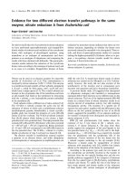
Validation of microarray data with quantitative real-time polymerase chain reaction (QRT-PCR) showing a representative experiment (repeated one additional time)Validation of microarray data with quantitative real-time polymerase
chain reaction (QRT-PCR) showing a representative experiment
(repeated one additional time). Three controls and three patients were
selected for QRT-PCR to validate microarray results. QRT-PCR was
carried out for individual samples, and then the average threshold cycle
(Ct) of the patients and the average Ct of the healthy controls were
used to calculate relative expression, expressed as fold change. The
fold changes of both microarray (open bars) and QRT-PCR (solid bars)
are shown. For all six genes selected, relative expression was higher in
healthy controls relative to patients as shown by microarrays and QRT-
PCR, thus confirming the microarray results.
Arthritis Research & Therapy Vol 8 No 5 Jarvis et al.
Page 10 of 14
(page number not for citation purposes)
seen on both a macroscopic and microscopic level in biologi-
cal systems. On the macroscopic level, the most obvious
examples would be heartbeat and respiration. However, levels
of key metabolites, including superoxide ion and NAD(P)H,
also have been shown to oscillate in neutrophils, and these
oscillations are causally linked to downstream neutrophil effec-
tor functions [30]. Known inflammatory mediators, including
TNF-α, IFN-γ, IL-2, and IL-8, regulate these oscillatory phe-
nomena. However, amplitude enhancement and frequency
enhancement are controlled by separate, independent, and
well-insulated metabolic pathways. IL-8 regulates changes in
oscillation frequency, and IFN-γ regulates changes in oscilla-
tion amplitude [31]. Thus, the finding that a subpopulation of
polyarticular JRA neutrophils exhibit loss of insulation separat-
ing the mechanisms that normally regulate amplitude and fre-
quency enhancement is both novel and intriguing. It is
important to point out that the defect in metabolic dynamics is
contingent upon activation of the hexose monophosphate
shunt pathway. That is, there is no defect in JRA until the shunt
is activated by LPS or fMLP (N-formyl-L-methionyl-L-leucyl-L-
phenylalanine) (data not shown). However, when the shunt is
activated in polyarticular JRA neutrophils, both metabolic path-
ways are triggered, which leads to an exaggerated cell
response. This process, like S100 protein levels, is likely tied
to enhanced secretory activity, in that myeloperoxidase, like
S100 proteins, is normally stored in intracellular granules and
not released in control cells, although surface expression is
seen for some polyarticular JRA neturophils. Precisely how this
occurs and how the defect relates (or is related) to the altered
expression of IL-8- and IFN-γ-regulated genes are now the
subject of investigation in our laboratories.
There are obviously some unanswered questions that emerge
from this study. The first is whether the neutrophil defect is
primary or secondary and how it relates (if at all) to adaptive
immune processes believed to be operative in polyarticular
Figure 3
Hierarchical cluster analysis of microarray data in juvenile rheumatoid arthritis (JRA) neutrophilsHierarchical cluster analysis of microarray data in juvenile rheumatoid arthritis (JRA) neutrophils. Data show clustering of control subjects to the left
of the grid based on patterns of gene expression. Data of children with JRA are scattered on the right side of the grid regardless of disease status.
That is, data of children with active disease (A) cluster together with those of children with partially responsive disease (P) and inactive disease (fully
responsive disease) (R).
Table 3
Summary of real-time polymerase chain reaction data
Fold change (control > patient)
Gene Microarray Polymerase
chain reaction
Directional
match
CD74 5.9 1.7 Yes
PTGS2 7.6 2.2 Yes
V-FOS 10.6 2.3 Yes
NFKB1A 19.1 7.2 Yes
SCYA3L1 10.8 23.7 Yes
SCYA4 12 27.3 Yes
Available online />Page 11 of 14
(page number not for citation purposes)
JRA. Previous studies reported from our group [13] support
the hypothesis that the defect may be primary. In earlier stud-
ies of JRA using whole blood buffy coats, we demonstrated
both by discriminant function analysis and connectivity analy-
ses identical to those shown in Figure 4 that JRA gene expres-
sion patterns return to normal after response to pharmacologic
therapy, regardless of the therapy used. In this larger study
using a more robust gene array, we demonstrate that abnormal
patterns of neutrophil gene expression persist even when the
disease is inactive.
Another question that arises is that of specificity. It is reason-
able to ask, for example, whether the same or similar abnormal-
ities of gene expression and neutrophil activation may not be
part of other JRA subtypes or any other chronic inflammatory
state. The elevation of S100A8/A9 complexes in the serum
are clearly not specific to polyarticular JRA, as such elevations
Figure 4
Contingency analyses of neutrophils from children with juvenile rheumatoid arthritis (JRA) and controlsContingency analyses of neutrophils from children with juvenile rheumatoid arthritis (JRA) and controls. Control samples are represented on the top
left panel. Families of genes whose expression levels correlated positively (red) or negatively (green) with one another are displayed on the grid.
These same relationships are distorted in neutrophils of children with active polyarticular JRA (bottom right panel) and are only partially restored after
full response to therapy (top right panel).
Arthritis Research & Therapy Vol 8 No 5 Jarvis et al.
Page 12 of 14
(page number not for citation purposes)
have been described in other JRA subtypes [32,33] and other
chronic inflammatory diseases [34,35]. However, even if these
findings are not specific for polyarticular JRA, they point to
important, previously unrecognised contributions of neu-
trophils in JRA pathogenesis and the need to examine in much
more detail than has been previously the case the role of
innate immunity in JRA pathogenesis. Despite their limits, our
data suggest that there may be pathogenic similarities
between polyarticular JRA and chronic autoinflammatory
states such as NOMID (neonatal onset multisystem inflamma-
tory disease) [36] and related disorders of neutrophil activa-
tion [37].
We have demonstrated through multiple lines of evidence that
polyarticular JRA is associated with chronic, dysregulated neu-
trophil activation. Further investigations into the role of
neutrophils in the JRA subset are likely to yield novel and unex-
pected insights into disease pathogenesis.
Conclusion
We provide evidence that the neutrophils in polyarticular JRA
are in a chronic, activated state. These findings suggest that
neutrophils play a critical role in disease pathogenesis.
Competing interests
The authors declare that they have no competing interests.
Authors' contributions
JNJ designed the study, enrolled patients, and assisted with
data analysis and interpretation. HRP designed and directed
the metabolic oscillation studies and assisted in their interpre-
tation. YT performed data analysis and interpretation of the
microarray studies. MBF assisted in the development of the
gene array used here, directed the labeling and scanning pro-
cedure, and assisted in data analysis and interpretation. PAT
directed the performance and interpretation of S100 protein
ELISAs. ID assisted YT in data analysis and interpretation. KJ
directed the cell separation and RNA purification procedures.
AK performed the metabolic oscillation studies. YC assisted
Figure 5
Scatterplots showing plasma levels of S100A8/A9 complexes in children with juvenile rheumatoid arthritis (JRA) and healthy controls (n = 10)Scatterplots showing plasma levels of S100A8/A9 complexes in children with juvenile rheumatoid arthritis (JRA) and healthy controls (n = 10). (a)
Comparison of all children with JRA (n = 24) at the time the initial sample was obtained for analysis. S100 proteins were markedly elevated in chil-
dren with JRA (662 ± 40 ng/ml) compared with controls (40 ± 9 ng/ml; p > 0.001). Although S100A8/A9 levels were higher in children with active
disease than with inactive disease (198 ± 60 ng/ml; p = 0.007) (b), S100 protein levels were significantly higher (p = 0.047) in children with inac-
tive JRA compared with controls (c). Ctr, control.
Available online />Page 13 of 14
(page number not for citation purposes)
with sample preparation and RNA purification. CC assisted
with primer design and real-time PCR assays. MT assisted
with primer design and real-time PCR assays. PS assisted in
labeling and scanning of microarrays as well as data analysis
and interpretation. JLM assisted with patient recruitment and
assignment of disease activity status and developed the data-
base used to record disease activity status and clinical param-
eters. MC directed hybridisation, scanning, and data analysis
activities. All authors read and approved the final manuscript.
Acknowledgements
This work was supported by grants R21-AR-48378, P20-RR15577,
P20-RR-16478, and R01-AI-51789 from the National Institutes of
Health. This work was also supported by National Institutes of Health,
National Center for Research Resources, General Clinical Research
Center Grant MO1 RR-14467, and grants NSM-66123 and MOP-
57777 from the Canadian Institutes of Health Research. JLM was sup-
ported through summer medical student research preceptorships from
the University of Oklahoma Native American Center of Excellence and
the American College of Rheumatology. JNJ is the recipient of a Clini-
cian Scholar Educator Award from the American College of Rheumatol-
ogy. PAT holds a scholarship from the Fonds de la Recherche en Santé
du Québec.
References
1. Førre Ø, Dobloug JH, Høyerall H, Thorsby E: HLA antigens in
juvenile arthritis. Genetic basis for the different subtypes.
Arthritis Rheum 1983, 26:35-38.
2. Jarvis JN, Pousak T, Krenz M, Iobidze M, Taylor H: Complement
activation and immune complexes in juvenile rheumatoid
arthritis. J Rheumatol 1993, 20:114-117.
3. Jarvis JN, Taylor H, Iobidze M, Krenz M: Complement activation
and immune complexes in children with polyarticular juvenile
rheumatoid arthritis: a longitudinal study. J Rheumatol 1994,
21:1124-1127.
4. Jarvis JN, Diebold MM, Chadwell MK, Iobidze M, Moore HT: Com-
position and biological behaviour of immune complexes iso-
lated from synovial fluid of patients with juvenile rheumatoid
arthritis. Clin Exp Immunol 1995, 100:514-518.
5. Eberhard BA, Laxer RM, Andersson U, Silverman ED: Local syn-
thesis of both macrophage and T cell cytokines in synovial
fluid cells from children with juvenile rheumatoid arthritis. Clin
Exp Immunol 1994, 96:260-266.
Figure 6
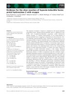
Representative kinetic traces illustrating the JRA-associated abnormality in metabolic oscillations of neutrophilsRepresentative kinetic traces illustrating the JRA-associated abnormality in metabolic oscillations of neutrophils. These traces show NAD(P)H
autofluorescence intensity (ordinate) versus time (abscissa); to conserve space, only a few oscillations are shown. Polarised cells were studied on
glass slides at 37°C. Using an anti-MPO antibody in immunofluorescence microscopy, JRA neutrophils can be classified as MPO-negative (left) and
MPO-positive (right). Untreated MPO-negative cells demonstrated NAD(P)H oscillations with a period of approximately 20 seconds (trace a). The
NAD(P)H oscillatory period of these cells decreased to 10 seconds in the presence of the activator LPS. In patients with JRA, a subpopulation of
neutrophils are MPO-positive. In the absence of cell stimulation, MPO-positive cells cannot be distinguished from MPO-negative cells. However, in
contrast to MPO-negative cells, MPO-positive cells undergo both a decrease in period to 10 seconds and a dramatic increase in the oscillatory
amplitude. JDA, juvenile rheumatoid arthritis; LPS, lipopolysaccharide; MPO, myeloperoxidase; NAD(P)H, nicotinamide adenine dinucleotide
(phosphate).
Arthritis Research & Therapy Vol 8 No 5 Jarvis et al.
Page 14 of 14
(page number not for citation purposes)
6. Petty RE, Cassidy JT: Juvenile rheumatoid arthritis. In Textbook
of Pediatric Rheumatology Philadelphia: WB Saunders Co;
2001:262.
7. Jarvis JN: Pathogenesis and mechanisms of inflammation in
the childhood rheumatic diseases. Curr Opin Rheumatol 1998,
10:459-467.
8. Witko-Sarsat V, Rieu P, Descamps-Latscha B, Lesavre P, Hal-
bwachs-Mecarelli L: Neutrophils: molecules, functions and
pathophysiological aspects. Lab Invest 2000, 80:617-653.
9. Foell D, Wittkowski H, Hammerschmidt I, Wulffraat N, Schmeling
H, Frosch M, Horneff G, Kuis W, Sorg C, Roth J: Monitoring neu-
trophil activation in juvenile rheumatoid arthritis by S100A12
serum concentrations. Arthritis Rheum 2004, 50:1286-1295.
10. Cassidy JT, Levinson JE, Brewer EJ: The development of classi-
fication criteria for children with juvenile rheumatoid arthritis.
Bull Rheum Dis 1989, 38:1-7.
11. Jarvis JN, Pousak T, Krenz M: Detection of IgM rheumatoid fac-
tors by ELISA in children with juvenile rheumatoid arthritis:
correlation with articular disease and laboratory
abnormalities. Pediatrics 1992, 90:945-949.
12. Giannini EH, Brewer EJ, Kuzmina N, Shaikov A, Wallin B:
Auranofin in the treatment of juvenile rheumatoid arthritis.
Results of a double-blind, placebo-controlled trial. Arthritis
Rheum 1990, 33:466-476.
13. Jarvis JN, Dozmorov I, Jiang K, Frank MB, Szodoray P, Alex P, Cen-
tola M: Novel approaches to gene expression analysis of active
polyarticular juvenile rheumatoid arthritis. Arthritis Res Ther
2004, 6:R15-R32.
14. Dozmorov I, Knowlton N, Tang Y, Shields A, Pathipvanich P, Jarvis
JN, Centola M: Hypervariable genes – experimental error or
hidden dynamics. Nucleic Acids Res 2004, 32:e147.
15. Kindzelskii AL, Eszes MM, Todd RF III, Petty HR: Proximity oscil-
lations of complement type 4 (alphaX beta2) and urokinase
receptors on migrating neutrophils. Biophys J 1997,
73:1777-1784.
16. Adachi Y, Kindzelskii AL, Ohno N, Yadomae T, Petty HR: Ampli-
tude and frequency modulation of metabolic signals in leuko-
cytes: synergistic role of IFN-gamma in IL-6- and IL-2-
mediated cell activation. J Immunol 1999, 163:4367-4374.
17. Complete normalized data comparing JRA and control neu-
trophils [ />neutrophil-data.xls]
18. Amit A, Kindelzelski A, Zanoni J, Jarvis JN, Petty HR: Complement
deposition on immune complexes reduces the frequencies of
metabolic, proteolytic, and superoxide oscillations in migrat-
ing neutrophils. Cell Immunol 1999, 194:47-53.
19. Edgeworth J, Gorman M, Bennett R, Freemont P, Hogg N: Identi-
fication of p8,14 as a highly abundant heterodimeric calcium
binding protein complex of myeloid cells. J Biol Chem 1991,
266:7706-7713.
20. Ryckman C, Gilbert C, de Medicis R, Lussier A, Vandal K, Tessier
PA: Monosodium urate monohydrate crystals induce the
release of the proinflammatory protein S100A8/A9 from
neutrophils. J Leukoc Biol 2004, 76:433-440.
21. Rouleau P, Vandal K, Ryckman C, Poubelle PE, Boivin A, Talbot M,
Tessier PA: The calcium-binding protein S100A12 induces
neutrophil adhesion, migration, and release from bone mar-
row in mouse at concentrations similar to those found in
human inflammatory arthritis. Clin Immunol 2003, 107:46-54.
22. Vandal K, Rouleau P, Ryckman C, Talbot M, Tessier PA: Blockade
of S100A8 and S100A9 suppresses neutrophil migration in
response to lipopolysaccharide. J Immunol 2003,
171:2602-2609.
23. Olsen LF, Kummer U, Kindzelskii AL, Petty HR: A model of the
oscillatory metabolism of activated neutrophils. Biophys J
2003, 84:69-81.
24. Grom AA, Hirsch R: T-cell and T-cell receptor abnormalities in
the immunopathogenesis of juvenile rheumatoid arthritis.
Curr Opin Rheumatol 2000, 12:420-424.
25. Carrasco R, Smith JA, Lovell D: Biologic agents for the treat-
ment of juvenile rheumatoid arthritis: current status. Paediatr
Drugs 2004, 6:137-146.
26. Foell D, Roth J: Proinflammatory S100 proteins in arthritis and
autoimmune disease. Arthritis Rheum 2004, 50:3762-3771.
27. Foell D, Wittkowski H, Hammerschmidt I, Wulffraat N, Schmeling
H, Frosch M, Horneff G, Kuis W, Sorg C, Roth J: Monitoring neu-
trophil activation in juvenile rheumatoid arthritis by S100A12
serum concentrations. Arthritis Rheum 2004, 50:1286-1295.
28. Jarvis JN, Wang W, Zhao L, Xu C, Moore HT: In vitro induction of
pro-inflammatory cytokine secretion by juvenile rheumatoid
arthritis synovial fluid immune complexes. Arthritis Rheum
1997, 40:2039-2046.
29. Xiao S, Xu C, Jarvis JN: C1q-bearing immune complexes induce
IL-8 secretion in human umbilical vein endothelial cells
(HUVEC) through protein tyrosine kinase- and mitogen-acti-
vated protein kinase-dependent mechanisms. Evidence that
the 126 kD phagocytic C1q receptor mediates immune com-
plex activation of HUVEC. Clin Exp Immunol 2001,
125:360-367.
30. Jiang K, Chen Y, Xu C, Jarvis JN: T Cell activation by soluble
C1q-bearing immune complexes: implications for the patho-
genesis of rheumatoid arthritis. Clin Exp Immunol 2003,
131:61-67.
31. Ryckman C, Roubichaud GA, Roy J, Cantin R, Tremblay MJ, Tess-
ier PA: HIV-1 Transcription and virus production are both
accentuated by the proinflammatory myeloid-related proteins
in human CD4
+
T lymphocytes. J Immunol 2002,
169:3307-3313.
32. Kindzelskii AL, Petty HR: Apparent role of traveling metabolic
waves in oxidant release by living neutrophils. Proc Natl Acad
Sci USA 2002, 99:9207-9212.
33. Petty HR: Neutrophil oscillations: temporal and spatiotemporal
aspects of cell behavior. Immunol Res 2001, 23:85-94.
34. Frosch M, Strey A, Vogl T, Wulffraat NM, Kuis W, Sunderkotter C,
Harms E, Sorg C, Roth J: Myeloid-related proteins 8 and 14 are
specifically secreted during interaction of phagocytes and
activated endothelium and are useful markers for monitoring
disease activity in pauciarticular-onset juvenile rheumatoid
arthritis. Arthritis Rheum 2000, 43:628-637.
35. Wulffraat NM, Haas PJ, Frosch M, De Kleer IM, Vogl T, Brinkman
DM, Quartier P, Roth J, Kuis W: Myeloid related protein 8 and 14
secretion reflects phagocyte activation and correlates with
disease activity in juvenile idiopathic arthritis treated with
autologous stem cell transplantation. Ann Rheum Dis 2003,
62:236-241.
36. Foell D, Kucharzik T, Kraft M, Vogl T, Sorg C, Domschke W, Roth
J: Neutrophil derived human S100A12 (EN-RAGE) is strongly
expressed during chronic active inflammatory bowel disease.
Gut 2003, 52:847-853.
37. Foell D, Seeliger S, Vogl T, Koch HG, Maschek H, Harms E, Sorg
C, Roth J: Expression of S100A12 (EN-RAGE) in cystic fibrosis.
Thorax 2003, 58:613-617.
38. Kilcline C, Shinkai K, Bree A, Modica R, Von Scheven E, Frieden
IJ: Neonatal-onset multisystem inflammatory disorder: the
emerging role of pyrin genes in autoinflammatory diseases.
Arch Dermatol 2005, 141:248-253.
39. Neven B, Callebaut I, Prieur AM, Feldmann J, Bodemer C, Lepore
L, Derfalvi B, Benjaponpitak S, Vesely R, Sauvain MJ, et al.: Molec-
ular basis of the spectral expression of CIAS1 mutations asso-
ciated with phagocytic cell-mediated autoinflammatory
disorders CINCA/NOMID, MWS, and FCU. Blood 2004,
103:2809-2815.