Báo cáo y học: "Is disturbed clearance of apoptotic keratinocytes responsible for UVB-induced inflammatory skin lesions in systemic lupus erythematosus" pot
Bạn đang xem bản rút gọn của tài liệu. Xem và tải ngay bản đầy đủ của tài liệu tại đây (437.55 KB, 7 trang )
Open Access
Available online />Page 1 of 7
(page number not for citation purposes)
Vol 8 No 6
Research article
PUMA-mediated apoptosis in fibroblast-like synoviocytes does
not require p53
Xin You, David L Boyle, Deepa Hammaker and Gary S Firestein
Division of Rheumatology, Allergy and Immunology, University of California at San Diego School of Medicine, 9500 Gilman Drive, La Jolla, California
92093, USA
Corresponding author: Gary S Firestein,
Received: 5 Jul 2006 Revisions requested: 14 Aug 2006 Revisions received: 13 Sep 2006 Accepted: 2 Oct 2006 Published: 2 Oct 2006
Arthritis Research & Therapy 2006, 8:R157 (doi:10.1186/ar2052)
This article is online at: />© 2006 You et al; licensee BioMed Central Ltd.
This is an open access article distributed under the terms of the Creative Commons Attribution License ( />),
which permits unrestricted use, distribution, and reproduction in any medium, provided the original work is properly cited.
Abstract
PUMA (p53-upregulated modulator of apoptosis) is a pro-
apoptotic gene that can induce rapid cell death through a p53-
dependent mechanism. However, the efficacy of PUMA gene
therapy to induce synovial apoptosis in rheumatoid arthritis
might have limited efficacy if p53 expression or function is
deficient. To evaluate this issue, studies were performed to
determine whether p53 is required for PUMA-mediated
apoptosis in fibroblast-like synoviocytes (FLS). p53 protein was
depleted or inhibited in human FLS by using p53 siRNA or a
dominant-negative p53 protein. Wild-type and p53
-/-
murine FLS
were also examined to evaluate whether p53 is required. p53-
deficient or control FLS were transfected with PUMA cDNA or
empty vector. p53 and p21 expression were then determined by
Western blot analysis. Apoptosis was assayed by ELISA to
measure histone release and caspase-3 activation, or by trypan
blue dye exclusion to measure cell viability. Initial studies
showed that p53 siRNA decreased p53 expression by more
than 98% in human FLS. Loss of p53 increased the growth rate
of cells and suppressed p21 expression. However, PUMA still
induced apoptosis in control and p53-deficient FLS after PUMA
cDNA transfection. Similar results were observed in p53
-/-
murine FLS or in human FLS transfected with a dominant-
negative mutant p53 gene. These data suggest that PUMA-
induced apoptosis in FLS does not require p53. Therefore,
approaches to gene therapy that involve increasing PUMA
expression could be an effective inducer of synoviocyte cell
death in rheumatoid arthritis regardless of the p53 status in the
synovium.
Introduction
Rheumatoid arthritis (RA) is a chronic inflammatory disease
characterized by synovial hyperplasia and invasion into carti-
lage and bone. Inadequate apoptosis of fibroblast-like synovi-
ocytes (FLS) could contribute to this process by increasing
the accumulation of cells in the intimal lining [1]. As a result of
the aggressive nature of rheumatoid synovium and the rela-
tively low level of apoptosis, interventions designed to
increase programmed cell death of synoviocytes have been
considered in treating RA. Several genes have been evaluated
as potential gene therapy targets, including Fas [2], TRAIL
(tumor necrosis factor-related apoptosis-inducing ligand) [3],
p53 [4], and PUMA (p53 up-regulated modulator of apopto-
sis) [5]. The latter is an especially interesting target because it
rapidly induces apoptosis in cultured synoviocytes [5]. PUMA
is a Bcl-2 homology 3 (BH3)-only pro-apoptotic Bcl-2 family
member recently identified as a principal mediator of p53-
dependent apoptosis [6]. The in vivo effects on apoptosis
observed in PUMA
-/-
mice are similar to those in p53
-/-
animals,
suggesting that PUMA can serve as an effector of p53 func-
tion [7,8]. However, our previous studies showed that p53 is
only a weak inducer of PUMA in FLS, which could account for
the variable pro-apoptotic effect of p53 in this cell lineage, with
no significant apoptosis induced by p53 overexpression in
some studies [9,10].
The mechanism of PUMA-mediated apoptosis has been exten-
sively evaluated. PUMA expression leads to apoptosis by dis-
placing p53 from Bcl-XL and allowing p53 to increase
mitochondrial permeability [6]. The need for functional p53
raises significant concerns about the utility of PUMA as a ther-
apeutic target in RA because deficient p53 expression or
BH3 = Bcl-2 homology 3; DMEM = Dulbecco's modified Eagle's medium; ELISA = enzyme-linked immunosorbent assay; FLS = fibroblast-like syn-
oviocytes; HA = hemagglutinin; PUMA = p53-upregulated modulator of apoptosis; RA = rheumatoid arthritis; siRNA = small interfering RNA.
Arthritis Research & Therapy Vol 8 No 6 You et al.
Page 2 of 7
(page number not for citation purposes)
function in the rheumatoid synovial intimal lining has been
described [11-14]. To address this issue, we determined
whether PUMA requires functional p53 in cultured FLS. These
studies show that PUMA-induced apoptosis can occur
despite defects in the p53 pathway.
Materials and methods
Human and murine cultured fibroblast-like synoviocytes
Synovial tissues were obtained from patients with rheumatoid
arthritis and osteoarthritis at joint replacement surgery. The
diagnosis of RA conformed to the American College of Rheu-
matology 1987 revised criteria [15]. The protocol was
approved by the University of California at San Diego Human
Subjects Research Protection Program. FLS were isolated
from individual tissues with 1 mg/ml collagenase and cultured
in DMEM supplemented with 10% fetal calf serum, penicillin,
streptomycin, and L-glutamine as described previously. Cell
lines were used from the third to ninth passage, when they are
a homogeneous population of fibroblast-like cells [16].
Although the origin of these cells cannot be certain, they prob-
ably derive from the intimal lining, on the basis of vascular cell
adhesion molecule (VCAM)-1 and CD55 expression. In addi-
tion to RA FLS, we also examined FLS derived from osteoar-
thritis FLS in most experiments. No differences were observed
between RA and osteoarthritis FLS in these assays. p53
+/+
and p53
-/-
murine synoviocytes were obtained as described
previously from DBA/1J wild-type mice (Jackson Laboratory,
Bar Harbor, ME, USA) and DBA/1J p53
-/-
mice [17].
Antibodies
Affinity-purified rabbit polyclonal anti-p53 (for immunohisto-
chemistry), mouse monoclonal anti-p53 (for Western blotting),
and rabbit polyclonal antibodies against p21 and hemaggluti-
nin (HA) were purchased from Santa Cruz Biotechnology
(Santa Cruz, CA, USA). Anti-mouse and anti-rabbit IgG sec-
ondary antibodies were purchased from Cell Signaling Tech-
nology, Inc. (Beverly, MA, USA). Rabbit anti-PUMA polyclonal
antibody was purchased from ProSci, Inc. (Poway, CA, USA).
Cell transfections
Scrambled RNA and p53 siRNA were purchased from Dhar-
macon Research, Inc. (Lafayette, CO, USA). Plasmids encod-
ing HA-tagged full-length PUMA (HA-PUMA) and PUMA with
a deletion of the BH3 domain (HA-PUMA-dBH3) were kindly
provided by Dr B Vogelstein (Johns Hopkins Oncology
Center, Baltimore, MD, USA) [5]. R213* encoding mutant p53
was isolated from a patient with RA and has previously been
characterized as dominant-negative [14]. Bax-luc (BF72-2
PGL3) is a reporter construct containing the p53-responsive
promoter for bax with the luciferase cDNA [14]. The control
construct contains the β-Gal cDNA and the cytomegalovirus
(CMV) promoter in pCI. Cells were transfected with the use of
the Amaxa Human Dermal Fibroblast Nucleofactor kit (NHDF-
adult) with program U-23 for human FLS. Murine FLS were
transfected with the use of the Mouse Embryonic Fibroblasts
kit (MEF1) with program T-20. Cells (2 × 10
5
to 10
6
) were
transfected with siRNAs, cDNAs, or control plasmids in each
reaction.
Western blot analysis
Cultured FLS were washed with phosphate-buffered saline,
and protein was extracted with lysis buffer (50 mM HEPES pH
8.0, 150 mM NaCl, 1% Triton X-100, 10% glycerol, 1 mM
MgCl
2
, 1.5 mM EDTA, 20 mM β-glycerophosphate, 50 mM
NaF, 1 mM Na
3
VO
4
, 10 µg/ml aprotonin, 1 µM pepstatin A, 1
mM phenylmethylsulphonyl fluoride). The protein concentra-
tions were determined with the DC protein assay kit (Bio-Rad,
Hercules, CA, USA). Whole cell lysates containing 50 µg of
protein were fractionated by 12% SDS-PAGE and transferred
to a nitrocellulose membrane. The membrane was blocked
with Tris-buffered saline plus 0.1% Tween 20 (TBST) contain-
ing 5% non-fat milk for 1 hour at room temperature followed by
incubation overnight with the appropriate antibody at 4°C. The
membrane was washed three times and incubated with horse-
radish peroxidase-conjugated secondary antibody for 1 hour.
Immunoreactive protein was detected by chemiluminescence
with Kodak X-AR film (Eastman Kodak, Rochester, NY, USA).
Immunohistochemistry
siRNA-transfected cells for immunostaining were cultured in
four-well chamber slides at 4.0 × 10
4
cells per well. They were
then fixed with methanol, permeabilized with 0.05% Triton X-
100 and blocked with 10% human serum. The fixed cells were
incubated overnight with anti-p53 antibody or matched control
antibody at 4°C. Endogenous peroxidase was then depleted
with 0.1% H
2
O
2
and 0.1% NaN
3
. The cells were then washed
and stained with biotinylated secondary antibody anti-mouse
or anti-rabbit IgG and Vectastain ABC and developed with
diaminobenzidine (Vector, Burlingame, CA, USA).
Cell viability and apoptosis assays
FLS were harvested and suspended in 0.2% trypan blue and
counted with a hemocytometer. Cells that excluded dye were
considered viable. Apoptosis was determined with a Cell
Death Detection ELISA
PLUS
kit (Roche Applied Science, Man-
nheim, Germany). FLS (4 × 10
3
) were seeded into each well
of a 96-well plate after transfection. Nine hours later, samples
were collected and ELISA was performed in accordance with
the manufacturer's instructions. Results are presented as the
fold induction compared with control. To confirm the role of
apoptosis, caspase-3 activation was also determined in trans-
fected cells with the use of the human active caspase-3 ELISA
(R&D Systems, Minneapolis, MN, USA). PUMA or PUMA-
dBH3 plasmids were transduced into p53 siRNA-transfected
or scrambled siRNA-transfected cells. The cells were then cul-
tured at 4.5 × 10
5
cells per well in six-well plates. Eight hours
later, the cells were lysed and assayed as described by the
manufacturer.
Available online />Page 3 of 7
(page number not for citation purposes)
Cell proliferation assay
Alamar Blue assays incorporate a fluorimetric/colorimetric
growth indicator based on the detection of metabolic activity.
FLS (3 × 10
3
) were plated into 96-well plate after siRNA trans-
fection. At various time points, medium was replaced by
DMEM without phenol red supplemented with 10% Alamar
Blue. After incubation for 4 hours at 37°C, fluorescence was
measured with a microplate reader at an excitation wavelength
of 530 nm and an emission wavelength of 590 nm. The
number of cells is expressed as relative fluorescence units.
Statistical analysis
Data are expressed as means ± SEM. Statistics were per-
formed with Student's t test, one-way analysis of variance and
repeated-measures analysis of variance. A comparison was
considered significant at p < 0.05.
Results
Use of siRNA to knockdown p53 protein in fibroblast-like
synoviocytes
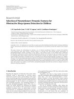
Initial studies were performed to determine whether siRNA
could knock down p53 expression in cultured FLS. A repre-
sentative time course and dose response are shown in Figure
1a. The residual expression of p53 protein was about 8 to
10%, 5%, and 1 to 2% for 1, 2.5, and 5 µg of siRNA, respec-
tively, as determined by Western blot analysis. Immunohisto-
chemistry also demonstrated a marked decrease in the
percentage of p53-positive cells after siRNA transduction
(Figure 1b). The percentage of cells with detectable p53 pro-
tein was 75.1 ± 2.9% for scrambled siRNA, 2.8 ± 0.6% for 1
µg of p53 siRNA, and 0.9 ± 0.5% for 5 µg of p53 siRNA.
Functional effects of p53 deficiency
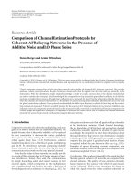
To confirm that p53 knockdown with siRNA was functionally
relevant, the effect on cell proliferation and p21 expression
was examined [18]. As shown in Figure 2a, growth of FLS was
increased by p53 siRNA. Figure 2b shows that siRNA also
blocked the expression of p21, which is normally induced by
p53. These data indicate that p53 knockdown leads to func-
tional alterations consistent with p53 deficiency.
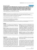
p53-independent apoptosis induced by PUMA
To determine whether p53 is required for PUMA-induced
apoptosis, FLS were transduced with p53 siRNA. After p53
expression reached its minimum 3 days later, cells were trans-
fected with the PUMA cDNA, PUMA-dBH3, or empty plasmid
Figure 1
p53 knockdown by siRNAp53 knockdown by siRNA. (a) Western blot analysis. Cultured fibroblast-like synoviocytes (FLS) were transfected with 1, 2.5, or 5 µg of siRNA or
non-silence scrambled siRNA (sc) as described in the Materials and methods section. Mock-transfected cells were treated in the same manner
except that no siRNA was added. FLS were then incubated for 3 or 5 days and Western blot analysis was performed. The p53 level was decreased
on day 3 and day 5, especially with the higher amounts of siRNA. For subsequent experiments, 2.5 µg of siRNA for 3 days was chosen as an opti-
mum condition. Two FLS lines were studied with similar results. (b) Immunohistochemistry staining of p53 protein expression in rheumatoid arthritis
FLS. Transfected FLS were seeded into four-well chamber slides, cultured for 5 days, and evaluated by immunohistochemistry. Prominent staining
was observed in the positive control (colon tumor cell line HCT116) as well as scrambled siRNA-transfected FLS. The number of p53-positive cells
was significantly reduced after transfection with 1 µg (not shown) or 5 µg of p53 siRNA (p < 0.0001 for p53 siRNA-transfected cells compared with
scrambled siRNA). Percentages of p53-positive cells are shown as means ± SEM. Two FLS lines were studied with similar results. An irrelevant con-
trol antibody was positive in less than 1% of cells (not shown).
Arthritis Research & Therapy Vol 8 No 6 You et al.
Page 4 of 7
(page number not for citation purposes)
pCEP4. As shown in Figure 3a,b, PUMA-induced apoptosis in
scrambled siRNA-transfected cells, as measured by either his-
tone release or cell viability, was similar to that in p53 siRNA-
transfected cells. PUMA-dBH3, which lacks the BH3 domain
and does not induce apoptosis, had no effect on cell viability.
As further evidence of PUMA-mediated apoptosis, caspase-3
activation was also evaluated in p53-deficient FLS. Figure 3c
shows that PUMA does not require p53 to engage this
pathway.
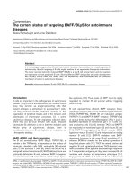
p53-independent apoptosis induced by PUMA in murine
fibroblast-like synoviocytes
Small amounts of residual p53 might contribute to PUMA
apoptosis in the siRNA studies, so we repeated the experi-
ments in p53
+/+
and p53
-/-
murine synoviocytes. Figure 4a
shows that cell viability overall was decreased in the trans-
fected cells. However, the effect of PUMA compared with
PUMA-dBH3 or empty vector was the same in p53
+/+
and
p53
-/-
synoviocytes (Figure 4).
PUMA-mediated apoptosis in the presence of mutant
p53
Because some RA synoviocytes could potentially express
dominant-negative p53 protein, we determined whether
PUMA could induce cell death in the presence of mutant p53.
A known dominant-negative gene that was isolated from a
patient with RA (R213*) was used for these experiments [14].
Sequential transfection of cultured human FLS with R213*
Figure 2
Effect of p53 siRNA on function of fibroblast-like synoviocytes (FLS)Effect of p53 siRNA on function of fibroblast-like synoviocytes (FLS). (a) Cell growth. p53 was knocked down by using siRNA, and cell growth was
determined with an Alamar Blue assay in triplicate wells (n = 3 separate cell lines) The relative number of cells is quantified using arbitrary fluores-
cence units, and the fold induction in p53 siRNA-transfected FLS is shown relative to mock-transfected cells. Similar results were observed with 5
µg of siRNA (data not shown). *p < 0.001 for p53 siRNA-transfected cells compared with scrambled (sc) siRNA. RFU, relative fluorescence units.
(b) p21 protein expression. p53 siRNA was knocked down by using siRNA, and p21 expression was determined by Western blot analysis. p21
expression decreased in cells with deficient p53 expression (n = 2 separate cell lines).
Available online />Page 5 of 7
(page number not for citation purposes)
and followed with PUMA 2 days later was performed. Figure 5
shows that PUMA effectively induced apoptosis in FLS even
in the presence of a dominant-negative p53.
Discussion
Several therapeutic approaches to RA have focused on induc-
ing apoptosis in the synovium, especially the intimal lining
[2,4]. This region is populated by macrophage-like and fibrob-
last-like synoviocytes and is a primary source of cytokines and
enzymes that degrade the extracellular matrix. The accumula-
tion of cells in the lining can be due to ingress of cells from the
blood, local proliferation, or insufficient deletion through
apoptosis. The latter is especially intriguing in view of the
observation that many pro-apoptotic genes are either defec-
tive or minimally expressed in RA, including p53, sentrin [19],
and PTEN (phosphatase and tensin homologue deleted on
chromosome 10 [20].
p53 is an interesting potential therapeutic gene because it can
induce apoptosis in many cell types. Although controversial,
defects in p53 structure and function in RA have been
described, suggesting that forced expression of the tumor
suppressor protein could be beneficial [11,12,21,22]. How-
ever, enhancing p53 gene expression in synovium with an ade-
noviral construct had only modest efficacy in a rabbit model of
arthritis [4], and a similar approach was not effective in the rat
adjuvant arthritis model (P.P. Tak, D.L. Boyle, G.S. Firestein,
unpublished data). One potential explanation for the limited
effect is that p53 does not readily induce apoptosis in synovi-
ocytes, probably because PUMA expression is not increased
[5]. In contrast, directly transducing cells with PUMA leads to
rapid synoviocyte death in vitro.
One issue that could potentially interfere with the efficacy of
PUMA gene therapy in RA is that this protein usually requires
the p53 to induce apoptosis [6]. Elegant studies have
Figure 3
Apoptosis induced by PUMA in p53-deficient human fibroblast-like synoviocytes (FLS)Apoptosis induced by PUMA in p53-deficient human fibroblast-like synoviocytes (FLS). Cultured FLS were transfected with siRNA and then, 3 days
later, with 10 µg of PUMA, PUMA-dBH3, or pCEP4. (a) DNA fragmentation as determined by histone release. Histone release was measured by
ELISA in samples collected 9 hours after the second transfection. The fold induction of DNA fragmentation in FLS transfected with PUMA plasmids
is shown relative to the control value of pCEP4-transfected cells. *p < 0.05, n = 3. (b) Cell viability. Trypan blue exclusion was evaluated at 24 hours
after cDNA transfection in 2.5 µg of siRNA-treated FLS. Data are presented as the percentage of non-viable cells. *p < 0.01, **p < 0.001, n = 3. (c)
Caspase-3 activation. Caspase-3 levels were determined in control (sc) and p53-deficient (p53) FLS 8 hours after transfection with PUMA. *p <
0.05, n = 3.
Arthritis Research & Therapy Vol 8 No 6 You et al.
Page 6 of 7
(page number not for citation purposes)
demonstrated that the mechanism of PUMA action is through
the release of p53 from inhibitory interactions with Bcl-XL in
the cytoplasm [6]. Unbound p53 protein can then directly acti-
vate Bax. If p53 is defective or deficient, the benefit of forced
PUMA expression would potentially be lost.
PUMA accounts for many of the apoptotic activities attributed
to p53 [9,10], although it can serve as a mediator of some
apoptotic pathways that do are not initiated by p53 induction,
including glucocorticoids and serum deprivation [7,23].
PUMA-mediated apoptosis can also bypass p53 in unusual
situations, especially in tumor cells. For instance, p53 expres-
sion does not require PUMA in melanoma and glioma cell lines
[24,25] or human leukemia cells [26]. Hence, the utility of
PUMA as an apoptosis-inducing protein and its relationship to
p53 depends on the cell lineage, the status of p53 (deficiency
versus mutation), and the type of stimulus. Therefore p53 has
a dual role related to PUMA gene expression and function. In
most cell types, p53 expression leads to increased PUMA
gene expression and subsequent PUMA-mediated apoptosis
requires functional p53. It is of interest that neither of these
relationships is effective in cultured FLS. This cell lineage can
also be distinguished from other cells in that p53 is expressed
constitutively [18] even though the short half-life of wild-type
p53 protein generally limits detection in non-cycling cells.
These highly variable data imply that tissue-specific cells
should be studied to determine the potential applicability of
PUMA gene therapy to RA. Our experiments using siRNA to
decrease p53 expression show that FLS are very sensitive to
PUMA-induced death and that p53 expression has no
influence on this effect. Because siRNA does not completely
deplete p53 levels, we confirmed these results in p53
-/-
murine
FLS. Finally, we showed that PUMA could function even in
cells transfected with a known dominant-negative p53 mutant.
These data demonstrate that PUMA-induced apoptosis in syn-
oviocytes does not require p53 and that PUMA gene transfer
could be effective regardless of the p53 status of the
synovium.
These data support the potential use of PUMA as a local gene
therapy approach to RA. By circumventing possible abnormal-
ities in p53 and inducing extensive apoptosis of synoviocytes,
intra-articular gene transfer could decrease the hyperplasia of
the synovial intimal lining. Although not feasible for systemic
administration, local therapy could debulk the synovium in RA
and serve as an alternative to synovectomy or intra-articular
corticosteroids.
Conclusion
PUMA efficiently induced apoptosis in control and p53 defi-
cient human FLS after PUMA overexpression. Similar results
were observed in p53
-/-
murine FLS and in human FLS trans-
fected with the R123* mutant p53 gene. PUMA-induced
apoptosis is therefore independent of p53 in FLS. These data
Figure 4
Apoptosis induced by PUMA in p53
+/+
and p53
-/-
murine fibroblast-like synoviocytes (FLS)Apoptosis induced by PUMA in p53
+/+
and p53
-/-
murine fibroblast-like
synoviocytes (FLS). Murine p53
+/+
and p53
-/-
FLS at passage 6 were
transfected as described in the Materials and methods section. Cell via-
bility by trypan blue staining (a) (*p < 0.05, n = 3) and histone release
(b) (*p < 0.05, n = 3) was used to evaluate PUMA-induced apoptosis
in both wild-type and p53-knockout FLS.
Figure 5
Apoptosis induced by PUMA in the presence of mutant p53Apoptosis induced by PUMA in the presence of mutant p53. Human
fibroblast-like synoviocytes (FLS) were transfected with dominant-neg-
ative p53 cDNA R213* or empty vector (Emp) followed by transfection
with PUMA cDNA. Apoptosis was induced in FLS despite expression
of the dominant-negative protein. *p < 0.05, n = 3.
Available online />Page 7 of 7
(page number not for citation purposes)
suggest that PUMA gene therapy could be effective in RA
regardless of the p53 status of the synovium.
Competing interests
The authors declare that they have no competing interests.
Authors' contributions
XY performed experiments, evaluated data and wrote the man-
uscript. DLB designed experiments and evaluated data. DH
performed experiments and evaluated data. GSF designed
experiments, evaluated data, and wrote the manuscript. All
authors read and approved the final manuscript.
Acknowledgements
This work was supported by grant R01 AR45347 from the National Insti-
tute of Arthritis and Musculoskeletal and Skin Diseases.
References
1. Firestein GS: Invasive fibroblast-like synoviocytes in rheuma-
toid arthritis. Passive responders or transformed aggressors?
Arthritis Rheum 1996, 39:1781-1790.
2. Okamoto K, Asahara H, Kobayashi T, Matsuno H, Hasunuma T,
Kobata T, Sumida T, Nishioka K: Induction of apoptosis in the
rheumatoid synovium by Fas ligand gene transfer. Gene Ther
1998, 5:331-338.
3. Yao Q, Seol DW, Mi Z, Robbins PD: Intra-articular injection of
recombinant TRAIL induces synovial apoptosis and reduces
inflammation in a rabbit knee model of arthritis. Arthritis Res
Ther 2005, 8:R16.
4. Yao Q, Wang S, Glorioso JC, Evans CH, Robbins PD, Ghivizzani
SC, Oligino TJ: Gene transfer of p53 to arthritic joints stimu-
lates synovial apoptosis and inhibits inflammation. Mol Ther
2001, 3:901-910.
5. Cha HS, Rosengren S, Boyle DL, Firestein GS: PUMA regulation
and proapoptotic effects in fibroblast-like synoviocytes. Arthri-
tis Rheum 2006, 54:587-592.
6. Chipuk JE, Bouchier-Hayes L, Kuwana T, Newmeyer DD, Green
DR: PUMA couples the nuclear and cytoplasmic proapoptotic
function of p53. Science 2005, 309:1732-1735.
7. Jeffers JR, Parganas E, Lee Y, Yang C, Wang J, Brennan J,
MacLean KH, Han J, Chittenden T, Ihle JN, et al.: Puma is an
essential mediator of p53-dependent and -independent apop-
totic pathways. Cancer Cell 2003, 4:321-328.
8. Villunger A, Michalak EM, Coultas L, Mullauer F, Bock G, Ausser-
lechner MJ, Adams JM, Strasser A: p53- and drug-induced apop-
totic responses mediated by BH3-only proteins puma and
noxa. Science 2003, 302:1036-1038.
9. Nakano K, Vousden KH: PUMA, a novel proapoptotic gene, is
induced by p53. Mol Cell 2001, 7:683-694.
10. Yu J, Zhang L, Hwang PM, Kinzler KW, Vogelstein B: PUMA
induces the rapid apoptosis of colorectal cancer cells. Mol
Cell 2001, 7:673-682.
11. Yamanishi Y, Boyle DL, Rosengren S, Green DR, Zvaifler NJ,
Firestein GS: Regional analysis of p53 mutations in rheumatoid
arthritis synovium. Proc Natl Acad Sci USA 2002,
99:10025-10030.
12. Firestein GS, Echeverri F, Yeo M, Zvaifler NJ, Green DR: Somatic
mutations in the p53 tumor suppressor gene in rheumatoid
arthritis synovium. Proc Natl Acad Sci USA 1997,
94:10895-10900.
13. Inazuka M, Tahira T, Horiuchi T, Harashima S, Sawabe T, Kondo M,
Miyahara H, Hayashi K: Analysis of p53 tumour suppressor
gene somatic mutations in rheumatoid arthritis synovium.
Rheumatology (Oxford) 2000, 39:262-266.
14. Han Z, Boyle DL, Shi Y, Green DR, Firestein GS: Dominant-neg-
ative p53 mutations in rheumatoid arthritis. Arthritis Rheum
1999, 42:1088-1092.
15. Arnett FC, Edworthy SM, Bloch DA, McShane DJ, Fries JF, Cooper
NS, Healey LA, Kaplan SR, Liang MH, Luthra HS, et al.: The Amer-
ican Rheumatism Association 1987 revised criteria for the
classification of rheumatoid arthritis. Arthritis Rheum 1988,
31:315-324.
16. Alvaro-Gracia JM, Yu C, Zvaifler NJ, Firestein GS: Mutual antago-
nism between interferon-γ and tumor necrosis factor-α on
fibroblast-like synoviocytes: paradoxical induction of IFN-γ
and TNF-α receptor expression. J Clin Immunol 1993,
13:212-218.
17. Yamanishi Y, Boyle DL, Pinkoski MJ, Mahboubi A, Lin T, Han Z,
Zvaifler NJ, Green DR, Firestein GS: Regulation of joint destruc-
tion and inflammation by p53 in collagen-induced arthritis. Am
J Pathol 2002, 160:123-130.
18. Pap T, Aupperle KR, Gay S, Firestein GS, Gay RE: Invasiveness
of synovial fibroblasts is regulated by p53 in the SCID mouse
in vivo model of cartilage invasion. Arthritis Rheum 2001,
44:676-681.
19. Franz JK, Pap T, Hummel KM, Nawrath M, Aicher WK, Shigeyama
Y, Muller-Ladner U, Gay RE, Gay S: Expression of sentrin, a
novel antiapoptotic molecule, at sites of synovial invasion in
rheumatoid arthritis. Arthritis Rheum 2000, 43:599-607.
20. Pap T, Franz JK, Hummel KM, Jeisy E, Gay R, Gay S: Activation of
synovial fibroblasts in rheumatoid arthritis: lack of expression
of the tumour suppressor PTEN at sites of invasive growth and
destruction. Arthritis Res 2000, 2:59-64.
21. Reme T, Travaglio A, Gueydon E, Adla L, Jorgensen C, Sany J:
Mutations of the p53 tumour suppressor gene in erosive rheu-
matoid synovial tissue. Clin Exp Immunol 1998, 111:353-358.
22. Kullmann F, Judex M, Neudecker I, Lechner S, Justen HP, Green
DR, Wessinghage D, Firestein GS, Gay S, Scholmerich J, et al.:
Analysis of the p53 tumor suppressor gene in rheumatoid
arthritis synovial fibroblasts. Arthritis Rheum 1999,
42:1594-1600.
23. Yu J, Wang Z, Kinzler KW, Vogelstein B, Zhang L: PUMA medi-
ates the apoptotic response to p53 in colorectal cancer cells.
Proc Natl Acad Sci USA 2003, 100:1931-1936.
24. Karst AM, Dai DL, Martinka M, Li G: PUMA expression is signifi-
cantly reduced in human cutaneous melanomas. Oncogene
2005, 24:1111-1116.
25. Ito H, Kanzawa T, Miyoshi T, Hirohata S, Kyo S, Iwamaru A, Aoki H,
Kondo Y, Kondo S: Therapeutic efficacy of PUMA for malignant
glioma cells regardless of p53 status. Hum Gene Ther 2005,
16:685-698.
26. Liu FT, Newland AC, Jia L: Bax conformational change is a cru-
cial step for PUMA-mediated apoptosis in human leukemia.
Biochem Biophys Res Commun 2003, 310:956-962.