Báo cáo y học: "The clinical utility of molecular diagnostic testing for primary immune deficiency disorders: a case based review" doc
Bạn đang xem bản rút gọn của tài liệu. Xem và tải ngay bản đầy đủ của tài liệu tại đây (732.15 KB, 9 trang )
ALLERGY, ASTHMA & CLINICAL
IMMUNOLOGY
Ameratunga et al. Allergy, Asthma & Clinical Immunology 2010, 6:12
/>Open Access
REVIEW
© 2010 Ameratunga et al; licensee BioMed Central Ltd. This is an Open Access article distributed under the terms of the Creative Com-
mons Attribution License ( which permits unrestricted use, distribution, and reproduc-
tion in any medium, provided the original work is properly cited.
Review
The clinical utility of molecular diagnostic testing
for primary immune deficiency disorders: a case
based review
Rohan Ameratunga*
1,2
, See-Tarn Woon
2
, Katherine Neas
3
and Donald R Love
2
Abstract
Primary immune deficiency disorders (PIDs) are a group of diseases associated with a genetic predisposition to
recurrent infections, malignancy, autoimmunity and allergy. The molecular basis of many of these disorders has been
identified in the last two decades. Most are inherited as single gene defects. Identifying the underlying genetic defect
plays a critical role in patient management including diagnosis, family studies, prognostic information, prenatal
diagnosis and is useful in defining new diseases. In this review we outline the clinical utility of molecular testing for
these disorders using clinical cases referred to Auckland Hospital. It is written from the perspective of a laboratory
offering a wide range of tests for a small developed country.
Introduction
Primary immune deficiency disorders (PIDs) were first
identified in 1952, with the description of agammaglobu-
linemia by Bruton [1]. In the last few years, the genetic
basis of many PID disorders has been identified [2,3].
Most are inherited as single gene defects. Several are X-
linked, which accounts for the preponderance of PIDs
amongst males. While rare, most are amenable to specific
treatment. For example, successful haematopoeitic stem
cell transplantation may be curative in patients with
severe combined immune deficiency syndrome (SCID).
In other PIDs, delayed diagnosis may be associated with
disabling complications such as bronchiectasis [4].
Clinical history and physical examination can be help-
ful in identifying PIDs and the need for further investiga-
tion. Once these patients are referred to an
immunologist, other tests including vaccine responses
can be undertaken [5,6]. Further advanced tests including
the enumeration of subsets of lymphocytes using flow
cytometry can be useful in evaluating patients [7,8]. For
instance, NKT cells may be absent in patients with some
forms of X-linked lymphoproliferative disease (XLP),
which is caused by mutations in the SH2D1A or XIAP
genes [9]. Elevated double-negative CD4
-
CD8
-
TCR
alpha/beta+ T (DNT) cells are useful markers for autoim-
mune lymphoproliferative syndrome due to mutations in
the fas gene [10]. Ultimately, however, identification of
the molecular basis of the disorder will secure the diagno-
sis. We believe molecular diagnostic testing is a critical
part of modern patient management and should be
regarded as the standard of care.
We have described the development of a customised
molecular testing service for PIDs at Auckland City Hos-
pital [11]. We offer full length (Sanger) sequencing with
results within a week if the test is established, or two to
three weeks for a customised test [11]. In patients with a
typical phenotype but normal genomic sequence we offer
cDNA sequencing to exclude the possibility of a complex
mutation such an inversion or a promoter mutation. To
date over twenty different PID genes have been
sequenced.
Clinicians have the opportunity to review actual labora-
tory data and discuss findings with the scientist perform-
ing the test. The technical limitations of the scientific
findings can be made clear. Proficiency testing is a critical
part of genetic analysis. Laboratory errors can have cata-
strophic consequences for the proband and the family
[12]. The service complies with the recent recommenda-
tions for quality assurance in laboratories performing
molecular diagnostic testing [13] and is accredited by
IANZ, the New Zealand laboratory accrediting agency
* Correspondence:
1
Department of Clinical Immunology Auckland City Hospital, Park Rd, Grafton,
Auckland New Zealand
Full list of author information is available at the end of the article
Ameratunga et al. Allergy, Asthma & Clinical Immunology 2010, 6:12
/>Page 2 of 9
[11]. The model we have described in New Zealand is
cost-effective for a developed country with a small popu-
lation of 4.3 million.
In this review, the value of genetic testing is explored
using patient cases referred to the molecular immunology
diagnostic service at Auckland Hospital, together with
selected examples from the literature.
The clinical utility of genetic testing in PID
disorders
The benefits of PID genetic testing are listed in Appendix
1. This distinction is artificial as the value to patients and
families cross these arbitrary boundaries. There are usu-
ally multiple indications for genetic testing as illustrated
by these cases. Most of the advantages (and disadvan-
tages) described here also apply to other genetic disor-
ders.
Distinguishing genetic from acquired disorders
Distinguishing congenital from acquired disorders is fun-
damentally important for patient management. Some-
times drug or virus induced disorders can mimic PIDs.
Removal of the causative drug or treating the viral infec-
tion may lead to clinical improvement.
Molecular diagnostic tests proved invaluable in charac-
terizing the cellular and molecular pathology of rubella
associated Hyper Immunoglobulin M syndrome (rHIM)
[14]. Prior to widespread rubella vaccination, cases of
dysgammaglobulinemia were described in patients who
had suffered congenital rubella. These cases have become
very rare since the advent of widespread rubella vaccina-
tion. We have had the opportunity to characterize in
detail a patient with this disorder.
The patient concerned is 54 years old with elevated
polyclonal IgM levels and absent IgA with low levels of
IgG. In 1984 before regular immunoglobulin replacement
was commenced, he had undetectable IgG. He suffered
recurrent lower and upper respiratory tract infections but
does not have bronchiectasis in spite of a chronic cough.
He has had sinus surgery for chronic rhinosinusitis.
On further questioning he had sensorineural deafness
and impaired vision. He wears a hearing aid. His mother
was thought to have suffered rubella during pregnancy.
Retinoscopy showed typical changes of congenital
rubella. The patient was noted to have a persistently ele-
vated rubella IgM titre. His rheumatoid factor was nega-
tive indicating the rubella IgM was from de novo
synthesis. He had normal in vitro T cell responses to lec-
tins and antigens. We have previously shown that patients
with X linked Hyper Immunoglobulin M syndrome
(XHIM) have impaired T cell antigen responses [15].
Examination of his immunophenotype confirmed the
presence of B cells bearing surface IgG, consistent with in
vivo class switching. In contrast to XHIM patients, he
was able to generate CD27+ memory B cells [14].
He had normal CD40 ligand expression by flow cytom-
etry. Given his age and relatively good health together
with laboratory results, it was felt it was unlikely he had
XHIM. This was confirmed by the presence of wild type
CD40 ligand sequence. The presence of normal CD40
ligand sequence confirmed that other family members
are not at risk of XHIM. It also provided reassurance to
the patient who may be at less at risk of complications
including lymphoma and liver failure.
Similarly, many drugs are also known to cause hypog-
ammaglobulinemia. We have described a patient with
epilepsy who developed profound hypogammaglobuline-
mia, which completely resolved on stopping his Lamotri-
gene [16]. In a similar situation, if a mutation was
identified in the BTK or SH2D1A genes, it would indicate
the presence of a PID rather than an acquired disorder
and would obviate the need to stop critical therapy such
as anti-epilepsy drugs.
Confirming the clinical diagnosis
Genetic testing plays a pivotal in confirming the clinical
diagnosis. This is illustrated by the case of an 18 year old
male who presented with fulminant infectious mononu-
cleosis in 2006. He suffered hepatic failure and died three
days after being transferred to Auckland City Hospital.
The history was remarkable in that he had been treated
for Burkitt type lymphoma and had made a complete
recovery after chemotherapy [17]. Prior to his death, very
high levels of EBV DNA (>8 × 10
6
copies/ml EBV DNA)
were detected in his serum and X-linked lymphoprolifer-
ative disorder (XLP) was strongly suspected. Sequencing
the SH2D1A gene in this patient revealed a point muta-
tion, c.261delT. This mutation causes a translational
frameshift and is predicted to result in expression of a
truncated protein, thus confirming the diagnosis of XLP.
XLP is a rare disorder characterised by susceptibility to
Epstein Barr virus infection [18]. Affected boys have a
catastrophic reaction and many die from fulminant infec-
tious mononucleosis. Mutations in the SH2D1A [19] and
XIAP [20] genes have been identified as the cause of these
syndromes. The initial study of the proband described
above was undertaken in Perth during the time the assay
was being developed in Auckland. We have undertaken
similar QA studies where mutations in blinded samples
have been identified.
Where presymptomatic diagnosis (at any age) is not
possible with protein-based tests
There are no reliable methods to identify presymptom-
atic XLP patients in the absence of molecular analysis.
Immunophenotype and immunoglobulin profiles do not
help identify these patients. Flow cytometry can be used
Ameratunga et al. Allergy, Asthma & Clinical Immunology 2010, 6:12
/>Page 3 of 9
to detect the presence and quantity of affected protein in
lymphocyte or lymphocyte subsets. Some investigators
[21] have found that XLP patients have decreased
SH2D1A protein expression compared to healthy individ-
uals; however, some patients with XLP have normal SAP
(SH2D1A) protein levels [21]. Missense mutations may
not abolish protein expression, thereby resulting in false
negative results. Mutations of the cytoplasmic tails of cell
surface receptors may impair signaling, while allowing
cell surface expression of defective proteins. Thus, even
when flow cytometry indicates a normal level of cell sur-
face protein, the results must be confirmed by molecular
analysis.
Cascade screening of at-risk relatives
Many PID disorders including XLP are inherited in an X-
linked fashion. Male patients will manifest the disorder,
while females are asymptomatic carriers of the mutation.
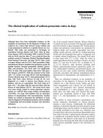
Figure 1 shows the family tree of this XLP kindred. The
male proband (V:1, described above) carries the disease-
causing mutation, which was inherited from his mother
(IV:1) [17]. Critically, analysis of the proband's sister (V:2)
and two maternal aunts (IV:4, IV:5) showed they had not
inherited the mutation. Therefore, their children are not
at risk of inheriting the disorder. In other disorders,
affected members may have phenotypic variation within
the same kindred. Genetic testing plays a crucial role in
family studies [22].
Identifying novel presentation of PIDs
A detailed study of this family also revealed that three
members had lymphomas before the fatal presentation of
the proband [17]. While lymphoma can complicate the
course of individual XLP patients, we have suggested that
familial lymphoma should be regarded as another presen-
tation of XLP. Supporting evidence comes from Brandau
et al [23] who reported SH2D1A gene mutations in boys
with non-Hodgkins lymphoma, but with no previous
EBV infection. Approximately 50% of Hodgkin's and 20%
of Burkitt's lymphomas contain EBV DNA [24]. There-
fore, it may be prudent to exclude XLP when multiple
family members, particularly males, develop lymphoma.
Identifying atypical presentation of PIDs
During the course of investigation, we found the
proband's grandmother (III:1) had died of non-Hodgkins
lymphoma at 51 years of age. Lymphoma is one of the
classical presentations of XLP in males [25]. We were able
to retrieve the grandmother's lymphoma tissue block and
extract DNA for further testing. Cloning of the amplified
DNA showed that several recombinants contained the
mutation, confirming she was a carrier of the disorder
Figure 1 Pedigree of a family segregating XLP[17].
Unaffected male
Non Hodgkins Lymphoma
Fulminant infectious mononucleosis
Myocardial infarction
+ SH2D1A mutation positive
- SH2D1A mutation negative
Ameratunga et al. Allergy, Asthma & Clinical Immunology 2010, 6:12
/>Page 4 of 9
[17]. This is the first example of a female who developed
an XLP phenotype. More distant members of the kindred
may be at-risk and have been advised to seek testing.
Again genetic testing played a critical role in confirming
symptomatic XLP in a carrier female.
Characterising the role of molecules in cellular function
Skewed X chromosome inactivation (lyonisation) in
symptomatic female carriers of PID genes is well docu-
mented [26-29]. In most cases this is a stochastic event
where the majority of X chromosomes bearing the wild
type allele are inactivated purely by chance. Females may
manifest X-linked disorders in this situation. More rarely,
skewed lyonisation may be a consequence of mutations at
the Xist locus, which initiates X-chromosome inactiva-
tion. In this situation the wild type allele may be selec-
tively inactivated in females of the kindred [30].
The identification of a female with an XLP phenotype
raised concern that other female carriers including the
mother (IV:1) were at risk of symptomatic XLP in this
kindred. Analysis of the methylation patterns of the
human androgen receptor locus (HUMARA) of the
mother (IV:1) did not suggest this family had a disorder of
X chromosome inactivation [17]. As a consequence, the
most likely explanation for the lymphoma in the affected
grandmother (III:1) was skewed X-inactivation. Progres-
sive skewing of lyonisation with aging may place female
carriers of X-linked disorders at risk of symptomatic dis-
ease [31].
The detection of abnormal lyonisation patterns
requires normal tissue. As the paraffin embedded tissue
block contained only lymphoma, we were unable to con-
firm this hypothesis. Our observation suggests that
female carriers of a mutation in one copy of the SH2D1A
gene in other kindreds should be considered at-risk of
symptomatic XLP, and hence may need to be monitored
for the development of phenotypic features of XLP.
Assisting treatment decisions
If male children with XLP can be identified before they
suffer Epstein-Barr virus infection, hematopoeitic stem
cell transplantation can be undertaken which is poten-
tially curative [32-34]. The prognosis after Epstein-Barr
virus infection is guarded. In this family, there are no
other male patients at risk of XLP.
Prognosis
Another patient with no family history of recurrent infec-
tions presented with a monoarthritis of his knee at the
age of 7. At the time he was noted to have absence of ton-
sils. Testing showed the presence of panhypogammaglob-
ulinemia and immunophenotyping revealed the absence
of B cells.
A clinical diagnosis of Bruton's agammaglobulinemia
(XLA) was made and the patient was treated with intra-
venous immunoglobulin (IVIG), even though he did not
suffer from frequent or severe infections. The monoar-
thritis resolved with IVIG treatment, as has been previ-
ously described [23]. He has subsequently been in
excellent health.
Analysis of the patient's DNA revealed the deletion of 4
nucleotides (TTTG) in exon 16 of the BTK gene
(c.1581_1584delTTTG), which is predicted to cause a
frameshift and premature truncation of the btk protein
(Figure 2). The molecular basis of the disorder was thus
confirmed. As there was no history of recurrent infec-
tions, the family was initially uncertain if the patient
needed long term IVIG. In this case, mutation analysis
confirmed the diagnosis of XLA and the need for life long
treatment.
Early identification of disorders which present later in
childhood
The phenotypic manifestations of some disorders are not
seen until patients are older. Conventional testing by pro-
tein analysis may not be helpful in some situations. This
is well-illustrated by type 1 hereditary angioedema (HAE
type 1), a disorder caused by autosomal dominant muta-
tions in the C1NH gene. Children with this disorder often
do not manifest symptoms until adolescence. These
patients suffer recurrent angioedema and may be at risk
of asphyxia from laryngeal swelling. Complement studies
in presymptomatic infants may not be diagnostic even in
those who have inherited the mutation [35].
Undertaking genetic studies may enable a presymptom-
atic diagnosis to be made in the majority of cases, provid-
ing prognostic information for the family, and earlier
treatment for an affected individual. However, molecular
analysis of the C1NH gene can be problematic as a signif-
icant number of patients have complex mutations includ-
ing inversions and rearrangements, which can be
confirmed by Southern blotting or multiplex fluorescence
PCR [36,37]. Genetic diagnosis may not be feasible in all
cases of Hereditary angioedema.
Urgent diagnosis in infancy where conventional diagnostic
tests are unreliable
Some PIDs such as XHIM due to CD40 ligand deficiency
can prove difficult to confirm in neonates. In normal neo-
nates, CD40 ligand expression in early infancy is reduced
and can be difficult to detect by flow cytometry [38].
Therefore, in the case of CD40 ligand deficiency, molecu-
lar testing is a more reliable diagnostic option.
A 5 month infant was referred to the service with pro-
gressive respiratory distress. Bronchoscopy confirmed
Pneumocystis jirovecii infection. He had an elevated IgM
of 1.4 g/l (0.2-1.0) with absent IgG < 0.33 g/l (2.0-7.0) and
absent IgA < 0.07 g/l (0.1-0.8) levels. XHIM was sus-
pected. He was treated with Co-trimoxazole and made a
Ameratunga et al. Allergy, Asthma & Clinical Immunology 2010, 6:12
/>Page 5 of 9
complete recovery. Full length sequencing of the CD40
ligand confirmed the presence of a missense mutation
(475G > A) leading to a stop codon. (figure 3) In the
absence of a suitable bone marrow donor, he has been
treated with IVIG and prophylactic antibiotics. He is in
good health.
Molecular studies confirmed the mother was a carrier.
Subsequently, she gave birth to another son. The region
of the (475G > A) mutation was amplified and sequenced.
The laboratory was able to confirm that her second child
did not carry the mutation.
Given that the familial mutation was known, amplifica-
tion and sequencing of the specific exon was undertaken
within 48 hours. The family was given a definitive diagno-
sis, which would not have been possible with flow cytom-
etry.
Prenatal Diagnosis
The identification of a disease-associated mutation offers
the possibility of prenatal diagnosis. Prenatal genetic test-
ing requires careful counseling of the family. The coun-
seling should include discussion about the possible
outcomes of testing (including the risk of an incorrect
result), the risks associated with the procedure, and the
options available to the family if an affected fetus is iden-
tified. A sample of the fetus' genetic material for such
testing is most commonly obtained by chorionic villus
sampling (CVS). It is critical that the familial mutation is
identified before considering prenatal diagnosis, and that
the mother is known to be a carrier of the mutation.
In the case of X-linked disorders, fetal gender is usually
determined first as in most cases mutation analysis would
only be performed to detect an affected male. PCR stud-
ies are undertaken on the sample after maternal tissue
contamination is excluded. Prenatal diagnosis enables a
couple to identify an affected fetus and then make deci-
sions about the outcome of that pregnancy. If the couple
chooses to continue the pregnancy, early treatment of an
affected child can occur. In the United Kingdom, prenatal
diagnosis is only undertaken if the pregnancy is to be ter-
minated in the case of an affected fetus. Prenatal diagno-
sis with a rapid turnaround time should be available
through a molecular immunology diagnostic service if
requested by the family and physicians.
Pre-implantation Genetic diagnosis
Pre-implantation genetic diagnosis (PGD) is a technique
which enables genetic diagnosis of an in vitro fertilized
embryo before it is implanted into the uterus. This proce-
dure has been undertaken for PIDs [39]. Once an embryo
has been created and cultured for between 3 and 5 days,
one or more cells are removed at the blastomere or blas-
tocyst stage. DNA is extracted, amplified using PCR, and
screened for the familial mutation. If the embryo has not
inherited the mutation, it can then be implanted in the
uterus.
PGD involves many technical and ethical complexities.
Currently the probability of a live-born infant from PGD
is approximately 20-25%. This is significantly lower than
the conception rate and ongoing pregnancy rate for cou-
Figure 2 Electropherogram of the BTK gene in a normal donor (A) and patient (B). A TGTT deletion in exon 16 leads to a frameshift resulting in
a premature stop codon in the BTK gene (g.66795_66798delTTGTT, c.1581_1584delTTTG).
A B
Normal
Patient
T T T G
Ameratunga et al. Allergy, Asthma & Clinical Immunology 2010, 6:12
/>Page 6 of 9
ples having prenatal diagnosis. However, this technology
has many benefits for the couple, and is likely to become
more successful in the future. Currently we have a
request for this procedure from a family. The many tech-
nical and ethical issues need to be carefully considered
before this service can be offered.
Gene therapy
Gene therapy offers the potential to replace a defective
gene with a wild type gene. Gene therapy is most likely to
succeed in autosomal recessive disorders or X-linked dis-
orders in males. Gene therapy trials have been under-
taken for SCID (Adenosine Deaminase deficiency,
Common Gamma chain deficiency) and Chronic Granu-
lomatous Disease (CGD) in several countries including
the USA, UK, Italy, France and Australia [40,41]. In order
to replace the defective gene, the mutation must be iden-
tified. Molecular diagnosis thus plays a critical role in any
gene therapy trial.
Assisting with the classification of primary
immunodeficiency disorders
The application of molecular techniques has broadened
the understanding of PIDs. Seemingly disparate disorders
such as Wiskott-Aldrich syndrome and X-linked neutro-
penia are caused by mutations of the same gene, encoding
the Wiskott-Aldrich syndrome protein (WASP) [42].
Allelic heterogeneity, as a result of mutations in different
parts of the same gene, can result in varying phenotypic
severity or distinct phenotypes as illustrated above. Simi-
larly, phenotypically identical disorders can have an
entirely different genetic basis (genocopy). This phenom-
enon is also known as locus heterogeneity and typically
occurs when mutations affect distinct molecules in the
same signaling pathway. This is illustrated by agamma-
globulinemia, where most male patients have a mutation
of the BTK gene. However, a similar disorder can be seen
in individuals with mutations in the BLNK [43] and v λ 5
pre-light chain (IGLL1) genes [44]. Similarly, chronic
granulomatous disease can be caused by mutations in any
of the five genes encoding components of the NADPH
oxidase complex (gp91, p47, p21 p40 and p67) [45,46].
Mutation analysis is thus critical in modern disease clas-
sification.
Identification of new genetic defects
Common variable immune deficiency (CVID) is the most
frequent symptomatic primary immune deficiency disor-
der in adults. Patients present with hypogammaglobu-
linemia, which is associated with an increase in
autoimmunity, malignancy and allergy. Approximately
Figure 3 Electropherogram of the CD40L gene in a normal donor (A) and patient (B). A point mutation in exon 5 leads to a premature stop
codon in the CD40L (c.475G > A, p.W140X).
A B
Normal Patient
Ameratunga et al. Allergy, Asthma & Clinical Immunology 2010, 6:12
/>Page 7 of 9
15-20% of patients have a family history of an immune
defect in an immediate family member. Over the last five
years, four genetic defects have been identified which
account for 10-15% of all CVID patients [47-50].
Recently, however, the role of TACI and the BAFF
receptor heterozygotes as causes of CVID has been ques-
tioned [51]. Many heterozygotes are asymptomatic with
no evidence of an immune defect. Many groups are
undertaking genetic studies to identify other genes which
may be mutated in these disorders. New mutations in
some of these CVID patients may lead to reclassification
of this group of disorders. Current thinking, however,
suggests that CVID may be a polygenic disorder in the
majority of affected individuals. High resolution DNA
melting analysis [52], whole exome analysis with tech-
niques such as massively parallel sequencing [53] and
other novel techniques will accelerate the pace of gene
discovery in the future.
Population based screening for PIDs
Community-based screening tests are well established for
disorders such as phenylketonuria, congenital hypothy-
roidism etc. Recently there has been interest in commu-
nity screening for Severe Combined Immunodeficiency
[54]. This is a rare condition for which effective treatment
is available, particularly if identified early. Testing
requires extraction of DNA from blood spots from new-
born screening cards (Guthrie cards) and the detection of
T Cell Receptor Excision Circles (TRECs). While specific
defects are not identified by screening, this technology
uses similar molecular techniques. The results of these
studies are awaited with great interest [55].
Discussion- the ethics of testing
All case studies described here illustrate the unparalleled
power of molecular techniques in solving clinical prob-
lems. As indicated above, apart from the examples drawn
from our own experience, there are many others in the
published literature.
We strongly encourage clinicians to refer their patients
for genetic counseling before testing for these disorders.
It is very important to discuss the potential advantages
and disadvantages of testing with patients before sending
blood samples for genetic testing.
The potential for genetic discrimination is widely dis-
cussed in the literature. A recent Australian study [56]
showed that 10% of over 1000 patients with a variety of
genetic diagnoses reported genetic discrimination. The
majority of the reported incidents related to life insur-
ance. The article concluded that genetic counseling is
essential as genetic professionals have a key role in pro-
viding information about the possible negative outcomes
of genetic testing in family, social, health and insurance
domains.
The technical difficulties in undertaking mutational
analysis have been previously described in detail [11,12].
In spite of the power of molecular testing, sometimes the
clinical significance of a sequence variation can be diffi-
cult to interpret. Molecular testing may add to the com-
plexity of confirming a diagnosis if the nature of the
sequence variation is unclear. Other complementary
techniques including functional studies may be needed to
determine the cellular consequences of a genetic variant
of unknown significance. Our own work has shown that
even with a classical phenotype, mutations can some-
times be difficult to identify [57]. The causative mutation
was identified in only 7 out of 27 patients with suspected
PID. Many of 20 undiagnosed patients may have had as
yet uncharacterized mutations in other genes. This
uncertainty may be difficult for patients and their fami-
lies, particularly if this possibility is not discussed in pre-
test counseling.
Presymptomatic and predictive genetic testing or car-
rier genetic testing of minors is the subject of multiple
international guidelines and position papers. Recent sys-
tematic reviews of these guidelines [58,59] suggest that in
the case of carrier testing of minors, testing should only
proceed with proper informed consent. This guideline
also applies to potential female carriers of an X-linked
disorder and for predictive genetic testing. It is important
to stress that such testing can be justified if the results are
of direct benefit to the minor, through either access to
treatment or to preventative therapy. Thus the testing of
asymptomatic males at-risk of XLP could be justified.
The diagnosis of a familial genetic disorder is a poten-
tial stressor on family relationships [60]. Parents may
report feelings of guilt about passing genetic mutations
onto their children. In addition some family members
who test negative for the familial mutation may experi-
ence survivor guilt.
In summary, the availability of molecular genetic test-
ing has profound implications for patients, their families
and their physicians. Genetic counseling plays a critical
role in the appropriate use of these tests, which have great
potential to improve treatment outcomes for patients.
Conflicts of interests
The authors declare that they have no competing inter-
ests.
Appendix 1
Advantages of Molecular analysis for PID diagnosis.
Diagnosis
• Distinguishing genetic from acquired disorders
• Confirming the clinical diagnosis
• Identifying novel presentations of PIDs
• Identifying atypical presentations of PIDs
Ameratunga et al. Allergy, Asthma & Clinical Immunology 2010, 6:12
/>Page 8 of 9
• Urgent diagnosis in infancy where conventional
diagnostic tests are unreliable
Treatment
• Assisting treatment decisions
• Gene therapy- identifying those who may benefit
from gene based therapy
Prognosis
• Prognosis
Pre-symptomatic testing
• Where presymptomatic diagnosis (at any age) is not
possible with protein based tests
• Early identification of disorders which present later
in childhood
Screening
• Cascade screening of at-risk relatives
• Population based screening
PID prevention
• Prenatal Diagnosis
• Pre-implantation Genetic diagnosis
Research
• Characterising the role of molecules in cellular func-
tion
• Assisting with the classification of primary immu-
nodeficiency disorders
• Identification of new genetic defects
Authors' contributions
RA conceptualized this review and wrote the first draft. This article is based on
an invited lecture given to the Royal Australasian College of Pathologists and
the World Associations of Pathology and Laboratory Medicine meeting, Syd-
ney 2009.
S-TW undertook most of the laboratory studies described in the paper. She
contributed references to the technical aspects of molecular analysis.
KN wrote the discussion section of the paper. She constructed the pedigree of
the family with XLP.
DL critically reviewed the manuscript and suggested changes to the final two
versions as well as suggesting changes to the references.
All authors have read approved the final manuscript.
Acknowledgements
We thank the late Dr Karen Snow-Bailey for her support for the Molecular
Immunology Diagnostic Service at Auckland Hospital. We are very grateful to
IDFNZ for their support in creating this service. We thank Octapharma for an
unrestricted educational grant. We thank Dr Kitty Croxson and LabPlus man-
agement for ongoing support. We thank Professors Jerry Winkelstein, Xavier
Bossuyt and Kate Sullivan for their comments. We are grateful to the patients
described in this paper for their generosity in allowing publication of data for
the benefit of others.
Author Details
1
Department of Clinical Immunology Auckland City Hospital, Park Rd, Grafton,
Auckland New Zealand,
2
LabPlus, Auckland City Hospital, Park Rd, Grafton,
Auckland New Zealand and
3
Central and Southern Regional Genetic Services,
Wellington Hospital, Private Bag 7902, Wellington, New Zealand
References
1. Bruton O: Agammaglobulinemia. Pediatrics 1952, 9:722-8.
2. Maródi L, Notarangelo L: Immunological and genetic bases of new
primary immunodeficiencies. Nat Rev Immunol 2007, 7:851-61.
3. Geha R, Notarangelo L, Casanova J, Chapel H, Conley M, Fischer A,
Hammarström L, Nonoyama S, Ochs H, Puck J, et al.: Primary
immunodeficiency diseases: an update from the International Union of
Immunological Societies Primary Immunodeficiency Diseases
Classification Committee. J Allergy Clin Immunol 2007, 120:776-94.
4. Notarangelo LD, Plebani A, Mazzolari E, Soresina A, Bondioni MP: Genetic
causes of bronchiectasis: primary immune deficiencies and the lung.
Respiration 2007, 74:264-75.
5. Paul ME: Diagnosis of immunodeficiency: clinical clues and diagnostic
tests. Curr Allergy Asthma Rep 2002, 2:349-55.
6. Fleisher TA, Oliveira JB: Functional and molecular evaluation of
lymphocytes. J Allergy Clin Immunol 2004, 114:227-34. quiz 235
7. Oliveira JB, Notarangelo LD, Fleisher TA: Applications of flow cytometry
for the study of primary immune deficiencies. Curr Opin Allergy Clin
Immunol 2008, 8:499-509.
8. Fleisher TA, Oliveira JB: Functional flow cytometry testing: an emerging
approach for the evaluation of genetic disease. Clin Chem 2009,
55:389-90. Epub 2009 Jan 15
9. Nichols KE, Hom J, Gong SY, Ganguly A, Ma CS, Cannons JL, Tangye SG,
Schwartzberg PL, Koretzky GA, Stein PL: Regulation of NKT cell
development by SAP, the protein defective in XLP. Nat Med 2005,
11:340-5. Epub 2005 Feb 13
10. Magerus-Chatinet A, Stolzenberg MC, Loffredo MS, Neven B, Schaffner C,
Ducrot N, Arkwright PD, Bader-Meunier B, Barbot J, Blanche S, et al.: FAS-L,
IL-10, and double-negative CD4- CD8- TCR alpha/beta+ T cells are
reliable markers of autoimmune lymphoproliferative syndrome (ALPS)
associated with FAS loss of function. Blood 2009, 113:3027-30. Epub
2009 Jan 27
11. Ameratunga R, Woon S-T: Customised molecular diagnosis of primary
immune deficiency disorders: an efficient strategy for New Zealand.
38th Annual Scientific Meeting of Australasian Society for Immunology 2008.
abstract 363
12. Morra M, Geigenmuller U, Curran J, Rainville IR, Brennan T, Curtis J,
Reichert V, Hovhannisyan H, Majzoub J, Miller DT: Genetic diagnosis of
primary immune deficiencies. Immunol Allergy Clin North Am 2008,
28:387-412.
13. Chen B, Gagnon M, Shahangian S, Anderson NL, Howerton DA, Boone JD:
Good laboratory practices for molecular genetic testing for heritable
diseases and conditions. MMWR Recomm Rep 2009, 58:1-37. quiz CE-1-4
14. Ameratunga R, Woon ST, Koopmans W, French J: Cellular and Molecular
Characterisation of the Hyper Immunoglobulin M Syndrome
Associated with Congenital Rubella Infection. J Clin Immunol 2008,
29:99-106.
15. Ameratunga R, Lederman HM, Sullivan KE, Ochs HD, Seyama K, French JK,
Prestidge R, Marbrook J, Fanslow WC, Winkelstein JA: Defective antigen-
induced lymphocyte proliferation in the X-linked hyper-IgM
syndrome. J Pediatr 1997, 131:147-50.
16. Smith J, Fernando T, McGrath N, Ameratunga R: Lamotrigine-induced
common variable immune deficiency. Neurology 2004, 62:833-4.
17. Woon S-T, Ameratunga R, Croxson M, Taylor G, Neas K, Edkins E, Browett P,
Gane E, Munn S: Follicular lymphoma in a X-linked lymphoproliferative
syndrome carrier female. Scan J Immunol 2008, 68:153-8.
18. Purtilo DT, Cassel CK, Yang JP, Harper R: X-linked recessive progressive
combined variable immunodeficiency (Duncan's disease). Lancet 1975,
1:935-40.
19. Sayos J, Wu C, Morra M, Wang N, Zhang X, Allen D, van Schaik S,
Notarangelo L, Geha R, Roncarolo M, et al.: The X-linked
lymphoproliferative-disease gene product SAP regulates signals
induced through the co-receptor SLAM. Nature 1998, 395:462-9.
20. Rigaud S, Fondanèche M, Lambert N, Pasquier B, Mateo V, Soulas P,
Galicier L, Le Deist F, Rieux-Laucat F, Revy P, et al.: XIAP deficiency in
humans causes an X-linked lymphoproliferative syndrome. Nature
2006, 2:110-4.
21. Tabata Y, Villanueva J, Lee SM, Zhang K, Kanegane H, Miyawaki T, Sumegi
J, Filipovich AH: Rapid detection of intracellular SH2D1A protein in
cytotoxic lymphocytes from patients with X-linked
lymphoproliferative disease and their family members. Blood 2005,
105:3066-71. Epub 2005 Jan 4
22. Morwood K, Bourne H, Gold M, Gillis D, Benson EM: Phenotypic
variability: clinical presentation between the 6th year and the 60th
year in a family with X-linked agammaglobulinemia. J Allergy Clin
Immunol 2004, 113:783-5.
Received: 16 March 2010 Accepted: 8 June 2010
Published: 8 June 2010
This article is available from: 2010 Ameratunga et al; licensee BioMed Central Ltd. This is an Open Access article distributed under the terms of the Creative Commons Attribution License ( ), which permits unrestricted use, distribution, and reproduction in any medium, provided the original work is properly cited.Allergy, Asthma & Clinical Immunology 2010, 6:12
Ameratunga et al. Allergy, Asthma & Clinical Immunology 2010, 6:12
/>Page 9 of 9
23. Brandau O, Schuster V, Weiss M, Hellebrand H, Fink FM, Kreczy A, Friedrich
W, Strahm B, Niemeyer C, Belohradsky BH, et al.: Epstein-Barr virus-
negative boys with non-Hodgkin lymphoma are mutated in the
SH2D1A gene, as are patients with X-linked lymphoproliferative
disease (XLP). Hum Mol Genet 1999, 8:2407-13.
24. Cohen JI: Benign and malignant Epstein-Barr virus-associated B-cell
lymphoproliferative diseases. Semin Hematol 2003, 40:116-23.
25. Nichols KE, Ma CS, Cannons JL, Schwartzberg PL, Tangye SG: Molecular
and cellular pathogenesis of X-linked lymphoproliferative disease.
Immunol Rev 2005, 203:180-99.
26. Tommasini A, Ferrari S, Moratto D, Badolato R, Boniotto M, Pirulli D,
Notarangelo LD, Andolina M: X-chromosome inactivation analysis in a
female carrier of FOXP3 mutation. Clin Exp Immunol 2002, 130:127-30.
27. Lewis EM, Singla M, Sergeant S, Koty PP, McPhail LC: X-linked chronic
granulomatous disease secondary to skewed X chromosome
inactivation in a female with a novel CYBB mutation and late
presentation. Clin Immunol 2008, 129:372-80. Epub 2008 Sep 6
28. Koker MY, Sanal O, de Boer M, Tezcan I, Metin A, Tan C, Ersoy F, Roos D:
Skewing of X-chromosome inactivation in three generations of carriers
with X-linked chronic granulomatous disease within one family. Eur J
Clin Invest 2006, 36:257-64.
29. Rosen-Wolff A, Soldan W, Heyne K, Bickhardt J, Gahr M, Roesler J:
Increased susceptibility of a carrier of X-linked chronic granulomatous
disease (CGD) to Aspergillus fumigatus infection associated with age-
related skewing of lyonization. Ann Hematol 2001, 80:113-5.
30. Plenge RM, Hendrich BD, Schwartz C, Arena JF, Naumova A, Sapienza C,
Winter RM, Willard HF: A promoter mutation in the XIST gene in two
unrelated families with skewed X-chromosome inactivation. Nat Genet
1997, 17:353-6.
31. Chen GL, Prchal JT: X-linked clonality testing: interpretation and
limitations. Blood 2007, 110:1411-9.
32. Gross T, Filipovich A, Conley M, Pracher E, Schmiegelow K, Verdirame J,
Vowels M, Williams L, Seemayer T: Cure of X-linked lymphoproliferative
disease (XLP) with allogeneic hematopoietic stem cell transplantation
(HSCT): report from the XLP registry. Bone Marrow Transplant 1996,
17:741-4.
33. Lankester AC, Visser LF, Hartwig NG, Bredius RG, Gaspar HB, van der Burg
M, van Tol MJ, Gross TG, Egeler RM: Allogeneic stem cell transplantation
in X-linked lymphoproliferative disease: two cases in one family and
review of the literature. Bone Marrow Transplant 2005, 36:99-105.
34. MacGinnitie A, Geha R: X-linked lymphoproliferative disease: genetic
lesions and clinical consequences. Curr Allergy Asthma Rep 2002,
2:361-7.
35. Farkas H, Varga L, Szeplaki G, Visy B, Harmat G, Bowen T: Management of
hereditary angioedema in pediatric patients. Pediatrics 2007,
120:e713-22.
36. Roche O, Blanch A, Duponchel C, Fontan G, Tosi M, Lopez-Trascasa M:
Hereditary angioedema: the mutation spectrum of SERPING1/C1NH in
a large Spanish cohort. Hum Mutat 2005, 26:135-44.
37. Duponchel C, Di Rocco C, Cicardi M, Tosi M: Rapid detection by
fluorescent multiplex PCR of exon deletions and duplications in the C1
inhibitor gene of hereditary angioedema patients. Hum Mutat 2001,
17:61-70.
38. Brugnoni D, Airo P, Graf D, Marconi M, Lebowitz M, Plebani A, Giliani S,
Malacarne F, Cattaneo R, Ugazio AG, et al.: Ineffective expression of CD40
ligand on cord blood T cells may contribute to poor immunoglobulin
production in the newborn. Eur J Immunol 1994, 24:1919-24.
39. Verlinsky Y, Rechitsky S, Sharapova T, Laziuk K, Barsky I, Verlinsky O, Tur-
Kaspa I, Kuliev A: Preimplantation diagnosis for immunodeficiencies.
Reprod Biomed Online 2007, 14:214-23.
40. Sokolic R, Kesserwan C, Candotti F: Recent advances in gene therapy for
severe congenital immunodeficiency diseases. Curr Opin Hematol 2008,
15:375-80.
41. Santilli G, Thornhill SI, Kinnon C, Thrasher AJ: Gene therapy of inherited
immunodeficiencies. Expert Opin Biol Ther 2008, 8:397-407.
42. Villa A, Notarangelo L, Macchi P, Mantuano E, Cavagni G, Brugnoni D,
Strina D, Patrosso M, Ramenghi U, Sacco M: X-linked thrombocytopenia
and Wiskott-Aldrich syndrome are allelic diseases with mutations in
the WASP gene. Nat Genet 1995, 9:414-7.
43. Minegishi Y, Rohrer J, Coustan-Smith E, Lederman H, Pappu R, Campana
D, Chan A, ME C: An essential role for BLNK in human B cell
development. Science 1999, 286:1954-7.
44. Minegishi Y, Coustan-Smith E, Wang YH, Cooper MD, Campana D, Conley
ME: Mutations in the human lambda5/14.1 gene result in B cell
deficiency and agammaglobulinemia. J Exp Med 1998, 187:71-7.
45. Assari T: Chronic Granulomatous Disease; fundamental stages in our
understanding of CGD. Med Immunol 2006, 5:4.
46. Matute JD, Arias AA, Wright NA, Wrobel I, Waterhouse CC, Li XJ, Marchal
CC, Stull ND, Lewis DB, Steele M, et al.: A new genetic subgroup of
chronic granulomatous disease with autosomal recessive mutations in
p40 phox and selective defects in neutrophil NADPH oxidase activity.
Blood 2009, 114:3309-15.
47. Warnatz K, Gutenberger S, Bossaller L, Schlesier M, Grimbacher B, Eibel H,
Peter H: Finally found: human BAFF-R deficiency causes CVID. XIth
Meeting of the European Society for Immunodeficiencies 21-24 October 2004;
Versailles., [Abstract #B. 72] 2004.
48. Salzer U, Chapel H, Webster A, Pan-Hammarström Q, Schmitt-Graeff A,
Schlesier M, Peter H, Rockstroh J, Schneider P, Schäffer A, et al.: Mutations
in TNFRSF13B encoding TACI are associated with common variable
immunodeficiency in humans. Nat Genet 2005, 37(8):820-8.
49. Grimbacher B, Hutloff A, Schlesier M, Glocker E, Warnatz K, Dräger R, Eibel
H, Fischer B, Schäffer A, Mages H, et al.: Homozygous loss of ICOS is
associated with adult-onset common variable immunodeficiency. Nat
Immunol 2003, 4:261-268.
50. van Zelm M, Reisli I, van der Burg M, Castaño D, van Noesel C, van Tol M,
Woellner C, Grimbacher B, Patiño P, van Dongen J, et al.: An antibody-
deficiency syndrome due to mutations in the CD19 gene. N Engl J Med
2006, 354:1901-12.
51. Chapel H, Cunningham-Rundles C: Update in understanding common
variable immunodeficiency disorders (CVIDs) and the management of
patients with these conditions. Br J Haematol 2009, 145:709-27.
52. Vossen RH, van Duijn M, Daha MR, den Dunnen JT, Roos A: High-
throughput genotyping of mannose-binding lectin variants using
high-resolution DNA-melting analysis. Hum Mutat 31:E1286-93.
53. Choi M, Scholl UI, Ji W, Liu T, Tikhonova IR, Zumbo P, Nayir A, Bakkaloglu A,
Ozen S, Sanjad S, et al.: Genetic diagnosis by whole exome capture and
massively parallel DNA sequencing. Proc Natl Acad Sci USA 2009,
106:19096-101.
54. Puck JM: Population-based newborn screening for severe combined
immunodeficiency: steps toward implementation. J Allergy Clin
Immunol 2007, 120:760-8.
55. Baker MW, Grossman WJ, Laessig RH, Hoffman GL, Brokopp CD, Kurtycz
DF, Cogley MF, Litsheim TJ, Katcher ML, Routes JM: Development of a
routine newborn screening protocol for severe combined
immunodeficiency. J Allergy Clin Immunol 2009, 124:522-7.
56. Taylor S, Treloar S, Barlow-Stewart K, Stranger M, Otlowski M:
Investigating genetic discrimination in Australia: a large-scale survey
of clinical genetics clients. Clin Genet 2008, 74:20-30.
57. Ameratunga R, Woon ST: Customised molecular diagnosis of primary
immune deficiency disorders in New Zealand: an efficient strategy for
a small developed country. N Z Med J 2009, 122:46-53.
58. Borry P, Fryns JP, Schotsmans P, Dierickx K: Carrier testing in minors: a
systematic review of guidelines and position papers. Eur J Hum Genet
2006, 14:133-8.
59. Borry P, Stultiens L, Nys H, Cassiman JJ, Dierickx K: Presymptomatic and
predictive genetic testing in minors: a systematic review of guidelines
and position papers. Clin Genet 2006, 70:374-81.
60. van Oostrom I, Meijers-Heijboer H, Duivenvoorden HJ, Brocker-Vriends
AH, van Asperen CJ, Sijmons RH, Seynaeve C, Van Gool AR, Klijn JG, Riedijk
SR, et al.: A prospective study of the impact of genetic susceptibility
testing for BRCA1/2 or HNPCC on family relationships. Psychooncology
2007, 16:320-8.
doi: 10.1186/1710-1492-6-12
Cite this article as: Ameratunga et al., The clinical utility of molecular diag-
nostic testing for primary immune deficiency disorders: a case based review
Allergy, Asthma & Clinical Immunology 2010, 6:12