Báo cáo y học: "Differential gene expression of bone anabolic factors and trabecular bone architectural changes in the proximal femoral shaft of primary hip osteoarthritis patients" pptx
Bạn đang xem bản rút gọn của tài liệu. Xem và tải ngay bản đầy đủ của tài liệu tại đây (661.18 KB, 12 trang )
Open Access
Available online />Page 1 of 12
(page number not for citation purposes)
Vol 8 No 6
Research article
Differential gene expression of bone anabolic factors and
trabecular bone architectural changes in the proximal femoral
shaft of primary hip osteoarthritis patients
Le-Hoa Truong
1,2
, Julia S Kuliwaba
1,2
, Helen Tsangari
1
and Nicola L Fazzalari
1,2
1
Bone and Joint Research Laboratory, Division of Tissue Pathology, Institute of Medical and Veterinary Science and the Hanson Institute, Frome Road,
Adelaide, 5000, Australia
2
Discipline of Pathology, School of Medical Sciences, The University of Adelaide, Frome Road, Adelaide, 5005, Australia
Corresponding author: Julia S Kuliwaba,
Received: 13 Oct 2006 Revisions requested: 7 Nov 2006 Revisions received: 4 Dec 2006 Accepted: 22 Dec 2006 Published: 22 Dec 2006
Arthritis Research & Therapy 2006, 8:R188 (doi:10.1186/ar2101)
This article is online at: />© 2006 Truong et al.; licensee BioMed Central Ltd.
This is an open access article distributed under the terms of the Creative Commons Attribution License ( />),
which permits unrestricted use, distribution, and reproduction in any medium, provided the original work is properly cited.
Abstract
Previous studies have shown a generalised increase in bone
mass in patients with osteoarthritis (OA). Using molecular
histomorphometry, this study examined the in vivo expression of
mRNA encoding bone anabolic factors and collagen type I
genes (COL1A1, COL1A2) in human OA and non-OA bone.
Bone samples were obtained from the intertrochanteric (IT)
region of the proximal femur, a skeletal site distal to the active
site of disease, from individuals with hip OA at joint replacement
surgery and from autopsy controls. Semi-quantitative reverse
transcription-polymerase chain reaction analysis revealed
elevated mRNA expression levels of alkaline phosphatase (p <
0.002), osteocalcin (OCN) (p < 0.0001), osteopontin (p <
0.05), COL1A1 (p < 0.0001), and COL1A2 (p < 0.002) in OA
bone compared to control, suggesting possible increases in
osteoblastic biosynthetic activity and/or bone turnover at the IT
region in OA. Interestingly, the ratio of COL1A1/COL1A2
mRNA was almost twofold greater in OA bone compared to
control (p < 0.001), suggesting the potential presence of
collagen type I homotrimer at the distal site. Insulin-like growth
factor (IGF)-I, IGF-II, and transforming growth factor-β1 mRNA
levels were similar between OA and control bone. Bone
histomorphometric analysis indicated that OA IT bone had
increased surface density of bone (p < 0.0003), increased
trabecular number (Tb.N) (p < 0.0003), and decreased
trabecular separation (Tb.Sp) (p < 0.0001) compared to control
bone. When the molecular and histomorphometric data were
plotted, positive associations were observed in the controls for
OCN/glyceraldehyde-3-phosphate dehydrogenase (GAPDH)
versus bone tissue volume (r = 0.82, p < 0.0007) and OCN/
GAPDH versus Tb.N (r = 0.56, p < 0.05) and a negative
association was observed for OCN/GAPDH versus Tb.Sp (r =
-0.64, p < 0.02). These relationships were not evident in
trabecular bone from patients with OA, suggesting that bone
regulatory processes leading to particular trabecular structures
may be altered in this disease. The finding of differential gene
expression, as well as architectural changes and differences in
molecular histomorphometric associations between OA and
controls, at a skeletal site distal to the active site of joint
degeneration supports the concept of generalised involvement
of bone in the pathogenesis of OA.
Introduction
Osteoarthritis (OA) is an age-related degenerative muscu-
loskeletal disease affecting both males and females and caus-
ing significant morbidity and immobility. OA is characterised
by loss of articular cartilage, subchondral bone architectural
changes, and altered joint biomechanical and biochemical
properties, which may be contributed to by environmental and
genetic influences [1]. The pathogenesis of OA is still
unknown.
Accumulating evidence supports the hypothesis that OA is a
bone disease instead of or in addition to a cartilage disease
[2]. There is substantial evidence from spontaneous OA ani-
mal models of a change in the density and metabolism of
ALP = alkaline phosphatase; BMD = bone mineral density; BS/BV = specific surface of bone; BS/TV = bone surface density; BV/TV = bone tissue
volume; COL1A = collagen type I alpha chain; ES/BS = eroded surface; GAPDH = glyceraldehyde-3-phosphate dehydrogenase; IGF = insulin-like
growth factor; IT = intertrochanteric; OA = osteoarthritis; OB = osteoblast; OCN = osteocalcin; OPN = osteopontin; OS/BS = osteoid surface; SD
= standard deviation; SQRT-PCR = semi-quantitative reverse transcription-polymerase chain reaction; Tb.N = trabecular number; Tb.Sp = trabecular
separation; Tb.Th = trabecular thickness; TGF-β1 = transforming growth factor-β1.
Arthritis Research & Therapy Vol 8 No 6 Truong et al.
Page 2 of 12
(page number not for citation purposes)
subchondral bone prior to any signs of cartilage damage
(reviewed in [2,3]). Human OA subchondral bone is sclerotic
yet mechanically weak due to hypomineralisation, increased
collagen metabolism, and altered bone remodelling [2,4-7]. An
increased secretion of type I collagen homotrimer from cul-
tured subchondral osteoblast (OB) cells may contribute to the
hypomineralisation in OA bone [8]. The ability of collagen to
provide a strong network and to fully mineralise depends on
the precise alignment of the type I collagen molecules in the
collagen fibre. With the presence of type I collagen homot-
rimer, collagen fibres have been observed to be narrower and
aligned in a disorganised manner in OA subchondral bone [8].
The OA bone changes observed at the subchondral region are
also present at skeletal sites distal to the active joint articular
cartilage degeneration, such as the intertrochanteric (IT) and
medial principal compressive regions of the proximal femur
and iliac crest. Studies investigating these distal skeletal sites
have found evidence of increased bone volume and trabecular
thickness (Tb.Th) and decreased trabecular separation
(Tb.Sp) in OA compared to non-OA individuals [9-12]. These
compositional and architectural alterations in OA bone reflect
differences in bone metabolism and remodelling compared to
normal bone physiology. A number of bone-related factors,
such as osteocalcin (OCN) and alkaline phosphatase (ALP),
both of which are commonly used as markers of bone forma-
tion, have been shown to be differentially expressed in OA
serum, in vitro, and ex vivo disease studies [6,13-17]. The
finding of altered bone anabolic factor expression levels
between normal and OA bone suggests abnormal bone cell
behaviour in OA [15,18]. Specifically, cultured OB cells from
OA subchondral bone have been shown to be capable of influ-
encing cartilage metabolism [19] and to have markedly altered
phenotypic characteristics [15]. The OA OB-like cells in cul-
ture are more biosynthetically active, producing increased pro-
tein levels of ALP, OCN, and insulin-like growth factor (IGF)-I
[15]. These OB-cell phenotypic and functional differences
may play an important role in the regulation of bone remodel-
ling in OA individuals.
Patients with primary or idiopathic OA of the hip have been
observed to have a higher bone mineral density (BMD) at local
and distal skeletal sites [20-22], suggesting generalised skel-
etal differences in OA individuals which are not necessarily
secondary to joint cartilage degeneration. A recent study indi-
cated that high-level hip and spinal BMD measurements at
baseline were associated with increased incidence and pro-
gression of knee OA, after adjustments for body mass index,
age, and gender [23]. Also, the mRNA expression level of reg-
ulators of osteoclastogenesis and catabolic factors at the IT
region of the proximal femur is decreased; consistent with this,
further histomorphometric analyses found decreased resorb-
ing surfaces and increased bone formation relative to bone
resorption in patients with primary hip OA [24]. The molecular
factors controlling the increase in bone formation at a distal
skeletal site in hip OA are yet to be fully understood.
This study used molecular histomorphometry to investigate
gene expression of a select group of bone anabolic factors, as
well as alpha chains corresponding to collagen type I, at a
skeletal site distal to the active site of disease in patients with
primary hip OA compared to non-OA controls. In addition,
associations between gene expression and bone architecture
were explored. The results of the data are indicative of the gen-
eralised skeletal distribution of primary OA pathology and sug-
gest that gene expression level differences may influence
trabecular architectural changes that ultimately lead to altered
bone biomechanical and biochemical properties that, in turn,
lead to susceptibility for the progression of joint articular carti-
lage degeneration.
Materials and methods
Human bone specimens
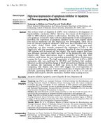
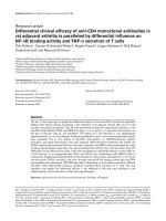
A 10-mm tube saw bone biopsy of approximately 30 mm in
length from the IT region of the proximal femur (Figure 1) was
obtained from 15 patients with primary hip OA (8 females, 48
to 82 years old, and 7 males, 50 to 85 years old; mean age =
65.1 ± 12.6 standard deviation [SD] years) undergoing total
hip arthroplasty surgery. The closely age- and gender-matched
control group, for which trabecular bone from the same site
was taken, comprised 13 autopsy cases (6 females, 57 to 83
years old, and 7 males, 44 to 71 years old; mean age = 63.5
± 11.2 years) known not to have suffered from any chronic
condition or disease that may have affected the skeleton. For
both the OA and control groups, cases with a known history
of medication that may have affected bone metabolism were
excluded. The mean age of the OA group did not differ signif-
icantly from that of the control group.
The surgical and autopsy femoral heads were macroscopically
graded for OA according to the criteria of Collins [25], as pre-
viously described [16,24]. At surgery, primary OA femoral
Figure 1
X-ray of a normal proximal femur, showing the intertrochanteric region (rectangle) used for samplingX-ray of a normal proximal femur, showing the intertrochanteric region
(rectangle) used for sampling.
Available online />Page 3 of 12
(page number not for citation purposes)
heads were either grade III or IV and the graded autopsy
femoral heads were not worse than grade II OA. The use of the
IT region of the proximal femur chosen for sampling has been
justified previously [24]. Furthermore, architectural and cata-
bolic gene expression differences have been observed
between OA and non-OA individuals at this distal skeletal site
[12,16,24]. Each trabecular IT bone specimen was divided
lengthwise for molecular and histology analyses. Informed
consent was obtained for the collection and use of bone spec-
imens, with approval by the Royal Adelaide Hospital Research
Ethics Committee.
Total RNA extraction
For total RNA extraction, the fresh surgical IT bone specimens
(stored at 4°C up to 12 hours in sterile RNase-free phosphate-
buffered saline) and control bone (obtained 24 to 96 hours
after death) were rinsed briefly in diethylpyrocarbonate-treated
water (Sigma-Aldrich, St. Louis, MO, USA) and then sepa-
rated into small fragments by using bone cutters. High-quality
total RNA was isolated using a modified guanidinium thiocy-
anate method of Chomczynski and Sacchi [26], as previously
detailed [16,24,27,28]. The procedure for specimen storage,
handling, and use of RNA extracted from the IT region from
both OA and autopsy controls has been validated by Kuliwaba
and colleagues [16,27]. Total RNA extracted from cultured
human OA OB cells, obtained from trabecular bone explants
[29], served as positive controls for the subsequent semi-
quantitative reverse transcription-polymerase chain reaction
(SQRT-PCR). The quality and integrity of the RNA extracted
were confirmed on 1% wt/vol ethidium bromide-stained for-
maldehyde-agarose gels.
Semi-quantitative reverse transcription-polymerase
chain reaction
First-strand cDNA synthesis of 1 μg of total RNA isolated from
the OA and control bone samples was prepared using a cDNA
synthesis kit, Superscript II (Invitrogen Corporation, Carlsbad,
CA, USA) with 250 ng of random hexamer primer
(GeneWorks Pty Ltd, Adelaide, Australia) according to the
manufacturer's instructions. cDNA was synthesised from RNA
samples of each group at the same time to limit differences in
the efficiency of the cDNA synthesis. Synthesised cDNA was
amplified by PCR using mRNA-specific primers to generate
products corresponding to mRNA encoding human ALP, col-
lagen type I alpha chain (COL1A) 1, COL1A2, IGF-I, IGF-II,
OCN, osteopontin (OPN), transforming growth factor-β1
(TGF-β1), and the housekeeping gene glyceraldehyde-3-
phosphate dehydrogenase (GAPDH) using reaction mixtures
and conditions as previously detailed [16,24,27,30]. Amplifi-
cation of GAPDH served as an internal positive control and
allowed normalisation of the various mRNA levels against the
total mRNA content in the samples. The human mRNA-spe-
cific primer sequences, predicted PCR product sizes, and
optimised PCR conditions are presented in Table 1. To allow
semi-quantitation of the PCR products, preliminary experi-
ments were performed to ensure that the PCR cycles were
within the exponential phase of the amplification curve. As pre-
viously reported in Tsangari and colleagues [28], results
obtained by the SQRT-PCR method are comparable to the
results obtained using the quantitative Taqman (Applied Bio-
systems, Foster City, CA, USA) PCR system. Amplifications of
each mRNA species for both the OA and control cases were
visualised on a single 2% wt/vol agarose gel post-stained with
SYBR
®
Gold (catalog no. S11494; Molecular Probes Inc.,
now part of Invitrogen Corporation) to minimise interassay var-
iability and were quantitated using the FluorImager/Typhoon
and ImageQuant software (Molecular Dynamics, now part of
GE Healthcare, Little Chalfont, Buckinghamshire, UK), as pre-
viously described [16,24,27].
Bone histomorphometry
For histology, trabecular bone samples were fixed in 70% eth-
anol, processed undecalcified through a graded series of eth-
anol, embedded in methylmethacrylate resin, and sectioned on
a microtome (Polycut-E, Leica SP2600; Leica Microsystems,
Wetzlar, Germany), as previously described [24,31]. There
was insufficient bone tissue for histology for one female OA
case (82 years old). Sections, 5 μm thick, were stained by the
von Kossa silver method and counterstained with haematoxylin
and eosin to distinguish between the mineralised bone, the
osteoid, and the cellular components of the marrow. Bone his-
tomorphometric analysis was performed using an ocular-
mounted 10 × 10 graticule at a magnification of ×100. Meas-
urements were made of the following parameters: bone tissue
volume (BV/TV) (percentage), bone surface density (BS/TV)
(square millimetres per cubic millimetre), specific surface of
bone (BS/BV) (square millimetres per cubic millimetre), Tb.Th
(micrometre), Tb.Sp (micrometre), trabecular number (Tb.N)
(number per millimetre), osteoid volume (OV/TV) (percent-
age), osteoid surface (OS/BS) (percentage), and eroded
surface (ES/BS) (percentage). It is worth noting that Tb.N is a
derived index of BS/TV and hence will have similar distribution
and significance level of data for both OA and control groups.
Data analysis
The data generated were tested for normality using the Sha-
piro-Wilk statistic. The statistical significance of the difference
between the OA and the control group was determined by
Student's t test for normally distributed data, expressed as
mean ± SD, and Mann-Whitney U test for non-parametric
data, expressed as median (25th percentile to 75th percen-
tile). Regression analysis using parametric Pearson (r)
statistics was used to examine age-related changes, the rela-
tionship between PCR product/GAPDH ratios and between
PCR product/GAPDH ratios, and bone histomorphometric
variables (PC-SAS statistical software; SAS Institute Inc.,
Cary, NC, USA). The critical value for significance was chosen
as p less than 0.05.
Arthritis Research & Therapy Vol 8 No 6 Truong et al.
Page 4 of 12
(page number not for citation purposes)
Results
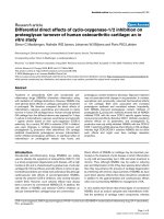
mRNA corresponding to each of the targeted genes was
found to be expressed in trabecular bone from the IT region of
the proximal femur for both OA and control individuals (relative
expression in OA and control is presented in Figure 2).
Gender differences in gene expression and
histomorphometric parameters
Few gene expression and histomorphometric differences were
observed between males and females when the OA and con-
trol groups were analysed separately. TGF-β1/GAPDH was
the only significant gene expression difference found between
the genders, with OA males having higher gene expression
levels than OA females (0.91 ± 0.24 versus 0.64 ± 0.07,
respectively; p < 0.03). In comparison to the respective female
group, OA males (118.8 ± 40.1 μm versus 78.2 ± 26.2 μm,
respectively; p < 0.05) and control males (130 ± 30 μm ver-
sus 90 ± 30 μm, respectively; p < 0.03) had increased Tb.Th.
Due to the few differences observed, further analyses of the
data determining relationships between gene expression and
histomorphometric indices were made independent of gender
for the OA and control groups.
Bone anabolic and collagen type I mRNA expression
between OA and control individuals
mRNA corresponding to ALP and OCN, both commonly used
as markers of bone formation, and the non-collagenous protein
OPN were significantly elevated in the OA group in compari-
Table 1
Primer design for semi-quantitative reverse transcription-polymerase chain reaction analysis
Target gene Primer sequence (5'-3') PCR product size (bp) Annealing temperature (°C) Number of PCR cycles GenBank accession
number
ALP
Sense ccaacgtggctaagaatgTC 434 58 30 NM_000478
Antisense catctcgttgtctgagtacc
OCN
Sense ggtgcagcctttgtgtccaagc 159 62 29 NM_199173
Antisense GTCAGCCAACTCGTCACAGTCC
OPN
Sense AGCCGTGGGAAGGACAGTTATG 472 62 29 NM_000582
Antisense GAGTTTCCATGAAGCCACAAAC
IGF-I
Sense GAGCCTGCGCAATGGAATAAAG 344 62 33 NM_000618
Antisense CCTGTCTCCACACACGAACTG
IGF-II
Sense GAGGAGTGCTGTTTCCGCAG 263 62 27 NM_000612
Antisense ACGTTTGGCCTCCCTGAACG
TGF-
β
1 [53]
Sense CTAGACCCTTTCTCCTCCAGGAGACG 224 62 26 NM_000660
Antisense GCTGGGGGTCTCCCGGCAAAAGGT
COL1A1
Sense CGGCAAGGTGTTGTGCGATG 339 62 33 NM_000088
Antisense CACGGAAATTCCTCCGGTTG
COL1A2
Sense CGCTGGTGAAGTTGGCAAACCA 778 66 31 NM_000089
Antisense GAGGACCACGAAGCCCTTCTTTC
GAPDH [16]
Sense CATGGAGAAGGCTGGGGCTC 415 62 23 NM_002046
Antisense CACTGACACGTTGGCAGTGG
ALP, alkaline phosphatase; COL1A, collagen type I alpha chain; GAPDH, glyceraldehyde-3-phosphate dehydrogenase; IGF, insulin-like growth
factor; OCN, osteocalcin; OPN, osteopontin; PCR, polymerase chain reaction; TGF-β1, transforming growth factor-β1.
Available online />Page 5 of 12
(page number not for citation purposes)
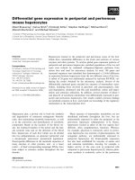
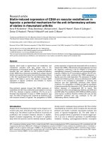
son with the controls (p < 0.002, p < 0.0001, and p < 0.05,
respectively; Figure 3a–c). In contrast, IGF-I/GAPDH, IGF-II/
GAPDH, and TGF-β1/GAPDH growth factor gene expres-
sions were similar between OA and control individuals. The
mean and median values for the growth factors, as well as the
differential gene expression of the α1 and α2 chain of collagen
type I (p < 0.0001 and p < 0.002, respectively) between OA
and controls, are shown in Table 2. Interestingly, the ratio of
COL1A1/COL1A2 was significantly greater in OA bone com-
pared to control (p < 0.0001; Figure 3d).
An age-related increase in OCN/GAPDH mRNA in OA (r =
0.57, p < 0.03) and an age-related decrease in controls (r = -
0.62, p < 0.03) were observed (Figure 4). A negative associ-
ation with age was found for COL1A2/GAPDH mRNA in the
OA group only (n = 15, r = -0.55, p < 0.04; data not shown).
No other significant age associations were found for the other
genes of interest in the two groups.
Because collagen type I consists of two α1 chains and one α2
chain, the positive association between the two alpha chains
observed in both the OA and control groups was expected (r
= 0.66, p < 0.008 and r = 0.70, p < 0.008, respectively; Fig-
ure 5). The slope of the regression line for the OA samples
was greater than the slope for the control samples (p <
0.001), such that for a given level of COL1A2 mRNA, the cor-
responding level of COL1A1 mRNA was greater in OA sam-
ples. Per unit of COL1A2 gene expression, the level of
COL1A1 gene expression in OA was almost double that in the
controls (1.71 versus 0.91, respectively).
In comparison to the controls, IGF-II/GAPDH mRNA expres-
sion was significantly higher than IGF-I/GAPDH mRNA (p <
0.0003; Table 2) in the OA group. When the IGF-I/GAPDH
mRNA and IGF-II/GAPDH mRNA data were plotted, a signifi-
cant positive association was observed for both OA and con-
trols (r = 0.64, p < 0.02 and r = 0.73, p < 0.005, respectively;
Figure 6).
Comparison of bone structural and turnover indices
between OA and control individuals
Bone histomorphometry was performed on IT trabecular bone
samples obtained from 14 out of the 15 OA cases from total
hip replacement (tissue sample size in one case was
insufficient) and the 13 controls without evidence of OA
pathology taken at autopsy. The mean and median values for
the structural and bone turnover parameters at this skeletal
site are shown in Table 3. OA bone had significantly increased
BS/TV (p < 0.0003) and Tb.N (p < 0.0003) and a significant
decrease in Tb.Sp (p < 0.0001). The static indices for bone
formation (OS/BS) and bone resorption (ES/BS) were similar
between the OA and control groups. A positive correlation
was found between OS/BS and ES/BS for both groups (OA:
n = 14, OS/BS = 0.97 [ES/BS] + 3.26; r = 0.57, p < 0.04;
control: n = 13, OS/BS = 0.56 [ES/BS] + 4.01; r = 0.62, p <
0.03). This finding suggests that the bone remodelling proc-
ess is still coupled in the two groups, consistent with previ-
ously reported data from the IT region [24].
When the histomorphometric measurements were plotted
with age, there was a significant increase in BS/BV (n = 13,
BS/BV = 0.49 [Age] – 10.7; r = 0.66, p < 0.02) and a
significant decrease in Tb.Th with age for the controls (n = 13,
Tb.Th = -1.8 [Age] + 223.4; r = -0.58, p < 0.04). These rela-
tionships were not observed for the OA group. Even though
there was no significant difference in OS/BS and ES/BS
between the OA and control groups, there was a significant
association for both of these parameters with age in the con-
trols (n = 13, OS/BS = 0.22 [Age] – 6.7; r = 0.58, p < 0.04
and n = 13, ES/BS = 0.29 [Age] – 12.7; r = 0.69, p < 0.01,
respectively), which is consistent with our previous findings
[24]. This indicated an increased extent of both bone forma-
tion and bone resorption with age, which together suggest an
increase in the rate of bone turnover with ageing in control
individuals. There were no significant correlations with age for
OS/BS or ES/BS in OA.
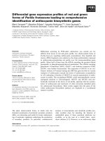
Figure 2
Representative gene expression as determined by semi-quantitative reverse transcription-polymerase chain reaction (PCR) using total RNA extracted from intertrochanteric trabecular boneRepresentative gene expression as determined by semi-quantitative
reverse transcription-polymerase chain reaction (PCR) using total RNA
extracted from intertrochanteric trabecular bone. Target genes included
alkaline phosphatase (ALP) (434 bp), osteocalcin (OCN) (159 bp),
osteopontin (OPN) (472 bp), insulin-like growth factor (IGF)-I (344 bp),
IGF-II (263 bp), transforming growth factor-β1 (TGF-
β
1) (224 bp),
COL1A1 (339 bp), COL1A2 (778 bp), and the housekeeping gene
GAPDH (415 bp). Specimens were obtained from a 60-year-old
female (F 60) and a 59-year-old male (M 59) undergoing total hip
replacement for primary osteoarthritis (OA). The control specimens
were obtained at autopsy from a 61-year-old female (F 61) and a 60-
year-old male (M 60) without any bone-related disease. PCR products
representing each mRNA species were visualised on SYBR Gold
®
-
stained 2% agarose gels. COL1A, collagen type I alpha chain;
GAPDH, glyceraldehyde-3-phosphate dehydrogenase.
Arthritis Research & Therapy Vol 8 No 6 Truong et al.
Page 6 of 12
(page number not for citation purposes)
Associations between OB marker gene expression and
histomorphometry
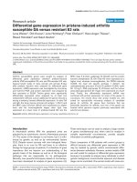
For the control group, OCN/GAPDH mRNA was found to
have a significant positive association with BV/TV (r = 0.82, p
< 0.0007; Figure 7a), BS/TV (r = 0.56, p < 0.05; data not
shown), and Tb.N (r = 0.56, p < 0.05; Figure 7b) and a signif-
icant negative association with Tb.Sp (r = -0.64, p < 0.02; Fig-
ure 7c). These relationships were not observed for the OA
group. However, it is interesting to note that the OA data for
Tb.N and Tb.Sp had segregated away from the controls such
that OA individuals have significantly elevated OCN gene
expression with increased Tb.N and decreased Tb.Sp, as
reflected in the group comparisons (Figure 3 and Table 3).
Furthermore, when the control and OA data were combined
for the Tb.N versus OCN/GAPDH (n = 27; Tb.N = 0.72
[OCN/GAPDH] + 0.33; r = 0.62, p < 0.0007) and Tb.Sp ver-
Figure 3
Relative polymerase chain reaction product/GAPDH ratios for alkaline phosphatase (ALP), osteocalcin (OCN), and osteopontin (OPN) and the rela-tive ratio of COL1A1/COL1A2Relative polymerase chain reaction product/GAPDH ratios for alkaline phosphatase (ALP), osteocalcin (OCN), and osteopontin (OPN) and the rela-
tive ratio of COL1A1/COL1A2. mRNA expression in intertrochanteric trabecular bone was compared between the osteoarthritis (OA) (n = 15) and
control (n = 13) groups. Patients with OA had significantly elevated (a) ALP/GAPDH (p < 0.002), (b) OCN/GAPDH (p < 0.0001), (c) OPN/
GAPDH (p < 0.05), and (d) COL1A1/COL1A2 (p < 0.0001) mRNA ratios versus controls. Data are expressed as parametric mean ± standard devi-
ation (open diamond) and non-parametric median (closed diamond) and quartile (dash) range. COL1A, collagen type I alpha chain; GAPDH, glycer-
aldehyde-3-phosphate dehydrogenase.
Available online />Page 7 of 12
(page number not for citation purposes)
sus OCN/GAPDH (n = 27; Tb.Sp = 0.91 [OCN/GAPDH] –
0.7; r = -0.73, p < 0.0001) plots, it appears that there is a con-
tinuum in the bone remodelling processes leading to particular
trabecular structures, with respect to OCN/GAPDH levels in
the IT region. In contrast, ALP, another OB-specific marker,
showed a significant decrease in BV/TV (n = 13; BV/TV = -9.7
[ALP/GAPDH] + 13.2; r = -0.61, p < 0.03), BS/TV (n = 13;
BS/TV = -1.08 [ALP/GAPDH] + 1.88; r = -0.56, p < 0.05),
and Tb.N (n = 13; Tb.N = -0.54 [ALP/GAPDH] + 0.9; r = -
0.56, p < 0.05), with increasing ALP/GAPDH mRNA expres-
sion in the controls (data not shown). No significant associa-
tions were found for OA individuals.
Discussion
Previous studies have reported higher BMD levels in patients
with early- to late-stage OA. However, there is limited
knowledge about the molecular and cellular mechanisms
involved in the increase or maintenance of bone mass in OA.
This study of the IT region of the proximal femur in primary hip
OA and non-OA postmortem controls investigated changes in
gene expression of bone anabolic factors and collagen type I
alpha chains and associations between gene expression and
bone micro-architecture.
The bone anabolic factors investigated in this study are known
to be involved in the bone remodelling process and there is
evidence for their involvement in OA disease
[6,15,17,18,24,32]. The results of this study indicated
significant elevation in the OB markers, OCN and ALP, as well
as OPN and the alpha chains of collagen type I, COL1A1 and
COL1A2, mRNA in OA individuals. The exact physiological
function of both OCN and ALP is still unknown. OCN, the
most abundant non-collagenous protein of the bone extracel-
Table 2
Semi-quantitative reverse transcription-polymerase chain reaction product/GAPDH ratios for OA and control individuals
Ratio OA Control
(n = 15) (n = 13)
IGF-I/GAPDH 0.52 (0.49–0.56)
a
0.63 (0.38–0.71)
IGF-II/GAPDH 0.79 (0.74–0.94)
a
0.58 (0.39–0.94)
TGF-β1/GAPDH 0.76 ± 0.22 0.72 ± 0.09
COL1A1/GAPDH 0.55 (0.47–0.59) 0.00 (0.00–0.24)
b
COL1A2/GAPDH 0.37 ± 0.04 0.22 ± 0.14
c
a
OA IGF-I/GAPDH versus OA IGF-II/GAPDH: p < 0.0003;
b
p < 0.0001;
c
p < 0.002. Parametric values are mean ± standard deviation. Non-
parametric values are median (quartiles). COL1A, collagen type I alpha chain; GAPDH, glyceraldehyde-3-phosphate dehydrogenase; IGF, insulin-
like growth factor; OA, osteoarthritis; TGF-β1, transforming growth factor-β1.
Figure 4
Changes in osteocalcin (OCN)/GAPDH mRNA with ageChanges in osteocalcin (OCN)/GAPDH mRNA with age. The relative
OCN/GAPDH ratios were determined in intertrochanteric trabecular
bone from individuals with osteoarthritis (OA) (n = 15) and control indi-
viduals (n = 13). In OA, OCN/GAPDH mRNA increased significantly
with age (OCN/GAPDH = 0.01 [Age] + 0.43; r = 0.57, p < 0.03). In
controls, OCN/GAPDH mRNA significantly declined with age (OCN/
GAPDH = -0.01 [Age] + 0.82; r = -0.62, p < 0.03). GAPDH, glyceral-
dehyde-3-phosphate dehydrogenase.
Figure 5
Association between the relative ratios of COL1A1/GAPDH mRNA and COL1A2/GAPDH mRNAAssociation between the relative ratios of COL1A1/GAPDH mRNA
and COL1A2/GAPDH mRNA. Gene expression was determined in
intertrochanteric trabecular bone from patients with osteoarthritis (OA)
(n = 15) and controls (n = 13). A significant correlation was observed
between the two parameters in patients with OA (COL1A1/GAPDH =
1.71 [COL1A2/GAPDH] – 0.10; r = 0.66, p < 0.008) and controls
(COL1A1/GAPDH = 0.91 [COL1A2/GAPDH] – 0.06; r = 0.70, p <
0.008). COL1A, collagen type I alpha chain; GAPDH, glyceraldehyde-
3-phosphate dehydrogenase.
Arthritis Research & Therapy Vol 8 No 6 Truong et al.
Page 8 of 12
(page number not for citation purposes)
lular matrix, is synthesised only by bone-forming OB cells.
OCN is suggested to be involved in the mineralisation process
of newly synthesised osteoid [33]. It is incorporated into the
bone matrix, where it is involved in calcium-binding [34]. Quan-
titative bone histomorphometry and combined calcium bal-
ance/calcium kinetics studies have validated the use of OCN
as a marker of bone formation [35,36]. Previous studies report
increased OCN levels in OA serum, protein, and mRNA gene
studies [14,16]. Our finding of differential OCN mRNA gene
expression between OA and non-OA individuals is consistent
with our previously reported data showing an age-related
increase in OCN gene expression in OA and a decrease in
controls [16]. This finding is supportive of an increase or main-
tenance of bone volume in OA individuals versus the age-
dependent bone loss in the general population [11]. ALP is
used as an enzymatic marker of bone formation, expressed by
early-differentiated OB cells. Elevated expression of this
enzyme in OA, as indicated by this study as well as previous
reports [5,6,15], might indicate a greater proportion of differ-
entiation of the pre-OB pool to the mature phenotype [5].
Even though collagen type I comprises the majority of the
organic matrix of bone, it is not unique to the tissue [37]. Thus,
non-collagenous proteins such as OCN and OPN have
become the focus of studies aimed at the elucidation of the
bone matrix mineralisation process in normal and pathological
conditions [14,38]. OPN is a cytokine currently understood to
be involved in cell attachment, cell migration, chemotaxis, and
intracellular signalling and is expressed by all three bone cell
types: OB, osteoclasts, and osteocytes [39-41]. In bone, OPN
produced by OB during bone matrix formation is subsequently
accumulated in the mineralised matrix. Increased OPN may
augment OB synthetic activity by increasing OB longevity and
surface extent of bone formation. Hence, increased OPN
mRNA expression in OA may contribute to the maintenance or
increase in bone mass in these individuals.
IGF-I, IGF-II, and TGF-β1 are established osteotropic growth
factors that play key roles in bone remodelling and are pro-
duced by the various bone marrow and bone cell types in the
bone microenvironment [42,43]. The role of these growth
factors suggests their involvement in the preservation of the
bone matrix. The two related IGFs are involved in inducing
matrix apposition and decreasing collagen degradation and
expression of interstitial collagenases [44,45]. The main bone
Figure 6
Association between the relative ratios of insulin-like growth factor (IGF)-II/GAPDH mRNA and IGF-I/GAPDH mRNAAssociation between the relative ratios of insulin-like growth factor
(IGF)-II/GAPDH mRNA and IGF-I/GAPDH mRNA. Gene expression
was determined in intertrochanteric trabecular bone from patients with
osteoarthritis (OA) (n = 15) and controls (n = 13). A significant correla-
tion was observed between the two parameters in patients with OA
(IGF-II/GAPDH = 1.49 [IGF-I/GAPDH] + 0.01; r = 0.64, p < 0.02) and
controls (IGF-II/GAPDH = 1.34 [IGF-I/GAPDH] – 0.09; r = 0.73, p <
0.005). GAPDH, glyceraldehyde-3-phosphate dehydrogenase.
Table 3
Trabecular bone structure and bone turnover indices in osteoarthritis and control intertrochanteric bone samples
Histomorphometric parameter Osteoarthritis Control
(n = 14) (n = 13)
BV/TV (percentage) 10.4 ± 4.1 7.6 ± 3.1
BS/TV (mm
2
/mm
3
)2.3 ± 0.71.3 ± 0.4
a
BS/BV (mm
2
/mm
3
) 20.5 (17.3–28.6) 18.0 (14.7–20.6)
Tb.Th (μm) 98 ± 39 111 ± 34
Tb.Sp (μm) 817 (760–892) 1,406 (1,203–2,202)
b
Tb.N (number/mm) 1.11 ± 0.36 0.63 ± 0.19
a
OV/TV (percentage) 0.12 (0.07–0.15) 0.05 (0.04–0.13)
OS/BS (percentage) 7.8 (3.7–9.7) 7.2 (4.2–9.7)
ES/BS (percentage) 4.6 (3.4–6.4) 4.5 (2.0–7.6)
a
p < 0.0003;
b
p < 0.0001. Parametric values are mean ± standard deviation. Non-parametric values are median (quartiles). BS/BV, specific
surface of bone; BS/TV, bone surface density; BV/TV, bone tissue volume; ES/BS, eroded surface; OS/BS, osteoid surface; OV/TV, osteoid
volume; Tb.N, trabecular number; Tb.Sp, trabecular separation; Tb.Th, trabecular thickness.
Available online />Page 9 of 12
(page number not for citation purposes)
anabolic roles for TGF-β1 include stimulating chemotaxis and
proliferation to increase the pool of committed OB precursors
[46,47]. These growth factors have been reported to be
upregulated in OA subchondral and iliac crest bone [6,15,18],
and this upregulation is hypothesised to be due to increased
OB biosynthetic activity [18]. The lack of differential gene
expression of these anabolic growth factors in our study is
most likely due to the gene expression being contributed by
the various cell types present in the bone microenvironment. In
addition to post-transcriptional and post-translational modifi-
cations, the well-known regulation of stored growth factors in
the extracellular matrix by binding proteins that are released
during bone resorption phases may account for the altered
protein expression levels in OA ex vivo studies. Bone cells pro-
duce both IGF-I and IGF-II, and IGF-II is reported to be
expressed at higher levels than IGF-I in OA and control
Figure 7
Associations between osteocalcin (OCN)/GAPDH mRNA and the histomorphometric parameters of bone tissue volume (BV/TV), trabecular number (Tb.N), and trabecular separation (Tb.Sp)Associations between osteocalcin (OCN)/GAPDH mRNA and the histomorphometric parameters of bone tissue volume (BV/TV), trabecular number
(Tb.N), and trabecular separation (Tb.Sp). The relative OCN/GAPDH mRNA expression and architectural parameters were determined in intertro-
chanteric trabecular bone from osteoarthritis (OA) (n = 14) and control (n = 13) individuals. (a) In controls, there was a significant increase in BV/TV
with increasing OCN/GAPDH mRNA (BV/TV = 27.7 [OCN/GAPDH] – 6.3; r = 0.82, p < 0.0007) in contrast to the patients with OA (BV/TV = -
7.57 [OCN/GAPDH] + 17.38; r = -0.31, p = not significant [NS]). (b) A significant increase in Tb.N with increasing OCN/GAPDH mRNA was
observed in controls (Tb.N = 1.16 [OCN/GAPDH] + 0.05; r = 0.56, p < 0.05). In OA, there was no significant association between Tb.N and OCN/
GAPDH mRNA (Tb.N = 0.09 [OCN/GAPDH] + 1.03; r = 0.04, p = NS). (c) In controls, there was a significant decline in Tb.Sp with increasing
OCN/GAPDH mRNA (Tb.Sp = -3,977.9 [OCN/GAPDH] + 3,626.1; r = -0.64, p < 0.02) and no significant change in Tb.Sp with OCN/GAPDH
mRNA in OA individuals (Tb.Sp = -353.5 [OCN/GAPDH] + 1,214.4; r = -0.19, p = NS). GAPDH, glyceraldehyde-3-phosphate dehydrogenase.
Arthritis Research & Therapy Vol 8 No 6 Truong et al.
Page 10 of 12
(page number not for citation purposes)
subchondral bone [32]. Interestingly, even though IGF-II
mRNA expression was significantly higher than IGF-I mRNA in
OA, this study indicated similar positive associations between
the two IGFs in both OA and non-OA individuals, indicating
that the probable co-expression of the two IGFs is not signifi-
cantly altered at the IT region in hip OA disease. The differen-
tial gene expression of ALP, OCN, OPN, and the alpha chains
of collagen type I in OA observed from this study suggests
support of the hypothesis of increased OB biosynthetic activ-
ity, as postulated by Dequeker and colleagues [18]. Further
experiments using in situ hybridisation and
immunohistochemical staining at this distal skeletal site would
confirm whether there is an increase in OB biosynthetic activ-
ity or increased OB cell number in OA individuals. Additionally,
the significant differential gene expression in OA versus con-
trol IT trabecular bone may be due to the presence of an
altered bone cell phenotype [19].
The trabecular bone samples obtained from patients with OA
were architecturally distinct, having elevated bone surface
density and Tb.N and decreased Tb.Sp compared to the age-
and gender-matched controls. These observations are con-
sistent with previous studies at the same distal skeletal site
[12,24] and indicate a more generalised distribution of OA
bone architectural changes in OA individuals. In contrast to
Fazzalari and colleagues [24], we did not observe age-related
changes in bone volume fraction in controls, due to the fact
that the aforementioned study analysed a broader age range
of subjects. Our control data indicated a significant increase in
bone surface density and a significant decrease in Tb.Th with
age.
Subchondral bone as well as cancellous bone from the central
regions of the femoral head and femoral neck of patients with
late-stage OA has been described as hypomineralised [4,6,7],
which may be due in part to increased bone remodelling due
to adaptation/repair of the diseased joint and therefore does
not allow sufficient time for complete mineralisation. In this
study of the IT region of the femur, however, the static histo-
morphometric indices for bone formation and bone resorption
were similar in magnitude between the OA and control groups,
and the data further indicated that the bone turnover process
remains coupled in both groups. These results are in contrast
to our previous study of a different OA and control sample set
of the IT region, in which we reported increased percentage of
bone forming surface for any given amount of bone resorption
in OA compared to a non-OA group [24]. The finding of
altered bone remodelling in OA [24], however, was confirmed
when the data set for OS/BS and ES/BS from Fazzalari and
colleagues [24] and the data set from the current study were
combined (data not shown). Dynamic histoquantitation of
bone remodelling would provide a more comprehensive
insight into the rate of turnover at this distal skeletal site.
Bailey and colleagues [8] have identified the presence of col-
lagen type I homotrimer in OA subchondral bone which con-
sists of three α1 chains instead of the usual α1/α2 chain ratio
of 2:1. By means of electron microscopy, the collagen fibres
were observed to be narrower and aligned in a disorganised
manner, which may contribute to the under-mineralisation in
OA [8]. Interestingly, the findings of the present study indi-
cated that the ratio of COL1A1/COL1A2 mRNA expression
was significantly elevated in OA bone compared to control,
suggesting the possible presence of collagen type I homot-
rimer at a skeletal site distal to the articular cartilage. However,
protein analysis determining the expression level of the two
alpha chains in the bone matrix at the IT region as well as
mineral density fractionation studies will be required to sup-
port this notion.
Our experimental approach of combining gene expression
analysis with histomorphometry allows the exploration of any
relationships between gene expression and indices of bone
architecture and bone remodelling. Interestingly, both OCN
and ALP gene expression significantly correlated with bone
micro-architecture at this distal skeletal site. In controls, there
is increased bone volume fraction, bone surface density, and
Tb.N, decreased Tb.Sp with increasing OCN mRNA, and an
apparent inverse involvement for ALP mRNA, with negative
associations with bone volume, bone surface density, and
Tb.N. Indeed, from our results, when OCN and bone volume
fraction were plotted for the controls, there was a significant
increase in bone volume with increasing OCN mRNA levels,
which is contradictory to the findings of the OCN knockout
mice experiments [48], which suggest that OCN limits bone
formation without directly impairing bone resorption or miner-
alisation. We can speculate that the results of our study may
reflect regulatory mechanisms of bone formation in the normal
bone remodelling process that are dysregulated in the skele-
ton of patients with primary hip OA. The pooled OA and con-
trol data suggest that there is a continuum in the bone
remodelling process leading to particular trabecular struc-
tures, with respect to OCN/GAPDH mRNA levels in the IT
region. However, the lack of association observed between
OCN mRNA expression and the architectural parameters in
the OA group may be due in part to altered bone cell response
indicated by an increased level of OCN gene expression in
OA. The altered cellular response may manifest as an
increased range of Tb.N for OCN gene expression levels in
OA when compared to controls. On the other hand, Tb.Sp
appeared to plateau in the amount of Tb.Sp change, with
respect to OCN/GAPDH mRNA levels in the OA group. This
is consistent with the reported observation that changes in
trabecular architecture are non-linear [49]. Consequently,
Tb.Sp is non-linearly associated with OCN/GAPDH mRNA
gene expression. The data in this study also indicated that with
increasing ALP gene expression there is decreasing bone vol-
ume, also inconsistent with ALP knockout studies that have
found decreased bone volume, hypomineralisation, and
Available online />Page 11 of 12
(page number not for citation purposes)
disordered mineral crystal alignment pattern and matrix archi-
tecture in metaphyseal bone trabeculae and cortical bone of
mice femora and upper tibias [50,51]. Our observations may
implicate an increase in bone turnover with increasing ALP
gene expression and, thus, a decrease in bone volume due to
decreasing Tb.N. Taken together, the observations from these
association analyses suggest that any potential regulatory
mechanisms between either OCN or ALP with bone architec-
tural parameters are clearly altered in individuals with primary
hip OA compared to non-OA controls.
The significant gene expression and bone architectural
changes observed between primary hip OA compared to non-
OA controls at a skeletal site distal to the active site of disease
support the concept that there is a generalised involvement of
bone in the pathogenesis of OA. This study, using molecular
histomorphometry, has also provided unique insight into pos-
sible perturbations or dysregulation of bone turnover proc-
esses in OA which differ from the norm. The changes present
at distal skeletal sites may be reflective of constitutive gene
expression and consequent skeletal structure that predis-
poses an individual to primary OA. Pre-existing genetic differ-
ences in individuals which lead to altered suboptimal loading
bone structures that adapt differently to the loads the skeleton
is subjected to may affect biomechanical and biochemical pro-
files, as well as accumulation of microdamage [52], and
enhance the initiation/progression of OA. Future research
investigating early OA and the diseased joint as a whole is
important to further the understanding of OA pathogenesis
and, hence, enable development of improved treatment and/or
preventative measures to delay joint degeneration or progres-
sion of OA.
Conclusion
Expression of mRNA corresponding to ALP, OCN, OPN, and
the two collagen type I alpha chains, COL1A1 and COL1A2,
is elevated in primary hip OA IT bone compared to postmortem
non-OA controls. The finding of differential gene expression,
as well as architectural changes and differences in molecular
histomorphometric associations between OA and controls, at
a skeletal site distal to the active joint cartilage degeneration
supports the concept of generalised involvement of bone in
the pathogenesis of OA.
Competing interests
The authors declare that they have no competing interests.
Authors' contributions
JSK and NLF contributed to the study design and coordina-
tion. LT performed the acquisition of the SQRT-PCR data. JSK
and HT performed the acquisition of the histomorphometry
data. LT and JSK performed the statistical analyses. LT, JSK,
and NLF analysed and interpreted the data. LT, JSK, and NLF
prepared the manuscript. All authors read and approved the
final manuscript.
Acknowledgements
The authors thank the donors and donors' families for their kind donation
of bone tissue used for this study and offer an extended appreciation to
the orthopaedic surgeons and nursing staff of the Department of Ortho-
paedics and Trauma in the Royal Adelaide Hospital for support and
cooperation in the collection of femoral specimens and to the mortuary
staff of the Institute of Medical and Veterinary Science for the collection
of autopsy specimens. The authors thank Professor David M Findlay
(Department of Orthopaedics and Trauma, Royal Adelaide Hospital,
Adelaide, Australia) for the kind use of his laboratory for the undertaking
of the molecular component in this study. This work was supported by
the National Health and Medical Research Council (Australia) and The
University of Adelaide.
References
1. Sarzi-Puttini P, Cimmino MA, Scarpa R, Caporali R, Parazzini F,
Zaninelli A, Atzeni F, Canesi B: Osteoarthritis: an overview of the
disease and its treatment strategies. Semin Arthritis Rheum
2005, 35:1-10.
2. Dequeker J, Luyten FP: Bone mass and osteoarthritis. Clin Exp
Rheumatol 2000, 18(suppl 21):S21-S26.
3. Ameye LG, Young MF: Animal models of osteoarthritis: lessons
learned while seeking the 'Holy Grail'. Curr Opin Rheumatol
2006, 18:537-547.
4. Grynpas MD, Alpert B, Katz I, Lieberman I, Pritzker KP: Subchon-
dral bone in osteoarthritis. Calcif Tissue Int 1991, 49:20-26.
5. Mansell JP, Tarlton JF, Bailey AJ: Biochemical evidence for
altered subchondral bone collagen metabolism in osteoarthri-
tis of the hip. Br J Rheumatol 1997, 36:16-19.
6. Mansell JP, Bailey AJ: Abnormal cancellous bone collagen
metabolism in osteoarthritis. J Clin Invest 1998,
101:1596-1603.
7. Li B, Aspden RM: Composition and mechanical properties of
cancellous bone from the femoral head of patients with oste-
oporosis or osteoarthritis. J Bone Miner Res 1997,
12:641-651.
8. Bailey AJ, Sims TJ, Knott L: Phenotypic expression of osteoblast
collagen in osteoarthritic bone: production of type I
homotrimer. Int J Biochem Cell Biol 2002, 34:176-182.
9. Gevers G, Dequeker J, Martens M, Van Audekercke R, Nyssen-
Behets C, Dhem A: Biomechanical characteristics of iliac crest
bone in elderly women according to osteoarthritis grade at the
hand joints. J Rheumatol 1989, 16:660-663.
10. Fazzalari NL, Parkinson IH: Femoral trabecular bone of osteoar-
thritic and normal subjects in an age and sex matched group.
Osteoarthr Cartil 1998, 6:377-382.
11. Crane GJ, Fazzalari NL, Parkinson IH, Vernon-Roberts B: Age-
related changes in femoral trabecular bone in arthrosis. Acta
Orthop Scand 1990, 61:421-426.
12. Fazzalari NL, Vernon-Roberts B, Manthey BA, Parkinson IH: Rela-
tionship between changes in articular cartilage and bone in
the femoral head in osteoarthritis of the hip. J Orthopaed
Rheumatol 1990, 3:
155-169.
13. Pagani F, Francucci CM, Moro L: Markers of bone turnover: bio-
chemical and clinical perspectives. J Endocrinol Invest 2005,
28:8-13.
14. Gevers G, Dequeker J: Collagen and non-collagenous protein
content (osteocalcin, sialoprotein, proteoglycan) in the iliac
crest bone and serum osteocalcin in women with and without
hand osteoarthritis. Coll Relat Res 1987, 7:435-442.
15. Hilal G, Martel-Pelletier J, Pelletier JP, Ranger P, Lajeunesse D:
Osteoblast-like cells from human subchondral osteoarthritic
bone demonstrate an altered phenotype in vitro: possible role
in subchondral bone sclerosis. Arthritis Rheum 1998,
41:891-899.
16. Kuliwaba JS, Findlay DM, Atkins GJ, Forwood MR, Fazzalari NL:
Enhanced expression of osteocalcin mRNA in human osteoar-
thritic trabecular bone of the proximal femur is associated
with decreased expression of interleukin-6 and interleukin-11
mRNA. J Bone Miner Res 2000, 15:332-341.
17. Raymaekers G, Aerssens J, Van den Eynde R, Peeters J, Geusens
P, Devos P, Dequeker J: Alterations of the mineralization profile
Arthritis Research & Therapy Vol 8 No 6 Truong et al.
Page 12 of 12
(page number not for citation purposes)
and osteocalcin concentrations in osteoarthritic cortical iliac
crest bone. Calcif Tissue Int 1992, 51:269-275.
18. Dequeker J, Mohan S, Finkelman RD, Aerssens J, Baylink DJ: Gen-
eralized osteoarthritis associated with increased insulin-like
growth factor types I and II and transforming growth factor
beta in cortical bone from the iliac crest. Possible mechanism
of increased bone density and protection against
osteoporosis. Arthritis Rheum 1993, 36:1702-1708.
19. Westacott CI, Webb GR, Warnock MG, Sims JV, Elson CJ: Alter-
ation of cartilage metabolism by cells from osteoarthritic bone.
Arthritis Rheum 1997, 40:1282-1291.
20. Nevitt MC, Lane NE, Scott JC, Hochberg MC, Pressman AR,
Genant HK, Cummings SR: Radiographic osteoarthritis of the
hip and bone mineral density. The Study of Osteoporotic Frac-
tures Research Group. Arthritis Rheum 1995, 38:907-916.
21. Bruno RJ, Sauer PA, Rosenberg AG, Block J, Sumner DR: The
pattern of bone mineral density in the proximal femur and radi-
ographic signs of early joint degeneration. J Rheumatol 1999,
26:636-640.
22. Goker B, Sumner DR, Hurwitz DE, Block JA: Bone mineral den-
sity varies as a function of the rate of joint space narrowing in
the hip. J Rheumatol 2000, 27:735-738.
23. Bergink AP, Uitterlinden AG, Van Leeuwen JP, Hofman A, Verhaar
JA, Pols HA: Bone mineral density and vertebral fracture his-
tory are associated with incident and progressive radiographic
knee osteoarthritis in elderly men and women: the Rotterdam
Study. Bone 2005, 37:446-456.
24. Fazzalari NL, Kuliwaba JS, Atkins GJ, Forwood MR, Findlay DM:
The ratio of messenger RNA levels of receptor activator of
nuclear factor kappaB ligand to osteoprotegerin correlates
with bone remodeling indices in normal human cancellous
bone but not in osteoarthritis. J Bone Miner Res 2001,
16:1015-1027.
25. Collins D: The Pathology of Articular and Spinal Diseases London,
UK: Edward Arnold and Co; 1949.
26. Chomczynski P, Sacchi N: Single-step method of RNA isolation
by acid guanidinium thiocyanate-phenol-chloroform
extraction. Anal Biochem 1987, 162:156-159.
27. Kuliwaba JS, Fazzalari NL, Findlay DM: Stability of RNA isolated
from human trabecular bone at post-mortem and surgery.
Biochim Biophys Acta 2005, 1740:1-11.
28. Tsangari H, Findlay DM, Kuliwaba JS, Atkins GJ, Fazzalari NL:
Increased expression of IL-6 and RANK mRNA in human
trabecular bone from fragility fracture of the femoral neck.
Bone 2004, 35:334-342.
29. Gronthos S, Zannettino AC, Graves SE, Ohta S, Hay SJ, Simmons
PJ: Differential cell surface expression of the STRO-1 and
alkaline phosphatase antigens on discrete development
stages in primary cultures of human bone cells. J Bone Miner
Res 1999, 14:47-56.
30. Hopwood B, Gronthos S, Kuliwaba JS, Robey PG, Findlay DM,
Fazzalari NL: Identification of differentially expressed genes
between osteoarthritic and normal trabecular bone from the
intertrochanteric region of the proximal femur using cDNA
microarray analysis. Bone 2005, 36:635-644.
31. Tsangari H, Findlay DM, Fazzalari NL: Structural and remodeling
indices in the cancellous bone of the proximal femur across
adulthood. Bone 2007, 40:211-217.
32. Sharp CA, Brown SJ, Davie MW, Magnussan P, Mohan S:
Increased matrix concentration of IGFBP-5 bone in
osteoarthritis. Ann Rheum Dis 2004, 63:1162-1165.
33. Roach HI: Why does bone matrix contain non-collagenous pro-
teins? The possible roles of osteocalcin, osteonectin, oste-
opontin and bone sialoprotein in bone mineralisation and
resorption. Cell Biol Int 1994, 18:617-628.
34. Hoang QQ, Sicheri F, Howard AJ, Yang DS: Bone recognition
mechanism of porcine osteocalcin from crystal structure.
Nature 2003, 425:977-980.
35. Eastell R, Delmas PD, Hodgson SF, Eriksen EF, Mann KG, Riggs
BL: Bone formation rate in older normal women: concurrent
assessment with bone histomorphometry, calcium kinetics,
and biochemical markers. J Clin Endocrinol Metab 1988,
67:741-748.
36. Lee AJ, Hodges S, Eastell R: Measurement of osteocalcin. Ann
Clin Biochem 2000, 37(Pt 4):432-446.
37. Rossert J, de Crombrugghe B: Type I collagen: structure, syn-
thesis, and regulation. In Dynamics of Bone and Cartilage
Metabolism Edited by: Seibel MJ, Robins SP, Bilezikian JP. San
Diego, CA: Academic Press; 1999:127-142.
38. Termine JD, Belcourt AB, Conn KM, Kleinman HK: Mineral and
collagen-binding proteins of fetal calf bone.
J Biol Chem 1981,
256:10403-10408.
39. Sodek J, Ganss B, McKee MD: Osteopontin. Crit Rev Oral Biol
Med 2000, 11:279-303.
40. Merry K, Dodds R, Littlewood A, Gowen M: Expression of oste-
opontin mRNA by osteoclasts and osteoblasts in modelling
adult human bone. J Cell Sci 1993, 104:1013-1020.
41. Dodds RA, Connor JR, James IE, Rykaczewski EL, Appelbaum E,
Dul E, Gowen M: Human osteoclasts, not osteoblasts, deposit
osteopontin onto resorption surfaces: an in vitro and ex vivo
study of remodeling bone. J Bone Miner Res 1995,
10:1666-1680.
42. Janssens K, ten Dijke P, Janssens S, Van Hul W: Transforming
growth factor-beta1 to the bone. Endocr Rev 2005,
26:743-774.
43. Hayden JM, Mohan S, Baylink DJ: The insulin-like growth factor
system and the coupling of formation to resorption. Bone
1995, 17:93S-98S.
44. Hock JM, Centrella M, Canalis E: Insulin-like growth factor I has
independent effects on bone matrix formation and cell
replication. Endocrinology 1988, 122:254-260.
45. Canalis E, Rydziel S, Delany AM, Varghese S, Jeffrey JJ: Insulin-
like growth factors inhibit interstitial collagenase synthesis in
bone cell cultures. Endocrinology 1995, 136:1348-1354.
46. Pfeilschifter J, Wolf O, Naumann A, Minne HW, Mundy GR, Ziegler
R: Chemotactic response of osteoblastlike cells to transform-
ing growth factor beta. J Bone Miner Res 1990, 5:825-830.
47. Erdmann J, Kogler C, Diel I, Ziegler R, Pfeilschifter J: Age-associ-
ated changes in the stimulatory effect of transforming growth
factor beta on human osteogenic colony formation. Mech Age-
ing Dev 1999, 110:73-85.
48. Ducy P, Desbois C, Boyce B, Pinero G, Story B, Dunstan C, Smith
E, Bonadio J, Goldstein S, Gundberg C, et al.: Increased bone
formation in osteocalcin-deficient mice. Nature 1996,
382:448-452.
49. Parkinson IH, Fazzalari NL:
Interrelationships between struc-
tural parameters of cancellous bone reveal accelerated struc-
tural change at low bone volume. J Bone Miner Res 2003,
18:2200-2205.
50. Anderson HC, Sipe JB, Hessle L, Dhanyamraju R, Atti E, Camacho
NP, Millan JL: Impaired calcification around matrix vesicles of
growth plate and bone in alkaline phosphatase-deficient mice.
Am J Pathol 2004, 164:841-847.
51. Tesch W, Vandenbos T, Roschgr P, Fratzl-Zelman N, Klaushofer K,
Beertsen W, Fratzl P: Orientation of mineral crystallites and
mineral density during skeletal development in mice deficient
in tissue nonspecific alkaline phosphatase. J Bone Miner Res
2003, 18:117-125.
52. Burr DB, Radin EL: Microfractures and microcracks in subchon-
dral bone: are they relevant to osteoarthrosis? Rheum Dis Clin
North Am 2003, 29:675-685.
53. Atkins GJ, Haynes DR, Graves SE, Evdokiou A, Hay S, Bouralexis
S, Findlay DM: Expression of osteoclast differentiation signals
by stromal elements of giant cell tumors. J Bone Miner Res
2000, 15:640-649.