Báo cáo y học: "Reactive oxygen species induce expression of vascular endothelial growth factor in chondrocytes and human articular cartilage explants" pps
Bạn đang xem bản rút gọn của tài liệu. Xem và tải ngay bản đầy đủ của tài liệu tại đây (366.84 KB, 8 trang )
Open Access
Available online />Page 1 of 8
(page number not for citation purposes)
Vol 8 No 6
Research article
Reactive oxygen species induce expression of vascular
endothelial growth factor in chondrocytes and human articular
cartilage explants
Jakob Fay
1
, Deike Varoga
2
, Christoph J Wruck
2
, Bodo Kurz
2
, Mary B Goldring
3,4
and
Thomas Pufe
2
1
Department of Trauma Surgery, University Hospital Schleswig-Holstein, Campus Kiel, Arnold-Heller-Strasse 7, 24105 Kiel, Germany
2
Department of Anatomy, Christian-Albrechts-University Kiel, Olshausenstrasse 40, 24098 Kiel, Germany
3
Beth Israel Deaconess Medical Center, New England Baptist Bone and Joint Institute, Harvard Institutes of Medicine, HIM-246, 4 Blackfan Circle,
Boston, MA 02115-5713, USA
4
Hospital for Special Surgery, Caspary Research Building, Room 528, 535 E. 70th Street, New York, NY 10021, USA
Corresponding author: Thomas Pufe,
Received: 13 Oct 2006 Revisions requested: 9 Nov 2006 Revisions received: 1 Dec 2006 Accepted: 23 Dec 2006 Published: 23 Dec 2006
Arthritis Research & Therapy 2006, 8:R189 (doi:10.1186/ar2102)
This article is online at: />© 2006 Fay et al.; licensee BioMed Central Ltd.
This is an open access article distributed under the terms of the Creative Commons Attribution License ( />),
which permits unrestricted use, distribution, and reproduction in any medium, provided the original work is properly cited.
Abstract
Vascular endothelial growth factor (VEGF) promotes cartilage-
degrading pathways, and there is evidence for the involvement
of reactive oxygen species (ROS) in cartilage degeneration.
However, a relationship between ROS and VEGF has not been
reported. Here, we investigate whether the expression of VEGF
is modulated by ROS.
Aspirates of synovial fluid from patients with osteoarthritis (OA)
were examined for intra-articular VEGF using ELISA.
Immortalized C28/I2 chondrocytes and human knee cartilage
explants were exposed to phorbol myristate acetate (PMA; 0–
20 μg/ml), which is a ROS inducer, or 3-morpholino-
sydnonimine hydrochloride (SIN-1; 0–20 μM), which is a ROS
donor. The levels of VEGF protein and nitric oxide (NO)
production were determined in the medium supernatant, using
ELISA and Griess reagent, respectively. Gene expression of
VEGF-121 and VEGF-165 was determined by splice variant RT-
PCR. Expression of VEGF and VEGF receptors (VEGFR-1 and
VEGFR-2) was quantified by real-time RT-PCR.
Synovial fluid from OA patients revealed markedly elevated levels
of VEGF. Common RT-PCR revealed that the splice variants
were present in both immortalized chondrocytes and cartilage
discs. In immortalized chondrocytes, stimulation with PMA or
SIN-1 caused increases in the levels of VEGF, VEGFR-1 and
VEGFR-2 mRNA expression. Cartilage explants produced similar
results, but VEGFR-1 was only detectable after stimulation with
SIN-1. Stimulation with PMA or SIN-1 resulted in a dose-
dependent upregulation of the VEGF protein (as determined
using ELISA) and an increase in the level of NO in the medium.
Our findings indicate ROS-mediated induction of VEGF and
VEGF receptors in chondrocytes and cartilage explants. These
results demonstrate a relationship between ROS and VEGF as
multiplex mediators in articular cartilage degeneration.
Introduction
Osteoarthritis (OA) is characterized by a breakdown of the
extracellular matrix (ECM) of articular cartilage in the affected
joints. The pathogenesis of OA involves multiple aetiologies,
including mechanical, genetic and biochemical factors. How-
ever, the precise signalling pathways in the degradation of
articular cartilage ECM and development of OA are still not
fully understood. Several studies have demonstrated the
involvement of cytokines, such as IL-1 and IL-6, or tumour
necrosis factor (TNF)-α, in addition to proteases, such as
AP-1 = activator protein 1; DMEM = Dulbecco's modified Eagle's medium; ECM = extracellular matrix; ELISA = enzyme-linked immunosorbent assay;
G3PDH = glyceroaldehyde-3-phosphate dehydrogenase; IL = interleukin; MMP = matrix metalloproteinases; NO = nitric oxide; NTC = no-template
control; OA = osteoarthritis; PMA = phorbol myristate acetate; ROS = reactive oxygen species; RT-PCR = reverse transcriptase polymerase chain
reaction; SEM = standard error of the mean; SIN-1 = 3-morpholino-sydnonimine hydrochloride; TIMP = tissue inhibitor of matrix metalloproteinase;
TNF = tumour necrosis factor; TPA = 12-O-tetradecanoylphorbol-13-acetate; VEGF: vascular endothelial growth factor; VEGFR = VEGF receptor.
Arthritis Research & Therapy Vol 8 No 6 Fay et al.
Page 2 of 8
(page number not for citation purposes)
matrix metalloproteases (MMPs), in the initiation and progres-
sion of articular cartilage destruction [1,2]. The imbalance
between activated proteinases and inhibitors ultimately leads
to an altered net proteolysis of cartilage components. Once
damaged, articular cartilage has a poor capacity for intrinsic
repair.
Angiogenesis, the development of new blood vessels by
sprouting from pre-existing endothelium, is a significant com-
ponent of a wide variety of biological processes [3,4]. How-
ever, in rheumatoid arthritis, new capillary blood vessels invade
the joints from the emerging synovial pannus and aid in the
destruction of articular cartilage [5], even in the absence of a
causative factor. The most important mediator of angiogenesis
is vascular endothelial growth factor (VEGF) [6], which stimu-
lates capillary formation in vivo and has direct mitogenic
actions on various cells in vitro [7]. Recent data reveal expres-
sion of VEGF in OA cartilage and reflect the ability of VEGF to
enhance catabolic pathways in chondrocytes by stimulating
MMP activity and reducing natural MMP inhibitors, that is tis-
sue inhibitors of MMPs (TIMPs) [8-11]. These data suggest
that, except from the effect of VEGF on proliferation of synovial
membranes, chondrocyte-derived VEGF promotes catabolic
pathways in the cartilage itself, thereby leading to a progres-
sive breakdown of the ECM of articular cartilage.
Recent investigations have revealed the participation of free
radicals in the pathogenesis of articular cartilage degradation
[12]. Free radicals are highly reactive in oxidative processes
and are essentially involved in physiological reactions, such as
the cellular respiratory chain. However, uncontrolled release of
free radicals can result ultimately in an imbalance, with respect
to their inhibitors or antioxidants. Moreover, free radicals can
stimulate inflammatory pathways or damage lipids, proteins or
DNA [13]. In the nomenclature of free radicals, the term 'reac-
tive oxygen species' (ROS) has prevailed, although ROS can
be differentiated into reactive nitrogen species and other oxi-
dant species. The relationship between ROS and articular car-
tilage degradation is complex and involves multiple pathways
[14]. ROS can induce changes in biosynthetic activity [15], in
addition to apoptosis [16]. In addition, ROS can influence
transcription factors in chondrocytes and induce the expres-
sion of catabolic cytokines [17]. However, the evidence for the
role of ROS is conflicting, because other investigators have
demonstrated anti-inflammatory properties of ROS in articular
cartilage [18].
The relationship between ROS and VEGF in articular cartilage
degradation has not been investigated previously. Investiga-
tions focusing on the effects of ROS have used ROS donors
as stimulants. Phorbol myristate acetate (PMA) activates pro-
tein kinase C and upregulates nicotinamide adenine dinucle-
otide phosphate (NADPH) oxidase, leading to enhanced
production of superoxide anions (O
2
) [19], one of the major
ROS. Another potent ROS donor is 3-morpholino-sydnon-
imine hydrochloride (SIN-1), which spontaneously decom-
poses to nitric oxide (NO) radicals and O
2
[20].
Therefore, the present study was performed to investigate the
expression of VEGF, VEGF-121 and VEGF-165 splice vari-
ants and VEGF receptors (VEGFR-1 and VEGFR-2) after stim-
ulation by ROS donors. Knowledge of this expression could
lead to a better understanding of the complex role of ROS and
VEGF in articular cartilage degeneration.
Materials and methods
Reagents
Chemical reagents were purchased from Sigma (Munich, Ger-
many), unless otherwise indicated.
Synovial fluid
Samples of synovial fluid from patients with OA (n = 8) were
obtained from the Department of Orthopaedic Surgery at the
University Hospital Schleswig-Holstein (Kiel, Germany). Syno-
vial fluid from healthy joints was collected from deceased
donors (n = 5) at the Department of Anatomy, Christian-Albre-
chts-University (Kiel, Germany).
Chondrocyte monolayer culture
The human C28/I2 chondrocyte cell line was used in monol-
ayer culture. These chondrocytes, which were immortalized
using SV-40 large T-antigen, continue to express chondro-
cyte-specific aggrecan and collagen type II mRNA after multi-
ple subculture [21]. Chondrocytes were seeded (500,000
cells/25 cm
2
) in DMEM supplemented with 10% foetal bovine
serum, 10 mM h-(2-hydroxyethyl)-1-piperazinethansulfonacid
(HEPES) buffer, 1 mM sodium pyruvate, 0.4 mM proline, 20
μg/ml ascorbic acid, 100 U/ml penicillin G, 100 μg/ml strep-
tomycin and 0.25 μg/ml amphotericin B. After reaching 80%
confluence, the cells were rinsed twice with HANK's solution
and placed in serum-free medium, containing 0.05% BSA, for
subsequent stimulation.
Human articular cartilage explants
The tissue harvest was approved by the Ethical Commission
of Christian-Albrechts-University of Kiel (Kiel, Germany). Knee
cartilage was obtained from the Institute of Pathology, Chris-
tian-Albrechts-University, (Kiel, Germany) as post-mortem
donor tissue. Donors who were 75 years of age or older were
excluded; the average age of the donors was 54 years. Knee
cartilage was scored, using a modified Collins scale [22], for
visual degeneration of grade 2 or less, otherwise the sample
was rejected.
Cartilage–bone cylinders (11 mm in diameter) from the femo-
ral condyles and femoropatellar groove were punched out per-
pendicular to the cartilage surface using Arthrex
®
(Arthrex
GmbH, Karlsfeld, Germany) instruments for osteochondral
transplantation (T-handle bar and punch; AR-1980D-11). The
cartilage–bone samples were removed, rinsed in HANK's
Available online />Page 3 of 8
(page number not for citation purposes)
solution (supplemented with antibiotics; see below) and
placed in a microtome holder. After creating a level surface by
removing superficial tissue, the cartilage tissue was sliced at a
thickness of 1 mm. Finally, up to eight explant discs (measur-
ing 3 mm in diameter and 1 mm in thickness) were punched
from each slice. In all subsequent experiments, treatment
groups were location-matched by distributing the explant
discs from a single slice to each of the different groups.
Cartilage explants were equilibrated for 2 days in culture
medium (250 μl of high-glucose DMEM with supplements (as
above) per explant in a 96-well plate) under free-swelling con-
ditions at 37°C in a standard cell-culture environment. Then,
cartilage explants were rinsed twice with HANK's solution and
placed in serum-free medium, containing 0.05% BSA, for sub-
sequent stimulation. For each group, we used eight cartilage
explants in five different experiments.
Stimulants
PMA was used at concentrations of 5, 10 and 20 μg/ml in
medium. SIN-1 concentrations were 1, 10 and 20 μM.
Chondrocytes in monolayer culture were exposed to PMA or
SIN-1 stimulation for 48 hours and cartilage explants were
similarly stimulated for 72 hours.
Isolation of RNA and cDNA synthesis
Total RNA was extracted from immortalized chondrocytes
using an RNeasy Total RNA Kit (Qiagen, Hilden, Germany).
Total RNA from tissue homogenates of cartilage explants was
extracted using the TriZOL Reagent (Invitrogen, Life Technol-
ogies, Karlsruhe, Germany). DNA contamination was
destroyed by digestion with RNase-free DNase-I (20 minutes
at 25°C; Boehringer, Mannheim, Germany), and cDNA was
generated from 100 ng RNA reacting with 1 μl (20 pmol) of
oligo(dT)
15
primer (Amersham Biosciences, Amersham, UK)
and 0.8 μl of superscript RNase H-reverse transcriptase
(Gibco, Paisley, UK) in 50 μl total volume for 60 minutes at
37°C. For each sample, a control without reverse tran-
scriptase was run in parallel to enable assessment of genomic
DNA contamination.
RT-PCR for VEGF splice variants
For PCR, 4 μl of cDNA was incubated with 30.5 μl water, 4 μl
25 mM MgCl
2
, 1 μl deoxynucleoside-triphosphate, 5 μl 10 ×
PCR buffer, 0.5 μl (2.5 U) Platinum Taq DNA polymerase
(Gibco) and 2.5 μl (10 pmol) of each primer pair. The following
primers and conditions were applied: VEGF splice variants, 5'-
CCA-TGA-ACT-TTC-TGC-TGT-CTT-3' (sense) and 5'-TCG-
ATC-GTT-CTG-TAT-CAG-TCT-3' (antisense), with 40 cycles
performed at a 55°C annealing temperature. A glyceralde-
hyde-3-phosphate-dehydrogenase (G3PDH)-specific primer
pair (5'-ATC-AAG-AAG-GTG-GTG-AAG-CAGG-3' (sense)
and 5'-TGA-GTG-TCG-CTG-TTG-AAG-TCG-3' (antisense),
with 40 cycles at 58°C) served as the internal control (983
bp).
Quantitative real-time RT-PCR for VEGF, VEGFR-1 and
VEGFR-2
Real-time RT-PCR was carried out using a one-step system,
according to the manufacturer's instructions (QuantiTect
SYBR Green RT-PCR; Qiagen), with 100 ng of total RNA in
an i-Cycler (Biorad, Munich, Germany). The temperature pro-
file included an initial denaturation for 15 minutes at 95°C, fol-
lowed by 37 cycles of denaturation at 95°C for 15 seconds,
annealing at a temperature of 60°C for 30 seconds, elongation
at 72°C (the elongation time depended on the size of the frag-
ment, that is the number of bp divided by 25 yielded the time
in seconds) and fluorescence monitoring at 72°C. Each cDNA
sample was analysed for expression of the gene of interest, in
addition to G3PDH, with the fluorescent TaqMan 5'-nuclease
assay, using 2 × TaqMan Master Mix (Applied Biosystems,
Foster City, CA, USA) and 20 × assay-on-demand TaqMan
primers and probes in a total volume of 20 μl. Each plate
included no-template controls (NTCs). TaqMan human prim-
ers and probes had the following identification numbers:
VEGF, Hs00173626_m1; VEGFR-1, Hs00176573_m1;
VEGFR-2, Hs00176676_m1; and G3PDH,
Hs99999905_m1. The cycle of threshold (C
T
) for each sam-
ple was averaged and normalized to G3PDH. The results were
then analysed by comparative ΔΔC
T
method (2
(-ΔΔCT)
) for rela-
tive quantification of gene expression:
ΔΔC
T
= ΔC
T
(sample) - ΔC
T
(control)
ΔC
T
(sample) = C
T
(sample; target) - C
T
(sample; G3PDH)
ΔC
T
(control) = C
T
(control; target) - C
T
(control; G3PDH)
ELISA
After stimulation with PMA or SIN-1, the conditioned medium
supernatant of each chondrocyte monolayer or cartilage
explant culture was collected. Aliquots were analysed using a
sandwich ELISA (R&D Systems, Minneapolis, MN, USA) to
detect VEGF, and signals were identified by a chemolumines-
cence reaction (ECL-Plus; Amersham-Pharmacia, Uppsala,
Sweden). Human recombinant VEGF
165
(Repro Tech, Rocky
Hill, NJ, USA) served as an internal standard. Aliquots of syn-
ovial fluid samples from OA patients were analysed by an iden-
tical procedure. VEGF concentrations were normalized using
Bradford reagent (Roti-Quant; Roth, Karlsruhe, Germany).
Biochemical analysis
Concentrations of nitrite, the stable end product of NO, were
analysed in the culture medium using Griess reagent, accord-
ing to the protocol described by Ailland and coworkers [23].
Results were corrected for the nitrite content of pure medium
with or without (blanks) the PMA or SIN-1 stimulants. Data
were calculated according to the amount of medium and
normalized to the number of cells or cartilage wet weight
(monolayer or tissue explants, respectively) and control group,
which was set at 100%.
Arthritis Research & Therapy Vol 8 No 6 Fay et al.
Page 4 of 8
(page number not for citation purposes)
Statistics
All data are shown as mean ± standard error of the mean
(SEM), unless indicated otherwise. Differences between ana-
lysed data were tested using the Student t test. Significance
was set to p value of < 0.05.
Results
Increased levels of VEGF in synovial fluids from patients
with OA
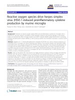
To characterize VEGF in vivo, aspirates of synovial fluid were
assessed for VEGF by ELISA. Compared with VEGF concen-
trations in healthy joints (36 pg/ml), VEGF concentrations in
the synovial fluid of patients with OA were significantly higher
(2,100 pg/ml, which was nearly 60-fold higher than healthy
synovial fluids; control versus OA, p ≤ 0.05 (Figure 1)).
The VEGF splice variants VEGF-121 and VEGF-165 are
detectable by splice-variant RT-PCR
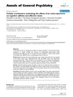
To determine whether the splice variants VEGF-121 (526 bp)
and VEGF-165 (658 bp) are expressed after stimulation of
chondrocytes with PMA, a known inducer of O
2
, semiquanti-
tative RT-PCR was performed (Figure 2). Both splice variants,
VEGF-121 (526 bp) and VEGF-165 (658 bp), are present in
immortalized chondrocytes and articular cartilage explants.
There were bold signals corresponding to VEGF-121 and fine
bands corresponding to VEGF-165. In general, more intense
signals were present in the chondrocytes compared with
those in cartilage explants. Stimulation with PMA (10 μg/ml)
increased the signals of the mRNAs encoding the VEGF
splice variants in monolayer chondrocytes and cartilage
explants.
Real-time RT-PCR revealed upregulation of VEGF,
VEGFR-1 and VEGFR-2 mRNA after stimulation with
ROS donors
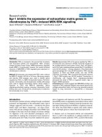
The levels of mRNA encoding VEGF and VEGF receptors
(VEGFR-1 (flt-1) and VEGFR-2 (KDR, flk-1)) were quantified
by real-time RT-PCR. After challenging with PMA, VEGF
mRNA was upregulated in monolayer chondrocytes (Figure
3a). The levels of VEGF were elevated dose-dependently, from
4.1-fold at 5 μg of PMA to 15.8-fold at 10 μg of PMA, with a
following decrease by 4.4-fold at 20 μg of PMA. Although
VEGFR-1 mRNA levels were increased only slightly (1.5-fold,
2.8-fold and 2.4-fold) after PMA stimulation (at 5, 10 and 20
μg, respectively), the levels of VEGFR-2 mRNA were elevated
2.6-fold, 10.4-fold and 4.6-fold at PMA concentrations of 5, 10
and 20 μg, respectively. Stimulation of cartilage explants with
10 and 20 μg of PMA upregulated VEGF mRNA by 2.3-fold
and 4.9-fold, respectively, compared with the control (Figure
3b). The level of VEGFR-2 mRNA was unaffected by 10 μg of
PMA and increased 2.7-fold by 20 μg of PMA. The level of
VEGFR-1 mRNA was undetectable in cartilage explants after
treatment with PMA.
Treatment of chondrocytes (monolayer) with SIN-1 resulted in
responses similar, in part, to those after PMA stimulation. After
treatment with 1, 10 and 20 μM of SIN-1, VEGF mRNA
expression was increased 2.2-fold, 19.6-fold and 17.2-fold,
respectively (Figure 3c), and VEGFR-2 mRNA expression was
elevated 2.0-fold, 15.4-fold and 13.5-fold, respectively. In con-
trast to PMA, 1, 10 and 20 μM of SIN-1 enhanced VEGFR-1
mRNA levels 1.2-fold, 9.2-fold and 8.1-fold, respectively, com-
pared with the control. In the cartilage explants, 3.1-fold and
9.0-fold increases in VEGF mRNA expression were apparent
after treatment with 10 and 20 μM of SIN-1, respectively
(Figure 3d). VEGFR-2 mRNA expression was increased by
1.8-fold and 6.1-fold at 10 and 20 μM of SIN-1, respectively.
In contrast to the undetectable level of VEGFR-1 in cartilage
explants after PMA treatment, VEGFR-1 mRNA levels slightly
increased after treatment with SIN-1 (1.2-fold and 1.8-fold
increases at 10 and 20 μM of SIN-1, respectively).
Figure 1
Comparison of the VEGF content of synovial fluid (as determined by ELISA) from healthy (Con) or OA patientsComparison of the VEGF content of synovial fluid (as determined by
ELISA) from healthy (Con) or OA patients. The level of VEGF is strongly
increased in the synovial fluid of OA patients. Results are shown as
mean ± standard error of the mean; n = 5 (Con) and n = 8 (OA). * p <
0.05. Con, control; OA, osteoarthritis; VEGF, vascular endothelial
growth factor.
Figure 2
VEGF splice variantsVEGF splice variants. Expression of VEGF-121 (526 bp) and VEGF-
165 (658 bp) in cartilage explants and immortalized chondrocytes after
stimulation with PMA (10 μg/ml) and SIN-1 (10 μM). The splice vari-
ants VEGF-121 and VEGF-165 are detected in cartilage explants and
C28/I2 cells. Con, control; PMA, phorbol myristate acetate; SIN-1, 3-
morpholino-sydnonimine hydrochloride; VEGF, vascular endothelial
growth factor.
Available online />Page 5 of 8
(page number not for citation purposes)
Enhanced VEGF production after stimulation with PMA
or SIN-1 (using ELISA)
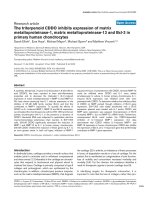
To determine whether the increased levels of VEGF mRNA
were reflected in the production of protein by chondrocytes,
an ELISA was performed to quantify the VEGF content in the
medium supernatants (Figure 4). Chondrocyte monolayer con-
trols released 1,100 pg of VEGF per 1 ml of medium and car-
tilage explant controls released 540 pg of VEGF per 1 ml of
medium. Treatment with PMA at concentrations of 5, 10 and
20 μg/ml increased VEGF production by 2.4-fold, 3.0-fold and
3.4-fold, respectively, in monolayer chondrocytes (control ver-
sus PMA, p ≤ 0.05) and treatment with SIN-1 at concentra-
tions of 1, 10 and 20 μM increased the level of VEGF by 1.7-
fold, 2.5-fold and 2.8-fold, respectively (control versus SIN-1,
p ≤ 0.05; Figure 4a). In cartilage explants, no increase in VEGF
was detected at 5 μg of PMA or 1 μM of SIN-1 (Figure 4b).
Using higher concentrations, VEGF production was increased
2.7-fold and 3.8-fold after treatment with PMA at concentra-
tions of 10 and 20 μg, respectively (control versus PMA, p ≤
0.05), and 2.4-fold and 3.7-fold after treatment with SIN-1 at
concentrations of 10 and 20 μM, respectively (control versus
SIN-1, p ≤ 0.05).
Increasing nitric oxide content of the medium
supernatant after stimulation with ROS donors
PMA dose-dependently increased the NO content of culture
medium from chondrocytes and, to a lesser extent, cartilage
explants. In monolayer cultures, NO levels were increased 2.6-
fold, 3.2-fold and 4.4-fold at 5, 10 and 20 μg/ml of PMA com-
pared with the control (control versus PMA, p ≤ 0.05; Figure
5a). By contrast, the cartilage explants showed no response to
low-dose PMA stimulation (5 μg) and only 1.7-fold and 2.4-
fold increases using 10 and 20 μg of PMA, respectively (con-
trol versus PMA, p ≤ 0.05; Figure 5b). After treatment with
SIN-1, the NO content showed similar results but the effect
was more extended (Figure 5a): in monolayer chondrocytes,
the NO content was 2.1-fold, 24.8-fold and 41.9-fold higher
than control after treatment with 1, 10 and 20 μM of SIN-1
(control versus SIN-1, p ≤ 0.05). In the cartilage explants, the
effects were attenuated compared with the monolayer cul-
tures, with 1.7-fold, 2.5-fold and 3.6-fold increases in NO
content reported after treatment with 1, 10 and 20 μM of SIN-
1, respectively (control versus SIN-1, p ≤ 0.05; Figure 5b).
Figure 3
Quantitative mRNA expression of VEGF, VEGFR-1 and VEGFR-2 normalized to the control (n = 1)Quantitative mRNA expression of VEGF, VEGFR-1 and VEGFR-2 normalized to the control (n = 1). Stimulation of immortalized chondrocytes (a)
and (c) and articular cartilage explants (b) and (d) with PMA (μg per 1 ml of medium) or SIN-1 (μM). VEGFR-1 is undetectable in (b). mRNA expres-
sion of VEGF, VEGFR-1 and VEGFR-2 is upregulated after stimulation with reactive oxygen species donors. Results are shown as mean ± standard
error of the mean for five separate experiments. * p < 0.05 versus control. G3PDH, glyceroaldehyde-3-phosphate dehydrogenase; PMA, phorbol
myristate acetate; SIN-1, 3-morpholino-sydnonimine hydrochloride; VEGF, vascular endothelial growth factor; VEGFR, VEGF receptor.
Arthritis Research & Therapy Vol 8 No 6 Fay et al.
Page 6 of 8
(page number not for citation purposes)
Discussion
Our findings show that the level of VEGF in synovial joint fluids
from patients suffering from OA is 60-fold higher than healthy
joints. Moreover, our in vitro model revealed that VEGF mRNA
and protein levels and VEGF receptors are increased by PMA
or SIN-1 stimulation in chondrocytes and human articular car-
tilage explants. We conclude that the presence of ROS, or
activation of production of O
2
, is responsible for the observed
results. Thus, VEGF accumulation in the synovial fluid is, at
least in part, cartilage-derived.
Inflammation in OA is known to be associated with activation
of host angiogenesis [24]. VEGF is one of the most potent
proangiogenic stimuli of neovascularization. Furthermore, the
capacity of VEGF to mediate chemotaxis, raise vascular per-
meability for neutrophil influx and activate MMPs in chondro-
cytes suggests a central function in catabolic pathways of OA
joints [9,25]. In addition to the involvement of VEGF in the
development of OA, VEGF has biological importance in carti-
lage metabolism during rheumatoid arthritis. The initial growth
and invasion of the synovial pannus tissue contributes to the
subsequent cartilage destruction. Blockade of VEGFR-I
resulted in reduced intensity of clinical manifestations and pre-
vented joint destruction in a mouse model of rheumatoid arthri-
tis [26]. It is obvious that tissues other than cartilage are
participating in these processes, especially the surrounding
synovial tissue. This reflects the findings of Felson and
coworkers [27], who declared OA to be a disease involving
the whole joint. In summary, these data support the role of
VEGF in mediating destructive processes in articular joints
and encouraged us to investigate the relationship between
VEGF expression and ROS in articular cartilage. Focusing on
other cell types, such as glomerular podocytes, endothelial
cells and skeletal muscle fibres, a correlation between ROS
and VEGF is described, but the results were contradictory
[28,29].
Figure 4
The VEGF content of medium supernatant (as determined by ELISA)The VEGF content of medium supernatant (as determined by ELISA).
Stimulation of immortalized chondrocytes (a) and articular cartilage
explants (b) with PMA (μg per 1 ml of medium) or SIN-1 (μM). The level
of VEGF protein is increased after stimulation with PMA or SIN-1.
Results are shown as mean ± standard error of the mean for five sepa-
rate experiments. * p < 0.05 versus Con. Con, control; PMA, phorbol
myristate acetate; SIN-1, 3-morpholino-sydnonimine hydrochloride;
VEGF, vascular endothelial growth factor.
Figure 5
The NO content of medium supernatant normalized to the Con (n = 1)The NO content of medium supernatant normalized to the Con (n = 1).
Stimulation of immortalized chondrocytes (a) and articular cartilage
explants (b) with PMA (μg per 1 ml of medium) or SIN-1 (μM). The level
of NO is increased after stimulation with reactive oxygen species
donors. Results are shown as mean ± standard error of the mean for
five separate experiments. * p < 0.05 versus Con. Con, control; NO,
nitric oxide; PMA, phorbol myristate acetate; SIN-1, 3-morpholino-syd-
nonimine hydrochloride.
Available online />Page 7 of 8
(page number not for citation purposes)
Here, we demonstrate increased VEGF mRNA expression and
production in cultured human chondrocytes and articular car-
tilage explants after challenge by ROS donors. We conclude
that the observed increase in VEGF content in synovial fluid of
OA joints is produced partly by articular chondrocytes and
consistent with previous findings from this and other laborato-
ries, which showed an increase in VEGF content in OA carti-
lage [8,10,11]. The observation that the concentration of
VEGF is positively correlated to joint destruction and vascular-
ization of synovial membrane in rheumatoid arthritis [30] sug-
gests the potential impact of VEGF in the pathophysiology of
OA.
Our demonstration of ROS-mediated induction of the ang-
iogenic factor VEGF in human chondrocytes and articular car-
tilage explants is consistent with prior reports showing that
NO stimulates VEGF production in chondrocytes [31]. How-
ever, we observed different responses to the two ROS donors.
The NO content after stimulation with SIN-1 was up to tenfold
higher than after stimulation with PMA and only SIN-1 induced
upregulation of VEGFR-1 mRNA. It could be speculated that
PMA induces enhanced production of O
2
within the cell,
whereas SIN-1 spontaneously decomposes to O
2
. A possible
mechanism by which PMA increases the level of VEGF is the
activation of AP-1 (activator protein 1), with subsequent acti-
vation of the TPA (12-O-tetradecanoylphorbol-13-acetate)
element of the VEGF promoter. The mode of action of SIN-1
is presumably by an increase in HIF-1α (hypoxia inducible fac-
tor subunit alpha). Future studies are necessary to investigate
the signal transduction pathways of PMA and SIN-1 in
chondrocytes.
Conclusion
By amplifying distinct ROS-dependent destructive pathways
in cartilage and joints, VEGF seems to have a crucial role in the
degeneration of articular cartilage by promoting neoangiogen-
esis in the emerging synovial tissue and stimulating cartilage
matrix-degrading pathways. Interestingly, do the splice vari-
ants of young donors differ from those of old donors? VEGF-
121 and VEGF-165 have high angiogenic potencies and lead
to a rapid and strong invasion of blood vessels. Therefore, in
the long term, a new understanding of the mechanisms
responsible for cartilage destruction in OA might enable the
development of novel strategies for intervention and treatment.
The functional knockout of VEGF with, for example, soluble
receptors might be potential therapeutic target for the treat-
ment of OA.
Competing interests
The authors declare that they have no competing interests.
Authors' contributions
JV, DV, CW, BK and TP performed the experiments and con-
tributed to the draft manuscript; DV and TP contributed
equally to the present work. MG established the C28/I2 cell
line, designed part of the study and contributed to the draft
manuscript. The manuscript has been read and approved by
all authors.
Acknowledgements
The authors would like to thank Inka Kronenbitter, Ursula Mundt, Frank
Lichte and Sonja Seiter for their excellent technical assistance. The T-
handle bar to perform tissue harvest from the donors' knees was a gen-
erous gift from Arthrex
®
, Karlsfeld, Germany. This work was funded, in
part, by the Research Promotion of Faculty of Medicine, Kiel University,
Kiel, Germany, and Deutsche Forschungsgemeinschaft (DFG; Pu 214/
4-2, Pu 214/3-2 and Pu 214/5-2). MG's research was funded by a grant
from the National Institutes of Health (R01-AG22021).
References
1. Fernandes JC, Martel-Pelletier J, Pelletier JP: The role of
cytokines in osteoarthritis pathophysiology. Biorheology 2002,
39:237-246.
2. Burrage PS, Mix KS, Brinckerhoff CE: Matrix metalloproteinases:
role in arthritis. Front Biosci 2006, 11:529-543.
3. Ferrara N: Role of vascular endothelial growth factor in the reg-
ulation of angiogenesis. Kidney Int 1999, 56:794-814.
4. Neufeld G, Cohen T, Gengrinovitch S, Poltorak Z: Vascular
endothelial growth factor (VEGF) and its receptors. FASEB J
1999, 13:9-22.
5. Firestein GS: Starving the synovium: angiogenesis and inflam-
mation in rheumatoid arthritis. J Clin Invest 1999, 103:3-4.
6. Robinson CJ, Stringer SE: The splice variants of vascular
endothelial growth factor (VEGF) and their receptors. J Cell
Sci 2001, 114:853-865.
7. Thomas KA: Vascular endothelial growth factor, a potent and
selective angiogenic agent. J Biol Chem 1996, 271:603-606.
8. Pufe T, Petersen W, Tillmann B, Mentlein R: The splice variants
VEGF121 and VEGF189 of the angiogenic peptide vascular
endothelial growth factor are expressed in osteoarthritic
cartilage. Arthritis Rheum 2001, 44:1082-1088.
9. Pufe T, Harde V, Petersen W, Goldring MB, Tillmann B, Mentlein
R: Vascular endothelial growth factor (VEGF) induces matrix
metalloproteinase expression in immortalized chondrocytes.
J Pathol 2004, 202:367-374.
10. Pfander D, Kortje D, Zimmermann R, Weseloh G, Kirsch T,
Gesslein M, Cramer T, Swoboda B: Vascular endothelial growth
factor in articular cartilage of healthy and osteoarthritic human
knee joints. Ann Rheum Dis 2001, 60:1070-1073.
11. Enomoto H, Inoki I, Komiya K, Shiomi T, Ikeda E, Obata K, Mat-
sumoto H, Toyama Y, Okada Y: Vascular endothelial growth fac-
tor isoforms and their receptors are expressed in human
osteoarthritic cartilage. Am J Pathol 2003, 162:171-181.
12. Henrotin Y, Kurz B, Aigner T: Oxygen and reactive oxygen spe-
cies in cartilage degradation: friends or foes?
Osteoarthritis
Cartilage 2005, 13:643-654.
13. Rice-Evans CA: Techniques in free radical research. In Labora-
tory techniques in biochemistry and molecular biology Edited by:
Burdon RH. Amsterdam: Elsevier; 1991:20.
14. Henrotin YE, Bruckner P, Pujol JP: The role of reactive oxygen
species in homeostasis and degradation of cartilage. Osteoar-
thritis Cartilage 2003, 11:747-755.
15. Panasyuk A, Frati E, Ribault D, Mitrovic D: Effect of reactive oxy-
gen species on the biosynthesis and structure of newly syn-
thesized proteoglycans. Free Radic Biol Med 1994,
16:157-167.
16. Del Carlo M Jr, Loeser RF: Nitric oxide-mediated chondrocyte
cell death requires the generation of additional reactive oxy-
gen species. Arthritis Rheum 2002, 46:394-403.
17. Ayache N, Boumediene K, Mathy-Hartert M, Reginster JY, Henrotin
Y, Pujol JP: Expression of TGF-betas and their receptors is dif-
ferentially modulated by reactive oxygen species and nitric
oxide in human articular chondrocytes. Osteoarthritis Cartilage
2002, 10:344-352.
18. Mathy-Hartert M, Martin G, Devel P, Deby-Dupont G, Pujol JP,
Reginster JY, Henrotin Y: Reactive oxygen species downregu-
Arthritis Research & Therapy Vol 8 No 6 Fay et al.
Page 8 of 8
(page number not for citation purposes)
late the expression of pro-inflammatory genes by human
chondrocytes. Inflamm Res 2003, 52:111-118.
19. Li JM, Mullen AM, Yun S, Wientjes F, Brouns GY, Thrasher AJ,
Shah AM: Essential role of the NADPH oxidase subunit
p47(phox) in endothelial cell superoxide production in
response to phorbol ester and tumor necrosis factor-alpha.
Circ Res 2002, 90:143-150.
20. de Groot H, Hegi U, Sies H: Loss of alpha-tocopherol upon
exposure to nitric oxide or the sydnonimine SIN-1. FEBS Lett
1993, 315:139-142.
21. Goldring MB, Birkhead JR, Suen LF, Yamin R, Mizuno S, Glowacki
J, Arbiser JL, Apperley JF: Interleukin-1 beta-modulated gene
expression in immortalized human chondrocytes. J Clin Invest
1994, 94:2307-2316.
22. Muehleman C, Bareither D, Huch K, Cole AA, Kuettner KE: Prev-
alence of degenerative morphological changes in the joints of
the lower extremity. Osteoarthritis Cartilage 1997, 5:23-37.
23. Ailland J, Kampen WU, Schunke M, Trentmann J, Kurz B: Beta irra-
diation decreases collagen type II synthesis and increases
nitric oxide production and cell death in articular
chondrocytes. Ann Rheum Dis 2003, 62:1054-1060.
24. Cerimele F, Brown LF, Bravo F, Ihler GM, Kouadio P, Arbiser JL:
Infectious angiogenesis: Bartonella bacilliformis infection
results in endothelial production of angiopoetin-2 and epider-
mal production of vascular endothelial growth factor. Am J
Pathol 2003, 163:1321-1327.
25. Gruber BL, Marchese MJ, Kew R: Angiogenic factors stimulate
mast-cell migration. Blood 1995, 86:2488-2493.
26. De Bandt M, Ben Mahdi MH, Ollivier V, Grossin M, Dupuis M,
Gaudry M, Bohlen P, Lipson KE, Rice A, Wu Y, et al.: Blockade of
vascular endothelial growth factor receptor I (VEGF-RI), but
not VEGF-RII, suppresses joint destruction in the K/BxN
model of rheumatoid arthritis. J Immunol 2003,
171:4853-4859.
27. Felson DT, Lawrence RC, Dieppe PA, Hirsch R, Helmick CG, Jor-
dan JM, Kington RS, Lane NE, Nevitt MC, Zhang Y, et al.: Oste-
oarthritis: new insights. Part 1: the disease and its risk factors.
Ann Intern Med 2000, 133:635-646.
28. Lee EY, Chung CH, Kim JH, Joung HJ, Hong SY: Antioxidants
ameliorate the expression of vascular endothelial growth fac-
tor mediated by protein kinase C in diabetic podocytes. Neph-
rol Dial Transplant 2006, 21:1496-1503.
29. Kosmidou I, Xagorari A, Roussos C, Papapetropoulos A: Reactive
oxygen species stimulate VEGF production from C(2)C(12)
skeletal myotubes through a PI3K/Akt pathway. Am J Physiol
Lung Cell Mol Physiol 2001, 280:L585-L592.
30. Nagashima M, Yoshino S, Ishiwata T, Asano G: Role of vascular
endothelial growth factor in angiogenesis of rheumatoid
arthritis. J Rheumatol 1995, 22:1624-1630.
31. Turpaev K, Litvinov D, Dubovaya V, Panasyuk A, Ivanov D,
Prassolov V: Induction of vascular endothelial growth factor by
nitric oxide in cultured human articular chondrocytes. Bio-
chimie 2001, 83:515-522.