Báo cáo khoa học: " Stereotactic radiosurgery may contribute to overall survival for patients with recurrent head and neck carcinoma" pptx
Bạn đang xem bản rút gọn của tài liệu. Xem và tải ngay bản đầy đủ của tài liệu tại đây (1.36 MB, 9 trang )
Kawaguchi et al. Radiation Oncology 2010, 5:51
/>Open Access
RESEARCH
© 2010 Kawaguchi et al; licensee BioMed Central Ltd. This is an Open Access article distributed under the terms of the Creative Com-
mons Attribution License ( which permits unrestricted use, distribution, and reproduc-
tion in any medium, provided the original work is properly cited.
Research
Stereotactic radiosurgery may contribute to overall
survival for patients with recurrent head and neck
carcinoma
Koji Kawaguchi*
1
, Kengo Sato
2
, Akihisa Horie
1
, Susumu Iketani
3
, Hiroyuki Yamada
1
, Yasunori Nakatani
3
, Junichi Sato
1
and Yoshiki Hamada
1
Abstract
Background: The aim of this study is to examine the effect of stereotactic radiosurgery (SRS) in the treatment of
advanced, recurrent lesions for head and neck carcinoma both with and without lymph node involvement.
Methods: Between April 2006 and July 2007, 22 patients (mean age 67 years) with advanced, recurrent head and neck
carcinoma were treated with stereotactic radiosurgery. All of the patients except one had biopsy confirmed disease
prior to stereotactic radiosurgery. Patients included 3 rT2, 8 rT3, and 9 rT4; 8 of the patients had lymph node metastases.
Marginal SRS doses were 20-42 Gy delivered in two to five fractions. Starting one month after SRS, all patients received
S-1 oral chemotherapy for one year.
Results: At an overall median follow-up of 24 months (range, 4-39 months), for the 14 locally recurrent patients
without lymph node metastases, 9 patients (64.3%) had a complete response (CR), 1 patient (7.1%) had a partial
response (PR), 1 patient (7.1%) had stable disease (SD), and 3 patients (21.4%) had progressive disease (PD). For the 8
patients with lymph node metastases, 1 patient with a single retropharyngeal (12.5%) had CR; the remaining 7 patients
(87.5%) all progressed. Nine patients have died from their cancer. The overall actuarial 2-year survival for the patients
with and without lymph node metastases is 12.5% and 78.6%, respectively.
Conclusions: These results show the benefit of stereotactic radiosurgery salvage treatment for advanced, recurrent
lesions, without lymph node metastases in previously irradiated head and neck cancer.
Background
The majority of head and neck region squamous cell car-
cinomas present at an advanced stage and are treated
with a combined-modality approach that often includes
surgery, radiation therapy, and chemotherapy. Despite
such aggressive approaches, advanced squamous cell car-
cinoma of the head and neck tends to recur locoregion-
ally and, thus, presents a significant clinical challenge [1-
3]. Surgery and/or conventional chemoradiotherapy sal-
vage is difficult in advanced, recurrent lesions of head
and neck carcinoma given the proximity of critical
organs. Similarly, radical treatment for wide recurrent
lesions is limited by overall radiation doses for the body
and the possibility of severe post-operative dysfunction.
In cases where further surgery is not feasible, reirradia-
tion offers the potential to gain locoregional control and
achieve palliation [4].
Several studies have confirmed the feasibility of reirra-
diation of recurrent head and neck tumors, with curative
intent using external beam radiation therapy (EBRT) [5-
8]. More recently, stereotactic radiosurgery (SRS) has
been employed in the treatment of head and neck cancers
both in primary cases [9,10] and in recurrent cases [11-
15]. The CyberKnife
®
(Accuray Incorporated, Sunnyvale,
California, USA) is a frameless robotic radiosurgery sys-
tem that has been utilized by numerous clinicians around
the world to treat intracranial and extracranial tumors
[9,13,16,17]. The CyberKnife image-guided radiosurgical
system can deliver isocentric or non-isocentric beams
with high precision and high dose conformity [18]. These
* Correspondence:
1
Department of Oral and Maxillofacial Surgery, Tsurumi University, School of
Dental Medicine. 2-1-3 Tsurumi, Tsurumi-ku, Yokohama, 230-8501, Japan
Full list of author information is available at the end of the article
Kawaguchi et al. Radiation Oncology 2010, 5:51
/>Page 2 of 9
abilities are especially important when treating irregu-
larly shaped tumors, or those in difficult locations close
to critical structures, as is the case with many of the
patients with advanced, recurrent head and neck cancer.
Here, we report on the tumor response and overall sur-
vival of stereotactic radiosurgery treatment using the
CyberKnife for advanced, recurrent head and neck carci-
noma lesions both with and without lymph node metas-
tases.
Methods
Between April 2006 and July 2007, 22 patients with
advanced, recurrent head and neck squamous cell carci-
noma were treated at the Yokohama CyberKnife Center,
Yokohama, Japan. All patients included in this study com-
pleted an informed consent form. Patients were exam-
ined using PET-CT, MRI, and Ultrasound. Prior to
radiation therapy, biopsies were performed for all
patients, except for one for whom the recurrence was
located deep in their temple muscle. For that patient,
diagnosis was determined based upon the high level of
uptake observed on PET scan. We assumed that this was
a metastasis from the original squamous cell carcinoma
that was located on the floor of their oral cavity. Hence,
we denoted this patient as M1.
Prior to stereotactic radiosurgery, 21 patients (95.5%)
had surgical treatment and 13 of those patients received
post-operative chemo-radiotherapy. The remaining
patient (4.7%) received prior irradiation, but neither prior
surgery nor chemotherapy. The prior conventional irradi-
ation was delivered as a wide field with doses of 40-65 Gy
delivered in 1.5-2.0 Gy daily fractions. In all cases, the
SRS was delivered within the prior irradiation field. The
overall interval between prior treatment and SRS treat-
ment was a median 11 months (range, 4-21 months). For
those patients that received prior irradiation, SRS treat-
ment occurred a median 11 months (range, 4-21 months)
later. For those that received surgery alone, SRS treat-
ment occurred a median 14 months (range, 7-26 months)
later. Table 1 provides an overview of the patient charac-
teristics.
Stereotactic radiosurgery was delivered with the
CyberKnife
®
(Accuray Inc., Sunnyvale, USA), an X-band
linear accelerator with an overall system targeting error
of less than 1 mm [18,19]. The lightweight linear accelera-
tor is capable of irradiating the target from 120 different
directions using image-guidance based on a treatment
plan created using a CT volume [13,20,21]. To assist with
treatment planning, the CT image was also fused with an
MRI or PET-CT image as applicable. During both treat-
ment planning and delivery, patients were imaged while
wearing a custom-made mouthpiece, to immobilize the
moving parts of the mouth, and a thermoplastic mask
fixed to the treatment couch, to minimize head move-
Table 1: Patient characteristics.
Sex Number (%)
Male 8 (36%)
Female 14 (64%)
Age, years
Median (range) 67 (42-91)
Initial treatment
Surgery alone 8 (36%)
Surgery + Post-operative
Chemo-Radiation
13 (59%)
Radiation 1 (5%)
Prior radiation dose (Gy)
Total (daily) dose range 40-65 (1.5-2)
Histology
Squamous cell carcinoma 21 (95.5%)
M1 case 1 (4.5%)
Regions of recurrence or
metastases
Tongue 7 (31.8%)
Mandible 5 (22.7%)
Maxilla 3 (13.6%)
Maxillary sinus 3 (13.6%)
Soft palate 2 (9.1%)
Limited with lymph node 1 (4.5%)
Temporal muscle 1 (4.5%)
Clinical Stage
rT0N1M0 1 (4.5%)
Kawaguchi et al. Radiation Oncology 2010, 5:51
/>Page 3 of 9
ment. Treatment was administered depending upon the
configuration and volume of the tumor as determined by
the treating radiation oncologist, neurosurgeon and oral
and maxillofacial surgeon. Dose constraints were applied
to nearby critical structures based upon the total dose
and fractionation scheme. Specifically, the dose to brain
stem, optic nerve, optic chiasm, retina, and spinal cord
were each limited to 21-25 Gy; the dose to the carotid
artery, esophagus, and larynx were each limited to 30-35
Gy; and the dose to the eye lens was limited to 7-10 Gy.
The prescribed dose of radiation was administered to the
clinical target volume without the addition of any margin,
corresponding to the 80-85% isodose contour. In the
cases of lymph nodes metastases, the lymph nodes were
treated. In general, the dose to those lesions which previ-
ously received irradiation was reduced by 20% from that
of those patients for which no prior radiation was deliv-
ered to the lesion. In those cases where the PTV was
more than 30 cc the dose was reduced by 30%. Overall,
patients were treated with a median marginal dose of
33.73 Gy (range, 20-42 Gy) in two to five fractions with
treatment delivered over consecutive days. The median
gross lesion diameter was 36.63 mm (range, 15.21-58.65
mm). The median irradiated volume was 24.5 cm
3
(range,
3.4-74.4 cm
3
). Table 2 provides a summary of the treat-
ment details.
In addition to stereotactic radiosurgery, a low dose of
oral chemotherapy S-1 (oral 5-FU prodrug) (Taiho Phar-
maceutical Company Limited, Tokyo, Japan) was admin-
istered to control micro-lymph node metastases and
distant metastases. The S-1 treatment began one month
after SRS and consisted of 40-80 mg/body of S-1 for 2
weeks followed by a one week break; the treatment
sequence was repeated for one year.
Following stereotactic radiosurgery, patients were
monitored at either Tsurumi University Hospital or
Toshiba Rinkan Hospital by oral and maxillofacial sur-
geons and at Yokohama CyberKnife Center by radiation
oncologists. The clinical follow-up interval was every 2
weeks for the first 3 months, and every 4 weeks thereafter
until the patient reached 2 years follow-up. Treatment
outcome was assessed based on the Response Evaluation
Criteria in Solid Tumors (RECIST) [22]. Response to
treatment was evaluated using MRI at one month follow-
up, contrast-enhanced CT at two months follow-up, PET-
CT and MRI at three months follow-up, and MRI or PET-
CT every three months thereafter. Toxicities were graded
using the National Cancer Institute Common Toxicity
Criteria Scale, Version 3.0. Overall survival after stereot-
actic radiosurgery was determined by Kaplan-Meier sur-
vival analysis.
Results
Clinical Outcomes
Twenty-two advanced, recurrent head and neck cancer
patients were treated with SRS. In these advanced, recur-
rent patients treatment options were limited and the
decision to treat these patients with SRS was based upon
Table 2: Treatment Characteristics: Summary of treatment
dose and treated tumor volume.
Dose (Gy) Number of Patients (%)
20 - 29 1 (4.5%)
30 - 34 7 (31.8%)
35 - 39 10 (45.5%)
40 - 42 4 (18.2%)
Median (range) 33.7 (20-42)
Tumor volume (cm3)
Median 24.5
Range 3.4 - 74.4
Tumor diameter (mm)
Median 36.63
Range 15.21 - 58.65
rT0N0M1 1 (4.5%)
rT2N0M0 3 (13.6%)
rT3N0M0 5 (22.7%)
rT3N1M0 2 (9.1%)
rT3N2M0 1 (4.5%)
rT4N0M0 5 (22.7%)
rT4N1M0 2 (9.1%)
rT4N2M0 2 (9.1%)
Table 1: Patient characteristics. (Continued)
Kawaguchi et al. Radiation Oncology 2010, 5:51
/>Page 4 of 9
a variety of issues including the nature of the tumor
recurrence, prior treatment approaches and patient pref-
erence. Specifically, the recurrent tumors were solid
masses that were well suited to a radiosurgical treatment
given the proximity of critical organs. For those patients
that previously received conventional radiotherapy (14/
22), the ability to target the radiation dose specifically to
the tumor and limit the dose to surrounding, previously
irradiated, tissue was also a strong indicator for SRS.
Lastly, the patients strongly preferred a treatment option
that did not require hospitalization.
Table 3: Clinical Outcomes: Summary of tumor response by RESIST criteria.
3A
N-patients Number of Patients (%)
Complete Response 9 (64.3%)
Partial Response 1 (7.1%)
Stable Disease 1 (7.1%)
Progressed Disease 3 (21.4%)
N+ patients
Complete Response 1 (12.5%)
Progressed Disease 7 (87.5%)
3B
Clinical Stage Complete Response Partial Response Stable Disease Progressed Disease
rT0N1M0 1
rT0N0M1 1
rT2N0M0 3
rT3N0M0 3 1 1
rT3N1M0 2
rT3N2M0 1
rT4N0M0 3 2
rT4N1M0 2
rT4N2M0 2
(A) Tumor response for patients without lymph node metastases (N-) and for patients with lymph node metastases (N+). (B) Tumor response
based on clinical stage.
Kawaguchi et al. Radiation Oncology 2010, 5:51
/>Page 5 of 9
The majority of patients (12/22) received SRS treat-
ment as outpatients with curative intent. Ten patients
(45%), however, received treatment with palliative intent
while in terminal care at the hospital. At an overall
median follow-up of 24 months (range, 4-39 months), 9
patients have died from their cancer. One additional
patient died from acute cardiac insufficiency. For surviv-
ing patients, the median follow-up was 32 months (range,
27-39 months).
Table A3A and Table B3B provide a summary of tumor
response as assessed by RECIST criteria based on lymph
node metastases and clinical stage, respectively. Specifi-
cally, for the 14 locally recurrent patients without lymph
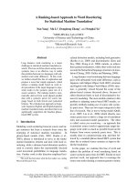
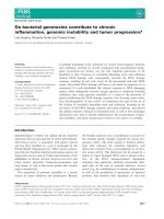
Figure 1 Kaplan-Meier plot showing the overall survival rate for patients without lymph node metastases (N-) and for patients with lymph
node metastases (N+).
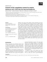
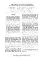
Figure 2 Case study of a 59 year old patient with a locally recur-
rent lesion with pterygopalatine fossa 3-years after maxillecto-
my. (A) Prior to treatment. (B) Treatment planning image. A total dose
of 40 Gy was delivered in 5 fractions. (C) At 4-months post-treatment a
complete response occurred.
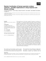
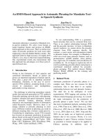
Figure 3 Case study of a T3N2cM0 patient, with tongue carcino-
ma that recurred as a distant metastasis in part of his temporal
muscle 5 years after surgery. (A) Treatment planning image. A total
dose of 30 Gy was delivered in 3 fractions. (B) Pre-treatment the recur-
rence is visible in his temporal muscle as denoted by the green circle.
(C) At 3-months post-treatment the lesion was stable (green circle).
Kawaguchi et al. Radiation Oncology 2010, 5:51
/>Page 6 of 9
node metastases, 9 patients (64.3%) had a complete
response (CR), 1 patient (7.1%) had a partial response
(PR), 1 patient (7.1%) had stable disease (SD), and 3
patients (21.4%) had progressive disease (PD). The three
PD patients developed new lymph node metastases on
the side opposite of SRS treatment. All three of these PD
patients subsequently died from these late-lymph node
metastases. For the 8 patients with lymph node metasta-
ses, one 1 patient, with only 1 retropharyngeal lymph
node metastasis (12.5%) had CR; the remaining 7 patients
(87.5%) all progressed. These seven patients each had 2 or
3 lymph node metastases located in their necks; upon
progression they did not undergo additional treatment.
Overall, at a median 2-years follow-up, 10 (45.5%) of
the 22 severe recurrent cases maintained a complete
response. All 10 of these patients have returned to society
and regained quality of life. The overall actuarial 2-year
survival for the locally recurrent patients with and with-
out lymph node metastases is 12.5% and 78.6%, respec-
tively (Figure 1). This difference was statistically
significant with p = 0.000019 by the log-rank test.
Complications
The first month following SRS 17 patients (77.3%) experi-
enced Grade 2 xerostomia and decreased taste; 5 patients
(22.7%), all of which were rT4 cases, experienced Grade 3
xerostomia and decreased taste. Of the 5 patients that
experienced Grade 3 toxicity, one received prior radiation
therapy (total dose 65 Gy) and four received prior surgery
followed by radiation therapy (total dose 50-56 Gy). After
the two month follow-up, there have been no serious
complications associated with the SRS re-irradiation.
Fourteen of the patients who had previously received
external beam radiation experienced Grade 1 (11
patients) and Grade 2 (3 patients) osteoradionecrosis at
10-18 months after SRS. None of the surgery-only
patients experienced any late complications.
Case reports
Case One (Figure 2): A 59-year-old male that was found
to have a locally recurrent lesion with pterygopalatine
fossa 3-years after maxillectomy. SRS was delivered to a
total dose of 40 Gy in 5 fractions. The patient experienced
Grade 2 xerostomia and decreased taste without osteora-
dionecrosis within the first month of treatment. Four
months after SRS a complete clinical response occurred.
At 30 months after SRS there is no evidence of recur-
rence.
Case Two (Figure 3): A patient with T3N2cM0 tongue
carcinoma that recurred as a distant metastasis in part of
his temporal muscle 5 years after surgery. SRS was deliv-
ered to a total dose of 30 Gy in 3 fractions. Three months
after SRS the lesion was assessed as stable and remains as
stable disease at 24 months. The patient experienced hair
loss at the temporal part of his head within 3 months after
SRS, after which the hair grew back.
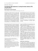
Case Three (Figure 4): A patient with T3N2cM0
tongue carcinoma that recurred with a retropharyngeal
lymph node metastasis 5 years after surgery. SRS was
delivered to a total dose of 23 Gy in 2 fractions. Three
months after SRS the lesion exhibited a complete clinical
response. At 26 months after SRS there is no evidence of
recurrence. This patient was the only one of eight treated
lymph node metastases in our study that had a complete
response.
Case Four (Figure 5): A patient with rT3N2cM0
tongue carcinoma recurred 6 months after irradiated
Figure 4 Case study of a patient with T3N2cM0 tongue carcinoma
that recurred as a retropharyngeal lymph node metastasis 5
years after surgery. (A) Pre-treatment the lymph node metastasis is
indicated by the green circle. (B) At 5-months post-treatment a com-
plete response occurred (green circle).
Figure 5 Case study of a patient with rT3N2cM0 tongue carcino-
ma that recurred 6 months after irradiation with 50 Gy in 25 frac-
tions by conventional external beam.
Kawaguchi et al. Radiation Oncology 2010, 5:51
/>Page 7 of 9
with 50 Gy in 25 fractions by conventional external beam.
SRS was delivered to a total dose of 35 Gy in 5 fractions.
Three months after SRS for the recurrent lesion of the
tongue and N+ lesions, these lesions increased in size and
were assessed as progressive disease. The patient experi-
enced Grade 3 xerostomia and decreased taste as well as
Grade 2 osteoradionecrosis of the mandible bone. This
patient died six months after SRS as a result of a large
number of lymph node metastases.
Discussion
Our results demonstrate that CyberKnife frameless ste-
reotactic radiosurgery for patients with recurrent head
and neck carcinoma is feasible and safe in the setting of
previous irradiation. In the case of local recurrence with-
out lymph node metastases, 9 out of 14 (64.3%) patients
had a complete response with a 2-year overall survival
rate of 78.6%. Overall, 10 out of 22 (45.5%) of the
advanced, recurrent patients maintain a complete
response at a median 2-years follow-up.
Several stereotactic radiosurgery results, also known as
fractionated stereotactic radiotherapy (fSRT), for re-irra-
diation of recurrent head and neck carcinomas have been
reported [13-15,23-25]. The complete response rates for
these studies vary from 8.6-54% with 2-year overall sur-
vival rates ranging from 14.3-41% and 1-year overall sur-
vival rates of 18-52.1% (see Table 4). In addition, one
study has reported 2-year overall survival rates of 58% for
reirradiation of recurrent head and neck cancer using
IMRT. As the ranges of these outcomes suggest, the het-
erogeneity between these various studies is large. Various
factors, including tumor stage, tumor volume, adequate
irradiation dose, prior treatment, and anatomical site
complexly, influenced these reported outcomes. In sev-
eral of these studies [13,14,23] patients with lymph node
metastases have been included, but the reported out-
comes have not been divided between those patients with
and without lymph node metastases. Our reported over-
all survival of 78.8% for our subset of patients without
lymph node involvement exceeds all of the prior pub-
lished survival rates.
Table 4: Overview of prior stereotactic radiosurgery results for the reirradiation of recurrent head and neck carcinoma.
Study Patients (#) SRS median
total dose/fx
Follow-up
median,
range
(months)
Tumor size
median,
range (cc)
Prior
irradiation
Dose, median,
range (Gy)
Toxicity Complete
Response
Rate
Overall survival
Voynov et al. [13] 22 24/1-8 19, 11-40* 19.1, 2.5-140.3 97.8, 70.1-
190.3 (BED
10
)
No Grade 4+ - 22% at 2-yrs
Heron et al. [15] 25 25-44/5 NS 44.8, 4.2-217 66-69.2 No Grade 3+ 8.6% 18% at 1-yr
Roh et al. [14] 36 30/3-5 17.3 22.6, 0.2-114.9 70.2, 39.6-
134.4
No Grade 4+
36% Grade 3
42.9% 52.1% at 1-yr,
30.9% at 2-yrs
Rwigema et al. [23] 85 35/1-5 6, 1.3-39 25.1, 2.5-162 70, 32-170.7 No Grade 4+
4.7% Grade 3
34% 48% at 1-yr, 16%
at 2-yrs
Sulman et al. [31]78
§
IMRT 60 Gy 25, 0-81 64.1, 2.9-425.4 60, 16-75 20% severe
Including 1%
Grade 5
- 58% at 2-yrs
Siddiqui et al. [25]44
§
† 13-18/1 or
36-48/5-8
6.8, 1.5-48 15.5, 1.7-155‡ 63.5, 50.4-74‡ 6.7% Grade 3‡
9% Grade 4‡
31% 38.1% at 1-yr
14.3% at 2-yrs
Unger et al. [24] 65 30/2-5 16* 75, 7-276 67, 32-120 11% Grade 4+ 54% 41% at 2-yrs
Current Study 22 (14 N-, 8 N+) 33.7/2-5 24, 4-39 24.5, 3.4-74.4 40-65 No Grade 4+
22.7% Grade 3
45.4%
(N- 64.3%,
N+ 12.5%)
N- 78.6% at 2-yrs
N+ 12.5% at 2-yrs
* Surviving patients
† Additional non-recurrent patients treated, data presented for recurrent patients only unless otherwise noted.
‡ Includes non-recurrent patients.
§ Includes patients with non-squamous cell carcinomas; data presented is for combined population.
Kawaguchi et al. Radiation Oncology 2010, 5:51
/>Page 8 of 9
Another factor affecting the reported overall survival
rates is the use of chemotherapy. Recent evidence sug-
gests that concurrent administration of chemotherapy
may reduce the risk of micrometastases. A study of
nasopharyngeal carcinoma demonstrated that progres-
sion-free survival among the patients who were treated
with radiation alone was 24% at 3 years, compared to 69%
in the combined treatment group [26]. In our study, start-
ing one month post-SRS and continuing for one year,
100% (22/22) of the patients received low dose S-1 oral
chemotherapy to maintain local control and to avoid
lymph node and distant metastases. We choose to use S-
1, an oral 5-fluorouracil (5-FU), based on several studies
showing promising safety and efficacy results with this
chemotherapy agent for the treatment of advanced head
and neck squamous cell carcinoma [27-30]. This addition
of S-1 to the SRS treatment may have also contributed to
the satisfactory outcomes observed in this study.
While our results are very promising for cases without
lymph node metastases, in the cases with lymph node
metastases only 1 of 8 patients had a complete response
and the 2-year overall survival rate was 12.5%. Since these
lymph node metastases were mostly locally advanced
lesions, neck dissection was not available. Given the low
observed control rate, we recommend that patients eligi-
ble for surgery under general anesthesia undergo a com-
bined salvage treatment strategy of neck dissection for
the regional lymph node metastases and SRS for the
locally recurrent lesion.
Reported toxicity rates for reirradiation of recurrent
head and neck carcinoma include late Grade 4 and higher
toxicity rates of 9% [25] and 11% [24], a rate of 36% Grade
3 toxicity [14], and a rate of 20% severe toxicity including
1% Grade 5 toxicity for an IMRT reirradiation study [31].
In comparison, our 22.7% rate of Grade 3 toxicity with no
higher grade toxicities is promising.
Conclusions
At a median 2-years follow-up, 45.5% (10/22) of the
advanced recurrent patients maintained a complete
response. For the local recurrent patients with non-
lymph node metastases 64.3% (9/14) of patients had a
complete response and the 2-year actuarial overall sur-
vival rate is 78.6%. Toxicity was acceptable with no
observed grade 4 or higher toxicity. Hypofractionated
robotic stereotactic CyberKnife radiosurgery treatment is
feasible, safe, and well-tolerated for patients with local
recurrence in head and neck carcinoma.
Conflict of interests statement
The authors declare that they have no competing inter-
ests.
Authors' contributions
KK and KS were responsible for the treatment of the patients and collection of
data. All authors were responsible for gathering and interpreting data, manu-
script revision and final manuscript approval.
Author Details
1
Department of Oral and Maxillofacial Surgery, Tsurumi University, School of
Dental Medicine. 2-1-3 Tsurumi, Tsurumi-ku, Yokohama, 230-8501, Japan,
2
Yokohama CyberKnife Center 574-1 Ishizawacyo, Asahi-ku, Yokohama, 241-
0014, Japan and
3
Department of Oral and Maxillofacial Surgery, Toshiba Rinkan
Hospital 7-9-1 Kamitsuruma, Sagamihara, 228-0802, Japan
References
1. Chuang SC, Scelo G, Tonita JM, et al.: Risk of second primary cancer
among patients with head and neck cancers: A pooled analysis of 13
cancer registries. Int J Cancer 2008, 123:2390-2396.
2. Forastiere A, Koch W, Trotti A, Sidransky D: Head and neck cancer. N Engl
J Med 2001, 345:1890-1900.
3. Licitra L, Vermorken JB: Is there still a role for neoadjuvant
chemotherapy in head and neck cancer? Ann Oncol 2004, 15:7-11.
4. De Crevoisier R, Bourhis J, Eschwege F: Modified fractionated
radiotherapy in head and neck squamous cell carcinoma (HNSCC) & re-
irradiation in recurrent head and neck carcinomas. Cancer Treat Res
2003, 114:199-212.
5. Kramer NM, Horwitz EM, Cheng J, et al.: Toxicity and outcome analysis of
patients with recurrent head and neck cancer treated with
hyperfractionated split-course reirradiation and concurrent cisplatin
and paclitaxel chemotherapy from two prospective phase I and II
studies. Head Neck 2005, 27:406-414.
6. Spencer S, Wheeler R, Peters G, et al.: Phase 1 trial of combined
chemotherapy and reirradiation for recurrent unresectable head and
neck cancer. Head Neck 2003, 25:118-122.
7. Oksuz DC, Meral G, Uzel O, Cagatay P, Turkan S: Reirradiation for locally
recurrent nasopharyngeal carcinoma: treatment results and
prognostic factors. Int J Radiat Oncol Biol Phys 2004, 60:388-394.
8. Yu KH, Leung SF, Tung SY, et al.: Survival outcome of patients with
nasopharyngeal carcinoma with first local failure: a study by the Hong
Kong Nasopharyngeal Carcinoma Study Group. Head Neck 2005,
27:397-405.
9. Tate DJ, Adler JR Jr, Chang SD, et al.: Stereotactic radiosurgical boost
following radiotherapy in primary nasopharyngeal carcinoma: impact
on local control. Int J Radiat Oncol Biol Phys 1999, 45:915-921.
10. Kawaguchi K, Yamada H, Horie A, Sato K: Radiosurgical treatment of
maxillary squamous cell carcinoma. Int J Oral Maxillofac Surg 2009,
38:1205-1207.
11. Pai PC, Chuang CC, Wei KC, et al.: Stereotactic radiosurgery for locally
recurrent nasopharyngeal carcinoma. Head Neck 2002, 24:748-753.
12. Low JS, Chua ET, Gao F, Wee JT: Stereotactic radiosurgery plus
intracavitary irradiation in the salvage of nasopharyngeal carcinoma.
Head Neck 2006, 28:321-329.
13. Voynov G, Heron DE, Burton S, et al.: Frameless stereotactic radiosurgery
for recurrent head and neck carcinoma. Technol Cancer Res Treat 2006,
5:529-535.
14. Roh KW, Jang JS, Kim MS, et al.: Fractionated stereotactic radiotherapy as
reirradiation for locally recurrent head and neck cancer. Int J Radiat
Oncol Biol Phys 2009, 74:1348-1355.
15. Heron DE, Ferris RL, Karamouzis M, et al.: Stereotactic Body Radiotherapy
for Recurrent Squamous Cell Carcinoma of the Head and Neck: Results
of a Phase I Dose-Escalation Trial. Int J Radiat Oncol Biol Phys 2009.
16. Le QT, Tate D, Koong A, et al.: Improved local control with stereotactic
radiosurgical boost in patients with nasopharyngeal carcinoma. Int J
Radiat Oncol Biol Phys 2003, 56:1046-1054.
17. Jansen EP, Keus RB, Hilgers FJ, et al.: Does the combination of
radiotherapy and debulking surgery favor survival in paranasal sinus
carcinoma? Int J Radiat Oncol Biol Phys 2000, 48:27-35.
18. Fu D, Kuduvalli G: A fast, accurate, and automatic 2D-3D image
registration for image-guided cranial radiosurgery. Med Phys 2008,
35:2180-2194.
Received: 3 March 2010 Accepted: 9 June 2010
Published: 9 June 2010
This article is available from: 2010 Kawaguchi et al; licensee BioMed Central Ltd. This is an Open Access article distributed under the terms of the Creative Commons Attribution License ( which permits unrestricted use, distribution, and reproduction in any medium, provided the original work is properly cited.Radiation Onc ology 2010, 5:51
Kawaguchi et al. Radiation Oncology 2010, 5:51
/>Page 9 of 9
19. Antypas C, Pantelis E: Performance evaluation of a CyberKnife G4
image-guided robotic stereotactic radiosurgery system. Phys Med Biol
2008, 53:4697-4718.
20. Romanelli P, Schaal DW, Adler JR: Image-guided radiosurgical ablation
of intra- and extra-cranial lesions. Technol Cancer Res Treat 2006,
5:421-428.
21. Hara W, Soltys SG, Gibbs IC: CyberKnife robotic radiosurgery system for
tumor treatment. Expert Rev Anticancer Ther 2007, 7:1507-1515.
22. Therasse P, Arbuck SG, Eisenhauer EA, et al.: New guidelines to evaluate
the response to treatment in solid tumors. European Organization for
Research and Treatment of Cancer, National Cancer Institute of the
United States, National Cancer Institute of Canada. J Natl Cancer Inst
2000, 92:205-216.
23. Rwigema JC, Heron DE, Ferris RL, et al.: Fractionated Stereotactic Body
Radiation Therapy in the Treatment of Previously-Irradiated Recurrent
Head and Neck Carcinoma: Updated Report of the University of
Pittsburgh Experience. Am J Clin Oncol 2009.
24. Unger KR, Lominska CE, Deeken JF, et al.: Fractionated Stereotactic
Radiosurgery for Reirradiation of Head-and-Neck Cancer. Int J Radiat
Oncol Biol Phys 2010 in press.
25. Siddiqui F, Patel M, Khan M, et al.: Stereotactic body radiation therapy for
primary, recurrent, and metastatic tumors in the head-and-neck
region. Int J Radiat Oncol Biol Phys 2009, 74:1047-1053.
26. Agulnik M, Epstein JB: Nasopharyngeal carcinoma: current
management, future directions and dental implications. Oral Oncol
2008, 44:617-627.
27. Tsukuda M, Kida A, Fujii M, et al.: [Long-term results of S-1 administration
as adjuvant chemotherapy for advanced head and neck cancer]. Gan
To Kagaku Ryoho 2007, 34:1215-1225.
28. Yamashita T, Shinden S, Watabe T, Shiotani A: Outpatient chemotherapy
with S-1 for recurrent head and neck cancer. Anticancer Res 2009,
29:577-581.
29. Suzuki S, Ishikawa K: Safety and efficacy of S-1 chemotherapy in
recurrent/metastatic head and neck cancer. J Infect Chemother 2009,
15:335-339.
30. Kawasaki M, Watanabe K, Watanabe J: [TS-1 and irradiation combination
therapy for head and neck cancer]. Gan To Kagaku Ryoho 2006,
33:1077-1080.
31. Sulman EP, Schwartz DL, Le TT, et al.: IMRT reirradiation of head and neck
cancer-disease control and morbidity outcomes. Int J Radiat Oncol Biol
Phys 2009, 73:399-409.
doi: 10.1186/1748-717X-5-51
Cite this article as: Kawaguchi et al., Stereotactic radiosurgery may contrib-
ute to overall survival for patients with recurrent head and neck carcinoma
Radiation Oncology 2010, 5:51