Báo cáo khoa học: "Single fraction radiosurgery using Rapid Arc for treatment of intracranial targets" potx
Bạn đang xem bản rút gọn của tài liệu. Xem và tải ngay bản đầy đủ của tài liệu tại đây (671.78 KB, 8 trang )
RESEARC H Open Access
Single fraction radiosurgery using Rapid Arc for
treatment of intracranial targets
Hendrik A Wolff
*
, Daniela M Wagner, Hans Christiansen, Clemens F Hess, Hilke Vorwerk
Abstract
Background: Stereotactic-Radio-Surgery (SRS) using Conformal-Arc-Therapy (CAT) is a well established irradiation
technique for treatment of intracranial targets. Although small safety margins are required because of very high
accuracy of patient positioning and exact online localisation, there are still disadvantages like long treatment time,
high number of monitor units (MU) and covering of noncircular targets. This planning study analysed whether
Rapid Arc (RA) with stereotactic localisation for single-fraction SRS can solve these problems.
Methods: Ten consecutive patients were treated with Linac-based SRS. Eight patients had one or more brain
metastases. The other patients presented a symptomatic vestibularis schwannoma and an atypic meningeoma. For
all patients, two plans (CAT/RA) were calculated and analysed.
Results: Conformity was higher for RA with additional larger low-dose areas. Furthermore, RA reduced the number
of MU and the treatment time for all patients. Dose to organs at risk were equal or slightly higher using RA in
comparison to CAT.
Conclusions: RA provides a new alternative for single-fraction SRS irradiation combining advantages of short
treatment time with lower number of MU and better conformity in addition to accuracy of stereotactic localisation
in selected cases with uncomplicated clinical realization.
Background
Stereotactic Radiosurgery (SRS) using Conformal Arc
Therapy (CAT) is a well established and commonly
used irradiation technique for applying high dose to the
target while sparing dose to surrounding critical struc-
tures via steep dose gradient outside the lesion [1,2].
A very high accuracy of patient positioning and exact
online localisation during treatment is required to
diminish the safety margin between gross tumour
volume (GTV) and planning target volume (PTV). How-
ever, there are still some disadvantages like long treat-
ment time, a large number of monitor units (MU), and
difficulties in covering of noncircular or ellipsoid targets.
In the past, conventional Intensity Modulated Radio-
therapy (IMRT) was tested to resolve the difficulties in
covering of noncircular or ellipsoid targets with mixed
success but without solving all described problems as
well in fractionated as in single fraction irradiatio n pro-
cedures [3-7].
In the next step, Rapid Arc (RA) - as an advanced
development of IMRT - was explored effectually for
hypo-fractionated irradiation of brain metastases or
benign intracranial diseases [8-10]. The RA technology
delivers an entire IMRT treatment in a single gantry
rotation around the patient. Three dynamic parameters
can be continuously varied to create IMRT dose distri-
butions: The speed of rotation, beam shaping aperture,
and delivery dose rate [11]. The variation of these three
dynamic parameters is used to cover the planning target
volume with clinical acceptable dose and to minimise
the dose to organs at risk (OAR) and normal tissue.
Because of the volumetric single arc, treatment time is
very short compared to IMRT or CAT including excel-
lent target coverin g, especially for complex and irregular
lesions. For example, Clivio et al. [12] found that RA
showed improvements in lowering the dose to the OAR
and healthy tissue with uncompromised target coverage
in irradiation of patients with anal cancer. In contrast,
the volume of low dose areas of the normal tissue is
higher in RA delivery, and should be considered for
* Correspondence:
Department of Radiotherapy and Radiooncology, Universitätsmedizin
Göttingen, Germany
Wolff et al . Radiation Oncology 2010, 5:77
/>© 2010 Wolff et al; licensee BioMed Central Ltd. This is an Open Access article distributed under the terms of the Creative Commons
Attribution License (http://creativecom mons.org/licenses/by/2.0), which permits unrestricted use, distribution, and reproduction in
any medium , provided the original work is properly cited.
selection of application technique, especially for young
patients.
However, RA has been evaluated for application of
hypo-fractionated radiotherapy but not for single fra c-
tion radiosurgery, yet. A treatment composed of single
fraction RA irradiation with stereotactic localisation
could possibly unify advantages of both treatment tech-
niques with accuracy of radiosurgery, shorter treatment
time, and better coverage of targets in selected cases.
Thus, aim of the present study was to compare quality
criteria of both techniques for ten patients with different
intracranial targets with special reference to feasibility,
critical structures, and target covering.
Patients and Methods
Ten consecutive patients with macroscopic intracranial
tumours were treated with Linac based SRS at our
department from 11/2008 to 10/2009. Two patients
were women, eight patients were men, and the median
age was 61.4 years (range 44 to 76 years). Eight patients
rec eived irradiation because of one or more intracra nial
metastases of a primary peripheral tumour. Five of these
presented 1 solitary, two 2 and one patient 4 brain
metastases, which were included into one treatment tar-
get volume (GTV) for treatment planning and later ana-
lysis. One patient showed a symptomatic vestibularis
schwannoma on the left side, and another patient was
treated because of an atypic meningeoma in the left area
of the clivus. Each patient was reviewed by a radiation
oncologist and neuroradiologist before SRS to verify
treatment eligibility. The presented consecutive 10 ca ses
showed varieties in numbe r of isocenters, shape, volume
and distances to critical structures and were consciou sly
selected to evaluate positive and negative factors for
both treatment modality options (patient and lesion
characteristics are summarized in table 1). All proce-
dures were followed in accordance with the ethical
standards of the responsible committee on human
experimentation and with Helsinki Declaration of 1975,
as revised in 2000.
Treatment planning
Lesions of each patient were evaluated on a 1.5 mm
slice magnetic resona nce imaging (MRI ) scan with con-
trast medium (Gadolinium). For Conformal Arc plan-
ning, image data set was transferred to the planning
workstation where the responsible radiation o ncologist
(same person H.A.W. for all ten cases with expertise i n
SRS) manually outlined the target volume and OAR on
axial imag es using FastPlan (version 5.5.1, Varian Medi-
calSystems,PaloAlto,CA,USA).TheGTVforCAT
was defined using the contrast-enhancing T1 weighted
MRI. The GTV should, as commonly recommended, be
covered with either the 80% isodose line for one isocen-
ter or the 70% isodose line for two or more isocenters
to minimize the maximum dose inside the GTV due to
the overlapping of two or more round treatment fields
outlined with the cones. To accomplish optimal target
covering different cone-widths from 5 mm to 25 mm
were tested during planning procedure for each isocen-
ter to achieve best results. No additional expansion of
the target volume was added. If one patient had two or
more targets, all separate targets were combined to one
GTV for posterior plan evaluation. Multiple arcs ( differ-
ent numbers and angles of beams) were designed to
take the best advantage of decreasing the dose to OAR’s
and normal brain tissue.
In the next step, a high resolution computer tomogra-
phy (CT) scan with 3 mm slices was performed with
SOMATOM Balance (Siemens Medical Systems, For-
chheim, Germany). For this examination, a customized
bite block for later localisation during treatment proce-
dure was prepared and patients were fixed on treatment
couch with an individual thermo plastic mask.
Table 1 Patient characteristics
Pat.
no
Gender Age
(years)
Diagnosis Summated
GTV (cm
3
)
Number of
isocenters
Prescribed SRS
dose (Gy)
Prescription isodose
CAT/RA (%)
Distance to nearest
OAR (cm)
1 M 58 1 metastasis 0.1 1 11.0 80/95 3.7
2 M 76 Vestibularis
schwannoma
0.9 2 13.0 70/95 0.6
3 F 44 2 metastases 0.3 2 22.0 80/95 4.2
4 M 55 1 metastasis 8.4 1 18.0 80/95 1.2
5 M 61 1 metastasis 3.2 1 18.0 80/95 2.8
6 M 60 1 metastasis 0.1 1 24.0 80/95 3.4
7 F 64 1 metastasis 0.7 1 24.0 80/95 4.0
8 M 72 Atypic
meningeoma
2.7 1 14.0 70/95 2.8
9 F 64 4 metastases 2.0 4 22.0 80/95 3.5
10 M 60 2 metastases 0.3 2 24.0 80/95 3.6
F: female, M: Male, GTV: Gross Tumour Volume, SRS: Stereotactic Radiosurgery, CAT: Conformal Arc Technique, RA: Rapid Arc, OAR: organ at risk.
Wolff et al . Radiation Oncology 2010, 5:77
/>Page 2 of 8
Afterwards, a simultaneous overlay in axial, coronal and
sagittal recons tru ctions for MRI-CT fusion of both data
sets was carried out to match the target volume on MRI
scan with the localisation system using CT scan by the
software FastPlan (see above).
Dose concept for each patient was assessed individu-
ally dependent on tumour entity, tumour volume and
involved critical structures: Metastases were irradiated
with a dose between 11 Gy and 24 Gy, whereas the
patient with vestibularis schwannoma received a dose of
13 Gy. Dose concept for one patient with atypic menin-
geoma was calcula ted to 14 Gy. Photon energy was
assessed to 6 MV for all plans.
For each patient another treatment plan using RA was
calculated on the same CT/MRI scan. All RA plans
were designed using a progre ssive resolution algorithm
(PRO, version 8.2.23, Varian, Medical Systems, Helsinki,
Finland). The dose distri bution was calculated using the
anisotropic analytical algorithm with a gr id size of 0.2
cm × 0.2 cm × 0.2 cm (AAA, version 8.2.23, Varian
Medical System, Helsinki, Finland). The AAA is a 3D
pencil beam convolution/superposition algorithm that
uses separate Monte Carlo derived modelling for pri-
mary photons, scattered extra-focal photons, and elec-
trons scattered from the beam limiting devices [13,14].
The single arc treatment field was split in 177 control
points. The modulation was achieved by delivering 177
control points. For each control point, the beam aper-
ture as defined by Millennium 120 multi leaf collimator
(MLC) (Varian Medical Systems, Palo Alto, CA, USA)
changed with the gantry angle to deliver the intensity
modulated dose to the patient. The dose rate was varied
between 0 MU/min to a maximum of 800 MU/min and
the gantry rotation between 0.0°/sec and a maximum of
about 4.8°/sec. To minimise the contribution of tongue
and groove effect during the treatment the collimator
was rotated to 45°. All plans were generated using the
Eclipse planning system (Version 8.5, Varian Medical
Systems, Helsinki, Finland). The quality assurance of
RapidArctreatmentfieldswasconductedwiththe
“I’mRT-MatriXX” (Scanditronix, Wellhöfer, Schwarzen-
bruck, Germany) and the Software: “ OmniPro I’mRT”
(version 1.5, Scanditronix, Wellhöfer, Schwarzenbruck,
Germany). Only in one patient a full rotation was neces-
sary to cover the GTV.
The GTV had to be covered by the 95% isodose line.
According to the ICRU 50 report [15] the maximum
dose should not exceed 107% of the prescribed dose.
Organs at risk including the brainstem, chiasm, optical
nerves, healthy brain and lenses were contoured manu-
ally on each single MRT slice for dose-volume-histo-
gram (DVH) a nalysis. The dose to OAR was aimed to
be as low as possible.
Stereotactic Radiosurgery Treatment Procedure
All patients received single session Linac based SRS.
Ther efor e, the patients were placed supine on the treat-
ment couch as before during CT scan. In the next step,
the previously constructed thermoplastic mask and the
bite block with reflecting fiducials was attached to the
patient. Patient position was registered by the reflecting
fiducials and an in room camera system. The camera
system was verified before patient setup. Due to the ver-
ification process the camera system saves the position of
the linac based isocenter in the treatment room. The
information about the treatment plan based isocenter
was send to the camera system. The camera system dis-
played the shift between the isocenter defined by the
bite block fiducials and the treatment plan based isocen-
ter. After the alignment of both isocenters the patient is
positioned exactly to the treatment plan based isocenter.
The irradiation took place at a Varian 2300 C/D Clinac
(Varian Medical Systems, Palo Alto, CA, USA) with fix
cones for CAT. For RA treatment the patients can be
localized within the isocenter via the same in room
camera system before single arc irradiation.
Dosimetric evaluation parameters and statistical analysis
Each treatment plan was evaluated with regard to target
coverage,dosetoOAR,treatmenttime,numberofMU
and irradiated normal tissue. PTV conformity index (CI)
was reviewed according to the technique dependent
standard const rai ns including commonly valid prescrip-
tion doses for each technique as follows: For CAT plans,
ratio of target volume covered by the 80% isodose line
foroneisocenterorthe70%isodoselinefortwoor
more isocenters divided by the total volume covered by
that isodose line was calculated. For all RA plans the
ratio of target volume covered by the 95% isodose line
divided by the total volume covered by that isodose line
was measured as usually recommended. The volume of
the body irradiated with 2 Gy was calculated to assess
low dose areas. The mean dose (D
mean
) to t he healthy
brain and the maximum dose (D
max,OAR
)toOARand
GTV were calculated and dosimetric results were com-
pared for both irradiation techniques. The maximum
dose (D
max,GTV
)wasdefinedasthemaximumdose
value measured within the target volume. Analyses of
inhomogeneity indices were not performed in detail
because of intended incomparable results through the
generated GTV for SRS using CAT with 70% or 80%
isodose line for target covering with a D
max,GTV
up to
140% of the prescribed dose in comparison to maximum
dose of 107% using RA according to ICRU report [15].
Because of these established, technique d ependent con-
strains RA homogeneity indices were clearly better and
would afford no reasonable information.
Wolff et al . Radiation Oncology 2010, 5:77
/>Page 3 of 8
Results
GTV coverage and conformity
Mean volume of GTV was 0.8 cm
3
,median1.78cm
3
,
minimum 0.1 cm
3
and maximum 8.4 cm
3
.Although
conflicts existed in some p lans resulting from the posi-
tion of OAR’s relative to the target volume (table 1),
GTV coverage was 100% in both different treatment
techniques for all patients. Thus, there was no need to
crop dose to the GTVs, even for central target localisa-
tions like vestibularis schwannomas with small distance
to the chiasm, brainstem or optical nerves.
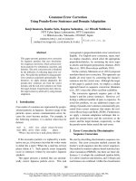
Conformity indices were clearly better for RA in all
analysed GTV localisation and treatment volumes with
amedianof0.56comparedto0.37forCAT(figure1).
Especially, irregularly formed tumours were framed
more precise with the prescribed do se including less
normal tissue or OAR in high dose area. Largest
improvement was achieved in patient 1 with a factor of
2.94 (RA: 0.50; CAT: 0.17).
Although inhomogeneity index was not be analysed rea-
sonable in detail (see above), the D
max
of GTV was reduced
dramatically for all patients using RA (19.9 Gy vs. 24.4 Gy).
Dose to organs at risk and normal tissue
In general, the dose to OAR is very low. However, in 7
of 10 analysed patients RA achieved a b etter dose pre-
servation of OAR in general comparison of all involved
tissues. Only patient 3, 4 and 8 showed a predominantly
better result for CAT (table 2). Two of these patients
had perip heral brain metastases with a large distance to
central OAR like brainstem, optical nerves or lenses
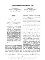
(patients 3 and 4). The other patient, treated because of
an atypic meningeoma in the area of the clivus, could
be irradiated with constant lower doses at all OAR
because of the steeper dose gradient using CAT. Patient
associated dose distributions of both techniques were
illustrated in (figure 2).
The D
mean
of the healthy brain tissue was lower using
RA in all patients. In contrast, low-dose areas could be
kept clearly smaller using CAT in all cases (table 2). In
maxi mum, low-do se volume was up to 12.5 times smal-
ler using CAT for treatment of 2 peripheral brain
metastases in patient 3 compared to RA.
Field Setup, Treatment Time and Monitor Units
The number of arcs using CAT depended on the num-
ber of required isocenters, whereas for RA single isocen-
ter plannin g was used. Patients with one isocenter were
treated with 5 arcs i n conformal therapy, whereas
patients with two isocenters received 10 to 12 arcs.
Additionally, patient 9 with four different isocenters was
irradiated with 20 arcs. Treatment time for delivering
prescribed dose was definitely l onger in all CAT cases
compared to single RA treatment (median time: 34.4
min vs. 4.5 min). Especially, for irradiation of patients
with more than one isocenter, treatment time was ≥ 17
times longer using CAT (patients 2 and 3) (figure 3).
Furthermore, median number of MU was 6504 MU
for CAT and 3455 MU for RA. In patient 6 with single
peripheral metastasis the number of MU was nearly the
same for both techniques (4618 MU (CAT) vs. 4663
MU (RA)), whereas for patient 4 CAT needed only 2964
MU compared to 3577 MU for RA f or one single per-
ipheral metastases. In all other cases, RA got along with
obvious less number of MU (figure 4).
Discussion
Our data show promising results analysing and imple-
menting a new approach for delivering single fraction
radiosurgery via RA with additional advantages in com-
parison to standard Conformal Arc application accuracy.
Similar results for IMRT were shown before by Baumert
et al. 2003 [16] by analyzing intensity modulated radio-
therapy compared to conformal static arc therapy in
treatment of meningioma of the skull base. In this work,
IMRT was superior in PTV coverage with lower doses
to the OAR admittedly in frac tionated therapy regime,
too. In another work from Wu et al. [6], results for
treatment of intracranial lesions using IMRT were clas-
sified as superior to a 3D-conformal static technique
and dynamic conformal arcs concerning dosimetric ben-
efits for SRS. However, these studies showed positive
results even without includ ing the new benefits evolving
through tested RA. In this context, Clark et al. [8] evalu-
ated the relative plan quality of single-isocenter vs.
multi-isocenter radiosurgical treatment of multiple cen-
tral nervous system metastases for VMAT irradiation. In
this planning study, plans were created using VMAT for
treatment of simulated patients with three brain metas-
tases. They concluded that radiosurgery for multiple tar-
gets using a single isoce nter can be efficiently delivered,
requiring less than one-half the beam time required for
multipl e isocenter set ups, too. In their opinion, VMAT
Figure 1 Diagram of Conformity Index for CAT (Conformal Arc
Therapy) in black and for RA (Rapid Arc) in white
Wolff et al . Radiation Oncology 2010, 5:77
/>Page 4 of 8
radiosurgery will likely replace multi-isocenter techni-
ques for linear accelerator-based treatment of multiple
targets in the future. Furthermore, Lagerwaard et al.
[10] used RA to plan and deliver whole-brain radiother-
apy (WBRT) with a simultaneous integrated boost in
patients with multiple brain metastases. In this study,
RA plans showed excellent coverage of planning target
volume for WBRT and PTV for the boost. These result
led the authors to the conclusion that RA treatment
planning and delivery of integrated plans of WBRT and
boosts to multiple brain metastases is a rapid and accu-
rate technique that has a higher conformity index than
conventional summation of WBRT and radiosurgery
boost.
In comparison, our conformity results were also better
for RA because of merely ellipsoid target shaping in
CAT using cones with circular fields. Because of this
fact, high-dose volumes could be kept significantly smal-
ler with RA. In contrast, low-dose volume was clearly
smaller using CAT in all patients. This fact could be
expected because of rotation around the patient with
continuous beam on time using RA with 177 control
points compared to step and shoot irradiation using
CAT with 5 to 20 arcs. Furthermore, the distance
between beam shaping aperture and patient is higher for
RA. For CAT, the cones minimise the distance between
beam shaping aperture and patient and therefore gener-
ate steeper dose gradients. This result may play no
Table 2 Summary of Organs at risk
Patient No. 1 2 3 4 5 6 7 8 9 10
Technique CAT RA CAT RA CAT RA CAT RA CAT RA CAT RA CAT RA CAT RA CAT RA CAT RA
Healthy brain
D
mean
[Gy]
0.1 0.3 0.4 0.5 0.6 3 1.0 1.4 2.1 5.2 0.3 0.6 0.6 0.7 0.6 1 1.3 3.2 0.5 2.2
V
2Gy
[cm
3
] 8.6 53 59.5 104.8 68.7 860.3 316.8 394.4 92.3 145.7 37.6 99.9 100.8 105.7 99.6 367.6 223.7 1057 71.4 601
OAR D
max
[Gy]
Lens left 0.0 0.5 0.0 0.1 0.1 1.3 0.0 1.3 0.6 0.1 0.3 0.2 0.0 0.0 0.0 1.9 0.6 0.3 0.3 0.6
Lens right 0.0 0.4 0.0 0.1 0.0 1.4 1.1 2.5 0.6 0.0 0.0 0.1 0.0 0.0 0.0 1.3 0.0 0.3 0.3 0.6
Brainstem 0.4 1.5 3.7 6.9 0.5 7.1 2.3 3.4 0.8 0.7 0.5 0.8 0.9 0.1 4.9 4.9 2.0 1.2 0.9 2.1
Chiasm 0.0 0.9 0.5 3.8 0.1 3.8 0.7 2.5 0.6 0.0 0.0 0.4 0.0 0.1 1.2 4.2 1.4 1.7 0.0 1.7
N. opticus right 0.0 0.6 0.0 0.2 0.2 3.2 2.4 1.4 0.7 0.1 0.0 0.3 0.0 0.0 0.3 2.6 0.5 1.0 0.4 1.3
N. opticus left 0.0 0.9 0.5 0.3 0.2 3.7 0.0 0.9 0.8 0.1 0.5 0.3 0.0 0.0 1.2 4.6 1.4 1.0 0.5 1.4
Healthy brain (D
mean
), low-dose volume (V
2Gy
) for all patients and both treatment techniques. The maximum dose to OAR is shown in Gy, the mean dose to
healthy brain in Gy, and the low dose volume (volume which is irradiated with maximum of 2 Gy) in cm
3
.
OAR = organs at risk, CAT = conformal arc therapy, RA = Rapid Arc, D
mean
= mean dose, V
2Gy
= volume irradiated with 2 Gy or higher, Fat marked fields indicate
a benefit for this value.
Figure 2 Comparison of representative dose distributions for conformal arc (left) and RapidArc (right) illustrating typical differences
between both techniques in patient 8 treated because of an atypic meningeoma in the area of the clivus.
Wolff et al . Radiation Oncology 2010, 5:77
/>Page 5 of 8
decisive role when ir radiation is indicated in palliative
situation. However, whenever younger patients with
longer estimated lifetime were analysed for irradiation,
risk of development of a secondary tumour should be
more weighted for final choice of treatment technique.
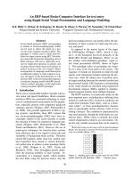
Especially, patient with benign disease should be ana-
lysed very carefully according to this complexity of pro-
blems (for example dose distribution of patient 2 with a
vestibularis schwannoma is illustrated in figure 5).
Higher cumulative dose in GTV as result of CAT can
be subordinated in treatment for malignant diseases like
metastases, but should be considered for irradiation of
benign targets, too. Whenever OARs are involved in
high dose areas risk of impairment with irreparable
damage rises [17-20]. For example, treatment of vestibu-
laris schwannoma involves the N. acousticus directly
into the target volume. In this case higher cumulative
doses using CAT should be considered. Similar results
were achieved from Lagerwaard et al. 2009 [9]. In their
work, RA irradiation for vestibular schwannomas was
compared to conformal arc therapy. In conclusion, they
found a better conformity and lower cumulative doses
with equal dose exposure to the OAR and significant
shorter treatment time for RA, too. These results had
led to RA replacing CAT for vestibula r schwannoma s in
their department.
In our study, treatment time was clearly shorter using
RA for all patients. This fact results from fewer patient
positioning procedures (1 time for RA; 1 time for every
single arc using CAT) and single arc irradiatio n techni-
que compared to several single arc using CAT. When-
ever patient constitution allows no long recumbency,
this parameter should be c onsidered very carefully for
choice of technique and could afford an important bene-
fit for RA.
In this context, number of MU was c learly lower for
RA in 8 of 10 patients. This item could be an advantage
for patients because of less scattered radiation and for
the daily routine of the department because of better
time utilisation of the accelerators.
Interestingly, size of target GTV had no clearly impact
on treatment choice in our patient population. For
example, patients 1, 6 and 10 had a small GTV with a
maximum of 0.3 cm
3
and were considered for RA treat-
ment, whereas patient 3 with a GTV of 0.3 cm
3
was
selected for CAT. In addition, in patients with larger
GTVs (patients 4, 5, 8) no definitely benefit for one
technique could be observed.
Doses at OAR were generally very low. Thus, this
parameter should not be overestimated for technique
selection but should be kept in mind especially for
patients with exposure due to pre-irradiation of the
brain or head. In these cases sparing of dose at OAR
could be very important to avoid serious side effects
during irradiation or in follow-up.
Comparing results of all parameters, choice of treat-
ment techniques could be reclassified retrospectively in
selected cases: patients 1, 5, 6, 7 and 9 achieved compar-
able dose expo sure to the OAR for a ll measured para-
meters for both treatment techniques. Dose maximum,
treatment time and number of MU showed clearly bet-
ter results for RA. Solely, irradiated low dose volume
was l ower in patients 1, 5, 6 and 9 for CAT. Because of
palliative indication, these five patients would have been
treated with RA with much shorter treatment time and
comparable OAR sparing, in future.
Patient 2 was irradiated because of a vestibularis
schwannoma. In this case, dose to the brain stem and
chiasm was higher using RA (figure 5). Furthermore,
benign indication of irradiation increased the impor-
tance of the fact that involved low dose volume for RA
was nearly twice as much as for CAT. An eventually
higher risk of tumour induction by low dose irradiation
areas [21-23] and higher OAR doses led to a final deci-
sion for CAT, although t reatment time and number of
MU were higher.
Analyzing patient 3 with two peripheral metastases led
in a clear decision for CAT. The dose to all OAR, low
Figure 3 Diagram of Treatment Time for CAT (Conformal Arc
Therapy) in black and for RA (Rapid Arc) in white. The
treatment time does not include the setup of patient and setup
between every single arc for CAT. The displayed treatment time is
the time where the beam is on.
Figure 4 Diagram of calculated Monitor Units (MU) for CAT
(Conformal Arc Therapy) in black and RA (Rapid Arc) in white.
The MU for each single arc using CAT were summed up and
displayed. For RA only one arc was used.
Wolff et al . Radiation Oncology 2010, 5:77
/>Page 6 of 8
dose volume and D
mean
of the healthy brain showed
clearly better results for CAT. Merely, treatment time
and number of MU would argue f or RA but were rea-
sonable for this patient using CAT.
For patient 8 CAT would be the treatment of choice,
as well. Dose to the O AR and low dose volume were
assessed as clear benefit for CAT, even though treat-
ment time and number of MU were better using RA.
Results of patient 4 showed comparable results for
both techniques. On the one hand, dose to OAR was
slightly better using CAT for 4 of 6 items (table 2), but
on the other hand, treatment time and dose maximum
were better for RA. In the palliative situation of this
patient, both choices of treatment technique should be
arguable without any major disadvantages for this
patient.
Analyses of patient 10 showed a retrospective decision
in aid of RA. Shorter treatment time with less number
of MU preponderated a slightly lower dose to the brain
stem and chiasm as well as smaller low dose volume by
use of CAT.
In summary, the choice of treatment technique should
be done with respect to target entity, dimension and
localisation as well as patient age and constitution. It is
recommended to evaluate both techniques prior to
treatment decision.
Conclusion
We conclude that RA is a new approach for single frac-
tion radiosurgery treatment. In selected cases, RA com-
bines advantages of short treatment time with less
number of MU and better conformity in addition to
accuracy of stereot actic locali sation, but with larger low
dose areas in comparison to conventional cone based
SRS. For this reason we successfully integrated this new
approach into our treatment routine and started to
irradiate patients with promising results.
Nevertheless, irradiation with CAT is not dis pensable
at the moment, but should rather kept in mind to be
another feasible approach with different advantages for
selected settings.
Authors’ contributions
All authors conceived of the study and participated in study design. HAW
carried out the clinical evaluation and performed the statistical analysis.
DMW and HV performed physical evaluation and technical implementing.
HC and CFH worked in study coordination and helped to draft the
manuscript. All authors read and approved the final manuscript.
Competing interests
The authors declare that they have no competing interests.
Received: 17 June 2010 Accepted: 13 September 2010
Published: 13 September 2010
References
1. De Salles AA, Gorgulho AA, Selch M, De Marco J, Agazaryan N:
Radiosurgery from the brain to the spine: 20 years experience. Acta
Neurochir Suppl 2008, 101:163-168.
2. Solberg TD, Boedeker KL, Fogg R, Selch MT, DeSalles AA: Dynamic arc
radiosurgery field shaping: a comparison with static field conformal and
noncoplanar circular arcs. Int J Radiat Oncol Biol Phys 2001, 49:1481-1491.
3. Ernst-Stecken A, Lambrecht U, Ganslandt O, Mueller R, Fahlbusch R, Sauer R,
Grabenbauer G: Radiosurgery of small skull-base lesions - No advantage
for intensity-modulated stereotactic radiosurgery versus conformal arc
technique. Strahlentherapie Und Onkologie 2005, 181:336-344.
Figure 5 Comparison of representative dose distributions for conformal arc (left) and RapidArc (right) illustrating typical differences
between both techniques in patient 2 treated because of a vestibularis schwannoma.
Wolff et al . Radiation Oncology 2010, 5:77
/>Page 7 of 8
4. Clark BG, Candish C, Vollans E, Gete E, Lee R, Martin M, Ma R, McKenzie M:
Optimization of stereotactic radiotherapy treatment delivery technique
for base-of-skull meningiomas. Med Dosim 2008, 33:239-247.
5. Lawson JD, Wang JZ, Nath SK, Rice R, Pawlicki T, Mundt AJ, Murphy K:
Intracranial application of IMRT based radiosurgery to treat multiple or
large irregular lesions and verification of infra-red frameless localization
system. J Neurooncol 97:59-66.
6. Wu QJ, Wang Z, Kirkpatrick JP, Chang Z, Meyer JJ, Lu M, Huntzinger C,
Yin FF: Impact of collimator leaf width and treatment technique on
stereotactic radiosurgery and radiotherapy plans for intra- and
extracranial lesions. Radiat Oncol 2009, 4:3.
7. Tobler M, Leavitt DD, Watson G: Optimization of the primary collimator
settings for fractionated IMRT stereotactic radiotherapy. Med Dosim 2004,
29:72-79.
8. Clark GM, Popple RA, Young PE, Fiveash JB: Feasibility of single-isocenter
volumetric modulated arc radiosurgery for treatment of multiple brain
metastases. Int J Radiat Oncol Biol Phys 76:296-302.
9. Lagerwaard FJ, Meijer OW, van der Hoorn EA, Verbakel WF, Slotman BJ,
Senan S: Volumetric modulated arc radiotherapy for vestibular
schwannomas. Int J Radiat Oncol Biol Phys 2009, 74:610-615.
10. Lagerwaard FJ, van der Hoorn EA, Verbakel WF, Haasbeek CJ, Slotman BJ,
Senan S: Whole-brain radiotherapy with simultaneous integrated boost
to multiple brain metastases using volumetric modulated arc therapy.
Int J Radiat Oncol Biol Phys 2009, 75:253-259.
11. Bush K, Townson R, Zavgorodni S: Monte Carlo simulation of RapidArc
radiotherapy delivery. Phys Med Biol 2008, 53:N359-370.
12. Clivio A, Fogliata A, Franzetti-Pellanda A, Nicolini G, Vanetti E, Wyttenbach R,
Cozzi L: Volumetric-modulated arc radiotherapy for carcinomas of the
anal canal: A treatment planning comparison with fixed field IMRT.
Radiother Oncol 2009, 92:118-124.
13. Ulmer W, Harder D: A triple gaussian pencil beam model for photon
beam treatment planning. Z Med Phys 1995, 5:25-30.
14. Ulmer W, Pyyry J, Kaissl W: A 3D photon superposition/convolution
algorithm and its foundation on results of Monte Carlo calculations. Phys
Med Biol 2005, 50:1767-1790.
15. Chavaudra J: [Last ICRU recommendations for the prescription, recording
and reporting of external bean therapy]. Cancer Radiother 1998, 2:607-614.
16. Baumert BG, Norton IA, Davis JB: Intensity-modulated stereotactic
radiotherapy vs. stereotactic conformal radiotherapy for the treatment
of meningioma located predominantly in the skull base. Int J Radiat
Oncol Biol Phys 2003, 57:580-592.
17. Belliere A, Girard N, Chapet O, Khodri M, Kubas A, Souquet PJ, Mornex F:
Feasibility of high-dose three-dimensional radiation therapy in the
treatment of localised non-small-cell lung cancer. Cancer Radiother 2009,
13:298-304.
18. Ganz JC, Reda WA, Abdelkarim K:
Adverse radiation effects after Gamma
Knife Surgery in relation to dose and volume. Acta Neurochir (Wien) 2009,
151:9-19.
19. Yang I, Sughrue ME, Han SJ, Fang S, Aranda D, Cheung SW, Pitts LH,
Parsa AT: Facial nerve preservation after vestibular schwannoma Gamma
Knife radiosurgery. J Neurooncol 2009, 93:41-48.
20. Foote KD, Friedman WA, Buatti JM, Meeks SL, Bova FJ, Kubilis PS: Analysis
of risk factors associated with radiosurgery for vestibular schwannoma.
J Neurosurg 2001, 95:440-449.
21. Schneider U: Mechanistic model of radiation-induced cancer after
fractionated radiotherapy using the linear-quadratic formula. Med Phys
2009, 36:1138-1143.
22. Hall EJ, Wuu CS: Radiation-induced second cancers: the impact of 3D-CRT
and IMRT. Int J Radiat Oncol Biol Phys 2003, 56:83-88.
23. Ruben JD, Davis S, Evans C, Jones P, Gagliardi F, Haynes M, Hunter A: The
effect of intensity-modulated radiotherapy on radiation-induced second
malignancies. Int J Radiat Oncol Biol Phys 2008, 70:1530-536.
doi:10.1186/1748-717X-5-77
Cite this article as: Wolff et al.: Single fraction radiosurgery using Rapid
Arc for treatment of intracranial targets. Radiation Oncology 2010 5:77.
Submit your next manuscript to BioMed Central
and take full advantage of:
• Convenient online submission
• Thorough peer review
• No space constraints or color figure charges
• Immediate publication on acceptance
• Inclusion in PubMed, CAS, Scopus and Google Scholar
• Research which is freely available for redistribution
Submit your manuscript at
www.biomedcentral.com/submit
Wolff et al . Radiation Oncology 2010, 5:77
/>Page 8 of 8