Báo cáo khoa học: "Radiation induced apoptosis and initial DNA damage are inversely related in locally advanced breast cancer patients" ppt
Bạn đang xem bản rút gọn của tài liệu. Xem và tải ngay bản đầy đủ của tài liệu tại đây (344.18 KB, 5 trang )
RESEARC H Open Access
Radiation induced apoptosis and initial DNA
damage are inversely related in locally advanced
breast cancer patients
Beatriz Pinar
1,2
, Luis Alberto Henríquez-Hernández
2,3*
, Pedro C Lara
1,2
, Elisa Bordon
2
, Carlos Rodriguez-Gallego
2,4
,
Marta Lloret
1,2
, Maria Isabel Nuñez
5
, Mariano Ruiz De Almodovar
5
Abstract
Background: DNA-damage assays, quantifying the initial number of DNA double-strand breaks induced by
radiation, have been proposed as a predictive test for radiation-induced toxicity. Determination of radiation-
induced apoptosis in peripheral blood lymphocytes by flow cytometry analysis has also been proposed as an
approach for predicting normal tissue responses following radiotherapy. The aim of the present study was to
explore the association between initial DNA damage, estimated by the number of double-strand breaks induced
by a given radiation dose, and the radio-induced apoptosis rates observed.
Methods: Peripheral blood lymphocytes were taken from 26 consecutive patients with locally advanced breast
carcinoma. Radiosensitivity of lymphocytes was quantified as the initial number of DNA double-strand breaks
induced per Gy and per DNA unit (200 Mbp). Radio-induced apoptosis at 1, 2 and 8 Gy was measured by flow
cytometry using annexin V/propidium iodide.
Results: Radiation-induced apoptosis increased in order to radiation dose and data fitted to a semi logarithmic
mathematical model. A positive correlation was found among radio-induced apoptosis values at different radiation
doses: 1, 2 and 8 Gy (p < 0.0001 in all cases). Mean DSB/Gy/DNA unit obtained was 1.70 ± 0.83 (range 0.63-4.08;
median, 1.46). A statistically significant inverse correlation was found between initial damage to DNA and radio-
induced apoptosis at 1 Gy (p = 0.034). A trend toward 2 Gy (p = 0.057) and 8 Gy (p = 0.067) was observed afte r 24
hours of incubation.
Conclusions: An inverse association was observed for the first time between these variables, both considered as
predictive factors to radiation toxicity.
Background
Radiation induced normal tissue damage is the most
important limitation for the delivery of a high poten-
tially curative radiation dose. Radiation doses are limited
by the tolerance of normal tissues included in the treat-
ment volume. Intrinsic variations in radiosensit ivity
determine most of the individual differences in normal
tissue damage [1-3]. It is possible to determine the indi-
vidual radiosensitivity before radiotherapy (RT) [4,5]. If
intrinsic differences in individual radiosensitivity were
responsible for the variation in severity of early or late
radio-induced toxicity [6-9], we could adjust the radia-
tion dose to be delivered. An association between DNA-
damage assays, quantifying the initial number of DNA
double-strand breaks (DSB) induced by radiation, and
radiation-toxicity has been reported [10,11]. Increasing
numbers of radiation induced DSB were related to
severe late toxicity in breast cancer patients [10]. Deter-
mination of radiation-induced apoptosis (RIA) in per-
ipheral blood lymphocytes (PBLs) by flow cytometry
analysis has been proposed as a possible prediction
value of normal tissue responses after RT [12,13]. RIA
waspredictiveoflatetoxicityinseveraltumourloca-
tions [14-16]. Patients suffering of late toxicity after RT
showed reduced rates of RIA. Furthermore, patients
* Correspondence:
2
Instituto Canario de Investigación del Cáncer (ICIC), Spain
Full list of author information is available at the end of the article
Pinar et al. Radiation Oncology 2010, 5:85
/>© 2010 Pinar et al; licensee BioMed Central Ltd. This is an Open Access article d istribute d under the terms of the Cr eative C ommons
Attribution License ( which permits unrestricted use, distribution, and reprod uctio n in
any medium, provided the original work is properly cited.
affected by the Ataxia-Telangiectasia (AT) syndrome
showed the lowest rates of RIA. Defective apoptotic
response to radiation in the PBLs of those sensitive
patients could help to explain this association. As
described above, late toxicity in breast cancer patients
treated with radiation therapy has been related to
increased radiosensitivity of lymphocytes, as shown by
increased number of DSB/Gy/DNA unit, and reduced
RIA. According to this, sensitive patients would show
increased nu mber of DSB and r educed RIA as results of
defective apoptotic processing of the initial damage
induced by x-rays. Considering the above background
and observations, the aim of the present study was to
analyze if there was any statistical relation between
initial DNA damage, estimated by the number of DSB,
and the apoptotic rates observed estimated by the
amount of RIA.
Methods
Patients
PBLs were taken from 26 consecutive patients with
locally advanced breast carcinoma (stage IIIa-IIIb), diag-
nosed and treated in our institution, and given inform
consent. All patients were referred to receive high-dose
hyperfr acti onated radical radiotherapy as follows: 60 Gy
to the whole breast over a period of 5 weeks in two
daily fractions of 1.2 Gy separated by at least 6 h on 5
days each week, and followed by a boost of 21.6 Gy to a
total dose of 81.6 Gy. The study was approved by the
Research & Ethics Committee of our institution. Mean
age of patients was 57.62 ± 12.99 years (range 30-83),
69.2% of them were menopause women.
Apoptosis assay and flow cytometry
RIA analyses were performed as previously reported
[13,17]. PBLs w ere irradiated with 0, 1, 2 and 8 Gy .
After irradiation, samples were incubated for 24 hours
at 37°C and 5% CO
2
. After extraction of cellular pellet,
it was resuspended in 100 μl Annexin V buffer Kit
(Pharmingen, Becton Dickinson). After the addition of 4
μl of Annexin-V-FITC and 10 μl of propidium iodure
(PI), cells were incubated during 15 minutes at room
temperature in the dark. Finally, 400 μlofAnnexinV
buffer Kit were added. Every assay was made in tripli-
cate. The flow cytometry analysis was performed in a
FACScalibur (Becton Dickinson,SanJosé,CA)usinga
488 nm argon laser. Each sample was analyzed using
5000 events/sample acquired in list mode by a Macin-
tosh Quadra 650 minicomputer (Apple computer Inc.,
Cupertino, CA). Data were analyzed using the CellQuest
program (Becton Dickinson, San José, CA) calculating
early and late apoptosis levels. RIA is defined as the per-
centage of total P BLs death induced by the radiation
dose minus the spontaneous cell death (control, 0 Gy).
DNA damage assay
Data related to initial DNA d amage were obtained from
our files [10]. Shortly, mononuclear cells were isolated
from blood of patients, resuspended in cold DMEM,
and mixed with 1% ultra-low-melting-point agarose to
obtain 250 μl plugs. Irradiation on ice was performed
using a
60
Co source (rate dose 1.5 Gy/min, approxi-
mately) as previously reported [10]. Plugs were held 1
hour at 4°C a nd incubated at 37°C for 24 hours. The
study of initial DNA damage wa s completed in the Uni-
versity of Granada (Spain). Initial radiation-induced
DNA damage in PBLs was measured as previously
described [18] and was considered an individual indica-
tor of the molecular radiosensitivity of normal cells.
Statistical analyses
Statistical analyses were performed using the SPSS Sta-
tistical Package (version 15.0 for Windows) as previously
reported [10,13,18]. The cut-off value for DSB/Gy/DNA
unit was the median. Additional cut-off values studied
were the tertiles of the distribution. All tests were two
sided and statistical significance level was established for
a p value less than 0.05.
Results
Radiation-induced apoptosis
Data of RIA were available in all 26 breast cancer
patients, as shown in Table 1. RIA values increased with
radiation dose (Table 1), and data fitted to a semi-loga-
rithmic equation as follows: RIA = a + b ln(Gy), confirm-
ing our previously observations [13,17,19]. The
increments in RIA were defined by two constants: the
coefficient in origin a (as the origin of the curve in the Y
axis determining the spontaneous apoptosis); and the
coefficient b defining the slope of the curve. a and b fol-
lowed a normal distribution (Kolmogorov-Smirnov test,
p > 0.05). Mean of b was 7.93 ± 2.68 standard deviation
(range 1.64-26.63; median, 12.64); mean of b was 7.93 ±
2.68 (range 3.18-12.57; median, 7.85) (Table 1). In this
way, we were able to establish an individual radiosensitiv-
ity value defined by two constants: a as the spontaneous
Table 1 Apoptosis data obtained after the irradiation of
PBLs at 1, 2 and 8 Gy
Absolute data Mean ± SD Median (range)
RIA 1 Gy 13.33 ± 7.26 12.36 (2.51-29.00)
RIA 2 Gy 18.20 ± 7.82 17.79 (4.17-32.08)
RIA 8 Gy 29.70 ± 10.05 30.44 (9.02-44.10)
DNA-DSB 1.70 ± 0.83 1.46 (0.63-4.08)
Defined model data
a Coefficient 13.08 ± 7.21 12.64 (1.64-26.63)
b Coefficient 7.93 ± 2.68 7.85 (3.18-12.57)
Regression coefficient 98.18 ± 4.58 99.58 (82.49-100)
Pinar et al. Radiation Oncology 2010, 5:85
/>Page 2 of 5
apoptotic rate, and b as the percentage of RIA per Gy. A
good correlation was found among RIA data at different
doses: 1 vs. 2 Gy, R
2
= 0.978 (p < 0.0001); 1 vs. 8 Gy, R
2
= 0.883 (p < 0.0001); 2 vs. 8 Gy, R
2
= 0.914 (p < 0.0001)
(Spearman Rho test, Figure 1). The experimental data
showed an excell ent fit to the described model (median
regression coefficient at 24 h was 99.58).
Relation to initial DNA damage
Mean ± standard deviation of DSB/Gy/DNA unit,
obtained from our files [10] was 1.70 ± 0.83 (range 0.63-
4.08; median, 1.46). No relation was found between the
number of DSB and the RIA at 1 (p = 0.406), 2 (p =
0.592) and 8 Gy (p = 0.619). In the same way, no rela-
tion was found between the number of DSB and the
model coefficient variables a (p = 0.457) and b (p =
0.901) , when they were analyzed as continuous variables
(Pearson test used in all correlations). When DSB values
were segregated in two groups (the lower third against
the two upper thirds of the distri bution), a mod est
inverse correlation was found, reaching statistical signifi-
cance for RIA at 1 Gy (p = 0.034). A similar trend was
found for RIA at 2 (p = 0.057) and 8 Gy (p = 0.067)
(Figure 2). a values also showed an inverse correlation
with DSB, that reached statistical significance (p =
0.041). No relation was found between the number of
DSB and b constant.
Discussion
We established for the first time, a statistical association
between initial DNA damage, measured as DSB, and
RIA. Ionizing radiation (IR) kills cells by damaging the
structure and function of genomic DNA. The response
of cells to this damage and their ability to restore DNA
sequence integrity remains unclear. Intrinsic radiosensi-
tivity is correlated in a first approach to the a bility of
the cell to detect and repair DNA damages. DSB can be
induced by a variety of DNA damaging agents, such as
x-rays [20]. Differences in cell survival may be related to
the number of initial DNA DSB, the DSB rejoining rate,
or the level of residual DNA damage [11,21-24]. Wide
variation in the level of initial radiation-induced DNA
damage suggests that variation in cell radiosensitivity
can be detected in vitro using radiosensitivity assays on
PBLs from normal tissues of cancer patients prior to RT
[11]. Patients with radiosensitive PBLs presented a sig-
nificant increased risk for develop late complications
[25]. Increasing numbers of radiation induced DSB were
related with severe late toxicity reactions in breast can-
cer patients [10]. In the other hand, RIA values, that
fitted to a semi logarithmic model defined by a and b
constants, increased with radiation dose [13]. Previously
studies were uniformly positive towards a relation
between RIA and radiation toxicity [14]. In fact, patients
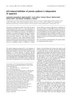
Figure 1 Correlation between radio-induced apoptosis data at the different doses of radiation. Panel A: 1 vs. 2 (Gy); Panel B, 1 vs. 8 (Gy);
Panel C: 2 vs. 8 (Gy). A linear correlation was established.
Figure 2 Box plot shows an association between DSB and RIA.
The lines connect the medians, the boxes cover the 25th to 75th
percentiles, and the minimal and maximal values are shown by the
ends of the bars. Patients with lower amount of DSB suffered higher
levels of RIA.
Pinar et al. Radiation Oncology 2010, 5:85
/>Page 3 of 5
suf fering of late toxicity af ter RT showed reduced levels
of RIA. The mechanism behind the relationship between
increased radiation toxicity and reduced apoptotic
response in PBLs is st ill unclear. Thus, lymphocyte s
from patients who suffered different syndrome s related
with radiosensitivity (i.e., Ataxia-telangiectasia , Bloom
syndrome, or Fanconi anaemia) showed absence of
induction of p53 [26,27] and lower levels of Bax [28].
This failure in the induction of the apoptosis response
in lymphocytes has been related with late toxicity [16].
So, defective apoptotic response to radiation in PBLs
could help to explain this inverse relation [14]. We
report here for the first time a statistical association
between these two predictive values f or radiation toxi-
city. Lowest values of i nitial DNA d amage were related
to higher values of RIA, at the same radiation dose. This
relation was also observed between DSB and the sponta-
neous apoptosis of cells (estimated by the a constant).
Cell response to x-rays is individual, and the amount of
initial DNA damage depends on each patient. The two
main mechanisms of DSB repair are 1) non-homologous
end joining (NHEJ) and homologous recombination
repair (HRR) [29,30], and 2) cell-cycle che ckpoints that
provide time for repair and apoptosis [31]. Depending
on the severity of the DNA damage, cells may undergo
apoptosis instead of attempting to repair the damage
[32]. Regulation of RIA and cell cycle arrests is achieved
primarily through p53 phosphorylation by ATM protein
[33]. T-lymphocytes from AT patients display severely
compromised apopto tic response, as well as non-induc-
tion of p53 after exposure to IR [26]. Moreover, PBLs
from AT patients are characterized by an elevated spon-
taneous level of apoptotic cells compared to normal
ones [26]. Extremely radiosensitive patients have
abnormalities in their ability to recognize or repair the
DNA DSB typically induced by IR [15]. Gene expression
profile of irradiated PBLs showed that the majority of
the strongly activated genes were p53 targets involved in
DNA repair and apoptosis [28]. The level of BAX activa-
tion correlated with the sensitivity of the cells to radia-
tion [28]. A link between RIA and cellular response to
DNA D SB arises because there are many proteins com-
mon to the execution of both processes. Anyhow, there
are yet unidentifie d proteins or complexe s that regulate
the cross-talk between the mutually exclusive pathways
of maintenance of life and initiation of death [31]. How
these pathways are integrated to provide a concerted
response to DSB is very complex, and could help to
understand the inverse relation between the initial DNA
damage to IR and RIA.
Conclusion
A statistical inverse association was observed for the
first time between DNA-DSB and RIA in 26 patients
diagnosed with locally advanced breast cancer. However,
these results must be verified in larger series of patients.
List of abbreviations
AT: Ataxia-Telangiectasia; DSB: Double-strand Break; PBLS: Peripheral Blood
Lymphocytes; PI: Propidium Iodide; RIA: Radio-induced Apoptosis; RT:
Radiotherapy.
Acknowledgements
This work was subsidized by Fundación del Instituto Canario de
Investigación del Cáncer (FICIC). LAHH and EB were supported by a grant
from the Instituto Canario de Investigación del Cáncer (ICIC).
Author details
1
Radiation Oncology Department, Hospital Universitario de Gran Canaria Dr.
Negrín, Spain.
2
Instituto Canario de Investigación del Cáncer (ICIC), Spain.
3
Clinical Sciences Department, Universidad de Las Palmas de Gran Canaria,
Spain.
4
Immunology Department, Hospital Universitario de Gran Canaria Dr.
Negrín, Spain.
5
Radiology Department, Hospital Universitario San Cecilio,
Universidad de Granada, Spain.
Authors’ contributions
BP has been involved in conception and design of the project and have
made the selection of patients, the evaluation of clinical variables and grade
of toxicity as well as all the aspects related with the patients selected,
including the treatment. LAHH has written the manuscript, has made tables
and figures and has been involved in type of packaging likewise in the
submission process. PCL has been involved in conception and design of the
study and in drafting the manuscript and has given final approval of the
version to be published. EB and CRG have made the cell experiments with
lymphocytes, irradiation of cells, flow cytometry experiments, data
acquisition and statistical analysis. ML has made the selection of patients,
the evaluation of clinical variables and grade of toxicity as well as all the
aspects related with the patients selected, including the treatment. MIN and
MRDA have been involved in conception and design of the study, in
drafting the manuscript, and have made the DNA-DSB experiments and
analyses. All authors read and approved the final manuscript.
Competing interests
The authors report no conflicts of interest. The authors alone are responsible
for the content and writing of the paper.
Received: 26 April 2010 Accepted: 24 September 2010
Published: 24 September 2010
References
1. Guirado D, Ruiz de Almodovar JM: Prediction of normal tissue response
and individualization of doses in radiotherapy. Phys Med Biol 2003,
48:3213-3223.
2. Moody AM, Mayles WP, Bliss JM, A’Hern RP, Owen JR, Regan J, Broad B,
Yarnold JR: The influence of breast size on late radiation effects and
association with radiotherapy dose inhomogeneity. Radiother Oncol 1994,
33:106-112.
3. Turesson I, Nyman J, Holmberg E, Oden A: Prognostic factors for acute
and late skin reactions in radiotherapy patients. Int J Radiat Oncol Biol
Phys 1996, 36:1065-1075.
4. Lopez E, Guerrero R, Nunez MI, del Moral R, Villalobos M, Martinez-Galan J,
Valenzuela MT, Munoz-Gamez JA, Oliver FJ, Martin-Oliva D, Ruiz de
Almodovar JM: Early and late skin reactions to radiotherapy for breast
cancer and their correlation with radiation-induced DNA damage in
lymphocytes. Breast Cancer Res 2005, 7:R690-698.
5. McMillan TJ, Tobi S, Mateos S, Lemon C: The use of DNA double-strand
break quantification in radiotherapy. Int J Radiat Oncol Biol Phys 2001,
49:373-377.
6. Baumann M, Holscher T, Begg AC: Towards genetic prediction of
radiation responses: ESTRO’s GENEPI project. Radiother Oncol 2003,
69:121-125.
Pinar et al. Radiation Oncology 2010, 5:85
/>Page 4 of 5
7. Bentzen SM, Tucker SL: Individualization of radiotherapy dose
prescriptions by means of an in vitro radiosensitivity assay. Radiother
Oncol 1998, 46:216-218.
8. Mackay RI, Hendry JH: The modelled benefits of individualizing
radiotherapy patients’ dose using cellular radiosensitivity assays with
inherent variability. Radiother Oncol 1999, 50:67-75.
9. Nunez MI, McMillan TJ, Valenzuela MT, Ruiz de Almodovar JM, Pedraza V:
Relationship between DNA damage, rejoining and cell killing by
radiation in mammalian cells. Radiother Oncol 1996, 39:155-165.
10. Pinar B, Lara PC, Lloret M, Bordon E, Nunez MI, Villalobos M, Guerrero R,
Luna JD, Ruiz de Almodovar JM: Radiation-induced DNA damage as a
predictor of long-term toxicity in locally advanced breast cancer
patients treated with high-dose hyperfractionated radical radiotherapy.
Radiat Res 2007, 168:415-422.
11. Ruiz de Almodovar JM, Guirado D, Isabel Nunez M, Lopez E, Guerrero R,
Valenzuela MT, Villalobos M, del Moral R: Individualization of radiotherapy
in breast cancer patients: possible usefulness of a DNA damage assay to
measure normal cell radiosensitivity. Radiother Oncol 2002, 62:327-333.
12. Barber JB, West CM, Kiltie AE, Roberts SA, Scott D: Detection of individual
differences in radiation-induced apoptosis of peripheral blood
lymphocytes in normal individuals, ataxia telangiectasia homozygotes
and heterozygotes, and breast cancer patients after radiotherapy. Radiat
Res 2000, 153:570-578.
13. Bordon E, Henriquez Hernandez LA, Lara PC, Pinar B, Fontes F, Rodriguez
Gallego C, Lloret M: Prediction of clinical toxicity in localized cervical
carcinoma by radio-induced apoptosis study in peripheral blood
lymphocytes (PBLs). Radiat Oncol 2009, 4:58.
14. Crompton NE, Miralbell R, Rutz HP, Ersoy F, Sanal O, Wellmann D, Bieri S,
Coucke PA, Emery GC, Shi YQ, et al: Altered apoptotic profiles in
irradiated patients with increased toxicity. Int J Radiat Oncol Biol Phys
1999, 45:707-714.
15. Crompton NE, Shi YQ, Emery GC, Wisser L, Blattmann H, Maier A, Li L,
Schindler D, Ozsahin H, Ozsahin M: Sources of variation in patient
response to radiation treatment. Int J Radiat Oncol Biol Phys 2001,
49:547-554.
16. Ozsahin M, Crompton NE, Gourgou S, Kramar A, Li L, Shi Y, Sozzi WJ,
Zouhair A, Mirimanoff RO, Azria D: CD4 and CD8 T-lymphocyte apoptosis
can predict radiation-induced late toxicity: a prospective study in 399
patients. Clin Cancer Res 2005, 11:7426-7433.
17. Bordon E, Henriquez-Hernandez LA, Lara PC, Ruiz A, Pinar B, Rodriguez-
Gallego C, Lloret M: Prediction of clinical toxicity in locally advanced
head and neck cancer patients by radio-induced apoptosis in peripheral
blood lymphocytes (PBLs). Radiat Oncol 2010, 5:4.
18. Nunez MI, Guerrero MR, Lopez E, del Moral MR, Valenzuela MT, Siles E,
Villalobos M, Pedraza V, Peacock JH, Ruiz de Almodovar JM: DNA damage
and prediction of radiation response in lymphocytes and epidermal skin
human cells. Int J Cancer 1998, 76:354-361.
19. Saavedra MM, Henriquez-Hernandez LA, Lara PC, Pinar B, Rodriguez-
Gallego C, Lloret M: Amifostine Modulates Radio-induced Apoptosis of
Peripheral Blood Lymphocytes in Head and Neck Cancer Patients. J
Radiat Res (Tokyo) 2010.
20. Ralhan R, Kaur J, Kreienberg R, Wiesmuller L: Links between DNA double
strand break repair and breast cancer: accumulating evidence from both
familial and nonfamilial cases. Cancer Lett 2007, 248:1-17.
21. Dickson J, Magee B, Stewart A, West CM: Relationship between residual
radiation-induced DNA double-strand breaks in cultured fibroblasts and
late radiation reactions: a comparison of training and validation cohorts
of breast cancer patients. Radiother Oncol 2002, 62:321-326.
22. Hoeller U, Borgmann K, Bonacker M, Kuhlmey A, Bajrovic A, Jung H,
Alberti W, Dikomey E: Individual radiosensitivity measured with
lymphocytes may be used to predict the risk of fibrosis after
radiotherapy for breast cancer. Radiother Oncol 2003, 69:137-144.
23. Kiltie AE, Orton CJ, Ryan AJ, Roberts SA, Marples B, Davidson SE, Hunter RD,
Margison GP, West CM, Hendry JH: A correlation between residual DNA
double-strand breaks and clonogenic measurements of radiosensitivity
in fibroblasts from preradiotherapy cervix cancer patients. Int J Radiat
Oncol Biol Phys 1997, 39:1137-1144.
24. Zhou PK, Sproston AR, Marples B, West CM, Margison GP, Hendry JH: The
radiosensitivity of human fibroblast cell lines correlates with residual
levels of DNA double-strand breaks. Radiother Oncol 1998, 47:271-276.
25. West CM, Davidson SE, Elyan SA, Swindell R, Roberts SA, Orton CJ,
Coyle CA, Valentine H, Wilks DP, Hunter RD, Hendry JH: The intrinsic
radiosensitivity of normal and tumour cells. Int J Radiat Biol 1998,
73:409-413.
26. Duchaud E, Ridet A, Delic Y, Cundari E, Moustacchi E, Rosselli F: [Changes
in the radiation-induced apoptotic response in homozygotes and
heterozygotes for the ataxia-telangiectasia gene]. C R Acad Sci III 1994,
317:983-989.
27. Rosselli F, Ridet A, Soussi T, Duchaud E, Alapetite C, Moustacchi E: p53-
dependent pathway of radio-induced apoptosis is altered in Fanconi
anemia. Oncogene 1995, 10:9-17.
28. Mori M, Benotmane MA, Tirone I, Hooghe-Peters EL, Desaintes C:
Transcriptional response to ionizing radiation in lymphocyte subsets. Cell
Mol Life Sci 2005, 62:1489-1501.
29. Thompson LH, Schild D: Recombinational DNA repair and human disease.
Mutat Res 2002, 509:49-78.
30. Valerie K, Povirk LF: Regulation and mechanisms of mammalian double-
strand break repair. Oncogene 2003, 22:5792-5812.
31. Cann KL, Hicks GG: Regulation of the cellular DNA double-strand break
response. Biochem Cell Biol 2007, 85:663-674.
32. Sionov RV, Haupt Y: The cellular response to p53: the decision between
life and death. Oncogene
1999, 18:6145-6157.
33. Banin S, Moyal L, Shieh S, Taya Y, Anderson CW, Chessa L, Smorodinsky NI,
Prives C, Reiss Y, Shiloh Y, Ziv Y: Enhanced phosphorylation of p53 by
ATM in response to DNA damage. Science 1998, 281:1674-1677.
doi:10.1186/1748-717X-5-85
Cite this article as: Pinar et al.: Radiation induced apoptosis and initial
DNA damage are inversely related in locally advanced breast cancer
patients. Radiation Oncology 2010 5:85.
Submit your next manuscript to BioMed Central
and take full advantage of:
• Convenient online submission
• Thorough peer review
• No space constraints or color figure charges
• Immediate publication on acceptance
• Inclusion in PubMed, CAS, Scopus and Google Scholar
• Research which is freely available for redistribution
Submit your manuscript at
www.biomedcentral.com/submit
Pinar et al. Radiation Oncology 2010, 5:85
/>Page 5 of 5