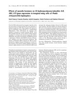
Báo cáo khoa học: "Effects of propranolol in combination with radiation on apoptosis and survival of gastric cancer cells in vitro" ppsx
Bạn đang xem bản rút gọn của tài liệu. Xem và tải ngay bản đầy đủ của tài liệu tại đây (1.9 MB, 8 trang )
RESEARC H Open Access
Effects of propranolol in combination with
radiation on apoptosis and survival of gastric
cancer cells in vitro
Xinhua Liao, Xiangming Che
*
, Wei Zhao, Danjie Zhang, Houlong Long, Prakash Chaudhary, Haijun Li
Abstract
Background: The National Comprehensive Cancer Network (NCCN) guidelines recommend radiotherapy as a
standard treatment for patients with a high risk of recurrence in gastric can cer. Because gastric cancer
demonstrates limited sensitivity to radiotherapy, a radiosensitizer might therefore be useful to enhance the
radiosensitivity of patients with advanced gastric carcinoma. In this study, we evaluated if propranolol, a b-
adrenoceptor (b-AR) antagonist, could enhance radiosensitivity and explored its precise molecular mechanism in
gastric cancer cells.
Methods: Human gastric adenocarcinoma cell lines (SGC-7901 and BGC-823) were treated with or without
propranolol and exposed to radiation. Cell viability and clonogenic survival assays were performed, and cell
apoptosis was evaluated with flow cytometry. In addition, the expression of nuclear factor B (NF-B), vascular
endothelial growth factor (VEGF), cyclooxygenase 2 (COX-2), and epidermal growth factor receptor (EGFR) were
detected by western blot and real-time reverse transcription polymerase chain reaction (PCR).
Results: Propranolol combined with radiation decreased cell viability and clonogenic survivability. Furthermore, it
also induced apoptosis in both cell lines tested, as determined by Annexin V staining. In addition, treatment with
propranolol decreased the level of NF- B and, subsequently, down-regulated VEGF, COX-2, and EGFR expression.
Conclusions: Taken together, these results suggested that propranolol enhanced the sensitivity of gastric cancer
cells to radiation through the inhibition of b-ARs and the dow nstream NF-B-VEGF/EGFR/COX-2 pathway.
Background
Gastric cancer is estimated to account for about 10% of
invasive cancers worldwide and is the second leading
cause of cancer deaths. Although the incidence of gastric
cancer has been decreasi ng, it remains a common malig-
nancy worldwide, especially in Asia [1]. Patients with gas-
tric cancer frequently experience recurrent tumors, even
aft er a curative surgical resection, because gastric cancer
is frequently diagnosed at an advanced stage. Surgical
treatment alone is not useful for patients with local and
distal recurrences. Therefore, another therapeutic modal-
ity might be useful to prevent the recurrence of advanced
gastric carcinoma. The National Comprehensive Cancer
Network (NCCN) guidelines on gastric cancer treatment
recommend radiotherapy as a standard treatment for
patients with a high risk o f recurrence, which is also sup-
ported by the clinical trial INT0116 [2]. Because gastric
cancer has limited sensitiv ity to radiotherapy, a radiosen-
sitizer is needed to overcome this problem.
It has be en reported that antagonists of cyclooxygenase
2 (COX-2), epidermal growth factor receptor (EGFR),
and vascular endothelial growth factor (VEGF) can act as
radiosensitizers to enhance therapeutic sensitivity in
many tumors [3-6]. Although associated with cell prolif-
eration, invasion, angiogenesis and metastasis, nuclear
factor B(NF-B) has been closely linked with radiore-
sistance in multiple tumors [7,8]. Numerous studies sug-
gest that prosurvival signaling mediated by NF-Bis
linked to radiation resistance and poorer clinical out-
comes among many cancers. Helen et al .reportedthat
activation of b-adrenoceptors (b-ARs) and the subse-
quent stimulation of COX-2 and VEGF expression was
* Correspondence:
Department of General Surgery, First Affiliated Hospital of Medical College of
Xi’an Jiao-Tong University, Yanta West Road 277, Xi’an 710061, PR China
Liao et al. Radiation Oncology 2010, 5:98
/>© 2010 Liao et al; licensee BioMed Ce ntral Ltd. This is an Open Access article distributed under the terms of the Creative Commons
Attribution License ( which pe rmits unrestricted use, distribution, and reproduction in
any medium, provided the original work is prope rly cited.
perhaps an important mechanism in the tumorigenic
action of nicotine in colon tumor growth [9]. It is not yet
known whether propranolol (a b-AR antagonist) can be
used as a radiosensitizer. The goal of this study was to
investigate radiosensitizing activities of propranolol in
human gastric cancer cell lines and to determine its
precise signaling pathway.
Methods
Cell culture and drug treatment
Two human gastric adenocarcinoma (HGC) cell lines,
BGC-823 and SGC-7901, were established in the Peo-
ple’ s Hospital of Peking University and China and
No.6 Hospital of Shanghai, China, respectively. These
two human gastric cancer cell lines were obtained
from the Medical Center Laboratory of Xi’an Jiaotong
University (Xi’an, China). Both cell lines were cultured
in complete Dulbecco’s modified eagle medium (Gibco,
Grand Island, NY) containing 10% (v/v) heat-inacti-
vated fetal bovine serum (Gib co, Grand Island, NY),
penicillin (100 U/mL) and streptomycin (100 mg/mL),
and they were maintained in a 37°C humidified incuba-
tor supplying 5% CO
2
. When cells reached the loga-
rithmic phase, t hey were treated with isoproterenol
(25 μmol/L) or propranolol (50 μmol/L). The concen-
trations of drugs were chosen from our previous
research. The b-AR a ntagonist propranolol and the
b-AR s timulator i soproterenol we re purchase d from
Sigma Chemical. After 24 h of drug exposure,
untreated and drug-treated cultures were irradiated at
different doses (0, 2, 4, 6, 8 and 10 Gy). X-irradiation
was performed with a n X-ray generator (Elekta Precise
Linear Accelerator, UK) at 4 Mev with a source-skin
distance of 100 cm and at a dose rate of 200 cGy/min.
Cell survival analysis
Colony formation assays were used to quantify the cyto-
toxicity of gastric cancer cells induced by treatments.
The cells were plated in six-well plates (Costar, USA) at
low densities. After overnight culture, the cells were
treated as de scribed above. The treate d cells were cul-
tured u ntil colonies formed. The colonies were washed
with PBS and stained with a crystal violet dye. The sur-
viving fraction of each irradiation dose was calculated as
the total number of colonies/(total cells inoculated×plat-
ing efficiency). A dose-survival curve was obtained for
each experiment and used f or calculating severa l survi-
val parameters. Parallel s amples were set at each radia-
tion dosage.
Cell apoptosis analysis
To detect phosphatidylserine externalization (on the
surface of cell membrane), an indi cator of early apopto-
sis, flow cytometry (FCM, BD Biosciences, USA) was
performed with PI and fluorescein isothiocyanate
(FITC)-labeled Annexin V (Joincare Biosciences, Zhuhai,
China) [10]. After treatment, the remaining intact cells
were incubated at 37°C for 24 hr, and then the cells
were washed with cold PBS at 4°C. After centrifugation
at 1500 rpm for 5 min, 500 μL of 1×binding buffer,
5 μL of FITC-lab eled Annexin V and 10 μLofPIwere
added to the cell suspension and gently mixed. After
incubation at 25°C for 10 min in the dark, the cells were
analyzed by FCM.
Real-time reverse transcription polymerase chain reaction
(real-time RT-PCR)
Total RNA was extracted from cultured cells by Tri-
Reagent (Sigma, MO, USA). To eliminate DNA contam-
ination, extracted RNA was treated with a genomic
DNA elimination mixture. Subsequently, the purified
RNA was reverse transcribed to cDNA. Expression of
b1- and b2-AR mRNA was quantified by RT-PCR
(Applied Biosystems, Inc., Foster City, CA). The expres-
sion of COX-2, VEGF and EGFR was quantified using a
real-time RT-PCR kit from Takara (Takara Biochem-
icals, Japan). Briefly, following a pre-heating step at 95°C
for 10 min, the reaction was carried out using an Icycler
(Bio-Rad, Hercules, CA) at a melting temperature of
95°C for 15 sec and an annealing temperature for 1 min
for 40 cycles. The primer sequences and annealing tem-
peratures for the six genes studied are given in Table 1.
Primers were designed according to Genbank, NCBI.
For validation, each experiment was done in triplicate.
Western blot assay
The primary antibodies recognizing the b1-adrenergic
receptor and b2-adrenergic receptor were purchased
from Abcam (Cambridge, Mass). Antibodies recognizing
COX-2, VEGF, NF-B (p65), and EGFR were purchased
from Santa Cruz Biotechnology (Santa Cruz, CA). The
nitrocellulose membrane was purchased from Millipore
(Bedford, Mass). The BCA assay kit and the chemilumi-
nescence kit we re purchased from Pierce (Rockford, Ill).
Equal amounts of protein (20 mg) of each sample, quan-
tified by the Bradford method, were electrophoresed on
10% SDS-PAGE and electrotransferred onto nitrocellu-
lose membranes (400 mA for 2 hr) using a Bio-Rad Mini
PROTEAN 3 System (Hercules, CA) according to the
standard protocol. Wet transfer was used for EGFR pro-
tein, and semi-dry transfer was used for other proteins.
The nitrocellulose membranes were then blocked with
TBS containing 10% milk powder and 0.1% Tween-20 at
37°C for 4 hr. Subsequently, the membranes were incu-
bated with a 1:200 dilution of the primary antibodies for
b1- AR, b2- AR, COX-2, VEGF, EGFR and NF- B (p65),
and a 1:500 dilution of anti-b-actin at 4°C overnight. An
antibody against rabbit or mouse IgG was used as the
Liao et al. Radiation Oncology 2010, 5:98
/>Page 2 of 8
secondary antibody corresponding to the appropriate pri-
mary antibody. Immunopositive bands were examined by
an enhanced chemiluminescence (ECL) detection system
(Amersham Bioscience, Piscataway, NJ, USA), and the
images were transferred onto an X-r ay film according to
the manufacturer’s instructions.
Statistical analysis
The results w ere expressed as the mean ± S.D. Statisti-
cal differences were estimated by one-way analysis of
variance (ANOVA) followed by Dunnett’stest.Those
p values that were less than 0.05 were co nsidere d statis-
tically significant. Analysis of the data and plotting of
the figures were performed with the aid of software
(Origin Version 7.5 and SPSS Version 13.0).
Results
Expression of b1- and b2-adrenergic receptors in SGC-
7901 and BGC-823 cells
Because propranolol is a b-adrenergic receptor antago-
nist, the expression of b1- a nd b2-ARs w as determined
at both the mRNA and protein level in SGC-7901 and
BGC-823 cells by RT-PCR and western blot. Our results
showed that b1- and b2-adrenergic receptors could be
detected at both the mRNA and protein level in both
cell lines. Figure 1 shows that expression of b1- and b2-
adrenergic receptors in SGC-7901 cells was higher than
that in BGC-823 cells.
Dose-survival curves of gastric cancer cells after different
doses of irradiation with or without propranolol pre-
treatment
To analyze the survival capability of gastric cancer cells
against propranolol induced cell death, the cell lines
SGC-7901 and BGC-823 were treated with propran olol
(50 μmol/L) 24 hr prior to irradiation, and the surviving
fraction of cells was determined in a clonogenic survival
assay. The survival curve of control and propranolol-
treated SGC-7901 and BGC-823 cells after irradiation is
shown in Figure 2. A significant difference in the colony
forming rate was found in combinati on with irradiation
and propranolol at 50 μmol/L i n SGC-7901 and BGC-
823 cells (p < 0.01) compared with irradiation alone.
Pre-treatment of SGC-7901 and BGC-823 c ells with
50 μ mol/L propranolol prior to irradiation resulted in a
significant decrease in the surviving fraction of cells and
an increase in radiation sensitivity at low irradiation
doses. The decreased survival rate in propranolol-treated
cells indicated that treatment with propranolol signifi-
cantly improved the biological effect of irradiation.
Propranolol enhances X-ray-induced gastric cancer cell
death by promoting apoptosis
To determine whether the radiosensitizing effect of pro-
pranolol is mediated by apoptosis, the effect of propranolol
on the induction of apoptosis was examined using flow
cytometric (FCM) analysis with Annexin V-PI staining.
After propranolol pre-treatment (50 μMfor24hr)and
following irradiation, FCM demonstrated an increase in
Annexin-V positive apoptotic BGC-823 and SGC-7901
cells compared with irradiation alone. Figure 3 shows that
when cells were subjected to 800 cGy irradiation in addi-
tion to propranolol, compared with irradiation alone, the
apoptosis rates were 39.73 ± 2.23% vs. 25.20 ± 0.99%, p <
0.01 (SGC-7901) and 38.69 ± 1.87% vs. 31.10 ± 1.83%, p <
0.01 (BGC-823). These data suggest that propranolol can
significantly increase cell death in both cell lines.
The effects of propranolol on radiation-induced gene
expression in gastric cancer cells
As measured by real-time RT-PCR and western blot assay,
we found that irradiation (last three groups) of BGC-823
and SGC-7901 cells down regulated the levels of NF-B
(p65) at the protein level with a subsequent decrease in
COX-2, VEGF a nd EGFR mRNA lev els (Figure 4) and
proteins (Figure 5) compared with controls. After pre-
treatment with propranolol, the expression of NF-B,
COX-2, VEGF, and EGFR was decreased and significantly
Table 1 The primer sequences and annealing temperatures for the seven genes studied
Gene Annealing temperature(°C) Primer sequence Amplicon (bp) Accession No.
b-actin 60 Forward
Reverse
ATCGTGCGTGACATTAAGGAGAAG
AGGAAGGAAGGCTGGAAGAGTG
179 NM_001101
b
1
-AR 60 Forward
Reverse
GGGAGAAGCATTAGGAGGG
CAAGGAAAGCAAGGTGGG
270 NM_000684
b
2
-AR 60 Forward
Reverse
CAGCAAAGGGACGAGGTG
AAGTAATGGCAAAGTAGCG
334 NM_000024
COX-2 57 Forward
Reverse
TTGACCAGAGCAGGCAGATG
CCAGAAGGGCAGGATACAGC
171 NM_000963.2
VEGF-A 57 Forward
Reverse
CTGGGCTGTTCTCGCTTCG
CTCTCCTCTTCCTTCTCTTCTTCC
140 NM_001025370.1
EGFR 53 Forward
Reverse
AGG ACA GCA TAG ACG ACA C
AGG ATT CTG CAC AGA GCC A
90 NM_005228.3
Liao et al. Radiation Oncology 2010, 5:98
/>Page 3 of 8
lower than the irradiation-only group. In addition, the pre-
treatment of isoproterenol had the opposite effect and
reduced the downregulation of gene expression caused by
irradiation. These results clearly suggested that treatment
with propranolol significantly improved the biological
effect of irradiation and down regulated expression of the
COX-2, VEGF and EGFR genes in gastric cancer cells,
which was mainly due to the decrease in expression of
NF-B via inhibited b-ARs.
Discussion
Gastric cancer is one of the major causes of cancer mor-
talities in the world, and radiotherapy is an important
treatment for gastric cancer patients wit h a high risk o f
recurrence. As we know, radiosensitizers have played a
key role in radiotherapy. In recent years, many research-
ers have focused on antagonists of VEGF, COX-2 and
EGFR expression as radiosensitizers [3-6], all of which
have the ability to e nhance the sensitivity to radiation.
Helen et al.reportedthatb-ARs and the downstream
COX-2 and VEGF genes played an important role in
colon tumor growth [9]. This suggests that prop ranolo l
(b-AR antagonist) may act as a radiosensiti zer of gastric
cancer. To our knowledge, this study was the first to
determine the propranolol radiosensitizing activities in
human gastric cancer cell lines a nd to investigate its
precise signaling pathway.
Based on results from the colony -forming assays, pro-
pranolol and irradiation cooperated to yield fewer and
smaller c olonies, suggesting that there was radiosensiti-
zation in the SGC-7901 and BGC-823 cell lines. In addi-
tion, propranolol showed a synergism of growth
inhibition in combination with irradiation in SGC-7901
and BGC-823 cells. On the co ntrary, isoproterenol
demonstrates anti-irradiation effects, which led to higher
Figure 1 Expression of b-ARs in human gastric cell lines SGC-7901 and BGC-823 by RT-PCR and western blotting. (A) Expression of b-
ARs in human gastric cell lines SGC-7901 and BGC-823 at the mRNA level by RT-PCR. Both of cell lines expressed b1- and b2-AR mRNA (contol
group had no cDNA). (B) Expression of b-ARs in human gastric cell lines SGC-7901 and BGC-823 at the protein level by western blotting. Both of
cell lines expressed the proteins of the b1- and b2-ARs.
Figure 2 Dose-survival curves of BGC-823 and SGC-7901 cells
after different doses of radiation with or without propranolol
(50 μmol/L) 24 hr before irradiation. Propranolol administration
before irradiation of BGC-823 (A) and SGC-7901 (B) cells; BGC-823
(C) and SGC-7901 (D) cells with irradiation. Compared with the
irradiation-only groups, the cells exposed to propranolol before
irradiation were more sensitive to irradiation.
Liao et al. Radiation Oncology 2010, 5:98
/>Page 4 of 8
survival rates than treatment with irradiation only by
using isoproterenol b efore irradiation. Furthermore, the
apoptosis assays show that the combination of propra-
nolol and irradiation leads to higher apoptosis rates
compared with irradiation only. In addition to this,
less apoptosis was observed in comparison to the
irradiation-only group caused by pre-treatment of iso-
proterenol. The apoptosis rates of these three groups
are higher than the control group. These results suggest
that propranolol (b-adrenergic receptor antagonist)
might be a useful irradiation sensiti zer in gastric cancer
therapy. Guidelines of the NCCN on gastric cancer
Figure 3 Apoptosis induction by isoproterenol or propranolol in combination with irradiation in BGC-823 and SGC-7901 cells. There was
an increasing rate of apoptosis in gastric cancer cell lines in response to the following treatments: isoproterenol before irradiation, irradiation only,
and propranolol before irradiation. The two cell lines that were treated with propranolol before irradiation had the highest apoptosis rates.
Figure 4 Quantification of mRNA expression of different genes. Analysis of mRNA expression of COX-2, VEGF and EGFR was performed on four
groups: control, radiotherapy (800 cGy) after isoproteronol (25 μM), radiotherapy (800 cGy) and radiotherapy (800 cGy) after propranolol (50 μM)
using an iCycler (Bio-Rad). Expression of COX-2, VEGF and EGFR was reduced significantly in different groups (*p < 0.05 versus the control group).
Liao et al. Radiation Oncology 2010, 5:98
/>Page 5 of 8
treatment show that r adiotherapy is a standard treat-
ment for gastric cancer patients. Taken together, radio-
therapy in combination with propranolo l can b e more
useful for patients with a high risk of recurrence in gas-
tric cancer.
Investigation of the specific mechanisms of NF-B
activation by radiation is currently a rapidly expanding
field o f research. It has been reported that NF-B plays
a key role in cellular protection against a variet y of gen-
otoxic agents including irradiation [11]. Radiation acti-
vates NF-B activity in cancer cells, thus making the
cells radioresistant [12]. Activation of NF-B by various
stimuli, including inflammation, stress and radiation,
involves degradation of the inhibitory subunit and trans-
location of activated NF-Btothenucleustoregulate
transcription [13,14]. Our results demonstrated that
treatment of BGC-823 and SGC-7901 cells with propra-
nolol reduced the levels of NF-B, suggesting that cellu-
lar r adiosensitivity is increased by propranolol-induced
NF-B inhi bition. It has been shown that NF-Bis
involved in the modulation of expression of several
proinflammatory, prometastatic and proangiogenic
genes, including COX-2, EGFR and VEGF [15]. Anti-
apoptotic COX-2 is an enzyme that con verts arachido-
nic acid to prostaglandins and is inducible by radiation
[16,17]. It is reported that COX-2 inhibitors act as
radiosensitizers in brain tumors [3]. EGFR is a member
of the ErbB family of receptors. Its stimulation by endo-
genous ligands, EGF or transforming growth factor-
alpha (TGF-a), results in activation of intracellular tyro-
sine kinases and promotes cell cycle progression. EGFR
was shown to play an influential role not only in cellular
growth and differentiation in healthy tissues, but also in
tumorigenesis and the progression of malignant disease
[18]. Now, in most studies, EGFR inhibitors are given as
a radiosensitizer to enhance the effect of radiotherapy
[19-21]. VEGF is th ought to be a critical angiogeni c fac-
tor for endothelial cell proliferation and blood vessel
formation. Thus, interfering with VEGF signaling has
become a major strategy to inhibit tumor growth and
Figure 5 Effects of isoproterenol, propranolol and/or radiotherapy on COX-2, VEGF, EGFR and NF-B (p65) proteins. SGC-7901 (A) and
BGC-823 (B) cells were treated with/without isoproterenol or propranolol for 24 hr prior to radiotherapy (800 cGy). The protein levels of COX-2,
VEGF, EGFR and NF-B were analyzed by western blot.
Liao et al. Radiation Oncology 2010, 5:98
/>Page 6 of 8
spread [22,23]. It has b een shown that anti-angiogenic
agents combined with radiotherapy improved tumor
oxygenation and increased treatment efficacy by killing
both cancer and endothelial cells [24]. It is well accepted
that the expression of EGFR, VEGF, and COX-2 is regu-
lated by NF-B [25-27]. In the present study, proprano-
lol radiosensitization effects were found to be associated
with changes in the levels of COX-2 and EGFR and
VEGF signaling molecules. It was observe d that prop ra-
nolol can act a s a radiosensitizer, which occurred via
inhibition of b-ARs and subsequent reduced NF-B
DNA-binding activity, which c oncomitantly inhibited
the expression of COX-2, EGFR and V EGF genes. In
this way, propranolol can enhance the effect of radio-
therapy on gastric cancer.
These findings, along with the present experimental
data, strongly suggest that propranolol, a b-adrenergic
receptor antagonist, plays an important role in the
radiotherapy of gast ric cancer. The present study
demo nstrates for the first time that b-adren ergic inhibi-
tion can enhance the effect of radiotherapy on gastric
cancer cells in vitro through the downregulation of NF-
B and modulation of downstream COX-2, EGFR and
VEGF gene expression. Furthermore, there is an oppo-
site effect caused by isoproterenol (b-adrenergic recep-
tor agonist) administration. These data suggest that
blockade of b-AR-stimulat ed signaling pathways could
have therapeutic implications for augmen ting the sensi-
tivity of radiotherapy on gastric cancer.
Conclusion
In conclusion, the addition of propranolol to radiother-
apy led to a decrease in gastric cancer cell survival in
vitro. Adding the drug will enhance the sensitivity of
gastric cancer cells to radiation through the inhibiti on
of b-ARs and the downstream NF-B -VEGF/EGFR/
COX-2 pathway.
Acknowledgements
The authors thank Dr. Dong Zhang for his technical assistance, who is from
the Hepatobiliary Department of First Affiliated Hospital and the Institution
of Genetic Disease Research of Xi’an Jiaotong University.
Authors’ contributions
XC and XL designed the study, coordinated the work and drafted the
manuscript. HLo, HLi and PC did the cytological work, helped with
irradiation tests, performed Western blots and PCR. WZ coordinated the
work, interpreted the data and helped drafting the manuscript. All authors
read and approved the final manuscript.
Competing interests
The authors declare that they have no competing interests.
Received: 5 August 2010 Accepted: 26 October 2010
Published: 26 October 2010
References
1. David M.R: The epidemiology of gastric cancer. Gastric Cancer 2002,
5:5-11.
2. Macdonald JS, Smalley SR, Benedeth J, Estes N, Haller DG, Ajani JA,
Gunderson LL, Jessup M, Martenson JA: Postoperative combined radiation
and chemotherapy improves disease-free survival (DFS) and overall
survival (OS) in resected adenocarcinoma of the stomach and
gastroesophageal junction: Update of the results of Intergroup Study
INT-0116 (SWOG 9008). Gastrointestinal Cancers Symposium 2004, Abstract
6.
3. Sminia P, Kuipers G, Geldof A, Lafleur V, Slotman B: COX-2 inhibitors act as
a radiosensitizer in tumor treatment. Biomed Pharmacother 2005,
59:272-275.
4. Geoerger B, Gaspar N, Opolon P, Morizet J, Devanz P, Lecluse Y, Valent A,
Lacroix L, Grill J, Vassal G: EGFR tyrosine kinase inhibition radiosensitizes
and induces apoptosis in malignant glioma and childhood
ependymoma xenografts. Int J Cancer 2008, 123:209-216.
5. Michel Z, Abderrahim Z, David A, Mahmut O: The epidermal growth factor
receptor (EGFR) in head and neck cancer: its role and treatment
implications. Radiation Oncology 2006, 1:11.
6. Wachsberger PR, Burd R, Cardi C, Thakur M, Daskalakis C, Holash J,
Yancopoulos GD, Dicker AP: VEGF trap in combination with radiotherapy
improves tumor control in U87 glioblastoma. Int J Radiat Oncol Biol Phys
2007, 67:1526-1537.
7. Graham W, Kris G, Yong X, Mahesh K, William ST: Clair Selectively
enhanced radiation sensitivity in prostate cancer cells associated with
proteasome inhibition. Oncology Reports 2006, 15:1287-1291.
8. Lee YY, Kao CL, Tsai PH, Tsai TH, Chiou SH, Wu WF: Caffeic acid phenethyl
ester preferentially enhanced radiosensitizing and increased oxidative
stress in medulloblastoma cell line. Childs Nerv Syst 2008, 24:987-994.
9. Helen PSW, Le Y, Emily KYL, Emily KKT, William KW, Cho CH: Nicotine
Promotes Colon Tumor Growth and Angiogenesis through β-Adrenergic
Activation. Toxicological Sciences 2007, 97 :279-287.
10. Vermes I, Clemens H, Helga SN, Chris R: A novel assay for apoptosis Flow
cytometric detection of phosphatidylserine expression on early
apoptotic cells using fluorescein labelled Annexin V. Journal of
Immunological Methods 1995, 17:39-51.
11. Ahmed KM, Li JJ: ATM-NF-kappaB connection as a target for tumor
radiosensitization. Curr Cancer Drug Targets 2007, 7:335-342.
12. Tamatani T, Azuma M, Motegi K, Takamaru N, Kawashima Y, Bando T:
Cepharanthin-enhanced radiosensitivity through the inhibition of
radiation-induced nuclear factor-kappaB activity in human oral
squamous cell carcinoma cells. Int J Oncol 2007, 31:761-768.
13. Voorhees PM, Dees EC, O’Neil B, Orlowski RZ: The proteasome as a target
for cancer therapy. Clin Cancer Res 2003, 9:6316-6325.
14. Adams J: The proteasome: structure, function, and role in the cell.
Cancer
Treat Rev 2003, 29:3-9.
15. Xiong HQ, Abbruzzese JL, Lin E, Wang L, Zheng L, Xie K: NF-κB activity
blockade impairs the angiogenic potential of human pancreatic cancer
cells. Int J Cancer 2004, 108:181-188.
16. Liao Z, Komaki R, Mason KA, Milas L: Role of cyclooxygenase 2 inhibitors
in combination with radiation therapy in lung cancer. Clin Lung Cancer
2003, 4:356-365.
17. Terakado N, Shintani S, Yano J, Chunnan L, Mihara M, Nakashiro K,
Hamakawa H: Overexpression of cyclooxygenase 2 is associated with
radioresistance in oral squamous cell carcinoma. Oral Oncol 2004,
40:383-389.
18. Arteaga C: Targeting HER1/EGFR: a molecular approach to cancer
therapy. Semin Oncol 2003, 30:3-14.
19. Wu RR, Wu SX, Zhao C, Xie FY, Gao JM, Hu WH, Gao YH, Li FY, Cui TT,
Lu TX: Phase II clinical trial of h-R3 combined radiotherapy for
locoregionally advanced nasopharyngeal carcinoma. Chin J Cancer 2007,
26:874-879.
20. Huang XD, Yi JL, Gao L, Xu GZ, Jin J, Yang WZ, Lu TX, Wu SX, Wu RR,
Hu WH, Xie WC, Han F, Gao YH, Gao JM, Pan JJ, Chen CB, Lang JY, Li T,
Dong Y, Fu YB, Fan L, Li BS, Li J, Wang XH, Chen BX, Gao XS, Zhang P,
Wu XW, Hu BQ: Multi-center phase II clinical trial of humanized anti-
epidermal factor receptor monoclonal antibody h-R3 combined with
radiotherapy for locoregionally advanced nasopharyngeal carcinoma.
Chin J Onco 2007, 29:197-202.
Liao et al. Radiation Oncology 2010, 5:98
/>Page 7 of 8
21. Bonner JA, Harari PM, Giralt J, Azarnia N, Shin DM, Cohen RB, Jones CU,
Sur R, Raben D, Jassem J, Ove R, Kies MS, Baselga J, Youssoufian H,
Amellal N, Rowinsky EK, Ang K: Radiotherapy plus cetuximab for
squamous-cell carcinoma of the head and neck. Engl J Med 2006,
354:567-578.
22. Ferrara N: Vascular endothelial growth factor as a target for anticancer
therapy. Oncologist 2004, 9:2-10.
23. Hicklin DJ, Ellis LM: Role of the vascular endothelial growth factor
pathway in tumor growth and angiogenesis. J Clin Oncol 2005,
23:1011-1027.
24. Teicher BA, Dupuis N, Kusomoto T, Robinson FM, Liu F, Menon K,
Coleman CN: Antiangiogenic agents can increase tumor oxygenation
and response to radiation therapy. Rad Oncol Invest 1995, 2:269-276.
25. Sclabas GM, Uwagawa T, Schmidt C, Hess KR, Evans DB, Abbruzzese JL,
Chiao PJ: Nuclear factor κB activation is a potential target for preventing
pancreatic carcinoma by aspirin. Cancer 2005, 103:2485-2490.
26. Takada Y, Kobayashi Y, Aggarwal BB: Evodiamine abolishes constitutive
and inducible NF-κB activation by inhibiting InBa kinase activation,
thereby suppressing NF-κB-regulated antiapoptotic and metastatic gene
expression, up-regulating apoptosis, and inhibiting invasion. J Biol Chem
2005, 280:17203-17212.
27. Takada Y, Murakami A, Aggarwal BB: Zerumbone abolishes NF-κB and
InBa kinase activation leading to suppression of antiapoptotic and
metastatic gene expression, upregulation of apoptosis, and
downregulation of invasion. Oncogene 2005, 24:6957-6969.
doi:10.1186/1748-717X-5-98
Cite this article as: Liao et al.: Effects of propranolol in combination
with radiation on apoptosis and survival of gastric cancer cells in vitro.
Radiation Oncology 2010 5:98.
Submit your next manuscript to BioMed Central
and take full advantage of:
• Convenient online submission
• Thorough peer review
• No space constraints or color figure charges
• Immediate publication on acceptance
• Inclusion in PubMed, CAS, Scopus and Google Scholar
• Research which is freely available for redistribution
Submit your manuscript at
www.biomedcentral.com/submit
Liao et al. Radiation Oncology 2010, 5:98
/>Page 8 of 8