Báo cáo khoa học: Effects of the antioxidant PycnogenolÒ on cellular redox systems in U1285 human lung carcinoma cells docx
Bạn đang xem bản rút gọn của tài liệu. Xem và tải ngay bản đầy đủ của tài liệu tại đây (369.52 KB, 9 trang )
Effects of the antioxidant Pycnogenol
Ò
on cellular redox
systems in U1285 human lung carcinoma cells
Valentina Gandin
1,
*, Christina Nystro
¨
m
1
, Anna-Klara Rundlo
¨
f
1
, Kerstin Jo
¨
nsson-Videsa
¨
ter
2
,
Frank Scho
¨
nlau
3
, Jarmo Ho
¨
rkko
¨
4
, Mikael Bjo
¨
rnstedt
1
and Aristi P. Fernandes
1
1 Division of Pathology, Department of Laboratory Medicine, Karolinska Institutet, Karolinska University Hospital Huddinge, Stockholm,
Sweden
2 Division of Haematology and Oncology, Department of Medicine, Karolinska University Hospital Huddinge, Stockholm, Sweden
3 Department of Pharmaceutical Chemistry, University of Mu
¨
nster, Germany
4 Almado, Turku, Finland
Oxidative stress is associated with many pathological
events, such as degenerative diseases, arteriosclerosis,
inflammatory diseases and cancer. It is believed to be
due to an imbalance in the formation and degradation
of reactive oxygen species (ROS), e.g. superoxide anion
(O
2
•)
), hydroxyl radical (OH
•)
) and hydrogen peroxide
(H
2
O
2
) [1]. ROS are highly reactive compounds derived
from oxygen, that in moderate amounts are needed for
intracellular signalling, redox regulation and as a
defence against infections. However, as the intracellular
environment is normally reduced, an excessive amount
of ROS can react with and harm important parts of the
cell such as DNA, proteins and lipids.
Pycnogenol
Ò
, a natural extract marketed as a food
supplement or herbal drug, has been shown to have
antioxidant and free radical scavenging activities [2].
Pycnogenol
Ò
is extracted from the bark of French mari-
time pine (Pinus maritima), which grows in the coastal
region of southwest France. The main constituents are
monomeric phenolic compounds (catechin, epicatechin
and taxifolin) and condensed flavonoids (procyanidines
and proanthocyanidines). These compounds are also
Keywords
antioxidant; glutathione peroxidise; oxidative
stress; Pycnogenol
Ò
; thioredoxin reductase
Correspondence
A. P. Fernandes, Division of Pathology,
Department of Laboratory Medicine,
Karolinska Institutet, Karolinska University
Hospital Huddinge, SE-14186 Stockholm,
Sweden
Fax: +46 8 58581020
Tel: +46 8 58582926
E-mail:
*Permanent address
Department of Pharmaceutical Sciences,
University of Padova, Italy
(Received 29 August 2008, revised 15
October 2008, accepted 13 November
2008)
doi:10.1111/j.1742-4658.2008.06800.x
Pycnogenol
Ò
, which is extracted from the bark of French maritime pine,
has been shown to have antioxidant and free radical scavenging activities.
Thioredoxin reductase (TrxR), glutathione peroxidase (GPx) and glutathi-
one reductase (GR) are three central redox enzymes that are active in
endogenous defence against oxidative stress in the cell. Treatment of cells
with Pycnogenol
Ò
decreased the activity of both TrxR and GPx in cells by
more than 50%, but GR was not affected. As previously reported, both
enzymes were induced after treatment with hydrogen peroxide and selenite.
The presence of Pycnogenol
Ò
efficiently decreased selenite-mediated reac-
tive oxygen species (ROS) production. Addition of Pycnogenol
Ò
after sele-
nite treatment reduced the mRNA expression and activity of TrxR to basal
levels. In contrast, the GPx activity was completely unaffected. The dis-
crepancy between TrxR and GPx regulation may indicate that transcription
of TrxR is induced primarily by oxidative stress. As TrxR is induced in
various pathological conditions, including tumours and inflammatory condi-
tions, decreased activity mediated by a non-toxic agent such as Pycnogenol
Ò
may be of great value.
Abbreviations
DTNB, 5,5¢-dithiobis(2-nitrobenzoic acid); GPx, glutathione peroxidase; GR, glutathione reductase; ROS, reactive oxygen species; SeMC,
Se-methyl-seleno-
L-cysteine; tert-BuOOH, t-butyl hydroperoxide; Trx, thioredoxin; TrxR, thioredoxin reductase.
532 FEBS Journal 276 (2009) 532–540 ª 2008 The Authors Journal compilation ª 2008 FEBS
important constituents of fruits, vegetables and other
plants [3]. Pycnogenol
Ò
has been shown to have excel-
lent radical scavenger and antioxidant properties in
model reactions that are superior to those of other fruit
and plant extracts and other antioxidants [2,3]. Pycno-
genol
Ò
has no mutagen activity according to the Ames
test, and has low acute and chronic toxicity [2]. In addi-
tion, Pycnogenol
Ò
has been shown to protect DNA
against oxidative damage in vivo [4]. Supplementation
with Pycnogenol
Ò
has been shown to have beneficial
effects on patients with retinopathy, reducing retinal
microbleeding and oedema and improving vision [5].
Pycnogenol
Ò
supplementation has also been shown to
improve endothelial function in hypertensive patients,
decreasing the hypertension [6], and to lower glucose
levels in type 2 diabetic patients [7].
Thioredoxin reductase (TrxR), glutathione peroxi-
dase (GPx) and glutathione reductase (GR) are three
important redox enzymes in the endogenous defence
against oxidative stress. As ROS are continuously
formed in all aerobic organisms during metabolism,
there is a need for effective defence systems that scav-
enge the excessive amounts of ROS. Apart from being
important in the defence against ROS, TrxR and GPx
are selenium-containing enzymes [8]. Because it is part
of important redox enzymes such as GPx and TrxR,
selenium is an essential trace element, and considered a
physiological antioxidant [9]. At low to moderate con-
centrations, selenium is known to induce the expression
of several selenoenzymes, including TrxR and GPx [10].
The biological effects of selenium are however strictly
concentration-dependent, with antioxidant properties at
low concentrations and powerful pro-oxidant effects at
moderate to high concentrations.
TrxR, together with thioredoxin (Trx) and NADPH,
comprises an important defence system against oxida-
tive stress, named the thioredoxin system [11,12]. Trx
is a small ubiquitous protein with a redox-active disul-
fide ⁄ dithiol that reduces disulfides, which is important
in many redox-regulated reactions. TrxR is an essential
protein that not only reduces Trx but also many other
substrates such as selenium compounds [12] and Q10
[13]. Mammalian thioredoxin reductases are oxido-
reductase flavoproteins, with a C-terminal active site
with a conserved GCUG sequence [14,15] that is neces-
sary for catalytic activity [16,17]. Three mammalian
genes for TrxR have previously been identified. The
first is the ‘classical’ cytosolic TrxR1 [18], with five
potential isoforms that differ at the C-terminal. The
other two, a mitochondrial and a glutaredoxin-
containing thioredoxin reductase, are named TrxR2
and TGR, respectively [19–22]. The thioredoxin system
is involved in many biological processes, such as DNA
synthesis, apoptosis and regulation of several transcrip-
tion factors [23]. Furthermore, it is known that thiore-
doxin family proteins are induced in several tumors,
and the thioredoxin system is thus suggested to be a
prime target in cancer therapy [24–26].
The aim of this study was to investigate the antioxi-
dant effects of Pycnogenol
Ò
on redox enzymes, with
emphasis on the regulation of TrxR.
Results
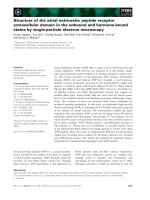
Effects of Pycnogenol
Ò
on cellular TrxR enzyme
activity
To investigate the effects of Pycnogenol
Ò
on the TrxR,
GPx and GR activity, the cells were cultivated for 48 h
supplemented with various concentrations of Pycno-
genol up to 25 lgÆmL
)1
(Fig. 1A–C). The applied
doses of Pycnogenol
Ò
(1–25 lgÆmL
)1
) did not signifi-
cantly affect cell viability (Table 1). Cells were har-
vested and homogenized and the enzyme activity was
determined. Pycnogenol
Ò
treatment decreased the
activity of both TrxR and GPx in cells by more than
50% compared to control cells, but the effect was less
pronounced for GPx compared to TrxR (Fig. 1). In
contrary to the selenoenzymes TrxR and GPx, Pycno-
genol
Ò
did not affect the enzyme activity of GR
(Fig. 1C). The reduction of TrxR and GPx activity
was dose-dependent, with maximal effects achieved at
concentrations of Pycnogenol
Ò
of 15 lgÆmL
)1
for
TrxR and 25 lgÆmL
)1
for GPx (Fig. 1).
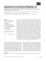
Time-dependent effects of Pycnogenol
Ò
The time-dependent effect of Pycnogenol
Ò
was inves-
tigated by cultivating U1285 cells with 25 lgÆmL
)1
Pyc-
nogenol
Ò
for 5 days. Cells were harvested at various
time points, and the TrxR and GPx enzyme activity was
measured (Fig. 2A,B). No time-dependent effects were
observed, as the activity of both enzymes decreased in
parallel with the duration of incubation for both treated
and non-treated cells, reflecting varying expression at
the various stages of the cell cycle. The presence of Pyc-
nogenol
Ò
(25 lgÆmL
)1
) resulted in a remarked decrease
in activity at all time points (Fig. 2).
Dose-dependent effects of Pycnogenol
Ò
on TrxR
enzyme activity in vitro
To determine any direct inhibition of Pycnogenol
Ò
on
TrxR enzyme activity, purified TrxR1 was incubated
with Pycnogenol
Ò
at various concentrations (up to
100 lgÆmL
)1
) and for various durations (0, 5, 10, 15
V. Gandin et al. Effects of Pycnogenol
Ò
on redox systems
FEBS Journal 276 (2009) 532–540 ª 2008 The Authors Journal compilation ª 2008 FEBS 533
and 30 min), and analysed spectrophotometrically.
There was, however, no significant effect of Pycnoge-
nol
Ò
on pure TrxR1 enzyme activity (data not shown).
Effects on the viability of U1285 cells
The toxicity of the compounds used was assessed by
means of determination of the IC
50
value for each
compound using a viability assay (Table 1). As shown
in Table 1, the IC
50
of Pycnogenol
Ò
was first reached
at doses exceeding 500 lgÆmL
)1
. This clearly demon-
A
B
C
Fig. 1. Levels of activity after treatment with Pycnogenol
Ò
in
U1285 cells. Activity of (A) TrxR, (B) GPx and (C) GR in U1285 cell
homogenates treated for 48 h with increasing concentrations of
Pycnogenol
Ò
(0–25 lgÆmL
)1
). Error bars represent the standard
error based on at least three independent experiments.
A
B
Fig. 2. TrxR and GPx activity in U1285 cell homogenates after
treatment with Pycnogenol
Ò
for various time periods. (A) TrxR
activity; (B) GPx activity. White bars indicate controls treated with
25 m
M Hepes in NaCl ⁄ P
i
. Black bars indicate cells treated with
25 lgÆmL
)1
Pycnogenol
Ò
. Error bars represent the standard error
based on at least three independent experiments.
Table 1. IC
50
values of various compounds in U1285 cells.
Compound IC
50
(mean ± SD)
a
Pycnogenol
Ò
511.12 ± 6.25 lgÆmL
)1
Na
2
SeO
3
13.25 ± 3.12 lM
Se-methyl-seleno-L-cysteine > 500 lM
tert-butyl hydroperoxide 341.58 ± 9.75 lM
a
IC
50
values were calculated by probit analysis (P < 0.05; v
2
test).
Effects of Pycnogenol
Ò
on redox systems V. Gandin et al.
534 FEBS Journal 276 (2009) 532–540 ª 2008 The Authors Journal compilation ª 2008 FEBS
strates that the antioxidant properties of Pycnogenol
Ò
(1–10 lgÆmL
)1
) occur at concentrations far lower than
the toxic dose.
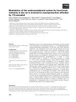
ROS production
To further explore the antioxidant effect of Pycnoge-
nol
Ò
, production of peroxide was measured in cells after
treatment with 10 lgÆmL
)1
Pycnogenol
Ò
in combination
with either selenite, Se-methyl-seleno-l-cysteine (SeMC)
or tert-butyl hydroperoxide (tert -BuOOH) (Fig. 3) at
doses marginally affecting cell viability. These com-
pounds are all known to cause oxidative stress, which is
also shown in Fig. 3. Pycnogenol
Ò
was able to efficiently
decrease ROS production by all compounds tested.
Effect of Pycnogenol
Ò
on cellular TrxR and GPx
activity during oxidative stress
To investigate the mechanisms of Pycnogenol
Ò
on
TrxR and GPx enzyme activity, the cells were exposed
to oxidative stress in the form of H
2
O
2
. The cells were
first incubated with Pycnogenol
Ò
for 48 h, followed by
H
2
O
2
treatment at a concentration of 0.1 mm and
incubation for an additional 48 h. Then the cells were
harvested and homogenized and the enzyme activity
was measured. Inhibition of enzyme activity of Pycno-
genol
Ò
in control cells is evident for both TrxR and
GPx (Fig. 4). The activity of TrxR was highly
increased after exposure to oxidative stress compared
to the control as shown in Fig. 4A. However, the
increase was reversible for TrxR after addition of
25 lgÆmL
)1
Pycnogenol
Ò
, declining to the basal level.
GPx, on the other hand, barely responded to treatment
with hydrogen peroxide, and Pycnogenol
Ò
did not
lower GPx activity in the presence of hydrogen perox-
ide (Fig. 4B).
Fig. 3. Determination of ROS production. ROS production was
detected using CM-H
2
DCFDA after 8 h treatment with 10 lgÆmL
)1
Pycnogenol
Ò
alone or in combination with 7.5 lM selenite (Se),
500 l
M Se-methyl-seleno-L-cysteine (SeMC), 750 lM tert-butyl
hydroperoxide (tert-BuOOH) or 40 l
M rotenone (positive control).
Error bars represent the standard error based on three independent
experiments. The Wilcoxon matched-pair test was used to
compare effects between control ⁄ treatment experimental set-ups:
**P < 0.01; ***P < 0.001.
A
B
Fig. 4. TrxR and GPx activity after treatment with Pycnogenol
Ò
in
combination with hydrogen peroxide. (A) TrxR activity and (B) GPx
activity in cell homogenates pre-treated with 25 lgÆmL
)1
Pycnoge-
nol
Ò
for 24 h, followed by treatment with or without 0.1 mM H
2
O
2
for an additional 48 h. Black bars indicate the addition of 0.1 mM
H
2
O
2
. Error bars represent the standard error based on three inde-
pendent experiments. The Wilcoxon matched-pair test was used to
compare effects between control ⁄ treatment experimental set-ups:
*P < 0.05; **P < 0.01; ***P < 0.001.
V. Gandin et al. Effects of Pycnogenol
Ò
on redox systems
FEBS Journal 276 (2009) 532–540 ª 2008 The Authors Journal compilation ª 2008 FEBS 535
Effect of Pycnogenol
Ò
on cellular TrxR and GPx
activity after treatment with selenite
As previously reported [27], both TrxR and GPx activ-
ity were elevated after selenite treatment (Fig. 5). In
contrast to GPx, TrxR was clearly affected by the
addition of Pycnogenol
Ò
, while GPx remained high
activity following selenite treatment even after addition
of Pycnogenol
Ò
(Fig. 5B).
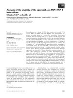
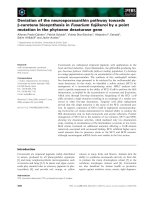
TrxR1 protein levels and TrxR1/TrxR2 mRNA
expression
As the TrxR activity was remarkably upregulated by
both selenite and hydrogen peroxide, with a reversible
affect after treatment with Pycnogenol
Ò
, the protein and
mRNA expression were investigated. TrxR1 protein lev-
els decreased remarkably, corresponding to the drop in
activity. A significant decrease in mRNA expression was
seen for both enzymes after treatment with 5 lgÆmL
)1
Pycnogenol
Ò
alone (Fig. 6). Moreover, the same pattern
with a decrease, as in mRNA expression was seen for
the activity when treated in combination with either sel-
enite or tert-BuOOH. The effect on TrxR1 mRNA was
nevertheless much more pronounced compared to TrxR2.
Discussion
Pycnogenol
Ò
has a wide variety of highly interesting
effects, including anti-inflammatory properties, benefi-
cial effects on the vascular system, and protection from
UV radiation [2]. Furthermore, Pycnogenol
Ò
is an
extremely efficient antioxidant, and it is likely that
many of its effects may be explained by its antioxidant
action. The antioxidant effects of Pycnogenol
Ò
are fur-
ther supported by our finding that Pycnogenol
Ò
decreases the production of hydrogen peroxide when
added in combination with compounds known to pro-
duce ROS, including selenite (Fig. 3). Even though our
findings are based on use of a lung carcinoma cell line,
we strongly believe that the action of Pycnogenol
Ò
is a
general mechanism and not only applicable in our
model cell system, as the antioxidant effects of Pycno-
genol
Ò
have been explored in other cell lines by us
(data not shown) and also described by others [2,3].
The antioxidant effects of Pycnogenol
Ò
clearly
resulted in a decrease in protein expression together with
a reduction in the cellular activity of TrxR. The effect
on GPx was much less pronounced, but still showed a
slight inhibition. The decrease in enzyme activity could
however not be explained by direct inhibition of TrxR,
as in vitro experiments with pure TrxR1 and Pycnoge-
nol
Ò
did not result in any inhibitory effect. Mammalian
TrxRs are complex selenoenzymes with many physiolog-
ical functions, including reduction of thioredoxin and
other low molecular weight substances, and reduction
and detoxification of hydroperoxides [9]. This reduction
may be direct or by regeneration of antioxidants includ-
ing Q10, vitamin C and lipoic acid [13,28]. Another
important function is to reduce selenium compounds,
thereby providing active selenide for the synthesis of
selenoproteins [29,30]. Our data show that addition of
hydrogen peroxide led to increased enzyme activity,
indicating that TrxR is regulated by the redox state of
the cell. Furthermore, hydrogen peroxide could in part
prevent the effect of Pycnogenol
Ò
and thus partly
restore the activity of TrxR. One possible mechanism
explaining the effect of Pycnogenol
Ò
on the activity of
TrxR is that Pycnogenol
Ò
is an extremely efficient anti-
oxidant that changes the redox status of the cell, thus
Fig. 5. TrxR and GPx activity after treatment with Pycnogenol
Ò
in
combination with selenite. (A) TrxR activity and (B) GPx activity in
U1285 cell homogenates treated with Pycnogenol
Ò
alone
(25 lgÆmL
)1
) or in combination with 5 lM selenite for 48 h. Black
bars indicate the addition of selenite. Error bars represent the stan-
dard error based on three independent experiments. The Wilcoxon
matched-pair test was used to compare effects between
control ⁄ treatment experimental set-ups: *P < 0.05; **P < 0.01.
Effects of Pycnogenol
Ò
on redox systems V. Gandin et al.
536 FEBS Journal 276 (2009) 532–540 ª 2008 The Authors Journal compilation ª 2008 FEBS
removing a stimulus for the synthesis of TrxR. How-
ever, Pycnogenol
Ò
did not suppress TrxR1 to sub-basal
levels. As shown in the ROS production experiment,
Pycnogenol
Ò
completely reversed the ROS formation
induced by both selenite and tert-BuOOH. However,
the activity of GPx was barely affected by the addition
of hydrogen peroxide after 48 h, implying a different
response to oxidative stress to that seen for TrxR.
Both TrxR and GPx are selenoproteins that are
known to be regulated by the selenium content in the
cell, with selenium generating a maximum expression
level for TrxR and GPx at around 1 lm. However,
TrxR1 is more readily saturated than GPx in response
to selenium supplementation [31], probably due to the
higher ranking of TrxR1 in terms of the hierarchy of
selenoenzymes [32]. Treatment of cells with an organic
selenium compound, SeMC, did not cause any cell
death even at very high concentrations. SeMC is a
natural selenium compound that is cleaved by b-lyase
to highly redox-active monomethyl selenol [33]. How-
ever, in the absence of b-lyase, monomethyl selenol is
not formed, explaining the low toxicity of SeMC in
U1285 cells. Despite the low cytotoxicity, SeMC caused
increased ROS production in the cells. By using an
inorganic selenium compound such as selenite as an
oxidative agent, the regulation of expression of the anti-
oxidant selenoproteins becomes much more complex.
We show here that regulation of TrxR appears to be
primarily dependent on the redox state of the cell,
rather than the selenium content. This is demonstrated
by the effect of addition of Pycnogenol
Ò
after selenite
treatment, which decreases the activity of TrxR to
levels below those of untreated cells, even though sele-
nite alone resulted in a higher response compared to
treatment with hydrogen peroxide. This was not the
case for GPx activity, which, although it was consider-
ably elevated after selenite treatment, was not at all
affected by addition of Pycnogenol
Ò
. These findings are
interesting and unexpected, as both enzymes have
strong antioxidant properties and are dependent on
selenium bioavailability. The decreased enzyme activity
and mRNA expression of TrxR caused by Pycnogenol
Ò
alone and in combination with oxidants also gives an
insight in the complex regulation of TrxR. These
observations indicate that there is a defined hierarchy
at various regulatory levels. The antioxidant-response
elements in the promoter region of TrxR, which are
targeted by nuclear erythroid-2-related factor (Nrf2)
[34], are probably one of the most important regulatory
elements for TrxR. Although our data suggest an effect
of Pycnogenol
Ò
primarily at the transcription level,
effects on translation may also occur. One example is
the strictly redox-regulated translation factor SBP2,
which is required for incorporation of selenocycstein
A
B
Fig. 6. Protein levels of TrxR1 and TrxR1 ⁄
TrxR2 mRNA expression in the U1285 cell
line. (A) Western blot comparing TrxR1
protein levels after treatment of cells for
48 h with Pycnogenol
Ò
(25 lgÆmL
)1
). Poly-
clonal TrxR1 antibodies are known to
generate more than one band due to the
existence of several protein isoforms.
(B) TrxR1 (black bars) and TrxR2 (grey bars)
mRNA levels after treatment with 5 or
10 lgÆmL
)1
Pycnogenol
Ò
alone or in
combination with 5 l
M selenite or 250 lM
tert-BuOOH for 48 h. Error bars represent
the standard error based on three indepen-
dent experiments. The Wilcoxon matched-
pair test was used to compare effects
between control ⁄ treatment experimental
set-ups: *P < 0.05; **P < 0.01;
***P < 0.001.
V. Gandin et al. Effects of Pycnogenol
Ò
on redox systems
FEBS Journal 276 (2009) 532–540 ª 2008 The Authors Journal compilation ª 2008 FEBS 537
into selenoproteins. SBP2 is translocated to the nucleus
when oxidized, resulting in translational inhibition of
TrxR [8]. The less pronounced effects seen for TrxR2
may be explained by the localization of TrxR2 in the
mitochondria, which may be slightly less affected than
the cytosol under these conditions. As TrxR is induced
in various pathological conditions, including tumours
and inflammatory conditions [35], decreased activity
mediated by a non-toxic agent such as Pycnogenol
Ò
would be beneficial, and may offer a mechanistic expla-
nation for the effects of Pycnogenol
Ò
.
Experimental procedures
Chemicals
5,5¢-dithiobis(2-nitrobenzoic acid) (DTNB), guanidine HCl,
sodium deoxycholate, sodium selenite, glutathione, GPx
from bovine erythrocytes, insulin from bovine pancreas, and
NADPH were all purchased from Sigma (St Louis, MO,
USA). Baker’s yeast GR was obtained from Fluka (Buchs,
Switzerland), Escherichia coli Trx1 was purchased from Pro-
mega, and recombinant rat TrxR1 was generously provided
by Elias Arne
´
r (Department of MBB, Karolinska Institutet,
Stockholm, Sweden). Pycnogenol
Ò
was kindly provided by
Horphag Research Ltd (Geneva, Switzerland). Se-methyl-
seleno-l-cysteine (SeMC) was purchased from PharmaSe
(Lubbock, TX, USA).
Cell line
The cells (U1285, a small-cell lung carcinoma cell line [36])
were cultured in RPMI-1640 medium with GlutaMAX-1
and 25 mm Hepes (Invitrogen, Paisley, UK), supplemented
with 10% heat-inactivated fetal bovine serum. The cells
were maintained in a humidified incubator with 5% CO
2
at
37 °C. The cells were cultured in the presence of indicated
concentrations of Pycnogenol
Ò
for various time periods.
The Pycnogenol
Ò
powder was solved in NaCl ⁄ P
i
containing
25 mm Hepes. The effect of Pycnogenol
Ò
was also studied
in combination with selenite, SeMC, tert-butyl hydroper-
oxide (tert-BuOOH) and H
2
O
2
. Selenite, SeMC or tert-
BuOOH, was added together with Pycnogenol
Ò
, while
H
2
O
2
was added after 48 h of Pycnogenol
Ò
pre-treatment.
Preparation of cell homogenates
Buffer (50 mm Tris ⁄ HCl pH 7.6 and 1 mm EDTA) was
added to the cell pellet, after which it was kept on ice. The
cells were sonicated three times for 15 seconds, followed by
centrifugation at 25 200 g for 10 min at 2 °C. The super-
natants were stored at )20 ° C for later enzyme activity stu-
dies. The protein concentrations in the cell homogenates
were determined by the Biuret method [37].
TrxR enzyme activity assay
The activity of the TrxR enzyme was measured through a
coupled reaction in the cell homogenates essentially as
described previously [38]. From each homogenate, 200 lg
proteins were incubated with 80 mm Hepes (pH 7.5),
0.9 mgÆmL
)1
NADPH, 6 mm EDTA, 2 mgÆmL
)1
insulin and
10 lm E. coli Trx at 37 °C for 20 min in a final volume of
120 lL. The reaction was terminated by addition of 500 lL
DTNB (0.4 mgÆmL
)1
) with 6 m guanidine HCl in 0.2 m
Tris ⁄ HCl pH 8.0. The absorbance at 412 nm was measured
using a SpectraMax 250 (Molecular Devices, Sunnyvale, CA,
USA) within 20 min. To determine the effects of Pycnoge-
nol
Ò
on pure TrxR1, 10 nm recombinant rat TrxR1 was used
instead of the homogenate, with the addition of only 5 lm
E. coli Trx1.
GR enzyme activity assay
Determination of the GR enzyme activity was performed as
for the TrxR1 enzyme activity except that insulin was
replaced by 1 mm oxidized glutathione, and 10 lgof
protein was incubated for each sample.
GPx enzyme activity assay
The GPx enzyme activity was measured through a coupled
reaction as described previously [39]. A modified protocol
was created to fit a 96-well plate. For each cell homoge-
nate, 200 lg of protein was incubated for 3 min at 25 °C
with 0.1 m Tris ⁄ HCl buffer (pH 7.6), 2 mm EDTA, 2 mm
NaN
3
,4mm glutathione, 10 units of GR and 0.8 mm
NADPH in a total volume of 195 lL. After incubation,
H
2
O
2
was added to a final concentration of 10 mm as
substrate for the GPx, and the absorbance at 340 nm was
recorded.
Viability assay
In order to investigate the susceptibility of the cells to
treatment with the various compounds, a Cell Prolifera-
tion Kit II (XTT) from Roche Diagnostics (Mannheim,
Germany) was used. Estimated IC
50
values from these
trials were used to decide an appropriate range of con-
centrations for treatment in all experiments conducted.
The viability assay was performed in 96-well flat-bot-
tomed culture dishes with 100 lL medium ⁄ well at
approximately 10% cell confluence. Cells were subjected
to treatment for 48 h, followed by an incubation time of
4 h before measurement. Absorbance was measured at
490 nm (with a reference wavelength of 650 nm sub-
tracted) using a SpectraMax 250. All samples were
measured in triplicate, and the entire assay was repeated
three times.
Effects of Pycnogenol
Ò
on redox systems V. Gandin et al.
538 FEBS Journal 276 (2009) 532–540 ª 2008 The Authors Journal compilation ª 2008 FEBS
ROS production
Intracellular ROS production was detected using non-
fluorescent compound 5(6)-chloromethyl-2¢,7¢-dichlorodihy-
drofluorescein diacetate (CM-H
2
DCFDA) (Invitrogen).
CM-H
2
DCFDA undergoes deacetylation by intracellular
esterases, and the product quantitatively reacts with oxygen
species inside the cell to produce the fluorescent dye 5(6)-
carboxy-2¢,7¢-dichlorofluorescein (CM-DCF). Briefly,
U1285 cells (10
4
per well) were grown for 24 h in a 96-well
plate in RPMI-1640 without phenol red. Subsequently, the
medium was removed and the cells were incubated for
45 min in the dark with 10 lm CM-DCFDA in NaCl ⁄ P
i
.
Excess probe was washed out with NaCl ⁄ P
i
, and cells were
incubated with the reported concentrations of tested com-
pounds for 8 h. The increase in fluorescence was deter-
mined at wavelengths of 485 nm (excitation) and 527 nm
(emission) on a SpectraMax 250.
Western blotting
Samples were analysed on 7.5% SDS–PAGE at 150 V,
followed by semi-dry electroblotting to a nitrocellulose mem-
brane for 1 h at 100 V. Membranes were probed with
anti-TrxR1 (1 : 2000, Upstate, Lake Placid, NY, USA) and
incubated for 1 h at room temperature. The membranes were
further incubated with a horseradish peroxidase-conjugated
secondary antibody (1 : 3000, DakoCytomation, Glostrup,
Denmark). Bound antibodies were detected using a chemilu-
minescence Western Lightning kit (PerkinElmer, Boston,
MA, USA) according to the manufacturer’s instructions.
Real-time PCR
Total RNA was isolated using and RNeasy mini kit (Qiagen,
Hilden, Germany), according to the manufacturer’s proto-
col. RNA quantification was performed using a Ribogreen
RNA quantification kit (Molecular Probes, Eugene, OR,
USA) according to the supplied ‘high range’ protocol with
rRNA as the standard. Synthesis of cDNA was performed
by reverse transcription of 2 lg of RNA using the Omni-
script reverse transcription kit (Qiagen) with oligo(dT)
12–18
as primer (final concentration 0.1 lg Æ lL
)1
). Real-time quan-
titative PCR was performed on a Bio-Rad ICycler (Bio-Rad,
Hercules, CA, USA) with 20 ng of cDNA per reaction in
triplicate on 96-well plates using Platinum SYBR Green
qPCR super mix (Invitrogen). Determination of TrxR1
mRNA levels was performed as described previously [40].
TrxR2 mRNA was analyzed using the same program as for
TrxR1 but with forward primer 5¢-TCAGAAGATCC
TGGTGGACTCC-3¢ and reverse primer 5¢-TCGTGGG
AACATTGTCGTAGTC-3¢, with concentration of 300 nm
for each primer. The results were analysed using the 2
ÀDDC
t
method. The C
T
value cut-off was set at 32 cycles. The effi-
ciency of the primer sets was 90 ± 5%.
Acknowledgements
This investigation was supported by Radiumhemmets
Research Society, Horphag Research Ltd, the Swedish
Medical Association, Karolinska Institutet Research
Grants, and the Swedish Cancer Society.
References
1 Nordberg J & Arne
´
r ES (2001) Reactive oxygen species,
antioxidants, and the mammalian thioredoxin system.
Free Radic Biol Med 31, 1287–1312.
2 Rohdewald P (2002) A review of the French maritime
pine bark extract (Pycnogenol
Ò
), a herbal medication
with a diverse clinical pharmacology. Int J Clin Phar-
macol Ther 40, 158–168.
3 Packer L, Rimbach G & Virgili F (1999) Antioxidant
activity and biologic properties of a procyanidin-rich
extract from pine (Pinus maritima) bark, pycnogenol.
Free Radic Biol Med 27, 704–724.
4 Rohdewald P (2005) Pycnogenol
Ò
protects DNA
against oxidative damage in vivo. Phytother Res 19, 262.
5 Spadea L & Balestrazzi E (2001) Treatment of vascular
retinopathies with Pycnogenol
Ò
. Phytother Res 15, 219–
223.
6 Liu X, Wei J, Tan F, Zhou S, Wurthwein G & Rohde-
wald P (2004) Pycnogenol
Ò
, French maritime pine bark
extract, improves endothelial function of hypertensive
patients. Life Sci 74, 855–862.
7 Liu X, Zhou HJ & Rohdewald P (2004) French maritime
pine bark extract Pycnogenol
Ò
dose-dependently lowers
glucose in type 2 diabetic patients. Diabetes Care 27, 839.
8 Papp LV, Lu J, Holmgren A & Khanna KK (2007)
From selenium to selenoproteins: synthesis, identity,
and their role in human health. Antioxid Redox Signal
9, 775–806.
9 Bjo
¨
rnstedt M, Hamberg M, Kumar S, Xue J & Holmgren
A (1995) Human thioredoxin reductase directly reduces
lipid hydroperoxides by NADPH and selenocystine
strongly stimulates the reaction via catalytically gener-
ated selenols. J Biol Chem 270, 11761–11764.
10 Gladyshev VN & Kryukov GV (2001) Evolution of sel-
enocysteine-containing proteins: significance of identifi-
cation and functional characterization of selenoproteins.
Biofactors 14, 87–92.
11 Arne
´
r ES & Holmgren A (2000) Physiological functions
of thioredoxin and thioredoxin reductase. Eur J Bio-
chem 267, 6102–6109.
12 Bjo
¨
rnstedt M, Kumar S, Bjo
¨
rkhem L, Spyrou G &
Holmgren A (1997) Selenium and the thioredoxin and
glutaredoxin systems. Biomed Environ Sci 10, 271–279.
13 Xia L, Nordman T, Olsson JM, Damdimopoulos A,
Bjo
¨
rkhem-Bergman L, Nalvarte I, Eriksson LC, Arne
´
r
ES, Spyrou G & Bjo
¨
rnstedt M (2003) The mammalian
cytosolic selenoenzyme thioredoxin reductase reduces
V. Gandin et al. Effects of Pycnogenol
Ò
on redox systems
FEBS Journal 276 (2009) 532–540 ª 2008 The Authors Journal compilation ª 2008 FEBS 539
ubiquinone. A novel mechanism for defense against
oxidative stress. J Biol Chem 278, 2141–2146.
14 Gladyshev VN, Jeang KT & Stadtman TC (1996) Selen-
ocysteine, identified as the penultimate C-terminal resi-
due in human T-cell thioredoxin reductase, corresponds
to TGA in the human placental gene. Proc Natl Acad
Sci USA 93, 6146–6151.
15 Tamura T & Stadtman TC (1996) A new selenoprotein
from human lung adenocarcinoma cells: purification,
properties, and thioredoxin reductase activity. Proc Natl
Acad Sci USA 93, 1006–1011.
16 Zhong L, Arne
´
r ES, Ljung J, Aslund F & Holmgren A
(1998) Rat and calf thioredoxin reductase are homolo-
gous to glutathione reductase with a carboxyl-terminal
elongation containing a conserved catalytically active
penultimate selenocysteine residue. J Biol Chem 273,
8581–8591.
17 Zhong L & Holmgren A (2000) Essential role of sele-
nium in the catalytic activities of mammalian thioredox-
in reductase revealed by characterization of
recombinant enzymes with selenocysteine mutations.
J Biol Chem 275, 18121–18128.
18 Holmgren A (1977) Bovine thioredoxin system. Purifica-
tion of thioredoxin reductase from calf liver and thymus
and studies of its function in disulfide reduction. J Biol
Chem 252, 4600–4606.
19 Spyrou G, Enmark E, Miranda-Vizuete A & Gustafs-
son J (1997) Cloning and expression of a novel mam-
malian thioredoxin. J Biol Chem 272, 2936–2941.
20 Damdimopoulos AE, Miranda-Vizuete A, Treuter E,
Gustafsson JA & Spyrou G (2004) An alternative splic-
ing variant of the selenoprotein thioredoxin reductase is
a modulator of estrogen signaling. J Biol Chem 279,
38721–38729.
21 Sun QA, Zappacosta F, Factor VM, Wirth PJ,
Hatfield DL & Gladyshev VN (2001) Heterogeneity
within animal thioredoxin reductases. Evidence for
alternative first exon splicing. J Biol Chem 276, 3106–
3114.
22 Lescure A, Gautheret D, Carbon P & Krol A (1999)
Novel selenoproteins identified in silico and in vivo by
using a conserved RNA structural motif. J Biol Chem
274, 38147–38154.
23 Gromer S, Urig S & Becker K (2004) The thioredox-
in system – from science to clinic. Med Res Rev 24,
40–89.
24 Powis G & Kirkpatrick DL (2007) Thioredoxin signal-
ing as a target for cancer therapy. Curr Opin Pharmacol
7, 392–397.
25 Urig S & Becker K (2006) On the potential of thiore-
doxin reductase inhibitors for cancer therapy. Semin
Cancer Biol 16, 452–465.
26 Nguyen P, Awwad RT, Smart DD, Spitz DR & Gius D
(2006) Thioredoxin reductase as a novel molecular
target for cancer therapy. Cancer Lett 236, 164–174.
27 Spyrou G, Bjo
¨
rnstedt M, Skog S & Holmgren A
(1996) Selenite and selenate inhibit human lymphocyte
growth via different mechanisms. Cancer Res 56,
4407–4412.
28 Arne
´
r ES, Nordberg J & Holmgren A (1996) Efficient
reduction of lipoamide and lipoic acid by mammalian
thioredoxin reductase. Biochem Biophys Res Commun
225, 268–274.
29 Kumar S, Bjo
¨
rnstedt M & Holmgren A (1992) Selenite
is a substrate for calf thymus thioredoxin reductase and
thioredoxin and elicits a large non-stoichiometric oxida-
tion of NADPH in the presence of oxygen. Eur J Bio-
chem 207, 435–439.
30 Bjo
¨
rnstedt M, Kumar S & Holmgren A (1992) Seleno-
diglutathione is a highly efficient oxidant of reduced thi-
oredoxin and a substrate for mammalian thioredoxin
reductase. J Biol Chem 267, 8030–8034.
31 Reeves PG, Leary PD, Gregoire BR, Finley JW, Lind-
lauf JE & Johnson LK (2005) Selenium bioavailability
from buckwheat bran in rats fed a modified AIN-93G
torula yeast-based diet. J Nutr 135, 2627–2633.
32 Berry MJ (2005) Insights into the hierarchy of selenium
incorporation. Nat Genet 37, 1162–1163.
33 Suzuki KT, Doi C & Suzuki N (2006) Metabolism of
76Se-methylselenocysteine compared with that of
77Se-selenomethionine and 82Se-selenite. Toxicol Appl
Pharmacol 217, 185–195.
34 Hintze KJ, Wald KA, Zeng H, Jeffery EH & Finley JW
(2003) Thioredoxin reductase in human hepatoma cells
is transcriptionally regulated by sulforaphane and other
electrophiles via an antioxidant response element.
J Nutr 133, 2721–2727.
35 Becker K, Gromer S, Schirmer RH & Muller S (2000)
Thioredoxin reductase as a pathophysiological factor
and drug target. Eur J Biochem 267, 6118–6125.
36 Bjo
¨
rkhem-Bergman L, Jonsson K, Eriksson LC, Olsson
JM, Lehmann S, Paul C & Bjo
¨
rnstedt M (2002) Drug-
resistant human lung cancer cells are more sensitive to
selenium cytotoxicity. Effects on thioredoxin reductase
and glutathione reductase. Biochem Pharmacol 63,
1875–1884.
37 Gornall AG, Bardawill CJ & David MM (1949) Deter-
mination of serum proteins by means of the biuret reac-
tion. J Biol Chem 177, 751–766.
38 Holmgren A & Bjo
¨
rnstedt M (1995) Thioredoxin and
thioredoxin reductase. Methods Enzymol 252, 199–208.
39 Lawrence RA & Burk RF (1976) Glutathione peroxi-
dase activity in selenium-deficient rat liver. Biochem
Biophys Res Commun 71, 952–958.
40 Rundlo
¨
f AK, Fernandes AP, Selenius M, Babic M,
Shariatgorji M, Nilsonne G, Ilag LL, Dobra K &
Bjo
¨
rnstedt M (2007) Quantification of alternative
mRNA species and identification of thioredoxin reduc-
tase 1 isoforms in human tumor cells. Differentiation
75, 123–132.
Effects of Pycnogenol
Ò
on redox systems V. Gandin et al.
540 FEBS Journal 276 (2009) 532–540 ª 2008 The Authors Journal compilation ª 2008 FEBS