Báo cáo khoa học: "Cell death by the quinoxaline dioxide DCQ in human colon cancer cells is enhanced under hypoxia and is independent of p53 and p21" pdf
Bạn đang xem bản rút gọn của tài liệu. Xem và tải ngay bản đầy đủ của tài liệu tại đây (2.01 MB, 13 trang )
RESEA R C H Open Access
Cell death by the quinoxaline dioxide DCQ in
human colon cancer cells is enhanced under
hypoxia and is independent of p53 and p21
Mona El-Khatib
1
, Fady Geara
2
, Makhluf J Haddadin
3
, Hala Gali-Muhtasib
1*
Abstract
Introduction: We have shown that the radio sensitizer DCQ enhances sensitivity of HCT116 human colon can cer
cells to hypoxia. However, it is not known whether the p53 or p21 genes influence cellular response to DCQ. In
this study, we used HCT116 that are either wildtype for p53 and p21, null for p53 or null for p21 to understand the
role of these genes in DCQ toxicity.
Methods: HCT116 cells were exposed to DCQ and incubated under normoxia or hypoxia and the viability, colony
forming ability, DNA damage and apoptotic responses of these cells was determined, in addition to the
modulation of HIF-1a and of p53, p21, caspase-2, and of the ataxia telangiectasia mutated (AT M) target PIDD-C.
Results: DCQ decreased colony forming ability and viability of all HCT116 cells to a greater extent under hypoxia
than normoxia and the p21
-/-
cell line was most sensitive. Cells had different HIF-1a responses to hypoxia and/or
drug treatment. In p53
+/+
, DCQ significantly inhibited the hypoxia-induced increases in HIF-1 a protein, in contrast
to the absence of a significant HIF-1a increase or modulation by DCQ in p21
-/-
cells. In p53
-/-
cells, 10 μM DCQ
significantly reduced HIF-1a expression, especially under hypoxia, despite the constitutive expression of this protein
in control cells. Higher DCQ doses induced PreG
1
-phase increase and apoptosis, however, lower doses caused
mitotic catastrophe. In p53
+/+
cells, apoptosis correlated with the inc reased expression of the pro-apoptotic
caspase-2 and inhibition of the pro-survival protein PIDD-C. Exposure of p53
+/+
cells to DCQ induced single strand
breaks and triggered the activation of the nuclear kinase ATM by phosphorylation at Ser-1981 in all cell cycle
phases. On the other hand, no drug toxicity to normal FHs74 Int human intestinal cell line was observed.
Conclusions: Collectively, our findings indicate that DCQ reduces the colony survival of HCT116 and induces
apoptosis even in cells that are null for p53 or p21, which makes it a molecule of clinical significance, since many
resistant colon tumors harbor mutations in p53.
Introduction
Hypoxia develops due to the inadequate vascularization
during early tumor development and is believed t o be
the major f actor causing tumor resistance to r adiother-
apy and chemotherapy [1]. Numerous gene products,
which are activated under hypoxia, are involved in
tumor metastasis and neoangiogenesis. On the other
hand, hypoxic cells contain high levels of bioreductive
enzymes and thus represent a therapeutic target if
directly targeted by hypoxia-activated drugs [2].
Quinoxaline 1,4-dioxides (QdNOs) are the prototype for
current heterocyclic N-oxide anticancer agents such as 3-
amino-1,2,4-benzotriazine 1,4-dioxide (Tirapazamine-
TPZ). Among four QdNOs tested, we found DCQ (2-
benzoyl-3-phenyl 6,7-dichloroquinoxaline 1,4-dioxide) to
be the most effective hypoxic cytotoxin [3-6]. Although
DCQ is not a benzotriazine 1,4-dioxide like TPZ, it resem-
bles TPZ in that these two compounds are electron-poor
by virtue of the formal positive charges held by the two
nitrogens of the N-O functions in each of them. In fact,
DCQ is believed to be more electron-poor than TPZ
because it has more electron attracting substituents: the 2-
benzoyl group and the 6,7-dichloro substituents. These
substituents render the quinoxa line 1,4- dioxid e moiety
* Correspondence:
1
Department of Biology, American University of Beirut, Beirut, Lebanon
Full list of author information is available at the end of the article
El-Khatib et al. Radiation Oncology 2010, 5:107
/>© 2010 El-Khatib et al; licensee BioMed Central Ltd. This is an Open Access article distributed under the terms of the Creative
Commons Attribution License ( nses/by/2.0), which permits unrestricted use, distribution, and
reprodu ction in any medium, provide d the original work is properly cited.
more receptive to an electron from a donor. Furthermore,
and in analogy with the mechanism of action of TPZ [7],
the radical that results from addition of an electron to C2
of DCQ is more stable, by resonance, and therefore longer
lasting and more damaging to DNA than the radical
resulting from the addition of an electron to TPZ.
DCQ was shown by our group to reduce cell growth
in T-84 human colon cancer cells, and in SP-1 keratino-
cyte cell line, under both normoxia a nd hypoxia; how-
ever, drug toxicity was greater in cells exposed to
hypoxia [3]. DCQ was found to decrease the expre ssion
levels of the hypoxia inducible factor (HIF-1a)mRNA
and protein in the human colon carcinoma cell line
T-84, and in EMT6 mouse mammary carcinoma cells
and Lewis Lung Carcinoma (LLC) cells [4,8]. We also
showed that DCQ inhibited cell proliferation and
induced apoptosis in colon T-84 cancer cell lines under
normoxia via the inhibition of the extracellular signal
regulated kinase (ERK) phosphorylation and reduction
in Bcl-2a protein [9]. While in adult T-cell leukemia,
DCQ reduced cell proliferation by decreasing Tumor
Growth Factor (TGF)-a, a key mediator of growth sti-
mulation with mitogenic effects, and by increasing the
mRNA and protein expression levels of the proapoptotic
TGF-b1 [6]. When studying the efficacy of DCQ as a
normoxic radiosensitizer, clonogenic survival assays in
LLC and EMT6 cell lines revealed an enhancement of
the radiation effect [8,10]. In vivo, DCQ in combination
with radiation delayed the growth of LLC tumors
injected in C57BL6 mice, reduced the mean tumor
volume by 80% and inhibited tumor angiog enesis [8]. In
a recent s tudy, DCQ was found to induce single strand
breaks (SSB) in DNA of DLD-1 human colon cancer
cells, and both SSB and double strand breaks (DSB) in
EMT6 cells [5,11].
DNA damage, in particular DSBs, imposes a critical
threat to the survival of cells if left unrepaired [12]. At
very early stages of the DNA damage response, cells acti-
vate the DNA damage checkpoint ATM, a member of
phosphoinositide 3 kinase-related kinase (PIKK) which is
involved in DNA repair [ 13]. ATM activation, in turn,
leads to the phosphorylation of p53, thereby blocking its
interactions with MDM2, and causing p53 stabilization.
This, in turn, stimulates the expression of the cyclin-
dependent kinase (CDKs) inhibitor p21. Through its
negative effects on various CDKs, p21 inhibits G1/S and
G2/M transitions. Thus, increased p53 levels due to the
ATM-p53-p21 pathway activation lead to cell-cycle
arrest, repair, and cell death [14]. Tumor cells that harbor
defective p53 have no such checkpoint mechanisms,
which favor their clonal outgrowth. The activation of
ATM also leads to the activation of PIDD (p53-induced
protein with a death domain), an important target gene
in a signaling pathway ini tiated by p53. The tumor
suppressor protein p53 has been also fo und to be acti-
vated in response to cellular stress, chemotherapeutic
drugs and hypoxia [15].
If DNA damage is severe, the initiator caspase-2 is
activated. This caspase possesses a caspase recruitment
domain (CARD) that allows it to interact with PIDD.
Caspase-2 activation can be initiated in the PIDDosome,
the assembly of which is mediated by PIDD autoproces-
sing to generate a PIDD-CC fragment necessary for cas-
pase-2 activation [16] . A recent study has demonst rated
that p53 controls the expression of PIDD that, in turns
recruits procaspase-2 by interaction with its prodomain
[16]. The resulting complex activates caspase-2 without
interdomain cleavage of cas pase-2 [16]. The activation
of caspase-2 within the PIDDosome complex results in
cytochrome c release and the activation of other cas-
pases which are involved in the mitochondria-mediated
apoptotic pathway [17]. Caspase-2 activation has been
shown to be involved i n metaphase-associated mitotic
catastrophe [17], which is characterized by multinu-
cleated giant cells with nuclear envelopes forming
around individual clusters of mis-segregated uncon-
densed chromosomes [17].
This project aimed to investigate the cytotoxicity of
DCQ in HCT116 human colorectal cancer cell lines that
are either wildtype for p53 and p21, null for p53, or null
for p21 to determine the role of these genes in cellular
response to DCQ. Since DCQ has been previously
shown to exhibit enhanced toxicity in hypoxic tumor
cells, its activity was determined in HCT116 cells
exposed to either normoxia or hypoxia. We also investi-
gated if DCQ causes apoptosis, induces SSB and acti-
vates the ATM repair pathway in human colon canc er
cells.
Methods
Chemicals
Propidium iodide (PI), YOYO-1 dye, fluorescein isothio-
cyanate (FITC) goat anti-mouse IgG (H+L), and
5-(and-6)-chloromethyl-2’,7’-dichlordihydrofluorescein
diacetate, acetyl ester (CM-H
2
DCFDA) were purchased
from Molecular Probes (Eugen e, Oregon, US). RNase A,
and dimethylsulfoxide (DMSO) were obtained from
Sigma Chemical Company (St. Loui s, Missouri, US).
Protease Inhibitor was from Roche Applied Science
(Penzberg, Germany). DCQ was synthesized from 5,6-
dichlorobenzofurazan oxide and dibenzoylmethane by
the Beirut Reaction [18].
Cell culture and treatments
FHs74Int normal human intestinal cells were cultured in
Hybri-Care medium supplemented with 30 ng/ml epi-
dermal growth factor. HCT116 (p53
+/+
)humancolon
cancer cells were maintained in RPMI 1640 with 25
El-Khatib et al. Radiation Oncology 2010, 5:107
/>Page 2 of 13
mM Hepes and L-Glutamine. HCT116 (p53
-/-
)and
HCT116 (p21
-/-
) cells were grown in Dulbecco’sModi-
fied Eagle Medium (DMEM) supplemented with sodium
pyruvate and 4500 mg/l glucose. All media were supple-
mented with 1% Penicillin-Streptomyci n (100 U/ml) and
10% heat-inactivated FBS. All cells were obtained from
ATCC and maintained in a humidified atmosphere of
5% CO
2
and 95% air. 10 mg of DCQ was dissolved in
1 ml of DMSO and stored in a brown eppendorf at 4°C
and then diluted in media to attain the drug concentra-
tions of u p to 10 μM. For hypoxia exposure, cells were
placed in a tightly sealed anaerobic gas chamber, Bac-
tron III (SHEL LAB, UK) at 37°C and oxygen level <
2%. T he desired oxygen level was monitored by an
Ohmeda Oxymeter (Datex-Ohmeda, Louisville CO,
USA) and maintaine d by pumping a gas mixture com-
posed of 1% O
2
,5%CO
2
,and94%N
2
.After6hrof
hypoxia exposure, cells were replated with drug-free
media and incubated under normal oxygen for clono-
genic survival assays.
Viability and clonogenic survival
For viability assays, HCT116 or FHs74Int (1.2 × 10
5
cells/ml) were cultured in 96-well plates and treated
with drugs 24 hr s after plating. Antineoplastic effects
were studied 6 hrs or 24 hrs after treatment by the non-
radioactive cell proliferation kit (Promega Corporation,
Madison, USA), an MTT-based method which measures
the ability of metabolically active cells to convert tetra-
zolium salt into a formazan product and its absorbance
is recorded at 570 nm [19]. For clonogenic survival stu-
dies, cells were treated with DCQ for 12 h r under nor-
moxia or hypoxia. The n they were trypsinized, replated
at low densities (300-5000 cells) in T-25 flasks, and left
for 8-14 days in the incubator. Subsequently, cells were
washed with PBS, and stained with 1 ml of aqueous
0.5% solution of crystal violet. Colonies having more
than 50 cells were counted. The plating efficiency (PE),
defined as the ability of control cells to survive and
grow into colonies, was calculated as: PE = colonies
counted in control/plating density of control. Surviving
fraction (SF) for each treatment was calculated as: SF =
colonies counted/[cells plated × (PE/100)]. The SF value
of each treatment was then plotted.
Flow cytometric analysis
Cells were plated in 60-mm dishes (1.2 × 10
5
cells/ml),
treated with different DCQ concentra tions at 50% con-
fluency, and i ncubated for 6 hrs under either normoxia
or hypoxia, then harvested and fixed in 70% ethanol.
Supernatants containing the dead cells were collected
and attached live cells were harvested by 2× trypsin and
added to the supernatant. Flow cytometry analysis of
Propidium Iodide-stained DNA was done as described
previously [19]. Cell Quest program was used to deter-
mine the percentages of cells in various cell cycle
phases. Pre-G
1
cells with DNA content < 2n represent
apoptotic or necrotic cells.
Hoechst staining
Cells were plated in 6-well plates at 1.2 × 10
5
and trea-
ted at 50% confluency with DCQ ( 2.5 or 5 μM) for
6 hrs under normoxia or hypoxia. The drug was then
removed, and cells were washed with 1× PBS and fixed
using 70% ethanol for 24 hrs. Next day, cells were
placed in wet chambers to prevent dehydration, a stock
of Hoechst stain (1:100) was prepared and 100 μlofthe
100× diluted Hoechst stain (from the stock) was added
to each slide and incubated for 10 min. A drop of fluor-
osave (antifade) was added on the slides which were
covered with coverslips and kept in the dark at 4°C.
Annexin V
Cells were collected along with the supernatant and cen-
trifuged at 1500 rpm for 10 min, 4°C. The pellet was
washed with PBS and centrifuged at 1500 rpm for
10 min, 4°C. The pellet was resuspended in 100 μl
Annexin-V-Fluos labeling solution (20 μl annexin
reagent and 20 μlPI(50μg/ml) in 1000 μlincubation
buffer pH 7.4 (10 mM Hepes/NaOH, 140 mM NaCl,
5mMCaCl
2
). The samples were incubated for 15 min
at room temperature and 0.4 ml incubation buffer was
added. The cellular f luorescence was then measured by
flow cytometry using a Fluorescence Activated Cell
Sorter (FACS) flow cytometer (Becton Dickinson,
Research Triangle, NC).
Western blot
Cellular protein extracts were prepared and proteins
were quantified as described previously [19]. 50 μgof
whole cell lysate was separated by SDS-PAGE (12%
gels) and transferred to PVDF membranes (Amersham
Pharmacia Biotech, Buckinghamshire, UK) in cold
transfer buffer at 30 Volts overnight. The membranes
were probed with the primary antibodies: p21 ((C-19)-
G), p53 (DO-1), caspase-2 (all from Santa Cruz, Cali-
fornia), ATM kinase phosphoser1981 antibodies (Che-
micon International, California), PIDD (Alexis
Biochemicals, Playmouth, USA), HIF-1a (Novus Biolo-
gicals, Littleton, USA), followed by horseradish peroxi-
dase-conjugated anti-mouse, anti-rabbit, or anti-goat
IgG-HRP (all from Santa-Cruz, California, US).
GAPDH (Biogenesis, Poole, UK) was used to ensure
equal protein loading. The immunoreactive bands were
visualized on X-ray film with chemiluminescent sub-
strate (Santa-Cruz). To quantify protein bands, densi-
tometry was done using LabWorks 4.0 software. Bands
were quantitated with ImageQuant software and the
El-Khatib et al. Radiation Oncology 2010, 5:107
/>Page 3 of 13
Molecular Dynamics Storm 860 System (Molecular
Dynamics,Sunnyvale,CA).
Alkaline comet
Thealkalinecometassayusedisamodificationofthe
method developed by Singh [20]. This method which
was described by us previously [11] detects the fre-
quency of SSBs and alkaline-labile lesions in DNA.
Images of a minimum of 50 cells per treatment were
analyzed using the CometScore™ software. Percentage of
DNA in the tail region, and tail moment (% DNA in tail
×bytaillength(μm)) were us ed as parameters to assess
DNA damage.
p-ATM immunocytochemistry
Ser-1981-phosphorylated ATM was detected immunocy-
tochemically by multiparameter cytometry with respect to
the cell cycle phases, using the method developed by
Huang and Darzynkiewicz [21]. Cells were collected by
trypsinization, centrifuged, washed with PBS, and fixed
with ice-cold 70% ethanol for a minimum of 2 hr at -20°
C. Ethanol was discarded by centrifugation at a speed of
10000 rpm for 5 min, and the pellets were washed with
BSA-T-PBS contain ing 1% BSA and 0.2% Triton X-100
dissolved in PBS. T he pellets w ere blocked in BSA-T-PBS
for 5 min at room temperature. After removal of the 1%
BSA solution by centrifugation, the cells were incubated
with the primary antibody Ser-1981-p-ATM at a dilution
of 1:100 overnight at 4°C. The cells were washed twice
with BSA-T-PBS, and the pellets were then incubated in
the dark with fluorescein isothiocyanate (FITC)-conjugated
secondary anti-mouse antibody (1:30) for 1 hr at room
temperature. BSA-T-PBS (5 ml) was added to the cell
suspension and kept for 2 min before centrifugation at
12000 rpm f or 4 min . Finally , the c ells were coun ter-
stained with PI (5 μg/ml) solution containing RNase A
(0.1 mg/ml) for 30 min at room temperature in the dark.
Both the fluorescence of PI and FITC of 10
4
cells/tre at-
ment w ere measured using t he FACS cytometer, and a na-
lyzed u sing Cell Quest.
Results
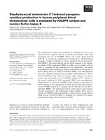
DCQ decreases colon cancer cell growth to a greater
extent under hypoxia
We have previously shown that DCQ is a hypoxic cyto-
toxic compound that induces apoptosis in several murine
and human cancer cell lines [4,5,8]. This is our first
attempt to understand the role of p53 and p21 in drug
efficacy using col on cancer cells that are wildtype or null
for p53 and p21. Before studying DCQ efficacy under
hypoxia, we determined the s ensitivit y of the colo n can-
cer cell lines to hypoxia. HCT116 (p53
+/+
,p53
-/-
,and
p21
-/-
) cells were exposed to 1% O
2
for6,12or24hrs,
after which cell viability was determined by the MTT-
based Cell Titer Promega assay (Figure 1A). Although up
to 12 hrs of hypoxia had no effect on viability, 24 hrs
reduced it by 50% in p53
+/+
cellsandbymorethan80%
in p53
-/-
and p21
-/-
cells (Figure 1A) . Therefore, all
further experiments were conducted by exposing cells to
6 or 12 hrs hypoxia. To determine the antineoplastic
effects of DCQ, cells were treated with 5 or 10 μMDCQ
for 6 hrs and cultured under normoxia or hypoxia. These
doses are in the IC
50
range for p53
+/+
cells [5]. As shown
in Figure 1B, DCQ inhibited the viability of all three
HCT116 cell lines in a dose-dependent fashion, and this
inhibition was 2-5 fold higher under hypoxia than nor-
moxia. p21
-/-
cells appeared to be more sensitive to DCQ
at 10 μM than the other two cell lines (Figure 1B).
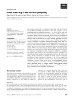
Further studies to confirm the higher drug activity
under the reducing conditions of a hypoxic environment
involved carrying out clonogenic survival assays. Cells
were treated with DCQ at concentrations ranging from
1-20 μM for 6 hrs (data not shown) or 12 hrs, and
exposed to normoxia or hypoxia, after which cells were
re-plated at low density and incubated for 8-14 days.
Colonies having more than 50 cells were counted and
surviving fractions were plotted (Figure 2A). DCQ
decreased the colony forming ability in a dose-
dependent fashion for all three cell lines under both
normoxic and hypoxic conditions; however, the effect
was more pronounced under hypoxia and in p21
-/-
cells.
In accordance with the MTT results, the clonogenic sur-
vival experiment indicated p21
-/-
as drug sensitive and
p53
+/+
as relatively more resistant.
DCQ modulates HIF-1a protein differently in the three
cell lines
To determine whether differences in drug efficacy was
related to the modulation of HIF-1 a,thethreecell
lines were exposed to DCQ (6 hr incubation with 5 μM
or 10 μM) under normoxia or hypoxia and the expres-
sion of HIF-1a protein was determined (Figure 2B). The
level of HIF-1a in hypoxic tumors is known to increase
to regulate metabolic adaptation to oxygen depr ivation
and angi oge nesis [22-24]. This renders cancer cells bet-
terabletosurviveintheharshhypoxicconditions[25].
Therefore, inhibiting HIF-1a-mediated signaling is
important for enhancing anticancer drug efficacy. Differ-
ences in HIF-1a responses to hypoxia exposure and/or
drug treatment were observed in the three cell lines. In
p53
+/+
cells, the HIF-1a protein levels increased by 3.5
fold when cell s were exposed to hypoxia, a nd this
increase was significantly inhibited by 10 μM DCQ (Fig-
ure 2B). This is in contrast to the observed increase in
HIF-1a in response to 5 μMor10μM DCQ under nor-
moxia in this cell line. In p53
-/-
cells, however, HIF-1a
protein was constitutively expressed under normoxia
and hypoxia, yet 10 μM DCQ reduced its expression
El-Khatib et al. Radiation Oncology 2010, 5:107
/>Page 4 of 13
especially under hypoxia (Figure 2B). It is interesting to
note that hypoxia selects for tumors that are mutant for
p53 [26]. In p21
-/-
cell, although DCQ altered the pro-
tein level pattern of HIF-1a, no dose-dependent increase
in HIF-1a was observed (Figure 2B).
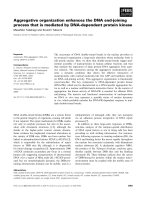
Low DCQ doses induce mitotic catastrophe while high
doses cause apoptosis
To determine the mode of cell death induced by DCQ,
we exposed HCT116 cells to low (2.5 μM) or high (5 or
10 μM) concentrations of DCQ under normoxia or
hypoxia and analyzed cells by flow cytometry, Hoechst
staining and Annexin V techniques 6-24 hrs later.
Depending on the severity of DNA damage, cancer cells
have been shown to die by apoptosis, necrosis or mitotic
catastrophe. Recent eviden ce has show n that low doses
of anticancer drugs, like paclitaxel, induce mitotic cata-
strophe followed b y apoptosis [27]. In our system, we
observed signs of mitotic catastrophe only in response
to lower concentrations o f DCQ (2.5 μM) for 48 hrs.
Under these treatment conditions, the nuclei of all three
HCT116 cells became significantly larger and some cells
contained several nuclei of unequal sizes, which are
characteristic of mitotic catastrophe (Figure 3A). Mitotic
catastroph e was not observed in cells exposed to higher
concentrations of DCQ (5 or 10 μM) under normoxia
or hypoxia (data not shown). The Pre-G
1
increase is
indicative of apoptosis and necrosis as evidenced by the
higher percentage of Annexin-positive apoptotic cells
(Figure 3C). I n Figure 3C, quadrant A represents apo p-
totic cells, B apoptotic and necrotic cells, C normal cells
and D necrotic cells. The percentage of apoptotic and
necrotic cells increased from 8% and 14% in control
normoxic and hypoxic cells respectively, to 31% and
34% in cells treated with 10 μM DCQ. The apoptotic
response and Pre-G
1
phase increase was 2-5 fold higher
under hypoxia than normoxia depending on the cell
line, which was in agreement with the clonogenic and
MTT assay observations (Figures 1 and 2). Again, the
p21
-/-
cells showed the greatest increase in the Pre-G
1
population (Figure 3B), fur ther confirming the higher
drug sensitivity of this cell line.
There is no dose-response toxicity by DCQ in normal
intestinal cells
To determine if DCQ is an effective anti-tumor drug that
specifically targets cancer cells and spares normal ones,
we investigated the dose-response toxicity of DCQ in
Figure 1 DCQ reduces the viability of HCT116 cells more so under hypoxia than normoxia.(A) The effect of hypoxia on HCT116 (p53
+/+
,
p53
-/-
, p21
-/-
) cell viability after 6, 12, or 24 hrs of exposure to 1% O
2
. Cells were plated in 96 well plates at 1.2 × 10
5
cells/ml and treated at 50%
confluency. Viability was determined using Cell Titer 96 non-radioactive proliferation assay. (B) Dose-dependent decrease in the viability of cells
exposed to DCQ for 6 hrs and cultured under normoxia or hypoxia. Values are averages ± SD of two independent experiments each done in
triplicates; (*) indicates p < 0.05 (one way ANOVA). ■ Normoxia □ Hypoxia. The experiment was repeated three times each in quadruplicates.
El-Khatib et al. Radiation Oncology 2010, 5:107
/>Page 5 of 13
normal human intestinal FHs74 cell line. Treatment of
cells with DCQ concentrations of up to 10 μMfor6hrs
was followed by measuring LDH release and cell viability
by the MTT-based assay. DCQ was not cytotoxic to the
normal intestinal cells (Figure 4A), and although cell via-
bility was reduced by 1 μM of the drug, it did not seem
to change much with dose increase (Figure 4B).
DCQ induces DNA damage and activates ATM in
p53
+/+
cells
Next we investigated whether DCQ causes cell death in
human colon cancer cells by inducing DNA damage and
activating ATM, as similar effects have been observed in
EMT6 mouse mammary carcinoma cell lines [11]. For
this, we used the p53
+/+
cells as model, since this cell line
harbors functional p53 and DCQ significantly decreased
the induction of HIF-1a by hypoxia in p53
+/+
cells (Figure
2B). Cells were treated with DCQ and exposed to nor-
moxia or hypoxia for 6 hrs after which they were subjected
to the alkaline comet assay for determining SSB formation
and to immunocytochemistry for measuring the extent of
ATM activation (an indication of DSB). The extent of SSB
formation in response to DCQ was evaluated and quanti-
fied using TriTek Comet Score, software which calculates
different parameters by assuming that the amount of DNA
at a certain location (or the intensity of the DNA stain) is
proportional to the pixel intensity at that position. Differ-
ent parameters were used to quantify the extent of DNA
damage induced by DCQ including % DNA in comet’s tail
(representing damaged DNA migrated away from
nucleus), and tail moment (% DNA in comet’stailmulti-
plied by the tail length). Under normoxia, DCQ induced a
significant increase in the level o f SSBs (Figures 5A, B),
however, under hypoxia, SSB were augmented by the
Figure 2 DCQ reduces the clonogenic survival of HCT116 cells more so under hypoxia than normoxia. (A) Cl onogenic survival of DCQ-
treated cells exposed to normoxic or hypoxic conditions. At 50% confluency, cells were treated for 12 hrs with different DCQ concentrations in
normoxia or hypoxia, after which they were replated at low densities and colonies (more than 50 cells) were stained and counted after 10-14
days in culture. Surviving fractions were calculated as mentioned in “Methods”. (*) indicates p < 0.05 (one way ANOVA). (B) Effect of DCQ on
HIF-1a protein expression. Cells were plated in 100 mm dishes and treated for 6 hrs with DCQ while in normoxia or hypoxia. Whole cell lysates
were immunoblotted for HIF-1a. GAPDH was used to ensure equal loading. Relative densitometry values are presented at the bottom of the
blots. All ratios were normalized to GAPDH and calculated relative to the control cells cultured under oxia. The experiment was repeated three
times each in triplicates.
El-Khatib et al. Radiation Oncology 2010, 5:107
/>Page 6 of 13
drug. The tail moment and % DNA in tail moment
increased significantly (p < 0.05) in comparison with that
of untreated cells (Figure 5B).
Upon DNA damage, cell cycle checkpoints are acti-
vated. These DNA repair processes are mediated via two
protein kinase pathways: the ATM through Chk2 and
ATR via Chk1 [ 28-30]. ATM, a memb er of the PIKK
family, is main ly activated upon DSB formation by the
autophosphorylation of the Ser-1981 [30]. Our results
indicated that control cells have basal levels of p-ATM
expression which are higher in the G
2
-M population
due to the critical role of ATM in mitosis. Exposure of
p53
+/+
cells to 5 or 10 μM DCQ triggered the activation
of ATM by its phosphorylation at Serine 1981 in all the
phases of cell cycle and this activation was more pro-
nounced under hypoxia (Figure 5C). Hy poxia alone
increased ATM expression, however, the combination of
DCQ and hypoxia treatment induced higher levels of
p-ATM expression in the G
2
-M phase in comparison
with control cells (Figure 5C). These results confirm
that DCQ induces DSBs in human colon cancer cells.
DCQ modulates protein expression of downstream
ATM effectors
Upon DNA damage, one of the important transcription
factors activated by ATM through phosphorylation is
Figure 3 DCQ induces mitotic catastrophe and apoptosis in HCT116 cells. (A) Low concentrations of DCQ triggered mitotic catastrophe in
all HCT116 cell lines. Cells were cultured on coverslips and treated at 50% confluency with 2.5 μM DCQ for 48 hrs after which they were fixed
and stained with Hoechst and viewed under a fluorescent microscope using UV. (**) indicates p < 0.001 (one way ANOVA) with respect to the
Ctrl. (B) Higher concentrations of DCQ (5 and 10 μM) induced increases in the PreG
1
phase population more so under hypoxia. Treatment with
DCQ in normoxia or hypoxia was for 6 hrs, after which cells were harvested immediately and DNA was stained with PI for analysis with FACScan
flow cytometry. The percentage of Pre G
1
cells was calculated using Cell Quest. (C) Annexin V assay showing the apoptotic/necrotic response of
p53
+/+
cells exposed to 5 or 10 μM DCQ for 6 hr in normoxia or hypoxia. Apoptosis was assayed 24 hr after drug treatment, and appeared to be
enhanced in hypoxia at higher drug concentrations. Quadrant A = apoptotic cells, B = apoptotic+necrotic, C = normal, D = necrotic. The
experiment was repeated twice each in duplicates.
El-Khatib et al. Radiation Oncology 2010, 5:107
/>Page 7 of 13
p53, the activation of which triggers G
1
or G
2
arrest (in
case of p21 increase) or apoptosis [29,30]. In addition,
ATM can lead to the activation of PIDD, an important
target gene in a signaling pathway that is initiated by
p53, subsequently causing either activation of NFB-
dependent cell survival through PIDD-C or apoptosis
through PIDD-CC [31,32]. To assess the effect of DCQ
on downstream targets of ATM, we investigated its abil-
ity to induce changes in the expression levels of p53,
p21, PIDD-C and caspase-2 proteins. Cells were exposed
to 5 or 10 μM DCQ and protein changes were moni-
tored 6 hrs post-treatment under normoxia or hypoxia.
The p53 protein increased in response to DCQ in all
three cell lines except in p53
+/+
cells exposed to hypoxia
(Figure 6A); p21 protein also increased in a ll cell l ines
except in p53
+/+
cells exposed to normoxia (Figure 6A).
Exposure of the p53
+/+
cells to 5 or 10 μM DCQ gradu-
ally increased the level of caspase-2, and the upregula-
tion was 8-10 fold higher under hypoxia (Figure 6C). In
p21
-/-
cells, DCQ treatment under normoxia increased
Figure 4 DCQ is not cytotoxic to norma l intestinal cells. DCQ at concentrations of up to 10 μM did not reduce FHs74 Int human normal
intestinal cell viability. At 50% confluency, cells were exposed to DCQ for 6 hr or were left untreated. Viability was assessed by the Cytotox 96
non-radioactive assay (A) and by the MTT-based Promega assay (B). Values are averages ± SE of two independent experiments each done in
triplicates. The experiment was repeated three times each in triplicates.
El-Khatib et al. Radiation Oncology 2010, 5:107
/>Page 8 of 13
caspase-2 expression levels. The exposure of p21
-/-
cells
to hypoxia alone increased caspase-2 expression, how-
ever the combination of DCQ and hypoxia reduced it
(Figure 6C). DCQ had no effect on caspase-2 protein
expression in p53
-/-
cells which is not surprising, since
p53 is known to regulate caspase-2 [33]. Although no
direct interaction between p53 and caspase-2 has been
observed, it is believed that a functional connection
between these two proteins is essential for the initiation
of drug-induced apoptosis [34]. Enforce d PIDD expres-
sion or the over expression of p53 have been shown to
promote cell dea th through the activation of caspase-2
[33,34]. In p53
+/+
and p53
-/-
cells, DCQ downregulated
PIDD-C protein expression under normoxia and
hypoxia (Figure 6B, C). PIDD-C was not detect ed in
p21
-/-
cells.
Discussion
The low oxygen tension in solid tumors is one major
factor for tumor resistance to radiotherapy and che-
motherapy; therefore there is interest in the discovery of
novel drugs that can specifically target tumor cells. In
this study, we showed that DCQ is a DNA damaging
and apoptotic agent that reduces the viability and colony
forming ability of colon cancer cells and is non-toxic to
normal intestinal cells. We have shown previously that
DCQ is not toxic to normal mouse intestinal Mode K
and IEC-6 cell s [9] or to normal mouse mammary SCP2
Figure 5 DCQ induces DNA damage and increases ATM expression in p53
+/+
HCT116 cells. SSB and DSB induced by DCQ in p53
+/+
cell
line. (A) Examples of comets induced by DCQ in cells subjected to the alkaline comet assay. Cells treated with DCQ for 6 hrs in normoxia or
hypoxia were collected directly after treatment, subjected to the alkaline comet assay and images were taken using a fluorescent microscope at
40× (oil immersion) magnification. The comets observed by each treatment are directly proportional to the amount of SSBs induced. (B) The
mean of the parameters (% DNA in comet’ s tail and tail moment) are shown in the graphs above. More than 50 cells per treatment were
photographed and quantified using TriTek CometScore software. (*) indicates p < 0.05 (one way ANOVA) with respect to control. (C) DCQ-
induced phosphorylation of ATM in p53
+/+
cells at 6 hrs as an indication of DSB. After treatment, cells were fixed and subjected to
immunocytochemical detection of ATM phosphorylated on Ser-1981, and stained with PI to detect at the same time p-ATM in each phase of
the cell cycle. The mean of the FL-1 intensity ± SD (reflecting the level of p-ATM expression) at the G
1
, S and G
2
M phases of the cell cycle are
shown in the table. The experiment was repeated twice each in duplicates.
El-Khatib et al. Radiation Oncology 2010, 5:107
/>Page 9 of 13
cell s [unpub lished findings], suggesting the selectivity of
this drug to cancer cells.
The reduction of viability and colony survival by DCQ
was more pronounced under hypoxia than normoxia and
was evident in al l HCT116 cell lines, particularly in p21
-/-
cells which showed greater drug-induced increases in
Pre-G
1
. The apoptotic effects of DCQ in p53
+/+
cells cor-
related with a n increase in the pro-apoptotic caspase-2
protein, inhibition of the pro-survival protein PIDD-C,
and increase in p-ATM expression, a major protein
kinase involved in repair of DSB.
DCQ belongs to a group of heterocyclic compounds
with potent hypoxic cytotoxic activities [4], of which the
heterocyclic di-N-oxide TPZ is in phase III clinical trials
[35]. The hypoxia toxicity of T PZ is due to the produc-
tion of radicals that form strand breaks in the DNA
[35,36]. Under normoxic conditions, the radical is back-
oxidized to the nontoxic original compound with the
related production of the much less toxic superoxide
radical [36]. Unlike TPZ which is active only under
hypoxia, DCQ appears to be equally active in HCT116
cells cultured in both normoxic and hypoxic environ-
ments which explains the low HCR ratios of (< 1.5) spe-
cific for this cell line. This is in contrast to the high
HCR ratios (> 100) in T-84 human colon cancer cells
[4], suggesting that the hypoxia potency of DCQ is cell-
type specific.
Hypoxia-Inducible Factor-1alpha (HIF-1a)isan
important cellular transcription factor that is stabilized
under hypoxia [reviewed in [37]]. HIF-1a regulates the
metabolic adaptation to O
2
deprivationintumors,and
playsanessentialroleinallowingtumorstoescape
Figure 6 DCQ modulates the protein expression levels of key mediators of apoptosis and mitotic catastrophe. At 50% confluency, cells
were treated with 5 or 10 μM DCQ for 6 hrs. Whole cell lysates were then immunoblotted with the different primary antibodies and probed
with GAPDH to ensure equal loading. (A) p53 and p21 protein expression and (B) caspase-2 and PIDD-C protein expression in HCT116 cell lines
in response to DCQ treatment under normoxic or hypoxic conditions. (C) Relative densitometry values of analyzed proteins are plotted. All
values were normalized to GAPDH and calculated relative to the control cells cultured under normoxia. The experiment was repeated twice each
in duplicates.
El-Khatib et al. Radiation Oncology 2010, 5:107
/>Page 10 of 13
apoptotic mechanisms and becoming angiogenic [37,38].
DCQ has previously been shown to decrease HIF-1a
mRNA and protein expression levels in mouse mam-
mary carcinoma cell lines [8]. Here we show that DCQ
decreased HIF-1a proteinexpressioninp53
+/+
and
p53
-/-
HCT116 cell lines, despite the constitutive expres-
sion of HIF-1a under both normoxic and hypoxic con-
ditions in the latter cell line (Figure 3B). This result is
interesting in light of the literature showing that hypoxia
selects for p53 mutant tumors [26] . Furthermore, inter-
fering with HIF-1a is important for effective antitumor
therapy [37].
Mitotic catastrophe occurs during, or shortly after,
dysregulated or failed m itosis, and is believed to be fun-
damentally different from apoptosis. Despite its distinc-
tive morphology, mitotic catastrophe may r epresent a
pre-stage of apoptosis [17]. Apoptosis, however, is not
always required for the lethal effect of mitotic cata-
strophe, since abnorma l mitosis can lead to cell death
through apoptosis and necrosis based on the molecular
profile o f cells [17]. In our system, we show that at low
doses of DCQ (2.5 μM), the three HCT116 cell lines
displayed an entirely different nuclear morphology with
enlarged nuclei, a morphology that has been previously
knowntoresultfrommitoticcatastrophe(Figure3A).
No signs of mitotic catastrophe were observed at higher
DCQ concentrations of 5 or 10 μMoratshorterincu-
bation times (data not shown). Rather high drug con-
centrations induced a significant increase in Pre-G
1
,a
sign apoptosis and necrosis (Figure 3B).
Having determined that DCQ induces mitotic cata-
strophe at lower drug concentrations and apoptosis at
higher concentrations, we next investigated whether it
causes DNA damage and activates ATM in the p53
+/+
cell line. DNA damage imposes a threat to the survival
of cells if the damage is unrepaired [12]. As a re sponse
to the damage, cells activate the DNA damage check-
point. DSBs are detected by two main players in the
DNA damage checkpoint: ATM and DNA-PK. Signal
transduction, induced by the activation of ATM, can
cause cell-cycle arrest, repair, and cell death. ATM plays
acriticalroleinSandG
2
-M phase arrest. Activated by
DSBs, ATM becomes phosphorylatedatSer-1981
[30,39]. Our experiments using the alkaline comet assay
show that DCQ causes SSB and DSB in p53
+/+
cells
under normoxic and hypoxic c onditions, however, the
extent of DNA strand breaks was higher under hypoxia.
Interestingly, ATM was activated in all phases of the
cell cycle in response to the DNA damage induced by
DCQ especially under hypoxia (Figure 5), suggesting a
positive correlation between the extent of DNA damage
and the activation of ATM.
The p21 gene is transcripti onally activated by p53 and
is responsible for the p53-dependent checkpoint which
induces cell cycle arrest after DNA damage. Enforced
p21 expression is known to result in a consistent, but
partial, protection of cells from apoptosis [40,41]. In
HCT116, a significant incre ase was observed in p21
expression in response to DCQ treatment under hypoxia
both in p53
+/+
and p53
-/-
cells suggesting that p21 acti-
vation is independent of p53. In addition, the decrease
in the expression levels of the prosurvival PIDD-C pro-
tein coupled with the increase in proapoptotic caspase-2
in p53
+/+
cells, appears to have committed the cells to
apoptosis. In p53
-/-
and p21
-/-
HCT116, the apoptotic
cell death occurred independent of caspase-2 activation
and/or PIDD-C downregulation (Figure 6B), suggesting
the involvement of other mediators of apoptosis.
It has been debated if mitotic catastrophe results i n
cell death via caspase2-dependent or - independent
mechanisms [42]. At least three lines of evidenc e indi-
cate that, in our cell system, mitotic catastrophe is inde-
pendent of p53 and/or caspase-2 activation. First,
mitotic catastrophe occurred in drug treated cells that
are null for p53 (Figure 3A). Second, in p53
+/+
cells,
higher doses of 5 and 10 μM DCQ induced caspase-2
activation (Figure 6B), while morphological changes of
mitotic catastrophe were observed at lower drug doses
(Figure 3A). Third, the three HCT116 cell lines dis-
played signs of mitotic catastrophe, yet only p53
+/+
cells
showed activation of caspase-2, and especially under
hypoxia (Figure 6B). Although , it was suggest ed that the
presence of functional p53 in cancer cells enhanced
their sensitivity to hypoxia, DCQ-induced apoptosis in
HCT116 was not dependent on the presence of the p53
gene, as the Pre-G
1
increase was evident even in cells
lacking the p53 gene (Figure 3B).
Conclusions
DCQ is a selective cytotoxin in HCT116 human colon
cancer cells and its toxicity is independent of p53 and
p21. DCQ toxicity is associated with enhanced DNA
damage, activation of the ATM damage repair pathway,
as well as induction of apoptosis or mitotic catastrophe
depending on the drug concentration used. The absence
of major toxicity to normal cell lines (human intestinal
cells in t his study and mouse intestinal cells and mouse
mammary cells in previous studies) makes DCQ an
interesting compound with potential anticancer activities
against colon cancer, and therefore a drug for further
testing.
Acknowledgements
We thank Dr. Youssef Mouneimne and Ms. Rania El-Osta, members of the
Kamal Shair Central Research Science Laboratory, for their valuable help with
data acquisition and analysis on the flow cyotmeter and fluorescence
microscope. This project was supported by the University Research Board of
the American University of Beirut.
El-Khatib et al. Radiation Oncology 2010, 5:107
/>Page 11 of 13
Author details
1
Department of Biology, American University of Beirut, Beirut, Lebanon.
2
Department of Radiation Oncology, American University of Beirut, Beirut,
Lebanon.
3
Department of Chemistry, American University of Beirut, Beirut,
Lebanon.
Authors’ contributions
ME carried out the experiments in the study and prepared the figures. FG
was involved in revising the manuscript. MH provided the compound and
revised the manuscript. HGM conceived of the study, designed the
experiments and drafted the manuscript. All authors read and approved the
final manuscript.
Competing interests
The authors declare that they have no competing interests.
Received: 16 August 2010 Accepted: 15 November 2010
Published: 15 November 2010
References
1. Brown JM: Tumor hypoxia in cancer therapy. Methods Enzymol 2007,
435:297-321.
2. McKeown SR, Cowen RL, Williams KJ: Bioreductive drugs: from concept to
clinic. Clin Oncol 2007, 19:427-442.
3. Gali-Muhtasib HU, Haddadin MJ, Rahhal DN, Younes I: Quinoxaline 1,4-
dioxides as anticancer and hypoxia-selective drugs. Oncology Reports
2001, 8:679-684.
4. Diab-Assaf M, Haddadin MJ, Yared P, Gali-Muhtasib HU: Quinoxaline 1,4-
dioxides: hypoxia selective therapeutic agents. Mol Carcinog 2002,
33:198-205.
5. Itani W, Geara F, Haykal J, Haddadin M, Gali-Muhtasib H: Radiosensitization
by 2-benzoyl-3-phenyl-6,7-dichloroquinoxaline 1,4-dioxide under
normoxia and hypoxia in human colon cancer cells. Radiat Oncol 2007,
2:1.
6. Harakeh S, Diab-Assef M, El-Sabban M, Haddadin M, Gali-Muhtasib H:
Inhibition of proliferation and induction of apoptosis by 2-benzoyl-3-
phenyl-6,7-dichloro quinoxaline 1,4-dioxide in adult T cell leukemia cells.
Chem Biol Interact 2004, 148 :101-113.
7. Junnotula V, Sarkar U, Sinha S, Gates KS: Initiation of DNA strand cleavage
by 1,2,4-benzotriazine 1,4-dioxide antitumor agents: mechanistic insight
from studies of 3-methyl-1,2,4-benzotriazine 1,4-dioxide. J Am Chem Soc
2009, 131:1015-1024.
8. Gali-Muhtasib HU, Sidani M, Geara F, Assaf-Diab M, Al-Hmaira J,
Haddadin MJ, Zaatari G: Quinoxaline 1,4-dioxides are novel angiogenesis
inhibitors that potentiate antitumor effects of ionizing radiation. Int J
Oncol 2004, 24:1121-1131.
9. Gali-Muhtasib HU, Diab-Assaf M, Haddadin MJ: Quinoxaline 1,4-dioxides
induce cell cycle arrest, apoptosis and changes in transforming growth
factors and extracellular signal-regulated kinase expression in human
colon cancer cells. Cancer Chemother Pharmacol 2005, 55:369-378.
10. Haykal J, Fernainy P, Itani W, Haddadin M, Geara F, Smith C, Gali-
Muhtasib H: Radiosensitization of EMT6 mammary carcinoma cells by 2-
benzoyl-3-phenyl-6,7-dichloroquinoxaline 1,4-dioxide. Radiother Oncol
2008, 86:412-418.
11. Haykal J, Geara F, Haddadin M, Smith C, Gali-Muhtasib H: The
radiosensitizer 2-benzoyl-3-phenyl-6,7-dichloroquinoxaline 1,4-dioxide
induces DNA damage in EMT6 mammary carcinoma cells. Radiat Oncol
2009, 4:25.
12. Vilenchik MM, Knudson AG: Endogenous DNA double-strand breaks:
production, fidelity of repair and induction of cancer. Proc Natl Acad Sci
USA 2003, 100:12871-12876.
13. Yang J, Yu Y, Hamrick HE, Duerksen-Hughes PJ: ATM, ATR and DNA-PK:
initiators of the cellular genotoxic stress responses. Carcinogenesis 2003,
24:1571-1580.
14. Vogelstein B, Lane D, Levine A: Surfing the p53 network. Nature 2000,
408:307-310.
15. Lakin ND, Jackson SP: Regulation of p53 in response to DNA damage.
Oncogene 1999, 18:7644-7655.
16. Tinel A, Tschopp J: The PIDDosome, a protein complex implicated in
activation of caspase-2 in response to genotoxic stress. Science 2004,
304:843-846.
17. Vakifahmetoglu H, Olsson M, Zhivotovsky B: Death through a tragedy:
mitotic catastrophe. Cell Death Differ 2008, 15:1153-1162.
18. Haddadin M, Issidorides C: The Beirut Reaction. Heterocycles 1993,
35:1503-1525.
19. Gali-Muhtasib H, Diab-Assaf M, Boltze C, Al-Hmaira J, Hartig R, Roessner A,
Schneider-Stock : Thymoquinone extracted from black seed triggers
apoptotic cell death in human colorectal cancer cells via a p53-
dependent mechanism. Int J Oncol 2004, 25:857-866.
20. Singh NP: Sodium ascorbate induces DNA single-strand breaks in human
cells in vitro. Mutat Res 1997, 375:195-203.
21. Huang X, Darzynkiewicz Z: Cytometric assessment of histone H2AX
phosphorylation: a reporter of DNA damage. Methods Mol Biol 2006,
314:73-80.
22. Semenza GL, Roth PH, Fang H-M, Wang LW: Transcriptional regulation of
genes encoding glycolytic enzymes by hypoxia-inducible factor 1. J Biol
Chem 1994, 269:23757-23763.
23. Wang GL, Jiang B-H, Rue EA, Semenza GL: Hypoxia-inducible factor 1 is a
basic-helix-loop-helix-PAS heterodimer regulated by cellular O
2
tension.
Proc Natl Acad Sci USA 1995, 92:5510-5514.
24. Ivan M, Kondo K, Yang H, Kim W, Valiando J, Ohh M, Salic A, Asara JM,
Lane WS, Kaelin WG Jr: HIFα targeted for VHL-mediated destruction by
proline hydroxylation: implications for O
2
sensing. Science 2001,
292:464-468.
25. Ryan HE, Poloni M, McNulty W, Elson D, Gassmann M, Arbeit JM,
Johnson RS: Hypoxia-inducible factor-1α is a positive factor in solid
tumour growth. Cancer Res 2000, 60:4010-4015.
26. Hammond EM, Mandell DJ, Salim A, Krieg AJ, Johnson TM, Shirazi HA,
Attardi LD, Giaccia AJ: Genome-wide analysis of p53 under hypoxic
conditions. Mol Cell Biol 2006, 26:3492-3504.
27. Morse DL, Gray H, Payne MClaire, Gillies JRobert: Docetaxel induces cell
death through mitotic catastrophe in human breast cancer cells. Mol
Cancer Ther 2005, 4:1495-1504.
28. Bruno T, Iezzi S, De Nicola F, Di Padova M, Desantis A, Scarsella M, Di
Certo MG, Leonetti C, Floridi A, Passananti C, Fanciulli M: Che-1 activates
XIAP expression in response to DNA damage. Cell Death Differ 2008,
15:515-520.
29. Sakasai R, Tibbetts R: Rnf8-dependent and independent regulation of
53BP1 in response to DNA damage. J Biol Chem 2008, 283:13549-13555.
30. Lavin M: Ataxia-telangiectasia: from a rare disorder to a paradigm for cell
signaling and cancer. Nature 2008, 9:759-769.
31. Halazonetis TD, Gorgoulis VG, Bartek J: An oncogene-induced DNA
damage model for cancer development. Science 2008, 319:1352-1355.
32. Tinel A, Janssens S, Lippens S, Cuenin S, Logette E, Jaccard B, Quadroni M,
Tschopp J: Autoproteolysis of PIDD marks the bifurcation between pro-
death caspase-2 and pro-survival NF-kappaB pathway. EMBO J 2007,
26:197-208.
33. Vakifahmetoglu H, Olsson M, Orrenius S, Zhivotovsky B: Functional
connection between p53 and caspase-2 is essential for apoptosis
induced by DNA damage. Oncogene 2006, 25:5683-5692.
34. Seth R, Yang C, Kaushal V, Shah SV, Kaushal GP: p53-dependent caspase-2
activation in mitochondrial release of apoptosis-inducing factor and its
role in renal tubular epithelial cell injury. J Biol Chem 2005,
280:31230-31239.
35. Reddy SB, Williamson SK: Tirapazamine: a novel agent targeting hypoxic
tumor cells. Expert Opin Investig Drugs 2009, 18:77-87.
36. Evans JW, Yudoh K, Delahoussaye YM, Brown JM: Tirapazamine is
metabolized to its DNA-damaging radical by intranuclear enzymes.
Cancer Res 1998, 58:2098-2101.
37. Liao D, Johnson RS: Hypoxia: a key regulator of angiogenesis in cancer.
Cancer Metastasis Rev 2007,
26:281-290.
38. Maxwell PH, Dachs GU, Gleadle JM, Nicholls LG, Harris AL, Stratford IJ,
Haninkson O, Pugh CW, Ratcliffe PJ: Hypoxia-inducible factor-1 modulates
gene expression in solid tumors and influences both angiogenesis and
tumor growth. Proc Natl Acad Sci 1997, 94:8104-8109.
39. Bakkenist CJ, Kastan MB: DNA damage activates ATM through
intermolecular autophosphorylation and dimer dissociation. Nature 2003,
421:499-506.
40. Meng LH, Kohn KW, Pommier Y: Dose-response transition from cell cycle
arrest to apoptosis with selective degradation of Mdm2 and p21
WAF1/
CIP1
in response to the novel anticancer agent, aminoflavone (NSC
686,288). Oncogene 2007, 26:4806-4816.
El-Khatib et al. Radiation Oncology 2010, 5:107
/>Page 12 of 13
41. Bunz F, Dutriaux A, Lengauer C, Waldman T, Zhou S, Brown JP, Sedivy JM,
Kinzler KW, Vogelstein B: Requirement for p53 and p21 to sustain G
2
arrest after DNA damage. Science 1998, 282:1497-1501.
42. Mansilla S, Priebe W, Portugal J: Mitotic catastrophe results in cell death
by caspase-dependent and caspase-independent mechanisms. Cell Cycle
2006, 5:53-60.
doi:10.1186/1748-717X-5-107
Cite this article as: El-Khatib et al.: Cell death by the quinoxaline dioxide
DCQ in human colon cancer cells is enhanced under hypoxia and is
independent of p53 and p21. Radiation Oncology 2010 5:107.
Submit your next manuscript to BioMed Central
and take full advantage of:
• Convenient online submission
• Thorough peer review
• No space constraints or color figure charges
• Immediate publication on acceptance
• Inclusion in PubMed, CAS, Scopus and Google Scholar
• Research which is freely available for redistribution
Submit your manuscript at
www.biomedcentral.com/submit
El-Khatib et al. Radiation Oncology 2010, 5:107
/>Page 13 of 13