Báo cáo khoa học: " Hypofractionated radiotherapy after conservative surgery for breast cancer: analysis of acute and late toxicity" ppt
Bạn đang xem bản rút gọn của tài liệu. Xem và tải ngay bản đầy đủ của tài liệu tại đây (671.63 KB, 7 trang )
RESEA R C H Open Access
Hypofractionated radiotherapy after conservative
surgery for breast cancer: analysis of acute and
late toxicity
Letizia Deantonio
1
, Giuseppina Gambaro
1
, Debora Beldì
1
, Laura Masini
1
, Sara Tunesi
2
, Corrado Magnani
2
,
Marco Krengli
1*
Abstract
Background: A variety of hypofractionated radiotherapy schedules has been proposed after breast conserving
surgery in the attempt to shorten the overall treatment time. The aim of the present study is to assess acute and
late toxicity of using daily fractionation of 2.25 Gy to a total dose of 45 Gy to the whole breast in a mono-
institutional series.
Methods: Eighty-five women with early breast cancer were assigned to receive 45 Gy followed by a boost to the
tumour bed. Early and late toxicity were scored according to the Radiation Therapy Oncology Group criteria. For
comparison, a group of 70 patients with similar cha racteristics and treated with conventional fractionation of 2 Gy
to a total dose of 50 Gy in 25 fractions followed by a boost, was retrospectively selected.
Results: Overall median treatment duration was 29 days for hypofractionated radiotherapy and 37 days for
conventional radiotherapy. Early reactions were observed in 72/85 (85%) patients treated with hypofractionation
and in 67/70 (96%) patients treated with conventional fractionation (p = 0.01). Late toxicity was observed in 8
patients (10%) in the hypofractionation group and in 10 patients (15%) in the conventional fractionation group,
respectively (p = 0.4).
Conclusions: The hypofractionated schedule delivering 45 Gy in 20 fractions shortened the overall treatment time
by 1 week with a reduction of skin acute toxicity and no increase of late effects compared to the conventional
fractionation. Our results support the implementation of hypofractionated schedules in clinical practice.
Background
Radiotherapy (RT) reduces th e risk of local relapse and
breast cancer mortality [1] and is offered to nearly all
patients after conservative surgery and to selected
patients after m astectomy. The international standard
RT regimen after breast conservative surgery for early
breast cancer delivers 25 daily fractions of 2 Gy to a
total dose of 50 Gy over 5 weeks followed by 5 fractions
of 2 Gy as a boost to the tumour bed [2]. The high
number of women with breast cancer, receiving post-
operative RT, led to think that a shorter course of irra-
diation would result in improved quality of life for
patients, in potentially better integration with systemic
treatments and in reduced costs. Therefore, alternative
schedules based on a lower total dose delivered in
fewer, larger fractions (hypofractionation) were firstly
introduced in Canada and the United Kingdom (UK)
[3,4]. The Canadian randomised trial [3] tested 42.5 Gy
in 16 fractions against 50 Gy in 25 fractions. Results
suggested equivalence in terms of local control and
breast cosmetic results for the 16-fractions regimen.
The two most recent randomized studies [5,6] were
conducted by the START Trials in order to test the
effects of radiotherapy schedules using fraction size lar-
ger than 2.0 Gy. The START Trial A tested two dose
levels of a 13-fractions regimen delivered over 5 weeks
and the START Trial B compared 40 Gy in 15 fractions
of 2.67 Gy in 3 weeks with a control group of 50 Gy in
* Correspondence:
1
Department of Radiotherapy, University Hospital Maggiore della Carità,
Novara, Italy
Full list of author information is available at the end of the article
Deantonio et al. Radiation Oncology 2010, 5:112
/>© 2010 Deantonio et al; licensee BioMe d Central Ltd. This is an Open Access article distributed under the terms of the Creative
Commons Attribution License ( which permits unr estricted us e, distribution, and
reproduction in any medium, provided the original work is properly cited.
25 fractions of 2.0 Gy over 5 weeks. These studies seem
to offer rates of late adverse effects and local-regional
tumour relapse at least as favourable as the standard
schedule.
The aim of the present study carried out in a mono-
institutional clinical setting is to assess acute and late
toxicity of hypofractionated radiotherapy after conserva-
tive surgery using a regimen of 2.25 Gy/fraction to a
total dose of 45 Gy to the whole b reast followed by a
boost comparing the results with those of a similar
group of patients treated with conventional fractionation
schedule.
Methods
Patients
From January 2006 to January 2008, 85 patients with
invasive carcinoma of the breast treated with conserva-
tive surgery and biopsy of sentinel lymph node or axil-
lary lymph node dissection were prospectively treated
with whole breast irradiation of 45 Gy in 20 fractions,
2.25 Gy/fraction, followed by 9 Gy in 3 fractions to the
tumour bed as a boost dose. Eligibility criteria were: age
≥ 60 years, T ≤ 2 cm, negative surgical margins and no
indication to lymph node RT (≤ 3 positive lymph-
nodes). Patients with history of contralateral breast can-
cer, multifocal disease, seri ous non-malignant disease
(e.g. cardiovascular or pulmonary), severe mental or
physical disorders w ere excluded from the study. The
initial work-up included chest radiogram, liver ultra-
sound, bone scan, full blood count, kidney and liver
function tests. Written informed consent was obtained
from patients before start treatment following the rules
of our institution.
A second group of 70 patients with similar character-
istics in terms of clinical history, staging and type of
surgery w as retrospectively selected from patients trea-
ted since 2005 with post-operative breast RT with con-
ventional schedule of 50 Gy in 25 fraction, 2 Gy/fraction
followed by 10 Gy in 5 fractions to the tumour bed as a
boost dose.
Radiotherapy
All patients underwent post-operative RT pla nned using
the three-dimensional treatment planning system, Pin-
nacle (Philips, Eindhoven, The Netherlands). Computed
Tomography (CT) images were obtained by helical CT
(Prospeed, General Electric Medical Systems, Milwau-
kee, WI), covering the entire thoracic region from the
apex of the lung to the diaphragm, with patients in
treat ment position. Target and non-targ et volumes were
outlined according to the criteria of the International
Commission of Radiation Units (ICRU) and Measure-
ments Reports 50 and 62 [7,8]. The clinical target
volume (CTV) was defined as the entire palpable breast
tissue starting 5 mm below the skin. The planning target
volume (PTV) was obtained by adding 10 mm margin to
the CTV, except in the direction of the skin. Ipsilateral
lung was automatically outlined from the apex to the
base and the left ventricle was manually outlined in case
of left sided cancer. The treatment technique consisted
of two opposed tangent ial fields by using 6-18 MV
photon beams. Radiation fields were appropriately cus-
tomized by multileaf collimator when needed in order
to spare the surrounding healthy tissues. The angle of
the beams was adjusted to minimize the irradiation of
lung parenchyma and left ventricle. Appropriate physical
wedge compensation was used to ensure a uniform dose
distribution throughout the target volume. The total
dose prescribed to the ICRU point was 45 Gy, delivered
with 2.25 Gy per fraction, 5 days a week. A boost dose
of 9 Gy in 3 fractions was given using a 6-9 MeV elec-
tron field, depending on the depth of the original tumor
site. Dose calculation with a grid of 3 mm was per-
formed using the collapsed cone convolution algorithm
of the treatme nt planning system, including the correc-
tion for tissue heterogeneity. For each patient, dose-
volume histograms (DVHs) for target, lung and left
ventricle for left-sided canc ers were calculated. The
same technique had been used for the patients treated
with convention al radiotherapy to a total dose of 50 Gy,
2 Gy/fraction and a boost dose of 10 Gy in 5 fractions
to the tumour bed.
Biologically effective dose (BED) was calculated
assuming alpha/beta ratio equals to 10 Gy for early reac-
tions, 3 Gy for late reactions. In the group assigned to
receive 45 Gy to the whole b reast, BED was 55 Gy for
early effects, 78 Gy for late effects versus 60 Gy for early
effects, 83 Gy for late effects in the group treated with
50 Gy.
Sequential chemotherapy (cyclophosphamide - metho-
trexate -5-fluorouracil, doxorubicin - cyclophosphamide,
5-fluorouracil - epirubicin - cyclophosphamide) and hor-
mone therapy given concomitantly to radiotherapy
(tamoxifen or aroma tase inhibit ors) were given in 20/85
patients (24%) in the hypofractionation group while 65/
85 patients (76%) received hormone therapy alone. The
patients of the group treated by conventional fractiona-
tion received sequential chemotherapy in 20/70 cases
(28%) and hormone therapy concomitant to radiother-
apy in 50/70 cases (71%).
Early and late toxicity were scored according to the
Radiation Therapy Oncology Group/European Organiza-
tion for Research and Treatment Cancer (RTOG/
EORTC) criteria in both groups of patients [9].
Statistical analysis
The chi-square test and Fisher’s exact test were used to
compare the two treatment groups.
Deantonio et al. Radiation Oncology 2010, 5:112
/>Page 2 of 7
The association of early and late toxicity with breast
volume, maximum radiation dose and chemotherapy
was analyzed in the two treatment groups using Chi-
square test and logistic regression. The Chi-square test
was used for comparing acute and late toxicity bet ween
the patients treated with hypofractionation and those
treated with conventional RT and for comparing the fre-
quency of breast volume < 500 cc and ≥ 500 cc in the
two treatment groups. Logistic regression was used to
adjust the effect on toxicity of covariates, such as breast
volume, maximum dose and chemotherapy. Chi-square
test for trend was used to compare acute toxicity
between the patients treated with hypofractionation and
those treated with conventional RT.
The t-test was used to compare breast volume
between the patients treated with hypofractionation and
those treated with conventional RT.
Surv ival curves were obtained to show the cumulative
probability of experiencing adverse effects during 6
months follow-up interval. The actuarial occurrence of
late toxicity was calculate d by the Kaplan-Meier method
and the two treatm ent groups were compared using the
Log-rank test.
Cox’s proportional hazards regression model was fitted
in order to obtain the haz ard ratio (HR) for hypofractio-
nation adjusted by volume and chemotherapy. A p value
of less than 0.05 was considered to be statistically signif-
icant. Statistical analysis was performed using the SAS
package version 8.02 (SAS Institute, Inc, Cary, NC,
USA).
Results
Both treatment groups were comparable in terms of age,
performance status, tumour stage, adjuvant chemother-
apy and hormone treatment. D ifferences were observed
for lymph node stage, breast volume and breast maxi-
mum dose (Table 1).
Median time from surgery to RT was 29 days for
hypofractionated RT and 37 days f or conventional RT.
No patient interrupted the treatment.
The mean follow-up was 15.0 months (median12.6;
25
th
quartile 7.8 and 75
th
quartile 20.8 months) in the
hypofractionation RT group and 28.6 months (med-
ian32.2; 25
th
quartile 22.1 and 75
th
quartile 40.0 months)
in the conventional fractionation group.
Acute toxicity
Early reactions, consisting in skin erythema, were
observed in 72 patients (85%) in the hypofractionation
group and in 67 (95%) in the conventional RT group
(chi-square p = 0.01; chi-square test for trend p = 0.001)
(Table 2).
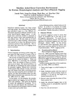
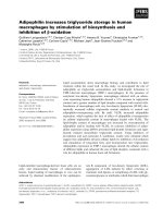
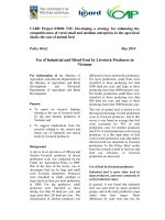
A significant correlation by chi-square test (p = 0.001)
between breast vo lume and maximum dose was found
for the occurrence of acute toxicity (Figure 1). Adjuvant
treatments did not influence acute toxicity.
Logistic regression analysis was carried on for adjust-
ing for potential confounders. In this analysis, acute skin
toxicity was classified in two categories: mild (G0 and
G1) and severe (G2 and G3). Hypofractionation reduced
the risk of severe acute toxicity: odd ratio (OR) adjusted
for volume was 0.45 (95% CI = 0.23-0.93). Including in
the analyses also maximum breast dose and chemother-
apy did not provide a significant contribution to the
model fit.
Late toxicity
Late toxicity was assessed after 6 months in 76/85
patients in the hypofractionation group and in 67/70
patients in the standard RT group since 9 and 3
patients, respectively, were lost at follow-up and were
not included in the statistical analysis. Late toxicity
according to the RTOG criteria was observed in 8
patients (10%) in the hypofractionation group and in 10
patients (15%) in the conventional fractionation group
(Table 3). The difference was not statistically significant
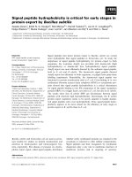
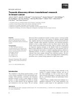
(chi-square p = 0.4). Cumulative occurrence of late toxi-
city over time was analyzed using Kaplan-Meier method
and compared b y log-rank test, resulting not statistically
significant (p = 0.17) (Figure 2). At 12 and 30 months,
the risk of late toxicity was 5.9% and 29.2% in the group
treated by hypofractionation, 8.2% and 10.6% in the
group treated by standard RT, respectively.
Cox’s proportional haz ards regression analysis showed
that hypofractionation, adjusted by volume and che-
motherapy, was not associated w ith the hazard of late
toxicity (HR = 2.16; 95% CI = 0.68-6.84; p = 0.19). In
thesamemodel,weobservedthatbreastvolume
increased the hazard of late toxicity over time (HR =
1.27; 95% CI = 1.04-1.55; p = 0.016).
Discussion
Although a number of preliminary data support the use
of partial breast irradiation in low risk patients [10],
whole breast irradiation will probably remain the stan-
dard treatment for intermediate and high risk cases.
Breast irradiation after conservative surgery is usually
given daily for 5-6 weeks. Results of many trials showed
that shorter fractionation schedules are as effective as
the conventional schedule of 50 Gy in 25 fractions in
terms of preventing recurrence of cancer in the breast
[3-5].
The linear-quadratic model is typically used to calcu-
late the biologically equivalent dose taking into account
a larger dose per fraction over a shorter period of time
[11]. As a matter of fact, the size of dose per fraction
may influence the tolerance of normal tissues and also
the therapeutic results [12].
Deantonio et al. Radiation Oncology 2010, 5:112
/>Page 3 of 7
This model predicts that the normal tissue toxicity is
not increased when the fraction dose is modestly
increased and the total dose is reduced accordingly to
the linear-quadratic formalism [11].
In the present study women with more than 60 years
were enrolled. In literature, some authors used age as a
selection criteria [13] while o ther studies, like START
trial, considered eligible women aged over 18 years.
As far as the incidence and the grade of acute skin toxi-
city are concerned, the present study showed a significant
difference (p = 0.01) between the two treatment groups
with lower toxicity in the hypofractionation group. A simi-
lar finding was reported in a Japanese retrospective study
[14] in which the authors observed that acute toxicity by
hypofra ctionated RT (40 Gy in 16 fractions, fraction size
2.5 Gy) was milder than that by the conventional schedule
Table 1 Patients’ baseline characteristics.
Hypofractionation No. Conventional fractionation No. P
Age ≥ 60 ≥ 60 0.688
Mean 71.9 71.6
IQR 9 11
KPS 0.742
100 34 40% 26 37%
90 51 60% 44 63%
T stage 0.054
1 70 82% 49 70%
2 14 17% 21 30%
3 1 1% 0 0%
N stage 0.045
0 69 83% 47 67%
1 16 17% 23 33%
Histologic type 0.357
Ductal 66 77% 54 77%
Lobular 13 15% 7 10%
Others 6 8% 9 13%
Histologic grade 0.781
1 17 20% 13 18%
2 48 56% 37 54%
3 20 24% 20 28%
Chemotherapy 0.375
yes 19 22% 20 29%
no 66 88% 50 71%
Hormone therapy 0.457
yes 66 78% 50 71%
no 19 22% 20 29%
Breast volume 0.039
≥ 500 cc 24 28% 29 41%
< 500 cc 61 72% 41 59%
Breast maximum dose 0.0009
> 107% ** 26 36% 40 65%
≤ 107% 59 64% 30 35%
Surgery 0.584
Quadrantectomy 84 99% 68 97%
Tumorectomy 1 1% 2 7%
IQR = interquartile range
KPS = Karnofsky performance status
T = tumor
N = lymph-node
** > 53.5 Gy for conventional fractionation and > 48.1 Gy for hypofractionation
P = chi-square test or Fisher’s exact test
Deantonio et al. Radiation Oncology 2010, 5:112
/>Page 4 of 7
(50 Gy in 25 fractions, fraction size 2 Gy) (p = 0.01). Other
literature studies reported substantially similar results.
Whelan et al. [3] in a randomized trial of 1234 w omen
with early stage negative nodes brea st cancer, comparing
conventi onal fractionation (50 Gy in 25 fractions) versus
hypofractionation (42.5 Gy in 16 fract ions) fou nd no di f-
ference regarding the early toxicity between the two regi-
mens. Substantially similar results were reported by
Olivotto et al. [ 15] in a non randomized study on 186
patients treated with 44 Gy in 16 fractions over 22 days.
The authors reported results to be comparable to their
historical patients. No significant differences in acute skin
reaction were showed also in an Egyptian study [13] ana-
lysing 30 patients randomized to receive adjuvant RT with
conventional schedule or hypofractionation of 42.5 Gy in
16 fractions.
Thepresentstudyshowedasignificantdifferencein
volume size (p = 0.039) between the two patient groups
and an association between severity of acute effects and
breast volume (p = 0.001). In o rder to overcome
Figure 1 The figure shows the relation between breast volume (expressed in cc × 100) and maximum dose (expressed in Gy). Standard
fractionation is represented by circles and hypofractionation by triangles.
Table 2 Acute radiation reactions (RTOG scale)
Grade Hypofractionation Conventional
fractionation
p
No % No %
0 13 16 3 4 < 0.001
1 51603449
2 19222941
32246
Deantonio et al. Radiation Oncology 2010, 5:112
/>Page 5 of 7
confounding analyses were adjusted by breast volume. In
this regard, the Egyptian study showed also a significant
correlation between breast volume and severity of acute
skin reac tions. In the present stu dy, sy stemic adjuvant
treatment h ad not significant correlati on with toxicity as
reported also in other literature series [12].
The reduction of acute toxicity in patients treated with
hypofractionated RT in the present series might be
explained by the BED value of acute reaction calculated
by linear quadratic model that was lower than that of
the conventional schedule and by the breast volume that
was larger in the group of conventional fractionation
(p = 0.039). As a matter of fact, large volume breasts are
frequently associated with dose inhomogeneity and this
fact may influence the occurrence of acute reactions. In
fact, the present study found that voluminous breasts
had more frequently a maximum dose higher than the
cut off of 48.1 and 53.5 Gy, i.e. the 107% of prescribed
dose for hypofractionation and standard RT, respectively
(Table 1).
Similarly to what reported in other literature studies,
no statistical difference (p = 0.4) for late toxicity was
found between patients treated by hypofractionation and
those treated by conventional fractionation [9]. How-
ever, the UK randomised trial [ 4] on hypofractionated
radiotherapy using fraction sizes of 2, 3 and 3.3 Gy in
1410 randomized patients found that the 3.3 Gy sche-
dule to a total dose of 42.9 Gy allowed to obtained the
worst cosmetic result meaning that late effects may wor-
senwhenthefractionsizeislargelyincreased.Inour
series, the actuar ial occurrence of late toxicity be tween
the two groups, by Kaplan-Meier, resulted not signifi-
cant (p = 0.17). At 12 m onths and 30 months, t he risk
of late toxicity was 5.9% and 29.2% respectively in the
group treated by hypofractionation, 8.7% and 10.6% in
the group treated by standard RT (Figure 2). Similarly
to what observed for acute toxicity’ s results, breast
volume increased the risk of late toxicity (p = 0.016)
when analysed with Cox’s proportional hazards regres-
sion model.
Main limitations of the present study were the non-
randomized design and consequently the possible pre-
sence of some minor bias such as the different breast
size, the different percentage of patients with maxi mum
dose higher than cut off in the two group s and the
relatively shorter follow-up time in the hypofractiona-
tion group that could affect the incidence of late side
effects. Nevertheless, statistical adjustment made possi-
ble to reach a conclusion in respect to the association of
fractionation and adverse effects.
Conclusions
The data reported in the present study confirm the fea-
sibility of the hypofractionated RT with 2.25 Gy per
fraction to a total dose of 45 Gy in patients with inva-
sive breast cancer in da ily practice. Patients well toler-
ated the treatment with excellent compliance and
nobody stopped the radiotherapy course that lasted 8
days less than that of conventional fractionation.
Acute dermatitis by hypofractionation was milder than
that by the conventional RT (p = 0.01). No significant
difference of late effects (p = 0.4) compared to the con-
ventional schedule was found. These results, like those
from other literature studies, support the implementa-
tion of hypofractionated radiation schedules in clinical
practice.
Acknowledgements
This work was supported by a grant from the “Lega Italiana per la lotta
contro i tumori”, Section of Novara, Italy.
Author details
1
Department of Radiotherapy, University Hospital Maggiore della Carità,
Novara, Italy.
2
Department of Epidemiology and Biostatistics, University
Hospital Maggiore della Carità, Novara, Italy.
Authors’ contributions
LD was the study coordinator, participated in the development of the study
and drafted the manuscript. CM and ST worked on analysis of data, GG, DB
and LM participated in the design of the study and were involved in
continuing optimization. MK was the study chairman, developed the design
of the study, was involved in continuing optimization and helped to draft
the manuscript. All authors read and approved the final manuscript.
Table 3 Late radiation reactions (RTOG scale).
Grade Hypofractionation Conventional fractionation p
No % No %
0 68 90 57 85 0.4
1 8 10 10 15
20000
30000
Figure 2 Time course of breast fibrosis as a cumulative risk of
late toxicity in patients treated with standard RT (continuous
line) and patients treated with hypofractionation (dotted line).
Deantonio et al. Radiation Oncology 2010, 5:112
/>Page 6 of 7
Competing interests
The authors declare that they have no competing interests.
Received: 3 September 2010 Accepted: 23 November 2010
Published: 23 November 2010
References
1. Clarke M, Collins R, Darby S, Early Breast Cancer Trialists’ Collaborative
Group (EBCTCG): Effects of radiotherapy and of differences in the extent
of surgery for early breast cancer on local recurrence and 15-year
survival: an overview of randomized trials. Lancet 2005, 366:2087-06.
2. Fisher B, Anderson S, Redmond R, Wolmark DL, Cronin W: Re-analysis and
results after 12 years of follow-up in a randomized clinical trial
comparing total mastectomy with lumpectomy with or without
irradiation in the treatment of breast cancer. N Engl J Med 1995,
333:1456-61.
3. Whelan TJ, MacKenzie R, Julian J, Levine M, Shelley W, Grimard L, Lada B,
Lukka H, Perera F, Fyles A, Laukkanen E, Gulavita S, Benk V, Szechtman B:
Randomized trial of breast irradiation schedules after lumpectomy for
women with lymph node-negative breast cancer. J Natl Cancer Inst 2002,
94(15):1143-50.
4. Owen JR, Ashton A, Bliss JM, Homewood J, Harper C, Hanson J, Haviland J,
Bentzen SM, Yarnold JR: Effect of radiotherapy fraction size on tumour
control in patients with early-stage breast cancer after local tumour
excision: long-term results of a randomised trial. Lancet Oncol 2006,
7:467-71.
5. The START Trialists’ Group: The UK Standardisation of Breast Radiotherapy
(START) Trial A of radiotherapy hypofractionation for treatment of early
breast cancer: a randomised trial. Lancet 2008, 9:331-41.
6. The START Trialists’ Group: The UK Standardisation of Breast Radiotherapy
(START) Trial B of radiotherapy hypofractionation for treatment of early
breast cancer: a randomised trial. Lancet 2008, 371:1098-17.
7. International Commission of Radiation Units and Measurements: ICRU
Report 50: Prescribing, recording, and reporting photon beam therapy.
Bethesda, MD: International Commission of Radiation Units and
Measurements; 1993. International Commission of Radiation Units and
Measurements.
8. ICRU Report 62: Prescribing, recording, and reporting photon beam
therapy (supplement to ICRU Report 50). Bethesda, MD: International
Commission of Radiation Units and Measurements; 1999.
9. Cox JD, Stetz J, Pajak TF: Toxicity criteria of the Radiation Therapy
Oncology Group (RTOG) and the European Organization for Research
and Treatment of Cancer (EORTC). Int J Radiat Oncol Biol Phys 1995,
31:1341-46.
10. Formenti SC: External-Beam partial breast irradiation. Semin Radiat Oncol
2005, 15:92.
11. Fowler JF: The linear-quadratic formula and progress in fractionated
radiotherapy. Br J Radiol 1989, 62(740):679-94.
12. Plataniotis GA, Dale RG: FIPEM., FRCR. Biologically effective dose-response
relationship for breast cancer treated by conservative surgery and
postoperative radiotherapy. Int J Radiat Oncol Biol Phys 2009, 75:512-7.
13. Taher AN, El-Baradie MM, Essa H, Zaki O, Ezzat S: Hypofractionation versus
conventional fractionation radiotherapy after conservative treatment of
breast cancer: Early Skin Reactions and Cosmetic Results. J. of the
Egyptian Nat. Cancer Inst 2004, 16:178-187.
14. Osako T, Oguchi M, Kumada M, Nemoto K, Iwase T, Yamashita T: Acute
radiation dermatitis and pneumonitis in Japanese breast cancer patients
with whole breast hypofractionated radiotherapy compared to
conventional radiotherapy. Jpn J Clin Oncol
2008, 38:334-338.
15. Olivotto IA, Weir LM, Kim-Sing C, Bajdik CD, Trevisan CH, Doll CM: Late
cosmetic results of short fractionation for breast conservation. Radiother
Oncol 1996, 41:7-13.
doi:10.1186/1748-717X-5-112
Cite this article as: Deantonio et al.: Hypofractionated radiotherapy after
conservative surgery for breast cancer: analysis of acute and late
toxicity. Radiation Oncology 2010 5:112.
Submit your next manuscript to BioMed Central
and take full advantage of:
• Convenient online submission
• Thorough peer review
• No space constraints or color figure charges
• Immediate publication on acceptance
• Inclusion in PubMed, CAS, Scopus and Google Scholar
• Research which is freely available for redistribution
Submit your manuscript at
www.biomedcentral.com/submit
Deantonio et al. Radiation Oncology 2010, 5:112
/>Page 7 of 7