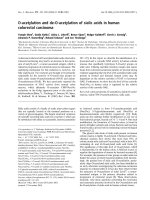
Báo cáo khoa học: "Chromosomal radiosensitivity and acute radiation side effects after radiotherapy in tumour patients a follow-up study" ppsx
Bạn đang xem bản rút gọn của tài liệu. Xem và tải ngay bản đầy đủ của tài liệu tại đây (255.85 KB, 8 trang )
RESEARCH Open Access
Chromosomal radiosensitivity and acute radiation
side effects after radiotherapy in tumour patients -
a follow-up study
Reinhard Huber
1*
, Herbert Braselmann
1
, Hans Geinitz
2
, Irene Jaehnert
1
, Adolf Baumgartner
1
, Reinhard Thamm
2
,
Markus Figel
3
, Michael Molls
2
and Horst Zitzelsberger
1
Abstract
Background: Radiotherapists are highly interested in optimizing doses especially for patients who tend to suffer
from side effects of radiotherapy (RT). It seems to be helpful to identify radiosensitive individuals before RT.
Thus we examined aberrations in FISH painted chromosomes in in vitro irradiated blood samples of a group of
patients suffering from breast cancer. In parallel, a follow-up of side effects in these patients was registered and
compared to detect ed chromosome aberrations.
Methods: Blood samples (taken before radiotherapy) were irradiated in vitro with 3 Gy X-rays and analysed by
FISH-painting to obtain aberration frequencies of first cycle metaphases for each patient. Aberration frequ encies
were analysed statistically to identify in dividuals with an elevated or reduced radiation response. Clinical data of
patients have been recorded in parallel to gain knowledge on acute side effects of radiotherapy.
Results: Eight patients with a significantly elevated or reduced aberration yield were identified by use of a t-test
criterion. A comparison with clinical side effects revealed that among patients with elevated aberration yields one
exhibited a higher degree of acute toxicity and two pat ients a premature onset of skin reaction already after a
cumulative dose of only 10 Gy. A significant relat ionship existed between translocations in vitro and the time
dependent occurrence of side effects of the skin during the therapy period.
Conclusions: The results suggest that translocations can be used as a test to identify individuals with a potentially
elevated radiosensitivity.
Background
So f ar, a central problem for radiotherapy is the nece s-
sity to avoid severe side effects to normal tissues.
Thus, the irradiation dose which can be normally
applied is limited by radiation response of the most
radiosensitive tumour patients. As a consequence of
such a protocol, lower than optimal irradiation doses
will be applied to m any patients. The lower doses affect
the chance to achieve a better local tumour control.
Suitable cytogenetic tests might provide a crucial basis
for an individualized radiotherapy. As a result, enhanced
cytogenetic effects in single individuals might refer to
enhanced tissue effects.
Thedoseresponsetoradiotherapymightsimplybe
analysed in peripheral blood cells before the beginning
of radiotherapy.
Introduction
Side effects in the normal tissues pose strong limitations
for efficient radiotherapy of malignant cancers [1].
Severe normal tissue reactions affect mostly radiosensi-
tive individuals who account for about 5% of all patients
[2]. Therefore, radiation doses in treatment of cancer
are generally restricted in order to minimize the inci-
dence of such severe side effects which conversely
imposes cure limitations for cancer treatment. For radia-
tion biology it is therefore a major goal to identify
* Correspondence:
1
Department of Radiation Cytogenetics, HelmholtzZentrum Muenchen -
German Research Center for Environmental Health, Neuherberg, Germany
Full list of author information is available at the end of the article
Huber et al. Radiation Oncology 2011, 6:32
/>© 2011 Huber et al; licensee BioMed Central Ltd. This is an Open Access article distributed under the terms of the Creative Commons
Attribution License (http://creativecom mons.org/licenses/by/2.0), which permits unrestricted use, distribution, and reproduction in
any medium, provided the original work is properly cited.
predictors for increased radiosensitivity before treatment
in order to allow an individualization of radiotherapy
[3], thus optimizing tumour control rates and minimiz-
ing severe radiotherapy effects.
In addition, cancer risk for the rise of secondary
tumours might increase in radiosensitive individuals
[4].
There are many biological endpoints which could be
used as a molecular predictor of radiosensitivity. Chro-
mosomal aberration frequency is regarded as a good
indicator because chromosomal aberrations are usually
related to a n altered DNA repair function which is in
turn linked to cellular radiosensitivity for whi ch dys-
function of many repair proteins have been demon-
strated [2]. De Ruyck et al. [5] reported an enhanced
chromosomal radiosensitivity detected by G2 assay as a
marker of genetic predisposition to head and neck can-
cer. Borgmann et al. [6] found an important heredital
impact with regard to radiation response detected by
different cytogenetic assays (G0 test, G2 test) in lympho-
cytes of a collective of t wins. Increased radiosensitivity
of chromosomes in peripheral lymphocytes from cancer
susceptibility syndrome patients, measured by chromo-
some breaks, was detected by Distel et al. [ 7]. The cited
effect seems in several patients to be due to genetic
instability [8]. Corr elations between chromosomal aber-
ration frequencies (chromosome aberrations or micro-
nucleus frequency) and acute tissue effects after
radiotherapy were reported by different authors [1,8,9].
In another study investigating radiation-induced DNA
primary damage and repair kinetic, by use of the
COMET assay [10], DNA effects were correlated with
acute tissue effects, whilst in a study of Popanda et a l.
[11] a correlation of acute side effects with DNA degra-
dation using the COMET assay could not be established.
For late tissue effects correlations with genomic altera-
tions detected by different assays have also been
reported [1,8,12-15], however, the influence of other fac-
tors could not be excluded before such late tissue effects
appeared in these clinical studies. Although the micro-
nucleus test is often regarded as highly suited in clinical
applications because of its simplicity, reproducibility and
promptness [2] it turned out in several studies [ 16-18]
that the analysis of chromosomal aberrations in FISH
(fluorescence in-situ hybridisation)-painted metaphases
isaverysensitivemarkercorrelated to tissue reactions
like acute skin effects or lesions. This leads us to investi-
gate whether chromosomal aberrations can be used as a
predictive marker to detect individuals showing a diver-
ging radiosensitivity. To make a FISH-based assay for
the detection of chromosomal aberrations more attrac-
tive for clinical applications we have combined the FISH
procedure with an automated scoring of FISH-painted
chromosome aberrations. This assay provides even
hardly detectable cytogenetic endpoints like transloca-
tions and colour junctions.
In the present study, chromosomal radiosensitivity has
been investigated in 47 breast tumour patients after in
vitro irradiation of blood samples. FISH-painting has
been applied to detect aberrations o n chromosome 1, 4
and 12 (partial genome analysis, [19]), whilst acute tis-
sue effects have been prospectively monitored during
radiotherapy of these patients.
Material and methods
Patients
The collective was selected from pa tients of the radiolo-
gical clinic t hat had to be subdued to radiotherapy
under similar schemes of radiotherapy, without applica-
tion of additional chemotherapeutic drugs. These condi-
tions delive red 47 patients examined in t he se quence of
their reception in the clinic, who received exclusively
radiotherapy due to a malignant breast tumour after
surgical lumpectomy. Individual blood sampling was
done within a follow-up period of six weeks.
The study was approved by the ethics committee of
the University hospital Rechts der Isar of the Technical
University Munich and done in accordance with the
revised Declaration of Helsinki.
Radiotherapy techniques
All patients were treated with 6 - 15 MeV photons from
a linear accelerator. Dose per fraction was 1.8 - 2.0 Gy
appli ed five times per week. Patients who received adju-
vant radiotherapy after breast conserving surgery for
breast cancer, were tre ated via t angential fields to the
ipsilateral breast. After a cumulative dose of 50 Gy an
electron boost with 10 -16 Gy to the former tumour
region was performed.
Side effects of radiotherapy
Clinical side effects of radiotherapy were evaluated
weekly during radiotherapy. Scoring was carried out
according to the Common Toxicity Criteria (NCI-CTC
scale; scale digits 0 , 1, 2, 3, 4). Mainly skin effects have
been identified as side reactions of radiotherapy.
Irradiation procedure in vitro and lymphocyte cultures
Whole blood samples (4ml fractionated i n 2× 2 ml syr-
inges) were irradiated in vitro with3Gyof220kVX-
rays (15 mA, 0.5 mm Cu and 4.05 mm Al filters, dose
rate 0.5 Gy min
-1
) at 37°C. Immediately after irradiation,
whole blood cultures were initiated according to our
published protocol [20]. Moreover, BrdU (final concen-
tration 9.6 x10
-6
μgml
-1
) was added to the cultures f or
identification of 1
st
cell cycle c hromosomes. Cultures
were incubated at 37° C for 48 h involving a colcemid
treatment (0.1 mg ml
-1
) for the final three hours.
Huber et al. Radiation Oncology 2011, 6:32
/>Page 2 of 8
Chromosome preparation wa s performed according to
standard procedures with slight modifications of our
published protocol [19]. Microscopic slides were stored
in a nitrogen atmosphere at -20°C until use.
FISH (fluorescence in-situ hybridisation)
For a homogeneous stai ning of three chromosome pairs,
FISH with painting probes for chromosomes 1, 4, and
12 directly labelled with FITC (probe set ID005, Chrom-
bios, Raubling, Germany), together with a pancentro-
meric DNA probe was applied according to
manufacturer’s manual. Counterstaining was performed
with propidium iodide (PI, 1 μgml
-1
)inantifadesolu-
tion. Before hybri disation, slides were treated with thio-
cyanate for 10 min at 90°C instead of pre-treatment
with pepsine [21]. For a discrimination between first
and second cycle metaphases (harlequin staining), prior
to painting, slides were treat ed with bisbenzimide
(H33258, Serva, Heidelberg, Germany) and UV light as
described by our published protocol [22].
Chromosome analyses
Metaphase finding and image capturing was performed
on a Metafer2 scanning system (Metasystems, Altlus-
sheim, Germany) with a Zeiss Axioplan2 MOT micro-
scope as described earlier [19]. Aberration analysis was
car ried out interactively on three-colour metaphase gal-
lery images or on full screen images, both providing
three colour channels on the display for the presentation
of FISH painted chromosomes, of counterstained chro-
mosomes, and of centromeric signals, using the PAINT
nomenclature syst em [23] to describe the observed
painting patterns. For the subsequent statistical analysis,
painted chromosomes bearing one centromere with a
colour junction were registered as t(Ab) or t(Ba), respec-
tively, painted chromosomes with two centromeres and
a colour junction as dicentrics. Painted chromosomes
exhibiting an insertion, ace(b), and other aberration
types, were regi stered but not subdued to statist ical
analysis.
Chromosome pairs 1, 4, and 12 appeared in green
(FITC), the centromeres were stained in blue (AMCA),
counterstaining of the complete metaphases appeared in
red (PI). Due to preceding harlequin staining, chromo-
somes in first cycle metaphases have a homogeneous
appearance, those in second cycle metaphases exhibit
differential staining of sister ch romatids. The latter were
excluded from chromosome analysis.
A mean of 140 in vitro irradiated lymphocytes (varia-
tion 5 0 - 467) per patient was analyse d. We protocoled
all types of structural aberrations in painted chromo-
somes as follows: all types of symmetrical translocations,
dicentrics, chromatid type aberrations, excess acentrics,
the numbers of metaphases with/without structural
aberrations, and colour junctions.
Statistical methods
For statistical analysis of the degree of skin side reaction
the maximum achieved scale digit during the follow-up
period was scored. The homogeneit y of chromosome
aberration frequencies among the patient samples was
examined by a c
2
test. Correlations w ere analysed by
Spearman’s rank correlation test. Outlying frequencies
were identified by a single classification t -test with p <
0.05 as criterion.
Results
47 patients have been investigated for clinical side reac-
tions and for in vitro response of peripheral lymphocytes
to 3 Gy X-rays irradiation.
Evaluation of clinical data
Skin reactions (NCI-CTC grading, common toxicity cri-
teria of the US National Cancer Institute) during and
after radiotherapy ha ve been classified according to the
following scale: grade 0: no skin reaction, grade 1: small
erythema, depilation, dry dandruff, reduced perspiration;
grade 2: moderate erythema, epitheliolysis <50% of
radiation field, moderate edema; grade 3: large erythema,
epitheliolysis >50% of radiati on field, strong edema;
grade 4: deep ulcer, haemorrhage or necrosis. 4 of 47
patients showed grade 0, 30 patients grade 1, 12 patients
grade 2, and 1 patient grade 3.
As an additional grouping patients were classified
according to the time-dependent occurrence of skin
reactions in the order “early reaction” if it occurred after
an accumulated dose of 10 Gy, as “in between reaction”,
if it occurred after 30 Gy accumulated dose, as “late
reaction”, if it occurred at the end of radiotherapy, and
as “ no reaction”. 4 of 47 patients showed no reactions,
13 patients late reactions, 23 patients in between reac-
tions, and 7 patients early react ions (individual data not
shown, total data presented in “Additional file 1 Table
S1”.
Evaluation of chromosome aberrations
FISH painting was performed on in vi tro irradiated
metaphase preparations which were further subdued to
aberration analysis using the semi-automated Metafer2
system (Metasystems GmbH, Altlussheim, Germany).
The following classifications of cytogenetic effects have
been used for statistical treatment:
(i) all metaphases containing structural aberrations, (ii)
translocations of the t(Ab) as well as t(Ba) types, (iii)
dicentrics (dic), (iv) colour junctions (cj). This classifica-
tion enables the detection of radiation-induced
Huber et al. Radiation Oncology 2011, 6:32
/>Page 3 of 8
chromosome aberrations in total and subclassification
into different aberration types.
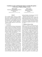
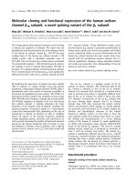
A total of 6829 metaphases were analysed and indivi-
dual chromosome aberration yields were compared for
47 patients. Aberration yields are shown for the respec-
tive cytogenetic effect in Figures 1 and 2.
Numerical data of aberrations are shown in table
“Additional file 2 Table S2”.
Statistical analyses revealed that for all patients inves-
tigated different aberration ty pes are correlated to each
other. This can be demonstrated for the yields of t(Ab)
corresponding t(Ba) (p < 0.0001), for t(Ba) and corre-
sponding dicentric yields (p < 0.0037) and for t(Ab) and
the c orresponding dicentric yields (p < 0.0072). More-
over, a significant ov erdispersion, i.e. a non-homoge-
neous distribution among patient samples (p < 0.0001)
was found for all cytogenetic effects (t(Ba), t(Ab),
dicentrics, colour junctions, cells containing aberra-
tions). The median frequencies were 0.20 per cell for t
(Ba), 0.21 for t(Ab) and 0.26 for dicentrics.
The chromosome analysis revealed several patients
that show a conspicuously higher or lower aberration
yield, respectively. A singl e classification test was further
used to identify single patients with a significant devia-
tion f rom the mean aberration frequencies. Results are
summarised in Table 1 showing significantly raised
aberration frequencies for patients 1 and 3 (t(Ba)/cell, t
(Ab)/cell, colour junctions/cell), for patient 7 (t(Ba)/cell)
and for patient 17 (t(Ab)/cell). Significantly reduced
aberration frequencies are revealed for patient 30
(dicentrics/cell), for patient 36 (t(Ab)/cell, structural
aberrations/aberrant cell, for patient 37 (t(Ba)/cell) and
for patient 41 (structural aberrations/aberrant cell).
Correlation between chromosome aberrations and clinical
side effects
A comparison of individual chromosome aberration data
with clinical side reactions revealed that among patients
with increased aberration yields (all of them treated
with identical doses of 50 Gy and 10 Gy boost) patient
1 e xhibited more severe side effects and patients 7 and
17 showed early reactions after 10 Gy. Patient 3 with an
increased chromosomal sensi tivity did not show an
increased acute side reaction. Apart from individual
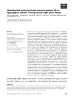
chromosomal outliers a significant o verall correlation
was found between the frequencies of t(Ba) in vitro and
the tim e-dependent occurence, i.e. latency of side effects
of the skin (Spearman’s rank corr elation test, p = 0.014).
The correlation is shown in Figure 3.
In practice, a discrimination of patients is done using
cut-off levels. The median t(Ba) frequency in the group
of patients showing a skin reaction already after 10 Gy
(short latency) is 0.21 per cell. In the group of patients
showing no skin reaction or not before 30 Gy (longer
Aberrant cells
(%)
0
10
20
30
40
50
60
70
Colour junctions per cell
0
.
0
0.2
0.4
0.6
0.8
1.0
1.2
1.4
1.6
Figure 1 Distribution of aberrant cells and of colour junctio ns in in vitro irradiated lymphocytes of 47 patients. Symbols represent
individual frequency of the respective cytogenetic endpoint. Filled symbols represent cases with significantly increased or decreased frequency.
Exposure 3 Gy.
Huber et al. Radiation Oncology 2011, 6:32
/>Page 4 of 8
latency) the t(Ba) median is 0.17 per cell. Taking the
mean 0.19 per cell as a cut-off, the low frequency group
(
<0.19, 19 patients) and high frequency group (> 0.19,
28 patients) are associated to the latency groups with a
Fisher’s exact test p-value of 0.015. With this cut-off 22
of the 30 short latency patients (73.3%) are correctly
detected (sensitivity) and 11 of the 17 longer latency
patients (64,7%) are correctly assigned (specifity). F or
the endpo ints t(Ab), dic and cj no co rrelation with side
effects or with latency was found (see test results in
“Additional file 3 Table S3”).
Discussion
The aim of this study was to investigate the relationship
of chromosomal radiosensitivity and acute clinical side
effects in 47 breast cancer patients who underwent
radiotherapy fo r tumour treatment. The extent of clini-
cal side effects has been used as an indicator for the
individual radiosensitivity of each patient. Such estab-
lished relationships would be of clinical relevance
because they could represent a predictive factor that is
required for an individualisation of radiotherapy [2].
Greve at al. [24] reasoned that neither measurement of
radiation-induced apoptotic and necrotic cell death is
detectable in immortalised lymphoblastoid derivatives
nor cell death in blood lymphocytes is suitable to
unequivocally predict the individual clinical radiosensi-
tivity of cancer patients.
Premature chromosome condensation (G2 test) reveals
practically indistinguishable levels of chromosomal
breaks in AT and normal lymphoblastoid cells or
Figure 2 Distribution of translocation types t(Ba), t(A b), and of dicentrics (dic) in in vitro irradiated lymphocytes of 47 patients.
Symbols represent respective individual frequency of respective aberration type. Filled symbols represent cases with significantly increased or
decreased frequency. Exposure 3 Gy.
Table 1 Patients exhibiting a significant deviation from
the mean at different aberration types (likelihood
quotient test, p < 0.01)
patients cytogenetic endpoint
C
A
(%) t(Ba)/cell t(Ab)/cell dic/cell cj/cell
significantly increased cytogenetic effects
patient 1 52.7 0.317 0.385 0.336 1.377
patient 3 53.7 0.347 0.355 0.238 1.285
patient 7 54.7 0.353 0.264 0.259 1.209
patient 17 51.7 0.313 0.381 0.224 1.136
significantly reduced cytogenetic effects
patient 30 49.1 0.171 0.239 0.107 0.585
patient 36 30.8 0.149 0.097 0.144 0.533
patient 37 26.0 0.020 0.060 0.140 0.320
patient 41 27.5 0.183 0.174 0.174 0.642
C
A
%: percentage of cells containing structural aberrations.
Italic: significant difference to mean value of patients.
Huber et al. Radiation Oncology 2011, 6:32
/>Page 5 of 8
lymphocytes, though lymphocytes of AT patients reveal
an increased radiosensitiv ity measured by PCC(prema-
ture chromosome condensation) chromosome breaks
[25].
Based o n the micronucleus assay in cytokinesis-
blocked lymphocytes, Mozdarani et al. [26] found signif-
icant differences between a control group and groups of
breast cancer or oesophageal cance r patients, respec-
tively, after in vitr o irradiation with 3 Gy; nevertheless,
radiosensitive individuals could not be identified in this
study.
Interindividual radiosensitivity in blood lymphocytes
of 14 healthy donors could not be detected with the
micronucleus assay, nor with the G2 assay. It could not
be decided whether the detected variation of both cyto-
genetic effects was due to interindividual variation of
radiosensitivity, or to i ntraindividual variation [27].
Hence it is promising to study chromosomal damage as
a marker for cellular radiosensitivity because it is well
established as a quantitative indicator for preceding
radiation exposure [28-33]. We therefore have quantified
chromosomal aberrations in blood samples from 47
tumour patients which h ave been irradiated with 3 Gy
X-rays in vitro. The measured aberration frequencies
showed for some patients significant deviations from the
mean value for each aberration category (Figures 1 and
2). The presented approach is novel because in this
study the use of an automated scoring system allowed
an evaluation of 6829 metaphases which would facilitate
to use this approach routinely in clinical testing. The
validity of these scoring results is indicated by the highly
significant correlations between each aberration
categories.
The statistical analyses further revealed that four out
of 47 patients exhibited a significantly elevated aberra-
tion frequency at least for one aberration category indi-
cating an increased radiation response at the DNA
repair level (Table 1). Interestingly, the dicentric fre-
quencies were not significantly elevated in each of the
four patients, but translocations showed a significant
increase. Such discrepancies between translocation and
dicentric yields after radiation exposure have already
been de scribed in seve ral studies quantifyin g radiation-
induced chromosome aberra tions [32,34]. In view of the
correlation, it means that translocations show a more
extensive response to radiation compared to dicentrics.
So far, Keller et al. [17] reported that among other
cytogenetic parameters, the paramete r “percentage of
dicentric chromosomes” could neither serve as meaning-
ful nor as significant criteria, since it showed a strong
interindividual variability, whereas translocations were
suitable indicators for detecting differences in blood
lymphocytes from patients and controls irradiated in
vitro with two different doses.
On the other hand there was found an indication for a
reduced radiation response since significantly reduced
aberration frequencies at least for one aberration cate-
gory have been detected in four patients (Table 1). Thus
based on cytogenetic results one w ould expect four
patients with an enhanced and four patients wi th a
reduced radiosensitivity in our study. In order to vali-
date this assumption, clinical phenotypes were also con-
sidered. The comparison with acute clinical side effects
(mainly skin reactions) demonstrated that none of the
patients exhibiting significantly reduced aberration yields
suffered from abnormal tissue reactions during or after
radiotherapy reflecting the initial finding of a reduced
radiosensitivity. However, among the four patients with
elevated aberration frequencies three patient s showed
either a more severe side reaction of radiotherapy
(patient 1) or a premature side reaction already after 10
Gy of irradiation (patients 7 and 17). Alt hough such a
co-incidence could not be found for patient 3, these
results let assume that a relationship between cellular
radiosensitivity measured as chromosome aberration
yield in peripheral lymphocytes and acute clinical side
rea ctions exists. Anyway, it could be demonstra ted with
statistical significance that a chromosome aberration
test investigating translocations by FISH is suitable to
identify individuals with shortened response time of
radiation-induced skin reactions.
Figure 3 Box plot analysis of t(Ba) frequencies in 4 patient
groups ordered according to temporal occurence of any side
effects of the skin during the period of radiation therapy. Box
area, 50% of data [lines in box denote medians; bars include at
most 1.5 of interquartile distance, difference between first and third
quartiles of data; circles indicate values out of the 1.5-fold box area
(outliers)]. A significant correlation between the frequencies of t(Ba)
from lymphocytes irradiated in vitro (3 Gy) and the time-dependent
occurence of side effects is demonstrated.
Huber et al. Radiation Oncology 2011, 6:32
/>Page 6 of 8
So far, only few studies exist reporting on similar rela-
tionships between acute clinical reactions and metaphase
chromosome radiosensitivity. Dunst et al. [12] demon-
strated that nine out of 26 radiotherapy patients showing
elevated chromos ome break frequencies suffered from an
increased acute skin damage. Compared to our patient
cohort they inv estigated more different tumour types
leading to higher heterogeneity after in vitro exposure
with0.7and2.0Gyinthestudygroup[12].Similar
results were reported by Popanda et al. [11] who detected
6 out of 113 r adiotherapy patients wit h excessive acut e
skin reactions also showi ng significantly increased radia-
tion-induced genomic changes detected by the COMET
ass ay. However, a statistical correlation between genome
alterationsandacutesideeffectscouldnotbedemon-
strated. Further studies reported an increased cellular
radiosensitivity in radiotherapy patients using G0 and G2
assays [27,35]. However, these did not register clinical
side effects which limits the impact of their r esults. On
the other hand in a recent study, Slonina et al. [36] could
not find elev ated acute or late side effects in cervix carci-
noma patients whose cultured keratinocytes and fibro-
blasts exhibited increased micronucleus frequencies.
Moreover, it has been demonstrated in several in vitro
studies that the G0 micronucleus assay in blood lympho-
cytes using 3 Gy in vitro exposure [37], usi ng 3.5 Gy in
vitro exposure [27], and blood l ymphocyte G2 assay
using 0.4 Gy in vitro exposure [27], have limited reprodu-
cibility due to extended intraindividual variability. Limita-
tions of the G2 assay, e.g. from interindividual variation,
were also reported in a compilation from data of different
studies [38].
In conclusion, a comparison o f our findings with sev-
eral published data suggests that measuring chromoso-
mal radiosensi tivity on t ranslocation level in blood
lymphocytes can be proposed to be used as a predictive
assay for detection of radiosensitive individuals which
should be developed further. Data from larger cohorts
are needed to assess whether a particular aberration
type is most sensitive to detect increased radiosensitiv-
ity. It would be also of interest to monitor chromosome
aberrations in blood lymphocytes ex vivo at different
times during radiotherapy to evaluate whether the
occurrence of acute clinical side effects is related to
increased aberration frequencies in a timely manner in
order to detect a potential timely correlation, which
wouldcorrespondtoourfindingsfromlymphocytes
exposed in vitro.
Additional material
Additional file 1: Radiotherapy’s side effects of 47 tumour patients.
Side effects of radiotherapy in 47 tumour patients (highest degree and
occurrence of skin reaction).
Additional file 2: Absolute numbers of cytogenetic effects in in vitro
irradiated blood lymphocytes of 47 tumour patients. Absolute
numbers of different types of cytogenetic effects from in vitro irradiated
(3 Gy) blood lymphocytes of 47 tumour patients.
Additional file 3: Correlation coefficients of different types of
chromosome aberrations from in vitro irradiated lymphocytes.
Correlation coefficients of different types of chromosome aberrations
from in vitro irradiated (3 Gy) lymphocytes compared to degree of side
effects and to latency of side effects in 47 patients (p-values for
Spearman’s rank correlation test).
Acknowledgements and Funding
We thankfully acknowledge the skilful technical assistance of S. Schroeferl
and E. Konhaeuser.
This study was supported in part financially by the Federal Office of Defense
Technology and Procurement, Grant E/B41G/Z0531/Z5803.
Author details
1
Department of Radiation Cytogenetics, HelmholtzZentrum Muenchen -
German Research Center for Environmental Health, Neuherberg, Germany.
2
Department of Radiation Oncology, Technische Universitaet Muenchen,
Munich, Germany.
3
Personal Monitoring Service, HelmholtzZentrum
Muenchen - German Research Center for Environmental Health, Munich,
Germany.
Authors’ contributions
RH has substantially contributed to acquisition and interpretation of data; he
has been involved in drafting the manuscript and has contributed to the
final version to be published. HB has made substantial contributions to the
conception and design of the study, to analysis and interpretation of the
study. He was responsible for the statistical treatment of data, kindly
delivering the manuscript’s Figures. He was involved in drafting the
manuscript and revising it critically, and has given final approval of the
version to be published. HG has made substantial contributions to
conception and design of the study. As a clinical radiologist, he supervised
the administration and delivery of patients’ blood samples. He has been
involved in revising the manuscript critically and has given final approval of
the version to be published. IJ has made substantial contributions to
acquisition of data, collecting blood samples, and lymphocyte culture
procedures. She has been involved in revising the protocol critically. AB has
made substantial contributions to acquisition of data, collecting blood
samples, lymphocyte culture and FISH procedures. RT has made substantial
contributions to conception and design of the study. As a clinical
radiologist, he supervised the administration and delivery of patients’ blood
samples. MF has delivered dosimetry for in vitro irradiation experiments, and
he provided practical advice for the handling of the irradiation device. MM
has made substantial contributions to conception and design of the study.
HZ has made substantial contributions to conception and design of the
study, and the interpretation of data. He has been involved in drafting the
manuscript and revising it critically. He has given final approval of the
version to be published.
All authors read and approved the final manuscript.
Competing interests
The authors declare that they have no competing interests.
Received: 25 November 2010 Accepted: 7 April 2011
Published: 7 April 2011
References
1. Barber JB, Burrill W, Spreadborough AR, Levine E, Warren C, Kiltie AE,
Roberts SA, Scott D: Relationship between in vitro chromosomal
radiosensitivity of peripheral blood lymphocytes and the expression of
normal tissue damage following radiotherapy for breast cancer.
Radiother Oncol 2000, 55:179-186.
2. Sprung CN, Chao M, Leong T, McKay J: Chromosomal radiosensitivity in
two cell lineages derived from clinically radiosensitive cancer patients.
Clin Cancer Res 2005, 11:6352-6358.
Huber et al. Radiation Oncology 2011, 6:32
/>Page 7 of 8
3. Sprung CN, Davey DS, Withana NP, Distel LV, McKay MJ: Telomere length
in lymphoblast cell lines derived from clinically radiosensitive cancer
patients. Cancer Biol Ther 2008, 638-644.
4. Dyomina EA, Ryabchenko NM: Increased individual chromosomal
radiosensitivity of human lymphocytes as a parameter of cancer risk. Exp
Oncol 2007, 29:217-220.
5. de Ruyck K, de Gelder V, van Eijkeren M, Boterberg T, De Neve W, Vral A,
Thierens H: Chromosomal radiosensitivity in head and neck cancer
patients: evidence for genetic predisposition? Br J Cancer 2008,
98:1723-1738.
6. Borgmann K, Haeberle D, Doerk T, Busjahn A, Stephan G, Dikomey E:
Genetic determination of chromosomal radiosensitivities in G0- and G2-
phase human lymphocytes. Radiother Oncol 2007, 83:196-202.
7. Distel LV, Neubauer S, Keller U, Sprung CN, Sauer R, Grabenbauer G:
Individual differences in chromosomal aberrations after in vitro
irradiation of cells from healthy individuals, cancer and cancer
susceptibility syndrome patients. Radiother Oncol 2006, 81:257-263.
8. Keller U, Grabenbauer G, Kuechler A, Sprung CN, Mueller E, Sauer R, Distel L:
Cytogenetic instability in young patients with multiple primary cancers.
Cancer Genet Cytogenet 2005, 157:25-32.
9. Jones LA, Scott D, Cowan R, Roberts SA: Abnormal radiosensitivity of
lymphocytes from breast cancer patients with excessive normal tissue
damage after radiotherapy: chromosome aberrations after low dose-rate
irradiation. Int J Radiat Biol 1995, 67:519-528.
10. Sterpone S, Cornetta T, Padua L, Mastellone V, Giammarino D, Testa A,
Tirindelli D, Cozzi R, Donato V: DNA repair capacity and acute
radiotherapy adverse effects in Italian breast cancer patients. Mutat Res
2010, 684:43-48.
11. Popanda O, Ebbeler R, Twardella D, Helmbold I, Gotzes F, Schmezer P,
Thielmann HW, von Fournier D, Haase W, Sautter-Bihl ML, Wenz F,
Bartsch H, Chang-Claude J: Radiation-induced DNA damage and repair in
lymphocytes from breast cancer patients and their correlation with
acute skin reactions to radiotherapy. Int J Radiat Oncol Biol Phys 2003,
55:1216-1225.
12. Dunst J, Neubauer S, Becker A, Gebhart E: Chromosomal in vitro
radiosensitivity of lymphocytes in radiotherapy patients and AT-
homozygotes. Strahlenther Onkol 1998, 174:510-516.
13. Borgmann K, Roper B, El-Awady R, Brackrock S, Bigalke M, Dork T, Alberti W,
Dikomey E, Dahm-Daphi J: Indicators of late normal tissue response after
radiotherapy for head and neck cancer: fibroblasts, lymphocytes,
genetics, DNA repair, and chromosome aberrations. Radiother Oncol 2002,
64:141-152.
14. Hoeller U, Borgmann K, Bonacker M, Kuhlmey A, Bajrovic A, Jung H,
Alberti W, Dikomey E: Individual radiosensitivity measured with
lymphocytes may be used to predict the risk of fibrosis after
radiotherapy for breast cancer. Radiother Oncol 2003, 69:137-144.
15. Ramsay J, Birrell G: Normal tissue radiosensitivity in breast cancer
patients. Int J Radiat Oncol Biol Phys 1995, 31:339-344.
16. Keller U, Grabenbauer G, Kuechler A, Sauer R, Distel L: Technical report.
Radiation sensitivity testing by fluorescence in-situ hybridisation: how
many metaphases have to be analysed? Int J Radiat Biol
2004, 80:615-620.
17. Keller U, Kuechler A, Liehr T, Mueller E, Grabenbauer G, Sauer R, Distel L:
Impact of various parameters in detecting chromosomal aberrations
by FISH to describe radiosensitivity. Strahlenther Onkol 2004,
180:289-296.
18. Tucker JD: Sensitivity, specificity, and persistence of chromosome
translocations for radiation biodosimetry. Mil Med 2002, 167:8-9.
19. Huber R, Kulka U, Loerch T, Braselmann H, Engert D, Figel M, Bauchinger M:
Technical report: application of the Metafer2 fluorescence scanning
system for the analysis of radiation-induced chromosome aberrations
measured by FISH-chromosome painting. Mutat Res 2001, 492:51-57.
20. Huber R, Braselmann H, Kulka U, Schumacher-Georgiadou V, Bayerl A,
Molls M, Bauchinger M: Follow-up analysis of translocation and dicentric
frequencies, measured by FISH-chromosome painting in breast cancer
patients after partial-body radiotherapy with little bone marrow
exposure. Mutat Res 1999, 446:103-109.
21. Mueller I, Geinitz H, Braselmann H, Baumgartner A, Fasan A, Thamm R,
Molls M, Meineke V, Zitzelsberger H: Time-course of radiation-induced
chromosomal aberrations in tumor patients after radiotherapy. Int J
Radiat Oncol Biol Phys 2005, 63:1214-1220.
22. Kulka U, Huber R, Mueller P, Knehr S, Bauchinger M: Combined FISH
painting and harlequin staining for cell cycle-controlled chromosome
analysis in human lymphocytes. Int J Radiat Biol 1995, 68:25-27.
23. Tucker JD, Morgan WF, Awa AA, Bauchinger M, Blakey D, Cornforth MN,
Littlefield LG, Natarajan AT, Shasserre C: PAINT: a proposed nomenclature
for structural aberrations detected by whole chromosome painting.
Mutat Res 1995, 347:21-24.
24. Greve B, Dreffke K, Rickinger A, Koenemann S, Fritz E, Eckardt-Schupp F,
Amler S, Sauerland C, Braselmann H, Sauter W, Illig T, Schmezer P,
Gomolka M, Willich N, Boelling T: Multicentric investigation of ionising
radiation-induced cell death as a predictive parameter of individual
radiosensitivity. Apoptosis 2009, 14:226-235.
25. Terzoudi GI, Manola KN, Pantelias GE, Iliakis G: Checkpoint abrogation in
G2 compromises repair of chromosomal breaks in ataxia telangiectasia
cells. Cancer Res 2005, 65:11292-11296.
26. Mozdarani H, Mansouri Z, Haeri SA: Cytogenetic radiosensitivity of G
0
-
lymphocytes of breast and esophageal cancer patients as determined
by micronucleus assay. J Radiat Res (Tokyo) 2005, 46:111-116.
27. Vral A, Thierens H, Baeyens A, De Ridder L: The micronucleus and G2-
phase assays for human blood lymphocytes as biomarkers of individual
sensitivity to ionizing radiation: limitations imposed by intraindividual
variability. Radiat Res 2002, 157:472-477.
28. Schmid E, Bauchinger M, Bunde E, Ferbert HF, von Lieven H: Comparison
of the chromosome damage and its dose response after medical whole-
body exposure to 60Co gamma-rays and irradiation of blood in vitro. Int
J Radiat Biol Relat Stud Phys Chem Med 1974, 26:31-37.
29. Evans HJ, Buckton KE, Hamilton GE, Carothers A: Radiation-induced
chromosome aberrations in nuclear-dockyard workers. Nature 1979,
277:531-534.
30. Pantelias GE, Iliakis GE, Sambani CD, Politis G: Biological dosimetry of
absorbed radiation by C-banding of interphase chromosomes in
peripheral blood lymphocytes. Int J Radiat Biol 1993, 63:349-354.
31. Bauchinger M, Braselmann H, Savage JR, Natarajan AT, Terzoudi GI,
Pantelias GE, Darroudi F, Figgitt M, Griffin CS, Knehr S, Okladnikova ND,
Santos S, Snigiryova G: Collaborative exercise on the use of FISH
chromosome painting for retrospective biodosimetry of Mayak nuclear-
industrial personnel. Int J Radiat Biol 2001, 77:259-267.
32. Rao BS, Natarajan AT: Retrospective biological dosimetry of absorbed
radiation. Radiat Prot Dosimetry 2001, 95:17-23.
33. Montoro A, Rodriguez P, Almonacid M, Villaescusa JI, Verdú G, Caballín MR,
Barrios L, Barquinero JF: Biological dosimetry in a group of radiologists by
the analysis of dicentrics and translocations. Radiat Res 2005, 164:612-617.
34. Bauchinger M, Schmid E, Braselmann H: Time-course of translocation and
dicentric frequencies in a radiation accident case. Int J Radiat Biol 2001,
77:553-557.
35. Baeyens A, Thierens H, Claes K, Poppe B, Messiaen L, De Ridder L, Vral A:
Chromosomal radiosensitivity in breast cancer patients with a known or
putative genetic predisposition. Br J Cancer 2002, 87:1379-1385.
36. Slonina D, Biesaga B, Urbanski K, Kojs Z: Comparison of chromosomal
radiosensitivity of normal cells with and without HRS-like response and
normal tissue reactions in patients with cervix cancer. Int J Radiat Biol
2008, 84:421-428.
37. Huber R, Braselmann H, Bauchinger M: Intra- and inter-individual variation
of background and radiation-induced micronucleus frequencies in
human lymphocytes. Int J Radiat Biol 1992, 61:655-661.
38. Bryant PE, Gray L, Riches AC, Poppe B, Messiaen L, De Ridder L, Vral A: The
G
2
chromosomal radiosensitivity assay. Int J Radiat Biol 2002, 78:863-866.
doi:10.1186/1748-717X-6-32
Cite this article as: Huber et al.: Chromosomal radiosensitivity and acute
radiation side effects after radiotherapy in tumour patients - a follow-up
study. Radiation Oncology 2011 6:32.
Huber et al. Radiation Oncology 2011, 6:32
/>Page 8 of 8