Báo cáo khoa học: "Helical tomotherapy in the treatment of pediatric malignancies: a preliminary report of feasibility and acute toxicitty" doc
Bạn đang xem bản rút gọn của tài liệu. Xem và tải ngay bản đầy đủ của tài liệu tại đây (2.53 MB, 9 trang )
RESEARCH Open Access
Helical tomotherapy in the treatment of pediatric
malignancies: a preliminary report of feasibility
and acute toxicity
Latifa Mesbah
1
, Raúl Matute
1*
, Sergey Usychkin
1
, Immacolata Marrone
1
, Fernando Puebla
1
, Cristina Mínguez
1
,
Rafael García
1
, Graciela García
1
, César Beltrán
1
and Hugo Marsiglia
1,2,3
Abstract
Background: Radiation therapy plays a central role in the management of many childhood malignancies and
Helical Tomotherapy (HT) provides potential to decrease toxicity by limiting the radiation dose to normal
structures. The aim of this article was to report preliminary results of our clinical experience with HT in pediatric
malignancies.
Methods: In this study 66 consecutive patients younger than 14 years old, treated with HT at our center between
January 2006 and April 2010, have been included. We performed statistical analyses to assess the relationship
between acute toxicity, graded according to the RTOG criteria, and several clinical and treatment chara cteristics
such as a dose and irradiation volume.
Results: The median age of patients was 5 years. The most common tumor sites were: central nervous system
(57%), abdomen (17%) and thorax (6%). The most prevalent histological types were: medulloblastoma (16 patients),
neuroblastoma (9 patients) and rhabdomyosarcoma (7 patients). A total of 52 patients were treated for primary
disease and 14 patients were treated for recurrent tumors. The majority of the patients (72%) were previously
treated with chemotherapy. The median prescribed dose was 51 Gy (range 10-70 Gy). In 81% of cases grade 1 or 2
acute toxicity was observed. There were 11 cases (16,6% ) of grade 3 hematological toxicity, two cases of grade 3
skin toxicity and one case of grade 3 emesis. Nine patients (13,6%) had grade 4 hematological toxicity. There were
no cases of grade 4 non-hematological toxicities . On the univariate analysis, total dose and craniospinal irradiation
(24 cases) were significantly associated with severe toxicity (grade 3 or more), whereas age and chemotherapy
were not. On the multivariate analysis, craniospinal irradiation was the only significant independent risk factor for
grade 3-4 toxicity.
Conclusion: HT in pediatric population is feasible and safe treatment modality. It is characterized by an acceptable
level of acute toxicity that we have seen in this highly selected pediatric patient cohort with clinical features of
poor prognosis and/or aggressive therapy needed. Despite of a dosimetrical advantage of HT technique, an
exhaustive analysis of long-term follow-up data is needed to assess late toxicity, especially in this potentially
sensitive to radiation population.
Keywords: Helical Tomotherapy, Intensity-Modulated Radiation Therapy, pediatric malignancies, feasibility, acute
toxicity
* Correspondence:
1
Radiotherapy Department, Instituto Madrileño de Oncología (Grupo IMO), 7
Plaza Republica Argentina, Madrid, 28002, Spain
Full list of author information is available at the end of the article
Mesbah et al. Radiation Oncology 2011, 6:102
/>© 2011 Mesbah et al; licensee BioMed Central Ltd . This is an Open Access article distributed under the terms of the Creative Commons
Attribution License (http:// creativecommons.org/ licenses/by/2.0), which permits unrestricted use, distribution, and re production in
any med ium, provided the original work is properly cited.
Background
Radiation t herapy is an integral part in the treatment of
40-60% of childhood cancer patients [1]. Although many
childhood malignancies are cured, the acute toxicity of
therapy and significant late treatment effects make these
cancers a substantial burden for patients, their families,
and societ y [2]. Therefore, the goal of modern strategies
is not only to improve cancer cure rate, but also to
decrease adverse sequelae of treatment. The use of mod-
ern radiotherapy techniques may, potentially, decrease
the incidence and severity of radiation toxicity.
Intensity-Modulated Radiation Therapy (IMRT) has
shown to be a safe and effective treatment modality for
adult cancer patients. This radiothe rapy delive ry techni-
que has proven capability to create highly conformal
dose distributions allowing to escalate dose in target
volume and to spare adjacent organs at risk [3,4]. While
IMRT is widely used as a standard of care for many
adult cancers patients, this technique has been used less
frequently in childhood cancer patients, for several rea-
sons, such as a potentially augmented risk of carcino-
genesis due to increased volume of normal tissues
receiving low-dose radiation.
Helical Tomotherapy (HT) is a novel highly precise
IMRT technique with image-guidance using megavoltage
computed tomography (MVCT) that actually is used by
more than 150 institutions around the word. In Spain, it
was implemented for the first time in 2006, at the Instituto
Madrileño de Oncología (Grupo IMO), which is a referral
center of pedia tric radiation oncology in the country. In
this article we report our initial experience of HT in the
treatment of pediatric malignancies, focused on analysis of
tumor response and acute radiation toxicity. A critical
review of published studies of IMRT and HT in the treat-
ment of pediatric cancer patients is also presented.
Methods
From April 2006 through May 201 0, 66 consecutive
children younger than 14 years old underwent HT at
the Tomotherapy Unit of the Grupo IMO in the context
of multidisci plinary national and international treatment
protocols. All the patients were treated with curative
intent, including those who had recurrent disease. Two
patients previously had received external beam radiation
therapy, one of them underw ent reirradiation for local
recurrence of rhabdomyosarcoma (RMS), and the other
patient received reirradiation fo r spinal recurrence of
medullo blastoma. All patients were referred to our cen-
ter from their local radiot herapy departments due to
inability of conventional rad iotherapy techniques to
comply with dose restrictions in critical organs.
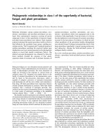
Individual immobilization was employed in all cases.
Depending on the site of the treatment, a customized
alpha-cradle mould was used for thoracic and
abdominopelvic tumor sites, whereas a ‘home-made’
non-invasive stereotactic frame system was used for
head and neck tumors (Figure 1).
Target volumes were defined using only computed
tomography images in 23 patients. In 43 patients co-
registration of 18-fluorodeoxyglucose positron emission
tomography and/or magnetic resonance images with
computed tom ography images was used. Target volumes
and organs at risk were contoured on a Pinnacle™
workstation version 8.0 (Philips Radiation Oncology Sys-
tems, Fitchburg, WI, USA) and defined according to the
criteria of the International Commission of Radiat ion
Units and Measuremen t [5,6]. As a rule 3 to 5 mm
CTV to PTV margins were applied. Data sets and struc-
tures were transferred to the Tomotherapy treatment
planning system (Tomotherapy Inc., Madison, WI) to
perform inverse treatment planning. The planning goal
was to deliver the prescription dose to at least 95% of
the PTV. The dose constraints for organs at risk (OARs)
were mainly those reported in of the National Cancer
InstitutePhysicianDataQuery[7].Dosevolumehisto-
grams for PTVs and OARs were recorded from the
dosimetric charts. Homogeneity index was calculated
dividing the maximal PTV dose by the prescription
dose; the coverage index was calculated dividing the
minimum PTV dose by the pr escription dose. Both
indexes were calculated accordingly to the recommenda-
tions established for evaluating tomotherapy treatment
plans [8].
All treatments were delivered by a Helical TomoTher-
apy™ HiArt™ II system treatment unit. Daily MVCT
acquisitions were performed for all patients to detect
set-up deviations and to correct them. All patients were
treated with once-daily fractions of 1.5-2 Gy, except for
one child with medulloblastoma who received twice-
daily fractionated radiotherapy.
Figure 1 “Home-made” non-invasive stereotactic frame.
Mesbah et al. Radiation Oncology 2011, 6:102
/>Page 2 of 9
All patients were examined at least weekly during treat-
ment. The acute and subacute toxicity was defined and
graded according to the RTOG criteria. After the radia-
tion therapy, all the patients underwent follow-up exami-
nations at 1, 3, 6 months after treatment and then yearly.
Statistical analysis
Univariate analysis was performed to test the association
between several clinical and treatment characteristics
and ≥ grade 3 acute toxicity. The t test or the non-para-
metric Mann-Whitney test (if the normal distribution
assumption was not fitted) was used for quantitative
variables and a chi-square test for qualitative variables.
For the multivariate analysis a regression logistic was
performed. Two-tailed p-values < 0.05 were considered
to be statistically significant. Analyses were performed
using SPSS version 15 (SPSS Inc., Chicago, IL).
Results
The median age at HT treatment was 5 years (range 1-
14 years); 20 patients (30%) were 3 years old or younger.
Patient characteristics are summarized in Table 1. The
most common tumor sites were central nervous system
(57%), abdom en (17%) and thorax (6%). The most pre-
valent histological types were medulloblastoma (16
patients), neuroblastoma (9 patients) and rhabdomyosar-
coma (7 patients). 52 patients were treated for primary
disease while 14 patients were treated for recurrence.
The majority of the patients (72%) received neoadjuvant
or concomitant chemotherapy. The median adminis-
tered radiation dose was 51 Gy (range 11 Gy - 70 Gy).
Sedation with inhalation of sevoflurane during radiother-
apy session was necessary in 4 1 p atients (6 2%). M edian a ge
of these patients was 4 years (range 1-9 years). They were
treated with craniospinal irradiation (n = 16, 40%) and
extended target volumes irradiation in thorax and abdom-
inal (n = 8, 20%) which were main indications for sedation.
It was well tolerated without severe side-effects and was
associated with fast recovery after treatment. General
anesthesia with intubation was not ne cessary.
AcutetoxicitydataissummarizedinTable2.In81%
of cases grade 1 or 2 acute toxicity was observed. There
Table 1 Patients characteristics
Characteristics n (%)
Gender Male 36 (55%)
Female 30 (45%)
Medulloblastoma 16 (24%)
Ependymoma and ependymoblastoma 8 (12%)
Glioma 7 (11%)
CNS Pineoblastoma 2 (3%)
Teratoid/Rhabdoid tumor 2 (3%)
Germinal tumor 1 (1%)
Choroid plexus tumor 1 (1%)
Craniopharyngioma 1 (1%)
Tumor site/histology Abdomen Neuroblastoma 7 (11%)
Nephroblastoma 2 (3%)
Rhabdomyosarcoma 1 (1%)
Clear cell sarcoma 1 (1%)
Thorax Ewing sarcoma 1 (1%)
Hodgkin lymphoma 1 (1%)
PNET (Askin’s) tumor 1 (1%)
Rhabdomyosarcoma 1 (1%)
Pelvis Rhabdomyosarcoma 2 (3%)
Ewing sarcoma 1 (1%)
PNET tumor 1 (1%)
Other sites Orbit Melanoma 1 (1%)
Rhabdomyosarcoma 1 (1%)
PNET tumor 1 (1%)
Spine Neuroblastoma 2 (3%)
Skull base Chordoma 1 (3%)
Oropharynx Rhabdomyosarcoma 1 (1%)
Extremity Rhabdomyosarcoma 1 (1%)
Sub- and supradiaphragmatic Hodgkin lymphoma 1 (1%)
Mesbah et al. Radiation Oncology 2011, 6:102
/>Page 3 of 9
were 11 cases (16,6%) of grade 3 hematological toxi city,
two cases of grade 3 skin toxicity and one case of grade
3 emesis. Nine patients (13 ,6%) ha d grade 4 hematologi-
cal toxicity. We have not seen any case of grade 4 non-
hematological toxicity.
Actual daily treatment was not recorded duri ng treat-
ment sessions. However it can be estimated approxi-
mately based on daily treatment practice of our
department. In analyzed cases of pediatric malignancies
daily treatment time was composed of ti me required for
patient set-up and anesthesia inside the treatment room,
time of MVCT acquisition, time of review/match and
applying couch c orrection inside the treatment room,
actual radiation delivery time and waiting time of
patient recovery (from end of irradiation until the
patientisawake)fromanesthesia.TimeofMVCT
acquisition and ac tual ra diation delivery time are factors
that mostly influence time of treatment session. It’ s
known that in helical tomotherapy these parameters
strongly depend on the longitudinal extension of irra-
diated volume and as well as on selected MVCT slice
thickness. For example, in case of craniospinal irradia-
tion typical time of MVCT acquisition in our depart-
ment is about 300-500 seconds. Time needed for review
and match of images is no more than 1-3 minutes.
Radiation delivery time was recorded for each patient in
treatment chart. It varied from 158 to 1991 seconds and
medianwas390secondsthusshowingstrongdepen-
dence on the extension of treated volume. Radiation
delivery time for selected “challenging” tumor sites is
presented in Table 3. Patient set-up and anesthesia
requirements prolong daily treatment time for about 5-
10 min and generally do not compromise treatment
time frame of these patients.
In a great proportion of patients (39%) we were able
to deliver radiation to extended volumes without field
junctions: craniospinal irradiation was performed in 23
patients ; two patients underwent hemithorax irradiation,
one for thoracic Askin’s tumor and the other for thor-
acic Ewing sarcoma; in one case of advanced Hodgkin
lymphoma the patient received near total lymphatic
irradiation.
Mean coverage index for entire group of patients and
all PTVs was 0,82 ± 0,13. Mean homogeneity index was
1,07 ± 0,02. Mean PTV doses, coverage and homogene-
ity indexes for selected challenging cases or groups of
patients are presented in Table 3. Even for challenging
cases of craniospinal irra diation and extended thoracic
and abdominal v olumes irradiation coverage and homo-
geneity of delivere d dose were acceptable. Mean doses
for selected OARs are presented in Table 4. It shows
that substantial sparing of critical structures was
achieved in all patients although major variability in
OARs mean doses in this very heterogeneous patient
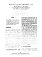
population is evident. In Figures 2 and 3 examples of
treatment plan for medulloblastoma and perineal rhab-
domyosarcoma with metastases to inguinal nodes are
presented.
On the univariate analysis, total dose and craniospinal
irradiation were associated significantly with toxicity
grade 3 or more, whereas age and chemotherapy were
not (Table 5). On the multivariate anal ysis, craniospinal
irradiation was the only significant independent risk fac-
tor for grade 3/4 toxicity.
While at present follow-up time is not sufficient (med-
ian 15 months; range 2-59 months) for reliable conclu-
sions of survival, the tumor response of 51 patients
could be analyzed: in 30 patients (59%) a complete
response was obtained, in 5 patients (9%) a partial
response, 7 patients (11%) showed stabilization and 5
patients (9%) died due to progressive disease. It’ s
remarkable that actually seven patients with primary
rhabdomyos arcoma are alive and free from local or dis-
tance relapse of disease.
Discussion
Helical To motherapy is a radiation delivery technique,
which is able to create highly conformal dose distribu-
tionsintargetvolume.HTwasdesignedasaninte-
grated system for volumetric IGRT and IMRT [9].
Reproducibility of patient positioning is especially
important in highly conformal radiotherapy techniques
such as HT. The use of daily pretreatment imaging with
MVCT allows to reduce the PTV margins and thereby
to reduce the amount of normal tissues receiving high
doses [10]. That in turn may lead to reduced rate of the
long-term side effects. It also allows monitoring of
changes in target volumes or patient anatomy during
the treatment course, i.e. an adaptive radiotherapy. In
addition, the possibility of daily deformable dose regis-
tration pote ntially permits to obtain a true representa-
tion of the dose delivered to the patient throughout the
course of treatment.
This study aimed to address the feasibility of HT in
the treatment of various pediatric tumor sites. We pre-
sent a very heterogeneous group of young children with
Table 2 Rate of acute toxicity by grade
Toxicity (Grade)
1 2 3 4 total
Hematological 8 5 11 9 33 (29%)
Skin 30 3 2 0 35 (31%)
Gastrointestinal 13 20 1 0 34 (30%)
SNC 3 1 0 0 4 (3%)
Ear 1 1 0 0 2 (2%)
Eye 4 2 0 0 6 (5%)
Total 59 (51%) 34 (30%) 13 (11%) 9 (8%) 114 (100%)
Mesbah et al. Radiation Oncology 2011, 6:102
/>Page 4 of 9
tumors that are extremely difficult to treat with conven-
tional radiotherapy techniques. H T allowed u s to per-
form reirradiation in challenging tumor sites that could
not be performed safely before. HT was easily adminis-
tered, even for very young children who required
anesthesia. No anesthesia related toxicity associated with
prolongation of treatment session time due to MVCT
imaging verification was noted.
In all cases HT generated clinically acceptable plan
with highly conformal dose distribution and sufficient
avoidance of OARs. The a nalysis of acute toxicities
demonstrated that, except for one case of grade 3 gas-
trointestinal a nd two cases of grade 3 skin toxicity, no
grade 4 non-hematological toxicities were found. This
noticeable low rate of acute toxicity deserves attention,
since in our study we included highly selected pediatric
patient population with clinical features of poor prog-
nosis and/or aggressive therapy needed. For example,
30% of patients were very young (3 years old or less), in
39% of patients large volumes of normal tissues were
irradiated, some patients had tumors c lose to OARs
and/or in some cases tumors were reirradiated. Rela-
tively high radiation doses were prescribed (median 51
Gy) and the majority of patients (72%) also received
chemotherapy.
In our series, the unique significant factor associated
with high degree of hematological toxicity was craniosp-
inal irradiation. In accordance with usual practice, we
included all vertebral bodies in the craniospinal irradia-
tion PTV to prevent growth asymmetries. This approach
and high load of chemotherapy probably explain
observed events of hematological toxicity despite the
fact that p-value in the univariate analysis was non-
significant.
Due to high heterogenei ty and limited follow-up of
patient population in this study, we suppose that it
wouldbetooriskytomakeevenpreliminaryconclu-
sions about survival or local control for whole treatment
Table 3 Target volume coverage and homogeneity indices for selected challenging cases
Tumor site Histology
(number of cases)
Target volume Prescribed
dose, Gy
Mean PTV
dose, Gy*
Coverage
Index
§
Homogeneity
Index
§
Irradiation time
(sec) †
CNS (craniospinal
irradiation)
Medulloblastoma
(16)
Whole brain 23,4 23,98 ± 0,17 0,78 (0,53-0,95) 1,10 (1,07-1,21) 912,7
(367,4 - 1991,2)
36,0 36,96 ± 0,15 0,74 (0,47-0,90) 1,10 (1,08-1,12)
Cribriform plate 23,4 23,88 ± 0,07 0,86 (0,75-0,95) 1,07 (1,04-1,09)
36,0 36,86 ± 0,30 0,79 (0,62-1,00) 1,07 (1,06-1,09)
Spinal canal 23,4 23,90 ± 0,16 0,87 (0,73-0,91) 1,07 (1,06-1,09)
36,0 36,82 ± 0,45 0,90 (0,78-1,00) 1,07 (1,06-1,13)
Tumor bed 54,0 55,06 ± 0,49 0,81 (0,57-0,98) 1,05 (1,02-1,13)
CNS Glioma (7) Tumor/tumor bed 45,0-59,4 45,18-60,76 0,89 (0,81-0,98) 1,04 (1,02-1,06) 328,0
(211,8 - 957,0)
Abdomen Neuroblastoma (7) Tumor bed 21,0 21,34 ± 0,13 0,85 (0,48-0,94) 1,07 (1,03-1,08) 256,8
(158,8 - 293,2)
Thorax Rhabdomyosarcoma
(1)
Right pleura 50,4 50,11 ± 0,98 0,84 1,02 730,3
PNET (Askin’s tumor)
(1)
Hemithorax 14,40 14,83 ± 0,19 0,89 1,09 554,1
GTV 48,60 49,87 ± 0,79 0,74 1,06
Ewing sarcoma (1) Hemithorax 14,00 14,38 ± 0,24 0,77 1,07 519,0
Tumor 48,00 49,29 ± 0,22 0,90 1,08
Met L2-S1 48,00 49,22 ± 0,15 0,92 1,05
Pelvis Rhabdomyosarcoma
(1)
Inguinal nodes 41,40 41,94 ± 0,59 0,91 1,05 327,2
Tumor bed 50,40 51,10 ± 0,62 0,65 1,04
Total lymphatic
irradiation
Hodgkin lymphoma
(1)
Liver, spleen, total
lymphatic
12,00 12,46 ± 0,25 0,76 1,07 538,2
Total lymphatic 21,00 21,74 ± 0,22 0,74 1,09
Orbit PNET (1) Tumor 48,60 49,40 ± 0,86 0,55 1,05 344,1
Rhabdomyosarcoma
(1)
Tumor bed 50,40 51,93 ± 0,83 0,94 1,07 479,8
Melanoma (1) Tumor bed 50,40 51,16 ± 0,42 0,98 1,08 329,6
* Data are presented as mean ± SD or as a range of mean PTV dose
§
Data are presented as median (range) for groups and as single values for individual cases
† Data are presented as irradiation time for the phase of treatment with longest irradiation time and as median (range) for groups
Mesbah et al. Radiation Oncology 2011, 6:102
/>Page 5 of 9
cohort. With more extended follow-up a more reliable
analysis of clinical endpoints by tumor sites and histolo-
gical types will be feasible.
HT is particularly interesting f or craniospinal irradia-
tion because of the possibility to irradiate extended
volumes without the need for field junctions. Parker et
al. demonstrated that HT plan provides superior sparing
of critical structures from high doses (> 10 Gy) and
excellent target coverage [11]. Similar results had been
obtained early by Penagaricano and Bauman [12,13].
Penagaricano et al. recently have published a cohort o f
18 children who received craniospinal irradiation with
HT, reporting a good local control without any pulmon-
ary radiation-related toxicity [14]. Kunos reported a
decrease of hematological acute toxicity and dose to
growing vertebrae with HT [15].
HT offers also an advan tage for selected patients such
as those who require a whole-ventricular irradiation. A
dosimetrical study was conducted by Chen et al, com-
paring 3D conformal radiotherapy (3D-CRT), IMRT,
and HT techniques, for six pediatric patients. In this
study, a good PTV coverage was achieved in all patients
regardless of treatment technique. HT significantly
reduced mean dose to the temporal lobes, pituitary
gland and chiasm, but not to the brainstem [16].
Another indication HT is a whole abdominal irradia-
tion that involves treatment of large target volumes with
complex shape. In this setting HT can be superior to
other techniques. Conventional techniques produce
inhomogeneous dose distributions due to necessity of
kidneys and liver shielding. Rochet and al. explored the
potential of HT to lower the dose t o kidneys, liver and
bone marrow, while covering the peritoneal cavity with
a homogeneous dose. HT enabled a very homogeneous
dose distribution with excellent sparing of OARs and
coverage of the PTV [17].
HT may potentially improve irradiation in Hodgkin’s
disease (HD). Vlachaki et al. compared the dosimetr y of
3D-CRT with HT in pediatric patients with advanced
HD. HT decreased mean normal tissue dose by 22% and
20% for right and left breasts respectively, 20% for lung,
31% for heart and 23% for the thyroid gland. Integral
dose also decreased with HT by 47% [18].
Fogliata et al. compared HT, RapidArc™ and Intensity
Modulated Protons for five challenging pediatric cases
in terms of tumor location, anatomical boundar y condi-
tions, dose coverage, and tolerance requirements. All
techniques sufficiently complied with planning objec-
tives and generated clinically acceptable plans. As
expected, protons presented a significant improvement
in OARs sparing, at the price of slightly compromised
target coverage. The auth ors conclude that, since the
access to proton facilities is still relatively limited in the
world, it is of interest to explore advanced photon tech-
niques such as HT and RapidArc™ [19].
Still there is no a randomized study comparing IMRT
and the other radiotherapy techniques in the childhood
malignancies. The only available data are based on pro-
spective comparative studies or institutional experience
that have shown feasibility and in some studies a clinical
Table 4 Mean doses in OARs for selected tumor sites
Tumor site Craniospinal irradiation Intracranial
lesions
Abdominal
lesions
Thoracic
lesions
Pelvic
lesions
23,4 Gy (CSI) + 54 Gy (tumor
bed)
36 (CSI) + 54 Gy (tumor
bed)
50,4 - 54 Gy 21 Gy 48 - 50,4 Gy 50,4 - 63
Gy
Normal brain - - 14,99 ± 6,34 - - -
Chiasm - - 36,24 ± 9,27 - - -
Eyes 12,81 ± 5,38 19,81 ± 4,43 6,25 ± 3,17 - - -
Lens 4,56 ± 3,17 6,59 ± 0,99 3,73 ± 1,2 - - -
Cochleae 28,94 ± 9,45 42,42 ± 6,06 - - - -
Optic nerves 25,37 ± 1,53 37,23 ± 4,93 22,38 ± 12,16 - - -
Brainstem 47,40 ± 4,18 49,73 ± 2,64 33,17 ± 17,55 - - -
Kidneys 8,79 ± 2,25 11,68 ± 4,34 - 8,73 ± 1,19 - -
Liver 5,99 ± 0,84 9,11 ± 1,15 - 7,44 ± 1,66 20,23 ± 10,20 -
Lungs 7,27 ± 1,31 10,82 ± 2,64 - 3,25 ± 0,87 8,61 ± 5,37 -
Heart 6,50 ± 2,15 11,74 ± 1,04 - - 16,27 ± 13,38 -
Spinal cord - - - 20,13 ± 3,78 46,73 ± 2,97 -
Rectum - - - - - 32,60 ±
14,47
Urinary
bladder
- - - - - 30,83 ±
21,22
Femoral
heads
- - - - - 14,23 ±
11,63
Mesbah et al. Radiation Oncology 2011, 6:102
/>Page 6 of 9
benefit with the use of the IMRT. In a study of Bhatna-
gar et al favorable results of IMRT treatment in twenty-
two pediatric cancer patients were reported. They
reported substantial sparing of surrounding critical
structures in very difficult for irradiation cases of cra-
nial, abdominopelvic or spinal tumors [20]. Similar
results were demonstrated in a series of 31 patients
from Sterzing et al. [21]. Huang et al. reported reduced
rate ototoxicity in medulloblastoma patients when the
boost dose was delivered by IMRT in comparison to
conventional radiotherapy. Thirteen percent of the
Figure 2 Dose distribution for craniospinal irradiation.
Figure 3 Dose distribution for perineal rhabdomyosarcoma.
Table 5 Univariate analysis for factors associated with ≥
grade3 acute toxicity
Characteristic Grade 0-2 Grade 3-4 P value
Total dose* 43,1 (15,4) 52,0 (7,6) 0,005
§
Age
§
5,4 (+/- 3,1) 7,1 (+/- 4,2) 0,12
Craniospinal irradiation
†
Yes 8 (18%) 16 (78%) < 0,001
No 36 (82%) 6 (27%)
Chemotherapy
†
Yes 33 (79%) 19 (86%) 0,52
No 9 (21%) 3 (14%)
* Asymmetric distribution verified by Kolmogorov-Smirnov test. Mann-Whitney
test pe rformed.
§
Chi-square test.
†
t-test
Mesbah et al. Radiation Oncology 2011, 6:102
/>Page 7 of 9
IMRT Group had grade 3 or 4 hearing loss, compa red
to 64% of the conventional RT group [22].
Schroeder et al. reported on 22 children with localized
intracranial ependymoma treated with IMRT, a three
year local control of 68% [23]. These results are similar
to those reported by Merchant e t al with CRT radio-
therapy [24], but no patient developed serious complica-
tion in Schroeder series (visual loss, brain necrosis,
myelitis, or a second malignancy).
Krasin et al. presented a planning study comparing
diff erent conventional photon, electron and IMRT tech-
niques in the treatment of intraocular retinoblastoma.
IMRT plans achieved best sparing of the bony orbit.
The mean volume of bony orbit treated with IMRT
above 20 Gy was 60% in contrast to 90% with the con-
ventional technique [25].
In a study by Wolden et al., 28 patients with head and
neck rhabdomyosarcoma were treated with IMRT. The
three-year local control w as 95% with minimal side
effects. One patient developed a local recurrence in
treatment field [26]. Curtis et al analyzed the patterns of
failure in 19 pediatric patients treated with IMRT for
head and neck rhabdomyosarcoma. The 4-year overall
survival and local control r ates were 76% and 92.9%,
respectively. One patient developed a local failure in the
high-dose region of the radiation field, there were no
marginal failures [27].
Laskar et al presented a cohort of 36 children treated
with CRT (n = 17) or IMRT (n = 19) for nasopharyn-
geal carcinoma. After a median follow-up of 27 months,
the 2-year loco-regional control, disease-free and overall
survival rate was 76.5%, 60.6%, and 71.3%, respectively.
A significant reduction of acute Grade 3 skin, mucosa
and pharynx toxicity rate was noted with the use of
IMRT. The median time to the development of Grade 2
toxicity was also delayed with IMRT [28].
IMRT and HT allow irradiation of the pediatric
tumors with be tter quality, in particular when the target
volume has a complex sh ape or when is located close to
critical structures such as thoracic or pelvic Ewing sar-
coma [29].
Another potential advantage of HT in pediatric
patients, especially in those with frequent metastatic
spread of tumor such as rhabdomyosarcoma and Ewing
sarco mas, could be a possibility of simultaneous irradia-
tion of multiple separated lesions. In few pilot studies in
adult cancer patients a technical feasibility and clinical
efficacy of this technique was demonstrated [30-32].
Although HT can be an elegant way to deliver radia-
tion therapy to target and limit radiation dose to normal
structures, this benefit could be achieved at the cost of
increasing the volume of normal tissues exposed to
lower doses. Some authors have estimated that IMRT
may increase the risk of a second cancer by a factor of
1.2-8 due to both the elevated integral dose to normal
tissue and its dose distribution [33,34]. However, other
authors have found that the integral dose to non-tar-
geted tissues is relatively unchanged by IMRT and may
even be reduced. So, Parker at al. r eported a lower inte-
gral dose with IMRT than with conventional technique
for craniospinal irradiation [11]. Others have observed
lower scattered dose with HT compared with other
photon IMRT techniques [35]. On the other hand, some
authors have found that the integral dose cannot be
considered as a good predictor for radiocarcinogenesis
[36]. Since the process of radiocarcinogenesis is not yet
fully understood, and a quantitative risk assessment still
has a lot of uncert ainties [37], in absence of an accurate
risk model, prospe ctive recording of dosimetrical data
seems necessary to evaluate the impact of these novel
methods.
The analysis of published series proves that IMRT and
HT can be a good alternative for the administration of
radiation therapy in pediatric population. These techni-
ques allow good protection of OARs as well as local
control rates. These preliminary results should be con-
firmed in further clinical studies aimed to evaluate the
long-term results of HT treatment.
Conclusion
HT is clinically and technically efficient and feasible
technique for the treatment of childhood malignancies.
It is associated with an acceptable r ate of acute toxicity.
A longe r follow-up is needed to evaluate the long-term
clinical effectiveness and dosimetric advantages of HT
over conventional radiotherapy techniques in the treat-
ment of pediatric malignancies.
Author details
1
Radiotherapy Department, Instituto Madrileño de Oncología (Grupo IMO), 7
Plaza Republica Argentina, Madrid, 28002, Spain.
2
Breast Cancer Unit, Institut
de Cancerologie Gustave Roussy, 39 Rue Camille Desmoulins, Ville Juif, Paris,
94805, France.
3
University of Florence, 14 Via della Mattonaia, Florence,
50121, Italia.
Authors’ contributions
LM, RM, IM, FM, CM patients data collection, processing and draft of
manuscript. SU patient data collection, processing, statistical analysis and
elaboration of manuscript final version, LM statistical analysis, RF, GG, CB
study design, coordination of data processing. HM study design,
coordination, elaboration of manuscript final version.
All authors read and approved the final manuscript.
Competing interests
Latifa Mesbah, Immacolata Marrone and Sergey Usychkin had financial
support from the Grupo IMO Foundation
Received: 24 May 2011 Accepted: 26 August 2011
Published: 26 August 2011
References
1. Taylor RE: Cancer in children: radiotherapeutic approaches. Br Med Bull
1996, 52:873-86.
Mesbah et al. Radiation Oncology 2011, 6:102
/>Page 8 of 9
2. Mulhern RK, Wasserman AL, Friedman AG, Fairclough D: Social
competence and behavioral adjustment of children who are long-term
survivors of cancer. Pediatrics 1989, 83:18-25.
3. Pollack A, Zagars GK, Starkschall G, Antolak JA, Lee JJ, Huang E, von
Eschenbach AC, Kuban DA, Rosen I: Prostate cancer radiation dose
response: results of the M. D. Anderson phase III randomized trial. Int J
Radiat Oncol Biol Phys 2002, 53:1097-105.
4. Lee N, Puri DR, Blanco AI, Chao KS: Intensity-modulated radiation therapy
in head and neck cancers: an update. Head Neck 2007, 29:387-400.
5. International Commission of Radiation Units and Measurements: ICRU
Report 50: Prescribing, recording, and reporting photon beam therapy.
Bethesda, MD: International Commission on Radiation Units and
Measurements 1993.
6. International Commission of Radiation Units and Measurements: ICRU
Report 62: Prescribing, recording, and reporting photon beam therapy
(supplement to ICRU Report 50). Bethesda, MD: International Commission
of Radiation Units and Measurements 1999.
7. Late Effects of Treatment for Childhood Cancer (PDQ). Healthy
Professional Version. U.S. National Cancer Institute, Physician Data Query
Database. 2010 [], Accessed May 19, 2010.
8. Kantor G, Mahé MA, Giraud P, Lisbona A, Caron J, Mazal A: [Helical
tomotherapy: general methodology for clinical and dosimetric
evaluation (national French project)]. Cancer Radiother 2006, 10(6-
7):488-91.
9. Fenwick JD, Tomé WA, Soisson ET, Mehta MP, Rock Mackie T: Tomotherapy
and other innovative IMRT delivery systems. Semin Radiat Oncol 2006,
16:199-208.
10. Burnet NG, Adams EJ, Fairfoul J, Tudor GS, Hoole AC, Routsis DS, Dean JC,
Kirby RD, Cowen M, Russell SG, Rimmer YL, Thomas SJ: Practical aspects of
implementation of helical tomotherapy for intensity-modulated and
image-guided radiotherapy. Clin Oncol (R Coll Radiol) 2010, 22:294-312.
11. Parker W, Brodeur M, Roberge D, Freeman C: Standard and nonstandard
craniospinal radiotherapy using helical TomoTherapy. Int J Radiat Oncol
Biol Phys 2010, 77:926-31.
12. Penagaricano JA, Yan Y, Corry P, Moros E, Ratanatharathorn V:
Retrospective evaluation of pediatric cranio-spinal axis irradiation plans
with the Hi-ART tomotherapy system. Technol Cancer Res Treat 2007,
6:355-60.
13. Bauman G, Yartsev S, Coad T, Fisher B, Kron T: Helical tomotherapy for
craniospinal radiation. Br J Radiol 2005, 78:548-52.
14. Peñagarícano J, Moros E, Corry P, Saylors R, Ratanatharathorn V: Pediatric
craniospinal axis irradiation with helical tomotherapy: patient outcome
and lack of acute pulmonary toxicity. Int J Radiat Oncol Biol Phys 2009,
75:1155-61.
15. Kunos CA, Dobbins DC, Kulasekere R, Latimer B, Kinsella TJ: Comparison of
helical tomotherapy versus conventional radiation to deliver
craniospinal radiation. Technol Cancer Res Treat 2008, 7:227-33.
16. Chen MJ, Santos Ada S, Sakuraba RK, Lopes CP, Gonçalves VD, Weltman E,
Ferrigno R, Cruz JC: Intensity-modulated and 3D-conformal radiotherapy
for whole-ventricular irradiation as compared with conventional whole-
brain irradiation in the management of localized central nervous system
germ cell tumors. Int J Radiat Oncol Biol Phys 2010, 76:608-14.
17. Rochet N, Sterzing F, Jensen A, Dinkel J, Herfarth K, Schubert K,
Eichbaum M, Schneeweiss A, Sohn C, Debus J, Harms W: Helical
tomotherapy as a new treatment technique for whole abdominal
irradiation. Strahlenther Onkol 2008, 184:145-9.
18. Vlachaki MT, Kumar S: Helical tomotherapy in the radiotherapy treatment
of Hodgkin’s disease - a feasibility study. J Appl Clin Med Phys 2010,
11:3042.
19. Fogliata A, Yartsev S, Nicolini G, Clivio A, Vanetti E, Wyttenbach R,
Bauman G, Cozzi L: On the performances of Intensity Modulated Protons,
RapidArc and Helical Tomotherapy for selected paediatric cases. Radiat
Oncol 2009, 4:2.
20. Bhatnagar A, Deutsch M: The Role for intensity modulated radiation
therapy (IMRT) in pediatric population. Technol Cancer Res Treat 2006,
5:591-5.
21. Sterzing F, Stoiber EM, Nill S, Bauer H, Huber P, Debus J, Münter MW:
Intensity modulated radiotherapy (IMRT) in the treatment of children
and adolescents - a single institution’s experience and a review of the
literature. Radiat Oncol 2009, 4:37.
22. Huang E, Teh BS, Strother DR, Davis QG, Chiu JK, Lu HH, Carpenter LS,
Mai WY, Chintagumpala MM, South M, Grant WH, Butler EB, Woo SY:
Intensity-modulated radiation therapy for pediatric medulloblastoma:
early report on the reduction of ototoxicity. Int J Radiat Oncol Biol Phys
2002, 52:599-605.
23. Schroeder TM, Chintagumpala M, Okcu MF, Chiu JK, Teh BS, Woo SY,
Paulino AC: Intensity-modulated radiation therapy in childhood
ependymoma. Int J Radiat Oncol Biol Phys 2008, 71:987-93.
24. Merchant TE: Three-dimensional conformal radiation therapy for
ependymoma. Childs Nerv Syst 2009, 25:1261-8.
25. Krasin MJ, Crawford BT, Zhu Y, Evans ES, Sontag MR, Kun LE, Merchant TE:
Intensity-modulated radiation therapy for children with intraocular
retinoblastoma: potential sparing of the bony orbit. Clin Oncol (R Coll
Radiol) 2004, 16:215-22.
26. Wolden SL, Wexler LH, Kraus DH, Laquaglia MP, Lis E, Meyers PA: Intensity-
modulated radiotherapy for head-and-neck rhabdomyosarcoma. Int J
Radiat Oncol Biol Phys 2005, 61:1432-8.
27. Curtis AE, Okcu MF, Chintagumpala M, Teh BS, Paulino AC: Local control
after intensity-modulated radiotherapy for head-and-neck
rhabdomyosarcoma. Int J Radiat Oncol Biol Phys 2009, 73:173-7.
28. Laskar S, Bahl G, Muckaden M, Pai SK, Gupta T, Banavali S, Arora B,
Sharma D, Kurkure PA, Ramadwar M, Viswanathan S, Rangarajan V,
Qureshi S, Deshpande DD, Shrivastava SK, Dinshaw KA: Nasopharyngeal
carcinoma in children: comparison of conventional and intensity-
modulated radiotherapy. Int J Radiat Oncol Biol Phys 2008, 72
:728-36.
29. Fogliata A, Nicolini G, Alber M, Asell M, Clivio A, Dobler B, Larsson M,
Lohr F, Lorenz F, Muzik J, Polednik M, Vanetti E, Wolff D, Wyttenbach R,
Cozzi L: On the performances of different IMRT Treatment Planning
Systems for selected paediatric cases. Radiat Oncol 2007, 2:7.
30. Lee IJ, Seong J, Lee CG, Kim YB, Keum KC, Suh CO, Kim GE, Cho J: Early
clinical experience and outcome of helical tomotherapy for multiple
metastatic lesions. Int J Radiat Oncol Biol Phys 2009, 73:1517-1524.
31. Jang JW, Kay CS, You CR, Kim CW, Bae SH, Choi JY, Yoon SK, Han CW,
Jung HS, Choi IB: Simultaneous multitarget irradiation using helical
tomotherapy for advanced hepatocellular carcinoma with multiple
extrahepatic metastases. Int J Radiat Oncol Biol Phys 2009, 74:412-418.
32. Kim JY, Kay CS, Kim YS, Jang JW, Bae SH, Choi JY, Yoon SK, Kim KJ: Helical
tomotherapy for simultaneous multitarget radiotherapy for pulmonary
metastasis. Int J Radiat Oncol Biol Phys 2009, 75:703-710.
33. Hall EJ: Intensity-modulated radiation therapy, protons, and the risk of
second cancers. Int J Radiat Oncol Biol Phys 2006, 65:1-7.
34. Kry SF, Salehpour M, Followill DS, Stovall M, Kuban DA, White RA, Rosen II:
Out-of-field photon and neutron dose equivalents from step-and-shoot
intensity-modulated radiation therapy. Int J Radiat Oncol Biol Phys 2005,
62:1204-16.
35. Soisson ET, Tomé WA, Richards GM, Mehta MP: Comparison of linac based
fractionated stereotactic radiotherapy and tomotherapy treatment plans
for skull-base tumors. Radiother Oncol 2006, 78:313-21.
36. Nguyen F, Rubino C, Guerin S, Diallo I, Samand A, Hawkins M, Oberlin O,
Lefkopoulos D, De Vathaire F: Risk of a second malignant neoplasm after
cancer in childhood treated with radiotherapy: correlation with the
integral dose restricted to the irradiated fields. Int J Radiat Oncol Biol Phys
2008, 70:908-15.
37. Hall EJ: Is there a place for quantitative risk assessment? J Radiol Prot
2009, 29:A171-A184.
doi:10.1186/1748-717X-6-102
Cite this article as: Mesbah et al.: Helical tomotherapy in the treatment
of pediatric malignancies: a preliminary report of feasibility and acute
toxicity. Radiation Oncology 2011 6:102.
Mesbah et al. Radiation Oncology 2011, 6:102
/>Page 9 of 9