Báo cáo khoa học: " Does Intensity Modulated Radiation Therapy (IMRT) prevent additional toxicity of treating the pelvic lymph nodes compared to treatment of the prostate only?" ppt
Bạn đang xem bản rút gọn của tài liệu. Xem và tải ngay bản đầy đủ của tài liệu tại đây (2.58 MB, 12 trang )
BioMed Central
Page 1 of 12
(page number not for citation purposes)
Radiation Oncology
Open Access
Research
Does Intensity Modulated Radiation Therapy (IMRT) prevent
additional toxicity of treating the pelvic lymph nodes compared to
treatment of the prostate only?
Matthias Guckenberger*, Kurt Baier, Anne Richter, Dirk Vordermark and
Michael Flentje
Address: Department of Radiation Oncology, Julius-Maximilians University, Wuerzburg, Germany
Email: Matthias Guckenberger* - ; Kurt Baier - ;
Anne Richter - ; Dirk Vordermark - ;
Michael Flentje -
* Corresponding author
Abstract
Background: To evaluate the risk of rectal, bladder and small bowel toxicity in intensity
modulated radiation therapy (IMRT) of the prostate only compared to additional irradiation of the
pelvic lymphatic region.
Methods: For ten patients with localized prostate cancer, IMRT plans with a simultaneous
integrated boost (SIB) were generated for treatment of the prostate only (plan-PO) and for
additional treatment of the pelvic lymph nodes (plan-WP). In plan-PO, doses of 60 Gy and 74 Gy
(33 fractions) were prescribed to the seminal vesicles and to the prostate, respectively. Three
plans-WP were generated with prescription doses of 46 Gy, 50.4 Gy and 54 Gy to the pelvic target
volume; doses to the prostate and seminal vesicles were identical to plan-PO. The risk of rectal,
bladder and small bowel toxicity was estimated based on NTCP calculations.
Results: Doses to the prostate were not significantly different between plan-PO and plan-WP and
doses to the pelvic lymph nodes were as planned. Plan-WP resulted in increased doses to the
rectum in the low-dose region ≤ 30 Gy, only, no difference was observed in the mid and high-dose
region. Normal tissue complication probability (NTCP) for late rectal toxicity ranged between 5%
and 8% with no significant difference between plan-PO and plan-WP. NTCP for late bladder toxicity
was less than 1% for both plan-PO and plan-WP. The risk of small bowel toxicity was moderately
increased for plan-WP.
Discussion: This retrospective planning study predicted similar risks of rectal, bladder and small
bowel toxicity for IMRT treatment of the prostate only and for additional treatment of the pelvic
lymph nodes.
Published: 11 January 2008
Radiation Oncology 2008, 3:3 doi:10.1186/1748-717X-3-3
Received: 25 September 2007
Accepted: 11 January 2008
This article is available from: />© 2008 Guckenberger et al; licensee BioMed Central Ltd.
This is an Open Access article distributed under the terms of the Creative Commons Attribution License ( />),
which permits unrestricted use, distribution, and reproduction in any medium, provided the original work is properly cited.
Radiation Oncology 2008, 3:3 />Page 2 of 12
(page number not for citation purposes)
Background
In 2003 the randomized phase III Radiation Therapy
Oncology Group (RTOG) trial 94-13 showed improve-
ment of progression free survival (PFS) for whole pelvis
(WP) radiotherapy compared to treatment of the prostate
only (PO) [1]. Patients with elevated prostate-specific
antigen (PSA) ≤100 ng/ml and an estimated risk of lymph
node involvement >15% based on pre-treatment PSA
value and Gleason score [2] were randomized between
PO vs. WP radiotherapy and neoadjuvant and concurrent
vs. adjuvant androgen depression: 4-year PFS was 54%
and 47% in the WP and PO treatment arms, respectively.
However, this difference was smaller based on an updated
analysis from 2007 [3]. Consequently, radiotherapy treat-
ment of the pelvic lymphatics for patients with localized
prostate cancer remains controversy.
With conventional or three-dimensional conformal radi-
otherapy (3D-CRT), the treatment of the pelvic lymphat-
ics ultimately results in increased doses to the organs-at-
risk (OAR) rectum, bladder and small bowel compared to
treatment of PO. Whereas no correlation between field
size and late genitourinary toxicity was seen in the RTOG
94-13 trial, a positive correlation was observed for late
Grade 3+ gastrointestinal toxicity: larger field sizes with
larger volumes of the rectum within the high-dose region
resulted in increased rates of toxicity [1]. Updated results
showed only a higher rate of late grade 3+ gastrointestinal
toxicity for men treated with whole-pelvic RT with neao-
adjvant RT (5%) compared to patients treated with whole
pelvis RT and adjuvant androgen deprivation therapy
(2%) and prostate only (1% with androgen deprivation
therapy and 2% without androgen deprivation therapy)
[3]. The authors suggest there may be an unexpected rela-
tionship between the timing of androgen deprivation and
whole pelvis radiotherapy.
The close proximity of the prostate and the pelvic lym-
phatics to the bladder, rectum and small bowel encour-
aged the use of intensity-modulated radiotherapy (IMRT)
for prostate cancer [4,5]. Multiple planning studies dem-
onstrated more conformal dose distributions and
decreased doses especially to the rectum for IMRT com-
pared with 3D-CRT in treatment of PO [6-8]. Early clinical
results confirmed the potential of IMRT with low rates of
toxicity despite escalated treatment doses to the prostate
[9-12]. Analogously, planning studies reported reduced
doses to the rectum, small bowel and bladder for IMRT
compared with 3D-CRT in treatment of WP [13-15].
Though planning studies proved the advantage of IMRT
compared to 3D-CRT in treatment of PO as well as treat-
ment of the WP, it is not possible to estimate the addi-
tional risk of IMRT treatment of the pelvic lymphatics
compared to IMRT treatment of the PO. Differences in the
design of the planning and clinical studies (target volume
definition, treatment planning, treatment machine, single
fraction dose, total dose) make a comparison difficult. We
conducted this intra-individual planning study to evalu-
ate, whether there exists a risk of increased toxicity in treat-
ment of the pelvic lymph nodes in the IMRT era.
Methods
This retrospective planning study included ten consecu-
tive patients treated for localized prostate cancer at the
Department of Radiation Oncology of the University of
Wuerzburg, Germany, between August 2006 and Novem-
ber 2006. The target volume in real patient treatment had
been PO and WP in five and five patients, respectively.
A spiral planning CT scan was acquired in supine posi-
tion. Slice thickness was 3 mm. Additionally, a planning
MRI was acquired for all patients; slice thickness was iden-
tical to the planning CT with 3 mm. Patients were advised
to have an empty bowel and a full bladder at the time of
treatment planning and during the treatment.
ADAC Pinnacle treatment planning system (TPS) v8.1s
(Philips/ADAC, Milpitas, CA, USA) was used for registra-
tion of the planning CT and MRI, for target volume defi-
nition, treatment planning and plan evaluation.
The prostate and seminal vesicles were contoured in the
planning CT and the planning MRI and the sum of both
structures was defined as the clinical target volume (CTV).
The CTV-1 was the prostate including seminal vesicles and
the CTV-2 was the prostate and base of the seminal vesi-
cles. The CTV-1 was expanded with a 3D margin of 10 mm
resulting in the planning target volume 1 (PTV-1), to pos-
terior the margin was limited to 7 mm. A 3D margin of 5
mm was added to the CTV-2 resulting in the PTV-2, over-
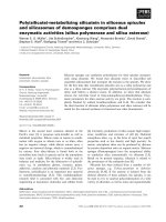
lap with the rectum was not allowed. The pelvic lymphatic
drainage comprised the obturator, peri-rectal, internal
iliac, proximal external iliac and common iliac lymph
nodes up to L5/S1 (Fig. 1). The delineation of the PTV-
LAG was based on the large pelvic vessels rather than the
bony anatomy as suggested by Shih et al.[16]. Definition
of target volumes is summarized in Table 1. The bladder,
rectum, small bowel and femoral heads were delineated
as OARs. The bladder and rectum were contoured as solid
rectal volume (RV) and solid bladder volume (BV) as well
as rectal wall (RW) and bladder wall (BW).
IMRT treatment planning
Treatment was planned for an Elekta Synergy S linac (Ele-
kta, Crawley, England) equipped with the beam modula-
tor with 4 mm leaf width and step-and-shoot IMRT
technique. The isocentre was placed in the geometrical
centre of the PTV-1 for treatment of PO (plan-PO) and in
the geometrical centre of PTV-LAG for treatment of the
Radiation Oncology 2008, 3:3 />Page 3 of 12
(page number not for citation purposes)
Composition of the target volumes PTV-1, PTV-2 and PTV-LAGFigure 1
Composition of the target volumes PTV-1, PTV-2 and PTV-LAG.
common iliac
external iliac
internal iliac
perirectal
external iliac
internal iliac
presacral
external iliac
internal iliac
obturator
PTV-1
PTV-1
PTV-2
Table 1: Definition of target volumes
Target volumes CTV definition PTV definition
PTV-1 Prostate and seminal vesicles + 10 mm uniform margin, 7 mm to posterior
PTV-2 Prostate and base seminal vesicles + 5 mm uniform margin, no overlap with rectum
PTV-LAG Obturator, peri-rectal, internal iliac, proximal external iliac, common iliac
lymph nodes up to L5/S1
+ 10 mm margin around large pelvic vessels
Radiation Oncology 2008, 3:3 />Page 4 of 12
(page number not for citation purposes)
WP (plan-WP). Seven beams were generated with gantry
angles of 0°, 51°, 103°, 155°, 206°, 258° and 309°. Pho-
ton energy was 10 MV.
For both plan-PO and plan-WP, IMRT class-solutions
with a simultaneous-integrated-boost (SIB) were devel-
oped. Schematic protocols of plan-PO and plan-WP are
shown in Figure 2. In plan-PO, the prescription dose [the
minimum dose that is delivered to 95% of the target vol-
ume (D95)] was 60 Gy to the PTV-1 and 74 Gy to the PTV-
2 in 33 fractions. This resulted in single fraction doses
(SFD) of 1.82 Gy to PTV-1 and 2.24 Gy to PTV-2. Based on
an α/β ratio of 1.5 Gy, 3 Gy or 10 Gy [17,18] for the pros-
tate this fractionation schema equates a 1.8 Gy equivalent
dose of 83.9 Gy, 80.7 Gy or 76.8 Gy, respectively.
For treatment of the WP, three plans were generated with
prescription doses of 46 Gy, 50.4 Gy and 54 Gy to the
PTV-LAG; prescription doses to PTV-1 and PTV-2 were
identical to plan-PO. Because of the large difference in the
total dose between PTV-LAG and PTV-2, one single IMRT
plan with SIB was not possible: the differences in the SFD
would be too large. Therefore, plan-WP was split into two
IMRT series, each with a SIB. The first series was an IMRT
plan with 25, 28 and 30 fractions for a total dose of 46 Gy,
50.4 Gy and 54 Gy to the PTV-LAG, respectively. Using the
SIB concept, the SFD to the PTV-1 and the PTV-LAG was
between 1.80 Gy and 1.84 Gy, respectively; SFD to the
PTV-2 was 2.24 Gy. The PTV-LAG was excluded from the
second IMRT series and the IMRT optimization objectives
were identical to plan-PO. However, only eight, five and
three fractions were prescribed for the plans with doses of
46 Gy, 50.4 Gy and 54 Gy to PTV-LAG in the first series,
respectively. This resulted in total doses of 60 Gy and 74
Gy to the PTV-1 and the PTV-2, respectively. Conse-
quently, the single fraction dose and total dose to the
prostate were identical between treatment of the PO and
WP: plan-PO and plan-WP differed in the treatment of the
pelvic lymph nodes, only.
Optimization objectives for plan-PO and plan-WP are
listed in Table 2 and 3. The minimum segment area was 4
cm
2
and the minimum number of monitor units was 4 for
one segment. The maximum number of segments was 30
for plan-PO and 50 for the first series in plan-WP. Direct-
machine-parameter-optimization (DMPO) was used with
sequencing simultaneously to the inverse optimization
process.
After plan generation, series one and series two of plan-
WP were accumulated and compared with plan-PO. All
plans were normalized to a mean dose of 76.5 Gy to the
PTV-2. Dose-volume histograms (DVH) were calculated
for target volumes and OARs. Vx was defined as the vol-
ume that is exposed to at least xGy. For the rectum (RV
and RW), the bladder (BV and BW) and the small bowel
normal-tissue complication probabilities (NTCP) were
calculated using the relative seriality model described by
Källman et al.[19]. Radiation tolerance data from Emami
et al. [20] were fitted to the relative seriality model [21]:
parameters for NTCP calculation are listed in Table 4. For
the small bowel a secondary set of tolerance data [22]
based on clinical results of small bowel toxicity published
by Letschert et al.[23] was applied.
Plan-PO and plan-WP were compared using student's t-
test. For statistical analysis Statistica 6.0 (Statsoft, Tulsa,
USA) was utilized. Differences were considered significant
for p < 0.05.
Results
Dose to the target volumes
Representative dose distributions for plan-PO and plan-
WP are shown in Figure 3 and Figure 4, respectively. After
Schematic protocols of plan-PO and plan-WP (dose prescription of 50.4 Gy to PTV-LAG)Figure 2
Schematic protocols of plan-PO and plan-WP (dose prescription of 50.4 Gy to PTV-LAG).
Plan-WP 50.4Gy
Plan-PO
series 1:
28 fractions
series 2:
5 fractions
PTV-2
PTV-1
PTV-LAG
PTV-2
PTV-1
-> 74Gy (SFD 2.24Gy)
-> 60Gy (SFD 1.82Gy)
-> 50.4Gy (SFD 1.8Gy)
-> 74Gy (SFD 2.24Gy)
-> 60Gy (SFD 1.82Gy)
Radiation Oncology 2008, 3:3 />Page 5 of 12
(page number not for citation purposes)
normalization of all plans to a mean dose of 76.5 Gy to
PTV-2, the D95 dose to the PTV-1 was higher than the pre-
scribed dose of 60 Gy and the D95 dose to the PTV-2 was
lower than 74 Gy (Table 5). This is explained by the small
distance between the structures PTV-1 and PTV-2 in poste-
rior direction where a dose gradient of 14 Gy was not pos-
sible. Plan-PO resulted in slightly higher D95 doses to
PTV-1 and PTV-2 compared to plan-WP; these differences
were in the range of 0.5 Gy or less and not statistically sig-
nificant. In plan-WP the D95 doses to the PTV-LAG were
close to the prescribed doses of 46 Gy, 50.4 Gy and 54 Gy
(Table 5).
Comparison of plan-PO and plan-WP 50.4 Gy
With the analysis based on the rectum as a solid organ,
differences between plan-WP and plan-PO were signifi-
cant in the low dose region (p < 0.001), only: plan-WP
resulted in increased rectal volumes exposed to 10 Gy
(V10: 97% ± 3% vs. 62% ± 14%) and exposed to 20 Gy
(V20: 83% ± 13% vs. 42% ± 12%). The difference between
plan-WP and plan-PO for V30 did not reach statistical sig-
nificance (45% ± 15% vs. 35% ± 12%) (p = 0.14). Vol-
umes of the RV exposed to mid and high doses of 40 Gy
to 70 Gy were almost identical (Fig. 5).
Results based on the RW were similar: treatment of the
pelvic lymphatics increased the dose to the rectal wall
only in the 10 Gy to 30 Gy region with no difference in the
mid and high dose region (Fig. 6). NTCP calculations of
late rectal toxicity confirmed data from DVH analysis: no
difference between plan-PO and plan-WP was observed
(Table 6). The risk for late rectal toxicity was 5% to 6%
based on the RV and 7% to 8% based on the RW.
Doses to the BV were increased for plan-WP compared to
plan-PO in the region of V10 to V40; no significant differ-
ence was observed for V50 to V70 (Fig. 7). At the 50 Gy
dose level, the prescription dose to the PTV-LAG, the dif-
Table 2: Objectives for IMRT treatment planning of plan-PO
Target Volumes
D min D 95 D max
PTV-1 56 Gy 60 Gy
PTV-2 70 Gy 74 Gy 80 Gy
Organs at risk
PTV-1 sine PTV-2 Max 74 Gy to 5% Max 66 Gy to 50% D
max
78 Gy
Help contour ring 1.5 cm Max 60 Gy to 10% Max 45 Gy to 50%
Bladder (BV) Max 70 Gy to 5% Max 50 Gy to 20% Max 30 Gy to 30%
Rectal volume sine PTV Max 60 Gy to 5% Max 40 Gy to 20% Max 20 Gy to 40%
Seminal vesicles Max 65 Gy to 20%
"PTV-1 sine PTV-2" is this proportion of the PTV-1 that does not overlap with PTV-2 – it is listed in organs-at-risk to limit doses >74 Gy to PTV-2.
"Rectal volume sine PTV" is the portion of the rectum located outside PTV-1. These objectives were adjusted for the patient's anatomy to achieve
optimal results. "Help contour ring" is a 15 mm wide ring shaped contour around the PTV-1 to confine high and mid doses to the target volume.
Table 3: Objectives for IMRT treatment planning of plan-WP with a prescribed dose of 50.4 Gy to PTV-LAG
Target Volumes
D min D 95 D max
PTV-1 47.5 Gy 50.9 Gy
PTV-2 59.4 Gy 62.8 Gy 68 Gy
PTV-LAG 47.5 Gy 50.4 Gy
Organs at risk
PTV-1 sine PTV-2 Max 62.8 Gy to 5% Max 56 Gy to 50% D max 66.2 Gy
Help contour ring 1 cm Max 50 Gy to 15% Max 40 Gy to 50% D max 54 Gy
Help contour ring 2–3 cm Max 30 Gy to 25% Max 20 Gy to 60% D max 40 Gy
Bladder (BV) Max 60 Gy to 5% Max 45 Gy to 20% Max 30 Gy to 50%
Rectal volume sine PTV Max 50 Gy to 5% Max 35 Gy to 20% Max 25 Gy to 70%
Seminal vesicles Max 55 Gy to 20%
"Help contour ring 1 cm" was a 10 mm wide ring shaped contour around the PTV-1 and PTV-LAG. "Help contour ring 2–3 cm" was a 20 mm wide
ring shaped contour around "Help contour ring 1 cm".
Radiation Oncology 2008, 3:3 />Page 6 of 12
(page number not for citation purposes)
ference between plan-WP and plan-PO did not reach sta-
tistical significance (22% ± 9% vs. 17% ± 7%). Results for
delineation of the BW were more pronounced. Plan-WP
resulted in greater volumes of the BW exposed to low and
mid doses from 10 Gy to 50 Gy: the difference at the 50
Gy dose level was significant with 30% ± 8% vs. 23% ± 6%
(Fig. 8). These increased doses to the bladder in the low
and mid dose region did not transfer into higher risk of
late bladder toxicity: NTCP calculations resulted in risk
values of <1% for both plan-PO and plan-WP.
No small bowel was exposed to doses of 36 Gy or higher
in plan-PO. In plan-WP the sparing of the small bowel
was successful with 7 ccm ± 8 ccm and 27 ccm ± 27 ccm
of small bowel exposed to 45 Gy and 36 Gy, respectively.
NTCP calculations for small bowel toxicity showed no risk
for plan-PO. Based on tolerance data I, plan-WP increased
the risk of small bowel toxicity to 2.3% ± 2.5%, maximum
6.4% in one single patient; based on tolerance data II, the
risk of small bowel toxicity was 0%. Detailed results are
shown in Table 7.
Different dose prescriptions to PTV-LAG
Increasing the prescription dose to PTV-LAG from 46 Gy
to 50.4 Gy and to 54 Gy resulted in mild increased doses
to the OARs rectum, bladder and small bowel. Similar to
previous results, the influence of the prescribed dose to
the PTV-LAG on the dose to the RW and BW was lager
than the influence on the dose to the RV and BV. The max-
imum effect was observed at the V30 dose level: a dose of
46 Gy, 50.4 Gy and 54 Gy to PTV-LAG resulted in V30 val-
ues of 43% ± 15%, 46% ± 15% and 49% ± 15% to the RW
(n.s.), respectively. For the BW, V30 values of 66% ± 8%,
71% ± 8% and 75% ± 8% were calculated (n.s.). At the
V50 dose level, no difference was observed for the rectum;
this was valid for delineation of the RV and of the RW. The
partial volume of the BW exposed to 50 Gy was 28% ±
8%, 30% ± 8% and 32% ± 8% for plan-WP with prescrip-
tion dose of 46 Gy, 50.5 Gy and 54 Gy, respectively.
NTCP calculations for late rectal toxicity showed only
small differences between plans with prescription doses
ranging between 46 Gy and 54 Gy to the PTV-LAG (Table
6). An escalation of the dose to the PTV-LAG from 46 Gy
to 54 Gy increased the risk of rectal toxicity from 5% ± 1%
to 6% ± 1% based on the RV and from 7% ± 2% to 8% ±
1% based on the RW (n.s.). The risk of late bladder toxic-
ity was <1% regardless of the dose to the PTV-LAG.
Doses to the small bowel were modestly increased by
escalation of the dose to PTV-LAG (Table 7). Based on tol-
erance data I, higher treatment doses to the PTV-LAG
increased the risk of small bowel toxicity, whereas no risk
of small bowel toxicity was calculated based on tolerance
data II.
Discussion
This retrospective planning study indicates that treatment
of the pelvic lymph nodes does not add significant rectal,
bladder or small bowel toxicity compared to treatment of
Table 4: Radiation tolerance data for calculation of NTCP using
the relative seriality model
D50 Gamma α/β ratio seriality
Rectum 80 Gy 2.2 3 Gy 1.5
Bladder 80 Gy 3 3 Gy 0.18
Small bowel I 53.6 Gy 2.3 3 Gy 1.5
Small bowel II 62 Gy 2.1 3 Gy 0.14
Representative dose distributions for plan-POFigure 3
Representative dose distributions for plan-PO.
Radiation Oncology 2008, 3:3 />Page 7 of 12
(page number not for citation purposes)
Representative dose distributions for plan-WPFigure 4
Representative dose distributions for plan-WP.
Radiation Oncology 2008, 3:3 />Page 8 of 12
(page number not for citation purposes)
the prostate only: IMRT treatment planning enabled
highly conformal dose distributions with excellent spar-
ing of these OARs.
Treatment of the pelvic lymph nodes increased doses to
the rectum in the low-dose region up to 30 Gy, only. These
results were similar for delineation of the rectum as solid
organ and for delineation of the rectal wall. This low dose
region is not considered to be of major relevance for acute
and late toxicity. Multiple studies reported a dose-volume
relationship for rectal toxicity in treatment of the prostate
cancer. All studies concluded that the high and mid dose
region is most predictive for rectal toxicity [24-31],
whereas no correlation between rectal toxicity and expo-
sure with doses of less than 40 Gy was observed. NTCP
calculations in our study are concordant with no signifi-
cant differences between treatment of PO and WP: the risk
of late rectal toxicity was in the range of 5% to 8% for pre-
scription doses of 46 Gy, 50.4 Gy or 54 Gy to the pelvic
lymphatics.
Treatment of the pelvic lymphatics influenced the dose
distribution to the bladder significantly more compared
to the dose to the rectum. This is explained by overlap of
the lymphatic target volume with the bladder superior to
the prostate and seminal vesicles whereas such overlap
with the rectum was completely avoided. Plan-WP
increased proportions of the bladder wall exposed to low
doses (10 Gy to 30 Gy) and to mid doses (40 Gy to 50
Gy); no difference in the high dose region was observed.
Despite these differences in the DVH, the risk of late blad-
der toxicity was less than 1% for both treatment of PO and
of WP based on NTCP calculations.
The dose-volume response of the urinary bladder is less
well understood compared to the rectum. Cheung et al.
investigated dose volume factors associated with an
increased risk of late urinary toxicity [32]; all patients had
been treated with 78 Gy in the MD Anderson dose escala-
tion study [33]. The hottest volume (hotspot) model was
found to be the best-fitting model. The analysis from
Peeters et al. was based on the Dutch dose escalation trial
[34]. Acute genitourinary toxicity grade 2 or worse was
correlated with the absolute bladder surfaces irradiated to
≥40 Gy, 45 Gy, and 65 Gy. The RTOG 94-06 data was ana-
lysed by Valicenti et al.[35]. The percent of the bladder
receiving doses higher than the reference dose (68.4 Gy,
73.8 Gy, or 79.2 Gy) was a significant predictor of acute
GU effects. Karlsdóttir et al. reported a retrospective single
institution experience with a prescribed dose of 70 Gy to
Dose-volume histogram of the rectal wall for plan-PO and plan-WPFigure 6
Dose-volume histogram of the rectal wall for plan-PO and
plan-WP.
0
10
20
30
40
50
60
70
80
90
100
110
0 1020304050607080
Volume (%)
Dose (Gy)
Rectal wall (RW)
PO
WP 46Gy
WP 50.4Gy
WP 54Gy
Table 5: Doses to the target volumes in plan-PO and plan-WP
PTV-2 D95 (Gy) PTV-1 D95 (Gy) PTV-LAG D95 (Gy)
Plan-PO 72.5 ± 0.5 62 ± 2.0
Plan-WP 46 Gy 72.3 ± 0.6 61.8 ± 1.8 45.6 ± 0.5
Plan-WP 50.4 Gy 72 ± 0.8 61.6 ± 1.6 49.7 ± 0.7
Plan-WP 54 Gy 71.9 ± 0.9 61.5 ± 1.6 53.2 ± 0.9
Doses to the target volumes PTV-1, PTV-2 and PTV-LAG in plan-PO and plan-WP. Averaged values for all ten patients.
Dose-volume histogram of the rectal volume for plan-PO and plan-WPFigure 5
Dose-volume histogram of the rectal volume for plan-PO
and plan-WP.
0
10
20
30
40
50
60
70
80
90
100
110
0 1020304050607080
Volume (%)
Dose (Gy)
Re ct al vol ume ( RV)
PO
WP 46Gy
WP 50.4Gy
WP 54Gy
Radiation Oncology 2008, 3:3 />Page 9 of 12
(page number not for citation purposes)
the prostate [36]. Contrary to previous results the toxicity
was correlated with rather low doses to the bladder: the
fractional bladder volume receiving more than 14 Gy-27
Gy showed the statistically strongest correlation with
acute GU toxicity. Nuyttens et al. did not find a dose-
response relationship for urinary toxicity after 3D-CRT
treatment of prostate cancer with 72 Gy – 80 Gy [37].
For the small bowel, the TD5/5, the dose at which 5% of
patients will experience toxicity within 5 years, has been
shown to be approximately 45 Gy to 50 Gy [23,38]. IMRT
treatment planning resulted in sufficient sparing of the
small bowel. No risk of small bowel toxicity is expected
after treatment of PO. Based on Emami tolerance data
[20], the risk of small bowel toxicity was moderately
increased for treatment of the pelvic lymphatic region
whereas no risk was calculated using updated tolerance
data [22] based on Letschert et al. [23].
These low risk estimations for rectal, bladder and small
bowel toxicity are remarkable in consideration of the esca-
lated dose to the prostate: a D95 dose of 74 Gy was pre-
scribed in 33 fractions. Based on an α/β ratio of 1.5 Gy, 3
Gy or 10 Gy for the prostate this hypo-fractionated
schema equates a 1.8 Gy equivalent dose of 83.9 Gy, 80.7
Gy or 76.8 Gy, respectively. It is discussed controversially
whether irradiation of the pelvic lymph nodes is required
in dose escalated treatment of the prostate [39,40] or with
long term hormonal therapy [41]. Nevertheless, data from
this study indicate that the IMRT technique enables dose
escalation to the prostate and simultaneous treatment of
the pelvic lymph nodes without increased risk of toxicity.
An integrated boost concept for the prostate while treating
the pelvic lymphatic region might be an issue of concern.
Motion of the prostate independently from the bony anat-
omy is well known [42,43] and the clinical significance of
these internal set-up errors has been proven [44,45]. Cor-
rection of such internal set-up errors by means of image-
guided treatment might decrease the coverage of the lym-
phatic target volume as the pelvic lymph nodes are not
expected to move synchronously with the prostate. Hsu et
al. investigated this issue, recently [46]: correction of set-
up errors was simulated by shifting the original isocenter
of the IMRT plan. The influence of these shifts on the dose
to the pelvic target volume was reported to be small; cov-
Dose-volume histogram of the bladder wall for plan-PO and plan-WPFigure 8
Dose-volume histogram of the bladder wall for plan-PO and
plan-WP.
0
10
20
30
40
50
60
70
80
90
100
110
0 1020304050607080
Volume (%)
Dose (Gy)
Bladder wall (BW)
PO
WP 46Gy
WP 50.4Gy
WP 54Gy
Table 6: NTCP for late rectal toxicity in plan-PO and plan-WP
NTCP for late rectal toxicity (%)
Rectal volume (RV) Rectal wall (RW)
Plan-PO 5.7% ± 1.8% 7.5% ± 2.2%
Plan-WP 46 Gy 5.8% ± 1.3% 7.6% ± 1.5%
Plan-WP 50.4 Gy 5.9% ± 1.1% 7.8% ± 1.1%
Plan-WP 54 Gy 6.1% ± 1.2% 8.1% ± 1.1%
Normal tissue complication probability (NTCP) for late rectal toxicity in plan-PO and plan-WP. Averaged values for all ten patients.
Dose-volume histogram of the bladder volume for plan-PO and plan-WPFigure 7
Dose-volume histogram of the bladder volume for plan-PO
and plan-WP.
0
10
20
30
40
50
60
70
80
90
100
110
0 1020304050607080
Volume (%)
Dose (Gy)
Bladder volume (BV)
PO
WP 46Gy
WP 50.4Gy
WP 54Gy
Radiation Oncology 2008, 3:3 />Page 10 of 12
(page number not for citation purposes)
erage of the pelvic target volume was decreased by less
than 1%. However, the small number of five patients cer-
tainly requires further investigation of this issue.
One limitation of this study is the fact that the calcula-
tions are based on one single planning CT study. All
patients were advised to empty their rectum about 1.5
hours prior to acquisition of the planning CT and prior to
every treatment fraction. The bladder was kept full by
drinking of about 500 ccm in that 1.5 hours interval. This
procedure has been chosen as an empty rectum was
proven to be most representative for the entire course of
treatment [47,48] resulting in lower variability of the
prostate position, improved target coverage and higher
rates of local control [44]. Our results of doses to the rec-
tum are consequently considered to be representative for
the total time of treatment. Treatment with a full bladder
has been standard protocol as this was shown to result in
lower rates of bladder toxicity [49]. However, several stud-
ies reported a time trend to decreased bladder filling dur-
ing treatment if the planning was based on a full bladder
[47,50,51]: a motion of the superior and anterior bladder
wall towards inferior into areas of higher doses might be
the consequence. Additionally, a synchronous motion of
small bowel might increase the risk of toxicity compared
to results of this study. As all patients at our department
are treated with soft-tissue image-guidance using a kV
cone-beam CT [52,53], we are currently investigating this
issue.
The target volume, which covers the lymphatic drainage
of the prostate adequately, has been discussed, inten-
sively. Compared to historical data, surgical series
reported a significantly higher incidence of lymphatic dis-
ease if the surgical dissection was extended beyond the
obturator and external iliac lymph nodes [54,55]. The
sentinel lymph node concept has been adapted from
breast cancer and malignant melanoma and surgical series
showed promising early results [56,57]. Most important
for radiotherapy was the finding that there was no uni-
form pattern of lymphatic drainage. Ganswindt et al. inte-
grated this sentinel lymph node concept into radiotherapy
treatment [58,59]. After intraprostatic injection of 250
MBq
99m
Tc-Nanocoll and SPECT imaging, sentinel lymph
nodes outside the standard target volume were detected in
17 of 25 patients. IMRT treatment planning ensured ade-
quate coverage of these complex shaped lymphatic target
volumes with significantly lower doses to the rectum and
bladder compared to 3D-CRT. As suggested by Shih et al.
the definition of the lymphatic target volume was based
on the major pelvic vasculature in our study, not on bony
landmarks [16]; adequate coverage of the lymphatic
drainage of the prostate is therefore expected.
Conclusion
This retrospective planning study showed similar risk of
rectal, bladder and small bowel toxicity for IMRT treat-
ment of the prostate only and for additional irradiation of
the pelvic lymph nodes. The decision whether to treat the
lymphatic drainage or not should therefore be based on
loco-regional control data rather than toxicity data of tri-
als using conventional techniques or 3D-CRT. Clinical
data will be necessary to prove this hypothesis.
Competing interests
The author(s) declare that they have no competing inter-
ests.
Authors' contributions
All authors read and approved the final manuscript.
MG designed the study and the analysis, generated the
treatment plans, performed the analysis drafted and
revised the manuscript.
KB participated in the study design and revised the manu-
script.
AR participated in the generation of IMRT plans and
revised the manuscript.
DV participated in the study design and revised the man-
uscript.
MF participated in the study design and revised the man-
uscript.
Table 7: DVH analysis and NTCP for the small bowel in plan-PO and plan-WP
Small bowel
V36 (ccm) V45 (ccm) NTCP (%) I NTCP (%) II
Plan-PO 0 0 0% 0%
Plan-WP 46 Gy 13 ± 16 3 ± 5 0.8% ± 1% 0%
Plan-WP 50.4 Gy 27 ± 27 7 ± 8 2.3% ± 2.5% 0%
Plan-WP 54 Gy 36 ± 38 10 ± 11 3.2% ± 3.5% 0%
DVH analysis and normal tissue complication probability (NTCP) for the small bowel in plan-PO and plan-WP. Averaged values for all ten patients.
Radiation Oncology 2008, 3:3 />Page 11 of 12
(page number not for citation purposes)
References
1. Roach M 3rd, DeSilvio M, Lawton C, Uhl V, Machtay M, Seider MJ,
Rotman M, Jones C, Asbell SO, Valicenti RK, Han S, Thomas CR Jr.,
Shipley WS: Phase III trial comparing whole-pelvic versus
prostate-only radiotherapy and neoadjuvant versus adjuvant
combined androgen suppression: Radiation Therapy Oncol-
ogy Group 9413. J Clin Oncol 2003, 21(10):1904-1911.
2. Roach M 3rd, Marquez C, Yuo HS, Narayan P, Coleman L, Nseyo UO,
Navvab Z, Carroll PR: Predicting the risk of lymph node
involvement using the pre-treatment prostate specific anti-
gen and Gleason score in men with clinically localized pros-
tate cancer. Int J Radiat Oncol Biol Phys 1994, 28(1):33-37.
3. Lawton CA, Desilvio M, Roach M 3rd, Uhl V, Kirsch R, Seider M, Rot-
man M, Jones C, Asbell S, Valicenti R, Hahn S, Thomas CR Jr.: An
Update of the Phase III Trial Comparing Whole Pelvic to
Prostate Only Radiotherapy and Neoadjuvant to Adjuvant
Total Androgen Suppression: Updated Analysis of RTOG 94-
13, with Emphasis on Unexpected Hormone/Radiation
Interactions. Int J Radiat Oncol Biol Phys 2007, 69(3):646-655.
4. Pirzkall A, Carol M, Lohr F, Hoss A, Wannenmacher M, Debus J:
Comparison of intensity-modulated radiotherapy with con-
ventional conformal radiotherapy for complex-shaped
tumors. Int J Radiat Oncol Biol Phys 2000, 48(5):1371-1380.
5. Guckenberger M, Flentje M: Intensity-Modulated Radiotherapy
(IMRT) of Localized Prostate Cancer : A Review and Future
Perspectives. Strahlenther Onkol 2007, 183(2):57-62.
6. De Meerleer GO, Vakaet LA, De Gersem WR, De Wagter C, De
Naeyer B, De Neve W: Radiotherapy of prostate cancer with or
without intensity modulated beams: a planning comparison.
Int J Radiat Oncol Biol Phys 2000, 47(3):639-648.
7. Ling CC, Burman C, Chui CS, Kutcher GJ, Leibel SA, LoSasso T,
Mohan R, Bortfeld T, Reinstein L, Spirou S, Wang XH, Wu Q, Zelef-
sky M, Fuks Z: Conformal radiation treatment of prostate can-
cer using inversely-planned intensity-modulated photon
beams produced with dynamic multileaf collimation. Int J
Radiat Oncol Biol Phys 1996, 35(4):721-730.
8. Guckenberger M, Meyer J, Baier K, Vordermark D, Flentje M: Dis-
tinct effects of rectum delineation methods in 3D-confromal
vs. IMRT treatment planning of prostate cancer. Radiat Oncol
2006, 1:34.
9. Zelefsky MJ, Fuks Z, Hunt M, Lee HJ, Lombardi D, Ling CC, Reuter
VE, Venkatraman ES, Leibel SA: High dose radiation delivered by
intensity modulated conformal radiotherapy improves the
outcome of localized prostate cancer. J Urol 2001,
166(3):876-881.
10. Kupelian PA, Thakkar VV, Khuntia D, Reddy CA, Klein EA,
Mahadevan A: Hypofractionated intensity-modulated radio-
therapy (70 gy at 2.5 Gy per fraction) for localized prostate
cancer: long-term outcomes. Int J Radiat Oncol Biol Phys 2005,
63(5):1463-1468.
11. De Meerleer GO, Fonteyne VH, Vakaet L, Villeirs GM, Denoyette L,
Verbaeys A, Lummen N, De Neve WJ: Intensity-modulated radi-
ation therapy for prostate cancer: late morbidity and results
on biochemical control. Radiother Oncol 2007, 82(2):160-166.
12. Zelefsky MJ, Chan H, Hunt M, Yamada Y, Shippy AM, Amols H: Long-
term outcome of high dose intensity modulated radiation
therapy for patients with clinically localized prostate cancer.
J Urol 2006, 176(4 Pt 1):1415-1419.
13. Cavey ML, Bayouth JE, Colman M, Endres EJ, Sanguineti G: IMRT to
escalate the dose to the prostate while treating the pelvic
nodes. Strahlenther Onkol 2005, 181(7):431-441.
14. Nutting CM, Convery DJ, Cosgrove VP, Rowbottom C, Padhani AR,
Webb S, Dearnaley DP: Reduction of small and large bowel irra-
diation using an optimized intensity-modulated pelvic radio-
therapy technique in patients with prostate cancer. Int J
Radiat Oncol Biol Phys 2000, 48(3):649-656.
15. Sanguineti G, Cavey ML, Endres EJ, Franzone P, Barra S, Parker BC,
Marcenaro M, Colman M, Agostinelli S, Foppiano F, Vitale V: Does
treatment of the pelvic nodes with IMRT increase late rectal
toxicity over conformal prostate-only radiotherapy to 76
Gy? Strahlenther Onkol 2006, 182(9):543-549.
16. Shih HA, Harisinghani M, Zietman AL, Wolfgang JA, Saksena M,
Weissleder R: Mapping of nodal disease in locally advanced
prostate cancer: rethinking the clinical target volume for
pelvic nodal irradiation based on vascular rather than bony
anatomy. Int J Radiat Oncol Biol Phys 2005, 63(4):1262-1269.
17. Brenner DJ, Hall EJ: Fractionation and protraction for radio-
therapy of prostate carcinoma. Int J Radiat Oncol Biol Phys 1999,
43(5):1095-1101.
18. Fowler JF:
The radiobiology of prostate cancer including new
aspects of fractionated radiotherapy. Acta Oncol 2005,
44(3):265-276.
19. Kallman P, Agren A, Brahme A: Tumour and normal tissue
responses to fractionated non-uniform dose delivery. Int J
Radiat Biol 1992, 62(2):249-262.
20. Emami B, Lyman J, Brown A, Coia L, Goitein M, Munzenrider JE, Shank
B, Solin LJ, Wesson M: Tolerance of normal tissue to therapeu-
tic irradiation. Int J Radiat Oncol Biol Phys 1991, 21(1):109-122.
21. Ågren-Cronqvist AK: Quantification of the response of hetero-
geneous tumours and organized normal tissues to fraction-
ated radiotherapy. Stockholm University, Department of Medical
Radiation Physics Stockholm, PhD Thesis 1995.
22. Ågren-Cronqvist AK, Källman P, Brahme A: Determination of the
relative seriality of a tissue from its response to non-uniform
dose delivery. In Modelling in clinical radiobiology Edited by: Baier K,
Baltas D. Freiburg , Albert-Ludwigs-University Freiburg; 1997.
23. Letschert JG, Lebesque JV, Aleman BM, Bosset JF, Horiot JC, Bartelink
H, Cionini L, Hamers JP, Leer JW, van Glabbeke M: The volume
effect in radiation-related late small bowel complications:
results of a clinical study of the EORTC Radiotherapy Coop-
erative Group in patients treated for rectal carcinoma. Radi-
other Oncol 1994, 32(2):116-123.
24. Lee WR, Hanks GE, Hanlon AL, Schultheiss TE, Hunt MA: Lateral
rectal shielding reduces late rectal morbidity following high
dose three-dimensional conformal radiation therapy for clin-
ically localized prostate cancer: further evidence for a signif-
icant dose effect. Int J Radiat Oncol Biol Phys 1996, 35(2):251-257.
25. Jackson A, Skwarchuk MW, Zelefsky MJ, Cowen DM, Venkatraman
ES, Levegrun S, Burman CM, Kutcher GJ, Fuks Z, Liebel SA, Ling CC:
Late rectal bleeding after conformal radiotherapy of pros-
tate cancer. II. Volume effects and dose-volume histograms.
Int J Radiat Oncol Biol Phys 2001, 49(3):685-698.
26. Fokdal L, Honore H, Hoyer M, von der Maase H: Dose-volume his-
tograms associated to long-term colorectal functions in
patients receiving pelvic radiotherapy. Radiother Oncol 2005,
74(2):203-210.
27. Greco C, Mazzetta C, Cattani F, Tosi G, Castiglioni S, Fodor A, Orec-
chia R: Finding dose-volume constraints to reduce late rectal
toxicity following 3D-conformal radiotherapy (3D-CRT) of
prostate cancer.
Radiother Oncol 2003, 69(2):215-222.
28. Heemsbergen WD, Hoogeman MS, Hart GA, Lebesque JV, Koper PC:
Gastrointestinal toxicity and its relation to dose distribu-
tions in the anorectal region of prostate cancer patients
treated with radiotherapy. Int J Radiat Oncol Biol Phys 2005,
61(4):1011-1018.
29. Fiorino C, Sanguineti G, Cozzarini C, Fellin G, Foppiano F, Menegotti
L, Piazzolla A, Vavassori V, Valdagni R: Rectal dose-volume con-
straints in high-dose radiotherapy of localized prostate can-
cer. Int J Radiat Oncol Biol Phys 2003, 57(4):953-962.
30. Storey MR, Pollack A, Zagars G, Smith L, Antolak J, Rosen I: Compli-
cations from radiotherapy dose escalation in prostate can-
cer: preliminary results of a randomized trial. Int J Radiat Oncol
Biol Phys 2000, 48(3):635-642.
31. Koper PC, Heemsbergen WD, Hoogeman MS, Jansen PP, Hart GA,
Wijnmaalen AJ, van Os M, Boersma LJ, Lebesque JV, Levendag P:
Impact of volume and location of irradiated rectum wall on
rectal blood loss after radiotherapy of prostate cancer. Int J
Radiat Oncol Biol Phys 2004, 58(4):1072-1082.
32. Cheung MR, Tucker SL, Dong L, de Crevoisier R, Lee AK, Frank S,
Kudchadker RJ, Thames H, Mohan R, Kuban D: Investigation of
bladder dose and volume factors influencing late urinary tox-
icity after external beam radiotherapy for prostate cancer.
Int J Radiat Oncol Biol Phys 2007, 67(4):1059-1065.
33. Pollack A, Zagars GK, Starkschall G, Antolak JA, Lee JJ, Huang E, von
Eschenbach AC, Kuban DA, Rosen I: Prostate cancer radiation
dose response: results of the M. D. Anderson phase III rand-
omized trial. Int J Radiat Oncol Biol Phys 2002, 53(5):1097-1105.
34. Peeters ST, Hoogeman MS, Heemsbergen WD, Slot A, Tabak H,
Koper PC, Lebesque JV: Volume and hormonal effects for acute
side effects of rectum and bladder during conformal radio-
therapy for prostate cancer. Int J Radiat Oncol Biol Phys 2005,
63(4):1142-1152.
Publish with BioMed Central and every
scientist can read your work free of charge
"BioMed Central will be the most significant development for
disseminating the results of biomedical research in our lifetime."
Sir Paul Nurse, Cancer Research UK
Your research papers will be:
available free of charge to the entire biomedical community
peer reviewed and published immediately upon acceptance
cited in PubMed and archived on PubMed Central
yours — you keep the copyright
Submit your manuscript here:
/>BioMedcentral
Radiation Oncology 2008, 3:3 />Page 12 of 12
(page number not for citation purposes)
35. Valicenti RK, Winter K, Cox JD, Sandler HM, Bosch W, Vijayakumar
S, Michalski J, Purdy J: RTOG 94-06: is the addition of neoadju-
vant hormonal therapy to dose-escalated 3D conformal radi-
ation therapy for prostate cancer associated with treatment
toxicity? Int J Radiat Oncol Biol Phys 2003, 57(3):614-620.
36. Karlsdottir A, Johannessen DC, Muren LP, Wentzel-Larsen T, Dahl
O: Acute morbidity related to treatment volume during 3D-
conformal radiation therapy for prostate cancer. Radiother
Oncol 2004, 71(1):43-53.
37. Nuyttens JJ, Milito S, Rust PF, Turrisi AT 3rd: Dose-volume rela-
tionship for acute side effects during high dose conformal
radiotherapy for prostate cancer. Radiother Oncol 2002,
64(2):209-214.
38. Gallagher MJ, Brereton HD, Rostock RA, Zero JM, Zekoski DA, Poyss
LF, Richter MP, Kligerman MM: A prospective study of treatment
techniques to minimize the volume of pelvic small bowel
with reduction of acute and late effects associated with pel-
vic irradiation. Int J Radiat Oncol Biol Phys 1986, 12(9):1565-1573.
39. Jacob R, Hanlon AL, Horwitz EM, Movsas B, Uzzo RG, Pollack A:
Role of prostate dose escalation in patients with greater than
15% risk of pelvic lymph node involvement. Int J Radiat Oncol
Biol Phys 2005, 61(3):695-701.
40. Vargas CE, Demanes J, Boike TP, Barnaba MC, Skoolisariyaporn P,
Schour L, Gustafson GS, Gonzalez J, Martinez AA: Matched-pair
analysis of prostate cancer patients with a high risk of posi-
tive pelvic lymph nodes treated with and without pelvic RT
and high-dose radiation using high dose rate brachytherapy.
Am J Clin Oncol 2006, 29(5):451-457.
41. Boehmer D, Maingon P, Poortmans P, Baron MH, Miralbell R, Remou-
champs V, Scrase C, Bossi A, Bolla M: Guidelines for primary radi-
otherapy of patients with prostate cancer. Radiother Oncol
2006, 79(3):259-269.
42. Litzenberg DW, Balter JM, Hadley SW, Sandler HM, Willoughby TR,
Kupelian PA, Levine L: Influence of intrafraction motion on
margins for prostate radiotherapy. Int J Radiat Oncol Biol Phys
2006, 65(2):548-553.
43. Roeske JC, Forman JD, Mesina CF, He T, Pelizzari CA, Fontenla E,
Vijayakumar S, Chen GT: Evaluation of changes in the size and
location of the prostate, seminal vesicles, bladder, and rec-
tum during a course of external beam radiation therapy. Int
J Radiat Oncol Biol Phys 1995, 33(5):
1321-1329.
44. de Crevoisier R, Tucker SL, Dong L, Mohan R, Cheung R, Cox JD,
Kuban DA: Increased risk of biochemical and local failure in
patients with distended rectum on the planning CT for pros-
tate cancer radiotherapy. Int J Radiat Oncol Biol Phys 2005,
62(4):965-973.
45. Heemsbergen WD, Hoogeman MS, Witte MG, Peeters ST, Incrocci
L, Lebesque JV: Increased risk of biochemical and clinical fail-
ure for prostate patients with a large rectum at radiotherapy
planning: results from the Dutch trial of 68 GY versus 78 Gy.
Int J Radiat Oncol Biol Phys 2007, 67(5):1418-1424.
46. Hsu A, Pawlicki T, Luxton G, Hara W, King CR: A study of image-
guided intensity-modulated radiotherapy with fiducials for
localized prostate cancer including pelvic lymph nodes. Int J
Radiat Oncol Biol Phys 2007, 68(3):898-902.
47. Lebesque JV, Bruce AM, Kroes AP, Touw A, Shouman RT, van Herk
M: Variation in volumes, dose-volume histograms, and esti-
mated normal tissue complication probabilities of rectum
and bladder during conformal radiotherapy of T3 prostate
cancer. Int J Radiat Oncol Biol Phys 1995, 33(5):1109-1119.
48. Stasi M, Munoz F, Fiorino C, Pasquino M, Baiotto B, Marini P, Malin-
verni G, Valdagni R, Gabriele P: Emptying the rectum before
treatment delivery limits the variations of rectal dose - vol-
ume parameters during 3DCRT of prostate cancer. Radiother
Oncol 2006, 80(3):363-370.
49. Pinkawa M, Fischedick K, Asadpour B, Gagel B, Piroth MD, Eble MJ:
Low-grade toxicity after conformal radiation therapy for
prostate cancer impact of bladder volume. Int J Radiat Oncol
Biol Phys 2006, 64(3):835-841.
50. Fiorino C, Foppiano F, Franzone P, Broggi S, Castellone P, Marcenaro
M, Calandrino R, Sanguineti G: Rectal and bladder motion during
conformal radiotherapy after radical prostatectomy. Radi-
other Oncol 2005, 74(2):187-195.
51. Pinkawa M, Asadpour B, Siluschek J, Gagel B, Piroth MD, Demirel C,
Eble MJ: Bladder extension variability during pelvic external
beam radiotherapy with a full or empty bladder. Radiother
Oncol 2007, 83(2):163-167.
52. Guckenberger M, Meyer J, Vordermark D, Baier K, Wilbert J, Flentje
M: Magnitude and clinical relevance of translational and rota-
tional patient setup errors: A cone-beam CT study. Int J
Radiat Oncol Biol Phys 2006, 65(3):934-942.
53. Guckenberger M, Meyer J, Wilbert J, Baier K, Sauer O, Flentje M:
Precision of Image-Guided Radiotherapy (IGRT) in Six
Degrees of Freedom and Limitations in Clinical Practice.
Strahlenther Onkol 2007, 183(6):307-313.
54. Heidenreich A, Ohlmann CH, Polyakov S: Anatomical extent of
pelvic lymphadenectomy in patients undergoing radical
prostatectomy. Eur Urol 2007, 52(1):29-37.
55. Heidenreich A, Varga Z, Von Knobloch R: Extended pelvic lym-
phadenectomy in patients undergoing radical prostatec-
tomy: high incidence of lymph node metastasis. J Urol 2002,
167(4):1681-1686.
56. Wawroschek F, Vogt H, Wengenmair H, Weckermann D, Hamm M,
Keil M, Graf G, Heidenreich P, Harzmann R: Prostate lymphoscin-
tigraphy and radio-guided surgery for sentinel lymph node
identification in prostate cancer. Technique and results of
the first 350 cases. Urol Int 2003, 70(4):303-310.
57. Weckermann D, Hamm M, Dorn R, Wagner T, Wawroschek F, Harz-
mann R: [Sentinel lymph node dissection in prostate cancer.
Experience after more than 800 interventions]. Urologe A
2006, 45(6):723-727.
58. Ganswindt U, Paulsen F, Corvin S, Eichhorn K, Glocker S, Hundt I,
Birkner M, Alber M, Anastasiadis A, Stenzl A, Bares R, Budach W,
Bamberg M, Belka C: Intensity modulated radiotherapy for high
risk prostate cancer based on sentinel node SPECT imaging
for target volume definition. BMC Cancer 2005, 5:91.
59. Ganswindt U, Paulsen F, Corvin S, Hundt I, Alber M, Frey B, Stenzl A,
Bares R, Bamberg M, Belka C: Optimized coverage of high-risk
adjuvant lymph node areas in prostate cancer using a senti-
nel node-based, intensity-modulated radiation therapy tech-
nique. Int J Radiat Oncol Biol Phys 2007, 67(2):347-355.