Báo cáo khoa học: "Acute and late toxicity in prostate cancer patients treated by dose escalated intensity modulated radiation therapy and organ tracking" pps
Bạn đang xem bản rút gọn của tài liệu. Xem và tải ngay bản đầy đủ của tài liệu tại đây (245.03 KB, 8 trang )
BioMed Central
Page 1 of 8
(page number not for citation purposes)
Radiation Oncology
Open Access
Research
Acute and late toxicity in prostate cancer patients treated by dose
escalated intensity modulated radiation therapy and organ tracking
Pirus Ghadjar
†
, Jacqueline Vock
†
, Daniel Vetterli, Peter Manser,
Roland Bigler, Jan Tille, Axel Madlung, Frank Behrensmeier, Roberto Mini
and Daniel M Aebersold*
Address: Department of Radiation Oncology with Division of Medical Radiation Physics, University of Bern, Inselspital, Bern, Switzerland,
Freiburgstrasse, 3010 Bern, Switzerland
Email: Pirus Ghadjar - ; Jacqueline Vock - ; Daniel Vetterli - ;
Peter Manser - ; Roland Bigler - ; Jan Tille - ;
Axel Madlung - ; Frank Behrensmeier - ; Roberto Mini - ;
Daniel M Aebersold* -
* Corresponding author †Equal contributors
Abstract
Background: To report acute and late toxicity in prostate cancer patients treated by dose
escalated intensity-modulated radiation therapy (IMRT) and organ tracking.
Methods: From 06/2004 to 12/2005 39 men were treated by 80 Gy IMRT along with organ
tracking. Median age was 69 years, risk of recurrence was low 18%, intermediate 21% and high in
61% patients. Hormone therapy (HT) was received by 74% of patients. Toxicity was scored
according to the CTC scale version 3.0. Median follow-up (FU) was 29 months.
Results: Acute and maximal late grade 2 gastrointestinal (GI) toxicity was 3% and 8%, late grade
2 GI toxicity dropped to 0% at the end of FU. No acute or late grade 3 GI toxicity was observed.
Grade 2 and 3 pre-treatment genitourinary (GU) morbidity (PGUM) was 20% and 5%. Acute and
maximal late grade 2 GU toxicity was 56% and 28% and late grade 2 GU toxicity decreased to 15%
of patients at the end of FU. Acute and maximal late grade 3 GU toxicity was 8% and 3%,
respectively. Decreased late ≥ grade 2 GU toxicity free survival was associated with higher age (P
= .025), absence of HT (P = .016) and higher PGUM (P < .001).
Discussion: GI toxicity rates after IMRT and organ tracking are excellent, GU toxicity rates are
strongly related to PGUM.
Background
Prostate cancer (PCA) is the most commonly diagnosed
cancer among men in Europe and North America [1,2]
after skin cancer. In the last decade significant improve-
ment in planning and delivery of radiotherapy (RT) such
as three-dimensional conformal RT (3D-CRT) and inten-
sity-modulated radiation therapy (IMRT) have been
developed [3-5] allowing the delivery of higher radiation
doses. Dose escalated RT has been shown to improve bio-
chemical control rates in PCA, however, to date, this has
not translated into better overall survival [5-12]. Dose
escalated 3D-CRT has led to increased gastrointestinal
Published: 20 October 2008
Radiation Oncology 2008, 3:35 doi:10.1186/1748-717X-3-35
Received: 28 July 2008
Accepted: 20 October 2008
This article is available from: />© 2008 Ghadjar et al; licensee BioMed Central Ltd.
This is an Open Access article distributed under the terms of the Creative Commons Attribution License ( />),
which permits unrestricted use, distribution, and reproduction in any medium, provided the original work is properly cited.
Radiation Oncology 2008, 3:35 />Page 2 of 8
(page number not for citation purposes)
(GI) toxicity [10,13,14]. High-dose RT for prostate cancer
delivered by IMRT causes lower acute and late GI toxicity
rates as compared to 3D-CRT [4] and improvement in
tumor delineation such as matching of an magnetic reso-
nance imaging (MRI) of the pelvis with the planning com-
puted tomography (CT) has been shown to reduce GI
toxicity in dose escalated RT due to decreased dose to the
rectal wall [15].
On the other hand localization of the prostate by image
guided radiation therapy (IGRT) is a reasonable approach
to reduce toxicity in dose escalated RT of PCA, taking into
account the interfractional variability of the prostate posi-
tion. Therefore we treated prostate cancer patients with
dose escalated high precision RT by IMRT along with
organ tracking, to reduce toxicity, especially of the GI
tract. This work presents the pre-treatment GI and geni-
tourinary (GU) morbidity and acute and late toxicity of
the first 39 patients after a median follow-up of 29
months.
Methods
Patient selection and characteristics
A total of 46 consecutive men with histologically proven
adenocarcinoma of the prostate and cM0 stage were
treated by IMRT along with organ tracking after providing
informed consent in accordance with the standards of the
local ethics committee and with the Helsinki Declaration
of 1975, as revised in 1983. The pre-treatment staging
included a complete history, physical examination, digital
rectal examination, transrectal ultrasound (TRUS) of the
prostate, biopsy with specification of the Gleason score,
prostate-specific antigen (PSA) level, CT and/or MRI of
the abdomen and pelvis and a total-body bone scan.
When clinically indicated a chest x-ray and/or thoracic CT
was performed. Based on these examinations clinical stag-
ing was defined according to the 2002 American Joint
Committee on Cancer tumor, lymph nodes and metasta-
sis system [16]. Patients were grouped according to their
risk of recurrence as recommended by the National Com-
prehensive Cancer Network practise guidelines in oncol-
ogy
. Patients with multiple adverse
factors were not shifted to the next higher risk group.
Seven patients with previous implantation of hip prosthe-
sis (n = 3) with changing of the treatment plan during the
treatment (n = 1) and with different dose constraints than
described below (n = 1) were excluded from the study.
One patient died because of pancreatic cancer < 2 months
after RT and one patient died because of bile duct cancer
< 9 months after RT, both were also excluded because of
insufficient follow-up. The study population thus con-
sisted of 39 patients, treated between 06/2004 and 12/
2005, who were retrospectively analyzed. All patients
were Caucasian and no patient had received previous pel-
vic RT. All patients completed the planned course of radi-
ation, receiving treatment as outlined below. Median age
of the patients was 69 years (range, 54 – 81 years), median
follow-up was 29 months (range, 22 – 40 months). Risk
of recurrence was low in 7 (18%), intermediate in 8
(21%) or high in 24 (61%) patients.
Hormonal therapy
A total of 29 patients (74%) received hormonal therapy
(HT), either neoadjuvant, concomitant or adjuvant, with
a median duration of 6 months (range, 2 – 24 months)
but always during RT. Twenty-six patients received com-
bined androgen blockade (CAB) consisting of antiandro-
gen and gonadotropin releasing hormone analogue
(GnRH). Three patients received antiandrogen mono-
therapy. Pre-irradiation HT was given to 26 patients
(67%) with a median duration of 1.5 months (range, 0.3
– 3.5 months) before starting RT.
External beam radiotherapy
All 39 patients included in this study were treated using
IMRT with organ tracking as described previously [17].
Briefly, the dose was delivered in daily fractions of 2 Gy,
given five sessions per week, to a total of 80 Gy. Prior to
the treatment, three gold markers (diameter = 0.9 mm,
length = 7 mm) were implanted into the prostate of every
patient under TRUS guidance. Thirty six of 39 patients
underwent an MRI scan of the pelvis in our radiology
department. In these patients, digital data was used for
image fusion with the planning CT scan. To produce high-
resolution digitally reconstructed radiographs (DRRs),
consecutive CT images (GE Prospeed
®
) with an interslice
spacing and thickness of 3 mm were obtained. To estimate
the risk for seminal vesicle (SV) invasion, the Roach for-
mula [18] was calculated for each patient. If the risk for SV
invasion was > 15%, as it was the case in 21 patients, the
proximal third of the SV was included to the clinical target
volume (CTV) electively. If invasion of the base of the SV
was seen in the MRI (cT3b), the whole SV were electively
included to the CTV. The planning target volume (PTV)
was then delineated by encompassing the prostate with 5
mm safety margins in all directions but dorsal, where a 3
mm margin was added. In addition to the PTV, the walls
of the rectum and bladder, the femoral heads and the skin
surface were also identified. A coplanar five-field IMRT
technique with fields placed at angles of 45°, 90°, 180°,
270°, and 320° was used to treat prostate cancer patients
with 15MV X-rays (Clinac 2300C/D, Varian Medical Sys-
tems). The dose was applied by the dynamic MLC tech-
nique using an 80 leaf-MLC. The 80 Gy were prescribed to
the median of the PTV. More than 99% of the PTV volume
was included in the 95% isodose and the maximum dose
was limited to 105%. To limit the dose to the organs at
risk (OAR), a template was used with a set of dose con-
straints. Daily treatment was intended with a full bladder.
The isocenter was placed 7 cm behind the symphysis and
Radiation Oncology 2008, 3:35 />Page 3 of 8
(page number not for citation purposes)
at the height of the cranial end of the opening of the
foramen obturatorium. Partial DRRs were generated for
all treatment beams and in addition for two extra setup
beams from the anterior-posterior (AP) and the lateral
direction (LAT). The field size of the AP setup field was
chosen to encompass the implanted markers with a mar-
gin of about 1.5 cm. The LAT field enclosed the markers
and the symphysis. With a drawing tool the marker posi-
tions were manually highlighted, serving as a layer struc-
ture for subsequent portal image evaluation. The two
setup DRRs served as a reference for portal images taken
before daily treatment. For isocenter verification, the
patients were simulated (Ximatron
®
, Varian Medical Sys-
tems) using the two additional setup fields. The isocenter
was then marked on the patient's skin. In the treatment
room, the patients were aligned on a carbon-fiber couch
panel within their immobilization device using the skin
marks. Before each treatment fraction the two setup fields
were acquired using the aS500
®
portal imager (Varian
Medical Systems) with a dose saving acquisition mode.
The setup and correction of patients position was per-
formed as previously described [19]. Briefly, setup images
from the AP and LAT direction were taken with the 6 MV
beam. A comparison between positions of the fiducial
markers on the portal image with the corresponding refer-
ence DRR followed. Patient position was then manually
adjusted in all 3 dimensions until the best center of mass
(CM) alignment was found with a CM >1 mm limit in any
direction. Following this sequential setup procedure, the
patient was treated with the multi-field IMRT plan. Opti-
mization of the IMRT plan included a dose limit within
the central periurethral area of 99% of the prescribed
dose. The duration of IMRT reached a median of 56 days
(range, 52 – 63 days).
Dose constraints
A maximum dose of 100% to the urethra was allowed. For
the rectal wall (from the ischial tuberosity to the bottom
of the sacroiliac joint), constraints described by Leibel et
al. [5] and Boersma et al. [13] were used: the median V47
(percentage of the rectal wall receiving at least 47 Gy) was
set to <53% and the median V75 to <5%. For the bladder
the constraint from Leibel et al. was used: the median V47
(percentage of bladder wall receiving at least 47 Gy) was
set to <53%.
To assess coverage, homogeneity and conformity of the
PTV the V95 (percentage of PTV receiving at least 95% of
dose, corresponding to 76 Gy) and the Dmax and Dmin
(maximum and minimum dose within the PTV) were cal-
culated from the dose volume histograms.
Follow-up protocol
Patients were seen by a radiation oncologist at least
weekly during the RT. Follow-up visits were arranged 2–4
weeks after completion of IMRT and every 3 to 6 months
Table 1: Patient characteristics
Total patients (n = 39)
Age
≤ 69 years 20 (51.3%)
> 69 years 19 (48.7%)
Tumor stage
cT1 12 (30.8%)
cT2 5 (12.8%)
cT3a 13 (33.4%)
cT3b 7 (17.9%)
cT4 2 (5.1%)
Gleason score
2–6 20 (51.3%)
7 13 (33.3%)
8–10 6 (15.4%)
Pre-treatment PSA
≤ 10 ng/mL 20 (51.3%)
>10 ng/mL 19 (48.7%)
Risk group
Low 7 (17.9%)
Intermediate 8 (20.5%)
High 24 (61.6%)
Hormonal therapy
no 10 (25.6%)
yes 29 (74.4%)
Median FU months† (range) 29 (22–40)
Abbreviations: PSA = prostate- specific antigen; FU = follow-up;
†period after completion of treatment.
Radiation Oncology 2008, 3:35 />Page 4 of 8
(page number not for citation purposes)
for the first 2 years and annually thereafter with a digital
rectal examination and a serum PSA level obtained at each
visit. Patients alternated follow-up visits between their
urologist and radiation oncologist. A minority of patients
who did not attend these visits were contacted and data
were successfully retrieved.
Toxicity scoring
Toxicity was graded using the common terminology crite-
ria for adverse events (CTC AE) version 3.0 http://
ctep.cancer.gov/forms/CTCAEv3.pdf from the National
Cancer Institute. Late toxicity was defined as complica-
tions occurring three months after the end of treatment.
Late toxicity at last follow-up visit was determined and
was termed last late toxicity. This was done to assess
whether the late toxicity was persistent or was transient.
Statistical analysis
Descriptives include absolute and relative frequencies for
categorical variables, median and range for quantitative
variables.
The primary endpoint was the occurrence of acute and late
≥ grade 2 GI and GU toxicity. For statistical analysis age (≤
vs > 69 years), PTV (≤ vs > 105 cm
3
), PTV Dmax (≤ vs >
84.3 Gy) and bladder V47 (≤ vs >28%) were grouped
according to the median. Pre treatment GU toxicity
(PGUM) (grade 0–1 vs grade 2–3) and acute and late GU
toxicity (grade 0–1 vs grade 2–3) were grouped. Acute GU
toxicity and PGUM were compared using the Fisher's exact
test. Estimation of actuarial rates for late ≥ grade 2 GU tox-
Table 3: Pre-treatment genitourinary morbidity and genitourinary acute and late toxicity
Pre-Tx Acute† Late‡ Last late§
Toxicity Grade n (%) n (%) n (%) n (%)
Dysuria 0 36 (92) 20 (51) 35 (90) 38 (97)
1 3 (8) 13 (33) 4 (10) 1 (3)
20 5 (13)0 0
30 1 (3) 0 0
Incontinence 0 34 (87) 33 (84) 34 (87) 37 (94)
1 4 (10) 5 (13) 4 (10) 1 (3)
2 1 (3) 1 (3) 1 (3) 1 (3)
Retention 0 20 (51) 16 (41) 23 (59) 34 (86)
1 13 (33) 9 (23) 11 (28) 3 (8)
2 5 (13) 12 (31) 4 (10) 1 (3)
3 1 (3) 2 (5) 1 (3) 1 (3)
Frequency/urgency 0 23 (59) 6 (15) 17 (44) 23 (59)
1 13 (33) 16 (41) 15 (38) 14 (36)
2 2 (5) 15 (39) 7 (18) 2 (5)
3 1 (3) 2 (5) 0 0
Hematuria 0 39 (100) 39 (100) 36 (92) 37 (95)
10 0 1 (3)2 (5)
20 0 2 (5)0
Highest GU* 0 15 (39) 1 (3) 11 (28) 19 (49)
1 14 (36) 13 (33) 16 (41) 13 (33)
2 8 (20) 22 (56) 11 (28) 6 (15)
3 2 (5) 3 (8) 1 (3) 1 (3)
Abbreviations: Pre-Tx = pre-treatment; * The highest toxicity in a patient was counted as a single event; † During therapy and until 3 months after
completion; ‡ maximal late toxicity > 3 months after completion of therapy; § Incidence of late toxicity at last follow-up visit
Table 2: Pre-treatment gastrointestinal morbidity and
gastrointestinal acute and late toxicity
Pre-Tx Acute† Late‡ Last late§
Toxicity Grade n (%) n (%) n (%) n (%)
Diarrhea 0 39 (100) 29 (74) 36 (92) 38 (97)
1 0 9 (23) 3 (8) 1 (3)
201 (3)0 0
Rectal pain 0 38 (97) 34 (87) 37 (96) 38 (97)
1 1 (3) 5 (13) 1 (3) 1 (3)
20 01 (3)0
Rectal bleeding 0 36 (92) 36 (92) 33 (85) 34 (87)
1 3 (8) 3 (8) 4 (10) 5 (13)
20 02 (5)0
Highest GI* 0 36 (92) 25 (64) 30 (77) 32 (82)
1 3 (8) 13 (33) 6 (15) 7 (18)
201 (3)3 (8)0
Abbreviations: Pre-Tx = pre-treatment; * The highest toxicity in a
patient was counted as a single event; † During therapy and until 3
months after completion; ‡ maximal late toxicity > 3 months after
completion of therapy; § Incidence of late toxicity at last follow-up
visit
Radiation Oncology 2008, 3:35 />Page 5 of 8
(page number not for citation purposes)
icity free survival was calculated using the Kaplan-Meier
product limit methodology. Kaplan-Meier rates were
compared using the log-rank test [20]. Cox regression
models were used to determine independent prognostic
factors for decreased late ≥ grade 2 GU toxicity free sur-
vival [21]. Variables were included to the model if the uni-
variate P-value was <.1, thus, age, HT and PGUM were
included. Backward model selection was performed in
order to identify predictors for decreased ≥ grade 2 late GU
toxicity free survival. Statistical significance was consid-
ered on a two-sided level of α = 0.05. Statistical analysis
was performed with SPSS version 16.0 (SPSS Inc., Chi-
cago, IL).
Results
Coverage, homogeneity and conformity of the PTV
For the PTV, the median V95 was 97.9% (range, 89.2 –
99.8%). Median Dmax and Dmin were 84.3 Gy (range,
82.2–85.4 Gy) and 72.1 Gy (range, 67.5 – 74.6 Gy),
respectively.
Acute and late GI Toxicity
Patient characteristics are summarized in Table 1. The pre-
treatment GI morbidity, acute and late GI toxicity and
incidence of late GI toxicity at last follow-up visit are sum-
marized in Table 2. Incidence of single symptoms
(diarrhea, rectal pain, rectal bleeding) as well as the high-
est toxicity occurred in a given patient concluding all sin-
gle symptoms but counting only the highest toxicity as a
single event are depicted. Acute grade 2 GI toxicity was 1
(3%) and maximal late grade 2 GI toxicity was 3 (8%). No
grade 3 GI toxicity occurred. The median time from the
completion of RT to the occurrence of ≥ grade 1 late GI
was 14 months (range, 6 – 30 months). Late GI toxicity
decreased as time from treatment elapsed. At last follow-
up visit the incidence of late grade 2 GI toxicity was 0%.
There was no association observed between clinical or
dosimetric parameters and acute GI toxicity or late GI tox-
icity free survival.
Acute and late GU Toxicity
The PGUM and acute and late GU toxicity are summarized
in Table 3. Incidence of single symptoms (dysuria, incon-
tinence, retention, frequency/urgency, hematuria) as well
as the highest toxicity occurred in a given patient conclud-
ing all single symptoms but counting only the highest tox-
icity as a single event are depicted. Before treatment 8
patients (20%) had grade 2 and 2 patients (5%) had grade
3 PGUM. After treatment acute grade 2 GU toxicity
occurred in 22 (56%) and maximal late grade 2 in 11
(28%) patients. Acute and maximal late grade 3 GU toxic-
ity occurred in 3 (8%) and 1 (3%) patients, respectively.
There was no grade 4 GU toxicity observed. The median
time from the completion of RT to the occurrence of late
≥ grade 2 GU toxicity was 12 months (range, 4 – 26
months).
Interestingly late GU toxicity decreased as time from treat-
ment elapsed. At the last follow-up visit only 6 (15%) of
patients had late grade 2 and 1 (3%) had late grade 3 GU
toxicity, being fewer patients than initially observed with
grade 2 or 3 PGUM. The patient who developed late grade
3 GU toxicity had a bulbar urethral stricture and was
treated twice by urethral dilatation, however obstructive
Table 5: Multiple Cox regression analysis of factors associated
with late ≥ grade 2 GU toxicity free survival
Factor RR CI p
Age > 69 years versus ≤ 69 years 2.7 0.7–10.9 .143
Hormonal therapy no versus yes 12.4 2.4–64.6 .003
PGUM grade 2-3 versus grade 0–1 29.1 5.4–156.8 <.001
Abbreviation: RR = relative risk; CI = 95% RR confidence intervals;
PGUM = pre-treatment genitourinary morbidity.
Table 4: Factors correlating with three year grade ≥ 2 GU toxicity free survival in univariate analysis
Factor Group No. of Patients % TFS No. at Risk* p†
All patients 39 69.2 5
Age (years) ≤ 69 20 85.0 4
> 69 19 52.6 1 .025
Hormonal therapy no 10 40.0 0
yes 29 79.3 5 .016
PGUM (grade) 0–1 29 86.2 4
2–3 10 20.0 1 <.001
PTV (cm
3
) ≤ 105 20 80.0 2
> 105 19 57.9 3 .128
PTV Dmax (Gy) ≤ 84.3 21 76.2 3
> 84.3 18 61.1 2 .199
Bladder V47 (%) ≤ 28 20 75.0 3
> 28 19 63.2 2 .437
Abbrevations: TFS = toxicity free survival; PGUM = pre-treatment genitourinary morbidity; PTV = planning target volume; PTV Dmax = maximum
dose within the PTV; Bladder V47 = percentage of bladder wall receiving at least 47 Gy * Number at risk at three years; † Log-rank test
Radiation Oncology 2008, 3:35 />Page 6 of 8
(page number not for citation purposes)
symptoms were persistent and ever since the patient uses
daily self-catheterism.
Factors associated with GU toxicity
There was an association of PGUM and acute ≥ grade 2
GU toxicity (P = .007) but not with the other tested
parameters age, HT, PTV, PTV Dmax or bladder V47.
Decreased grade ≥ 2 late GU toxicity free survival was asso-
ciated with higher age (P = .025), absence of HT (P = .016)
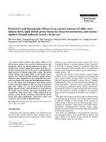
and higher PGUM (P < .001) but not with the PTV, PTV
Dmax or bladder V47 (Table 4; Figure 1). In multiple Cox
regression analysis higher PGUM (P < .001) and absence
of HT (P = .003) were independently associated with
decreased late ≥ grade 2 GU toxicity free survival (Table 5).
PSA nadir
With a median follow-up of 29 months it is too early to
report biochemical control rates. However, after treatment
93% (27 out of 29) of patients with and 90% (9 out of 10)
of patients without HT reached a PSA nadir value ≤ 0.5 ng/
mL. In patients treated by HT, time to nadir was shorter (P
< .001) and the nadir value was lower (P = .001) as com-
pared to patients without HT. The median time to nadir
was 23.6 months (range, 3.3 – 26.1 months) in patients
without and 4.0 months (range, 0.0 – 24.4 months) in
patients with HT. The median nadir value was 0.3 ng/mL
(range, 0.05 – 5.1 ng/mL) in patients without and 0.07
ng/mL (range, 0.0 – 0.95 ng/mL) in patients with HT.
Discussion
With the use of more conformal RT techniques, high dose
RT of PCA has become the accepted standard of care.
Simultaneously, the incidence of treatment related toxic-
ity has become even more important. Here we report the
pre-treatment GI and GU morbidity and acute and late
toxicity in patients treated by dose escalated high preci-
sion RT by IMRT along with organ tracking. The grade 2
acute and maximal late GI toxicity rates were 3% and 8%
and late grade 2 GI toxicity decreased to 0% at the end of
the follow-up with no grade 3 GI toxicity observed. In face
of the grade 2 and 3 PGUM rates of 20% and 5% the
observed acute and late grade 2 GU toxicity rates were
56% and 28%. Interestingly the late grade 2 GU toxicity
rate dropped to 15% at the end of follow-up being even
below the PGUM reported. Acute and late grade 3 GU tox-
icity was 8% and 3%, respectively. The use of different tox-
icity scales and RT techniques makes it difficult to
compare our data to other studies. Only two studies have
reported toxicity rates after PCA treatment with 3D-CRT or
IMRT using fiducial markers for position verification
[22,23]. Table 6 summarizes the observed toxicity in these
studies in comparison with the results of our study. Con-
cerning acute and late GI toxicity our results are excellent,
but the maximal GU toxicity rates are higher than those
described elsewhere. However, the PGUM in our patient
cohort was relatively high and if this is considered, the GU
toxicity rates observed in our patients are comparable to
those in the literature. The acute GU toxicity was associ-
ated with PGUM and the ≥ grade 2 late GU toxicity free
survival was significantly decreased in patients with
higher age, higher PGUM and in patients who were not
treated by HT. The decreased rate of GU late effects in
patients undergoing HT is in accordance with pooled data
from RTOG trials 85-31, 86-10 and 92-02, where the addi-
tional use of HT decreased GU toxicity [24]. The impact of
HT on the late GU toxicity might depend on the extent of
PGUM, being beneficial especially in patients with
obstructive symptoms. The high amount of patients with
pre-treatment obstructive symptoms (grade 1 – 3: 49%)
Actuarial analysis of three year late ≥ grade 2 GU toxicity free survival stratified by (a) pre-treatment genitourinary morbidity (PGUM) and (b) by history of hormonal therapyFigure 1
Actuarial analysis of three year late ≥ grade 2 GU
toxicity free survival stratified by (a) pre-treatment
genitourinary morbidity (PGUM) and (b) by history
of hormonal therapy.
Radiation Oncology 2008, 3:35 />Page 7 of 8
(page number not for citation purposes)
might therefore account for the association of HT with
decreased GU toxicity.
We are aware of the obvious limitations of our study,
being the relatively small number of patients analyzed
and the retrospective nature of the study. We also recog-
nize that longer follow-up will be needed to compare the
tumor control rates with those after different treatment
regiments. Nevertheless, this report provides strong evi-
dence for the need of reporting pre-treatment morbidity
rates along with the observed toxicity rates to enhance
comparability with other studies.
Conclusion
GI toxicity rates after dose escalated IMRT and organ track-
ing are excellent. Acute and late GU toxicity are compara-
ble to other reported series when the pre-treatment GU
morbidity is considered.
Competing interests
The authors declare that they have no competing interests.
Authors' contributions
Each author had participated sufficiently in the work to
take public responsibility for appropriate portions of the
content. PG, JV and DMA designed the study. PG, JT and
RB performed the statistical analysis. PG, JV, JT, RB, DV,
PM, RM, AM and FB collected the data and together with
DMA interpreted the data. The manuscript was written by
PG, all other authors helped and finally approved the
final manuscript.
References
1. Bracarda S, de Cobelli O, Greco C, Prayer-Galetti T, Valdagni R,
Gatta G, de Braud F, Bartsch G: Cancer of the prostate. Crit Rev
Oncol Haematol 2005, 56:379-396.
2. Jemal A, Siegel R, Ward E, Murray T, Xu J, Smigal C, Thun MJ: Cancer
statistics, 2006. CA Cancer J Clin 2006, 56:106-130.
3. D' Amico AV, Hanks GE: Three-dimensional conformal radia-
tion therapy for prostate cancer. In Radiotherapeutic management
of prostate adenocarcinoma Boston: Arnold; 1999:21-50.
4. Zelefsky MJ, Fuks Z, Happersett L, Lee HJ, Ling CC, Burman CM,
Hunt M, Wolfe T, Venkatraman ES, Jackson A, Skwarchuk M, Leibel
SA: Clinical experience with intensity modulated radiation
therapy (IMRT) in prostate cancer. Radiother Oncol 2000,
55:241-249.
5. Leibel SA, Fuks Z, Zelefsky MJ, Hunt M, Burman CM, Mageras GS,
Chui CS, Jackson A, Amols HI, Ling CC: Technological advances
in external-beam radiation therapy for the treatment of
localized prostate cancer. Semin Oncol 2003, 30(5):596-615.
6. Shipley WU, Verhey LJ, Munzenrider JE, Suit HD, Urie MM, McManus
PL, Young RH, Shipley JW, Zietman AL, Biggs PJ, et al.: Advanced
prostate cancer: The results of a randomized comparative
trial of high dose irradiation boosting with conformal pro-
tons compared with conventional dose irradiation using pho-
tons alone. Int J Radiat Oncol Biol Phys 1995, 32:3-12.
7. Hanks GE, Hanlon AL, Schultheiss TE, Pinover WH, Movsas B, Epstein
BE, Hunt MA: Dose escalation with 3D conformal treatment:
5 year outcomes, treatment optimization, and future direc-
tions. Int J Radiat Oncol Biol Phys 1998, 41:501-510.
8. Zelefsky MJ, Leibel SA, Gaudin PB, Kutcher GJ, Fleshner NE, Venkat-
ramen ES, Reuter VE, Fair WR, Ling CC, Fuks Z: Dose escalation
with three-dimensional conformal radiation therapy affects
the outcome in prostate cancer. Int J Radiat Oncol Biol Phys 1998,
41:491-500.
9. Zelefsky MJ, Fuks Z, Hunt M, Lee HJ, Lombardi D, Ling CC, Reuter
VE, Venkatraman ES, Leibel SA: High dose radiation delivered by
intensity modulated conformal radiotherapy improves the
outcome of localized prostate cancer. J Urol 2001,
166:
876-881.
10. Pollack A, Zagars GK, Starkschall G, Antolak JA, Lee JJ, Huang E, von
Eschenbach AC, Kuban DA, Rosen I: Prostate cancer radiation
dose response: Results of the M. D. Anderson phase III rand-
omized trial. Int J Radiat Oncol Biol Phys 2002, 53:1097-1105.
11. Sathya JR, Davis IR, Julian JA, Guo Q, Daya D, Dayes IS, Lukka HR,
Levine M: Randomized trial comparing iridium implant plus
external-beam radiation therapy with external-beam radia-
tion therapy alone in node-negative locally advanced cancer
of the prostate. J Clin Oncol 2005, 23:1192-1199.
12. Zietman AL, DeSilvio ML, Slater JD, Rossi CJ Jr, Miller DW, Adams
JA, Shipley WU: Comparison of conventional-dose vs high-
dose conformal radiation therapy in clinically localized ade-
nocarcinoma of the prostate: A randomized controlled trial.
JAMA 2005, 294:1233-1239.
13. Boersma LJ, Brink M van den, Bruce AM, Shouman T, Gras L, te Velde
A, Lebesque JV: Estimation of the incidence of late bladder and
rectum complications after high-dose (70–78 GY) conformal
radiotherapy for prostate cancer using dose-volume histo-
grams. Int J Radiat Oncol Biol Phys 1998, 41:83-92.
14. Storey MR, Pollack A, Zagars G, Smith L, Antolak J, Rosen I: Compli-
cations from radiotherapy dose escalation in prostate can-
cer: preliminary results of a randomized trial. Int J Radiat Oncol
Biol Phys 2000, 48:635-42.
15. Steenbakkers RJ, Deurloo KE, Nowak PJ, Lebesque JV, van Herk M,
Rasch CR: Reduction of dose delivered to the rectum and bulb
of the penis using MRI delineation for radiotherapy of the
prostate. Int J Radiat Oncol Biol Phys 2003, 57:1269-79.
16. Sobin LH, Wittekind CH: The prostate. In TNM Classification of
Malignant Tumors New York, Wiley-Liss; 2002:184-187.
17. Vetterli D, Riem H, Aebersold DM, Greiner RH, Manser P, Cossmann
P, Kemmerling L, Born EJ, Mini R: Introduction of a novel dose
saving acquisition mode for the PortalVision aS500 EPID to
facilitate on-line patient setup verification. Med Phys 2004,
31:828-31.
Table 6: Comparison of acute and late GI and GU toxicity rates with the literature
Study Pts RT Dose (Gy) FU
(months)
Tox Acute GI
grade 2/3
Late GI
grade 2/3
Acute GU
grade 2/3
Late GU
grade 2/3
Ref
Skala et al. 690 3D/IMRT ≤ 79.8 37# RTOG n.a. 2.5%/0.7% n.a. 8.8/0.9 22
Lips et al. 331 IMRT 76 47* CTC/RTOG
1
30%/0 9%/1% 47%/3%
2
21%/4%
2
23
Current
study
39 IMRT 80 29# CTC 3%/0 8%/0 61%/3% 26%/3%
Abbreviations: GI = gastrointestinal; GU = genitourinary; Pts = patients; RT = radiotherapy; FU = follow-up; Tox = toxicity score used; Ref =
reference; 3D = three-dimensional conformal radiation therapy; IMRT = intensity modulated radiation therapy; RTOG = Radiation Therapy
Oncology Group toxicity criteria; n.a. = not applicable; CTC = Common Toxicity Criteria; # median (months); * mean (months);
1
For acute
toxicity CTC criteria were used and for late toxicity RTOG criteria.
2
plus 0.3% of patients with grade 4 toxicity.
Publish with BioMed Central and every
scientist can read your work free of charge
"BioMed Central will be the most significant development for
disseminating the results of biomedical research in our lifetime."
Sir Paul Nurse, Cancer Research UK
Your research papers will be:
available free of charge to the entire biomedical community
peer reviewed and published immediately upon acceptance
cited in PubMed and archived on PubMed Central
yours — you keep the copyright
Submit your manuscript here:
/>BioMedcentral
Radiation Oncology 2008, 3:35 />Page 8 of 8
(page number not for citation purposes)
18. Roach M 3rd: Re: The use of prostate specific antigen, clinical
stage and Gleason score to predict pathological stage in men
with localized prostate cancer. J Urol 1993, 150:1923-24.
19. Vetterli D, Thalmann S, Behrensmeier F, Kemmerling L, Born EJ, Mini
R, Greiner RH, Aebersold DM: Daily organ tracking in intensity-
modulated radiotherapy of prostate cancer using an elec-
tronic portal imaging device with a dose saving acquisition
mode. Radiother Oncol 2006, 79:101-108.
20. Harris EK, Albert A: Survivorship analysis for clinical studies.
New York, NY: Marcel Dekker; 1991:5-125.
21. Cox DR, Oakes D: Analysis of survival data. London, United
Kingdom: Chapman and Hall; 1984:110-120.
22. Skala M, Rosewall T, Dawson L, Divanbeigi L, Lockwood G, Thomas
C, Crook J, Chung P, Warde P, Catton C: Patient-assessed late
toxicity rates and principal component analysis after image-
guided radiation therapy for prostate cancer. Int J Radiat Oncol
Biol Phys 2007, 68:690-698.
23. Lips IM, Dehnad H, van Gils CH, Boeken Kruger AE, Heide UA van
der, van Vulpen M: High-dose intensity-modulated radiother-
apy for prostate cancer using daily fiducial marker-based
position verification: acute and late toxicity in 331 patients.
Radiat Oncol 2008, 3:15.
24. Lawton CA, Bae K, Pilepich M, Hanks G, Shipley W: Long-term
treatment sequelae after external beam irradiation with or
without hormonal manipulation for adenocarcinoma of the
prostate: Analysis of Radiation Therapy Oncology Group
studies 85-31, 86-10, and 92-02. Int J Radiat Oncol Biol Phys 2008,
70:437-41.