Báo cáo khoa học: "Prognostic indices for brain metastases – usefulness and challenges" pptx
Bạn đang xem bản rút gọn của tài liệu. Xem và tải ngay bản đầy đủ của tài liệu tại đây (309.54 KB, 11 trang )
BioMed Central
Page 1 of 11
(page number not for citation purposes)
Radiation Oncology
Open Access
Review
Prognostic indices for brain metastases – usefulness and challenges
Carsten Nieder*
1,2
and Minesh P Mehta
3
Address:
1
Medical Department, Division of Oncology, Nordland Hospital, 8092 Bodø, Norway,
2
Faculty of Medicine, Institute of Clinical
Medicine, University of Tromsø, 9038 Tromsø, Norway and
3
Department of Human Oncology, University of Wisconsin Hospital Medical School,
Madison, WI 53792, USA
Email: Carsten Nieder* - ; Minesh P Mehta -
* Corresponding author
Abstract
Background: This review addresses the strengths and weaknesses of 6 different prognostic
indices, published since the Radiation Therapy Oncology Group (RTOG) developed and validated
the widely used 3-tiered prognostic index known as recursive partitioning analysis (RPA) classes,
i.e. between 1997 and 2008. In addition, other analyses of prognostic factors in groups of patients,
which typically are underrepresented in large trials or databases, published in the same time period
are reviewed.
Methods: Based on a systematic literature search, studies with more than 20 patients were
included. The methods and results of prognostic factor analyses were extracted and compared. The
authors discuss why current data suggest a need for a more refined index than RPA.
Results: So far, none of the indices has been derived from analyses of all potential prognostic
factors. The 3 most recently published indices, including the RTOG's graded prognostic assessment
(GPA), all expanded from the primary 3-tiered RPA system to a 4-tiered system. The authors' own
data confirm the results of the RTOG GPA analysis and support further evaluation of this tool.
Conclusion: This review provides a basis for further refinement of the current prognostic indices
by identifying open questions regarding, e.g., performance of the ideal index, evaluation of new
candidate parameters, and separate analyses for different cancer types. Unusual primary tumors
and their potential differences in biology or unique treatment approaches are not well represented
in large pooled analyses.
Background
Prognostic indices might represent a useful tool in pallia-
tive cancer treatment. Estimation of a patient's prognosis
in terms of overall survival might allow for tailored treat-
ment, i.e. more aggressive approaches when these are
likely to impact on survival and focus on disease stabilisa-
tion, symptom control and toxicity minimization when
the disease is more advanced, or comorbidity limits the
tolerability of aggressive therapy. In addition, prognostic
indices might also be used as inclusion/exclusion criteria
for clinical trials and for comparison of results across dif-
ferent studies in relatively homogeneous patient groups.
Brain metastases continue to represent a formidable chal-
lenge in oncology [1-3]. With increasing numbers of local
and systemic treatment options, the issue of patient selec-
tion gains importance. While surgery and stereotactic radi-
osurgery (SRS) provide long-term local control of
Published: 4 March 2009
Radiation Oncology 2009, 4:10 doi:10.1186/1748-717X-4-10
Received: 3 December 2008
Accepted: 4 March 2009
This article is available from: />© 2009 Nieder and Mehta; licensee BioMed Central Ltd.
This is an Open Access article distributed under the terms of the Creative Commons Attribution License ( />),
which permits unrestricted use, distribution, and reproduction in any medium, provided the original work is properly cited.
Radiation Oncology 2009, 4:10 />Page 2 of 11
(page number not for citation purposes)
macroscopic disease and in combination with whole-
brain radiotherapy (WBRT) the best available overall
brain control for the remaining life time [4-10], they rep-
resent overtreatment in patients with short survival, which
typically is caused by uncontrollable systemic disease.
This review will address the strengths and weaknesses of 6
different prognostic indices, published since the Radia-
tion Therapy Oncology Group (RTOG) developed and
validated the widely used 3-tiered prognostic index
known as recursive partitioning analysis (RPA) classes
[11,12], i.e. between 1997 and 2008. In addition, other
analyses of prognostic factors in groups of patients, which
typically are underrepresented in large trials or databases,
published in the same time period are reviewed. These
include patients with primary tumors that do not com-
monly metastasize to the brain, and the elderly, who are
often either excluded or under-represented in clinical tri-
als.
Methods
The present review compares different prognostic indices
and analyses of prognostic factors based on a systematic
literature search by use of Medline (Pub Med by the
National Library of Medicine, National Institutes of
Health, Bethesda, Maryland, USA). It is limited to adult
patients having received first-line treatment for parenchy-
mal brain metastases in the absence of leptomeningeal
disease. The key words used were "brain metastases",
"metastatic brain tumor" and "cerebral metastases". The
final search was performed on June 30, 2008. It also
included the reference lists of all articles and the appropri-
ate chapters in textbooks on brain metastases, neuro-
oncology and radiation oncology. Case reports and review
articles were not assessed. Only studies with more than 20
patients were included. If several subsequent reports were
published from the same institution, the most recent pub-
lication was evaluated. The methods and results of prog-
nostic factor analyses were extracted and compared.
Results
The search identified 6 different prognostic indices, which
are shown in Table 1. Comparison of the patients' charac-
teristics is shown in Table 2. Unfortunately, a considera-
ble amount of information can not be extracted from the
publications. The most widely used index over the last
decade is the RPA index originally described by Gaspar et
al. on behalf of the RTOG [11], which is based on 4
parameters (age, Karnofsky performance status (KPS),
presence or absence of extracranial metastases, and the
control status of the primary tumor), separating patients
into 3 different classes. Lutterbach et al. suggested expan-
sion of the classification by further dividing class III into
3 separate classes [13]. This was based on their multivari-
ate analysis of 916 patients from a single institution, but
was not adopted by other authors in subsequent publica-
tions. Their definition yielded class IIIa defined as age <65
years, controlled primary tumor and single brain metasta-
sis, class IIIc defined as age ≥ 65 years, uncontrolled pri-
mary tumor and multiple brain metastases, while other
patients would make up class IIIb. The original RPA clas-
sification has been validated by several authors, both in
selected and unselected patient groups, e.g., patients with
breast primary, lung primary (small cell and non-small
cell), malignant melanoma, unknown primary, or surgi-
cal resection and SRS as main local treatment modalities
[14-35].
Probably, the surgically treated patients represent the
most homogeneous cohorts assessed with the RPA sys-
tem, as these were patients with rather favourable progno-
sis, fit to undergo surgery and with limited brain disease.
Nevertheless, the differences in median survival between
the individual studies were large. In RPA class I, median
survival ranged from 15–29 months [31-35]. In class II, a
survival range of 5.5–11 months has been reported. In
class III, these figures reached 1.4–9 months. As illustrated
here, survival within the same RPA class might vary by a
factor of 2 or more between different studies (identical
treatment approach). In series where the majority of
patients were treated with WBRT, less variation between
studies can be found (Figure 1). As shown in Table 1, both
RPA class II and III contain quite heterogeneous groups of
patients. The factor determining class III is KPS<70, which
might result from many different causes including the
brain metastases themselves, advanced and treatment-
refractory extracranial metastases, severe pain or patho-
logical fracture in patients with bone metastases, atelecta-
sis or pneumonia from primary lung cancer, anemia
induced by chemotherapy, recovery from recent surgery,
and non-cancer-related comorbidity. In all the reports
reviewed variable proportions of patients in the most
favourable RPA class I unexpectedly died within 2
months, while some patients in class III survived for more
than 6 months. For these reasons, there obviously is a
need for a more refined index than RPA.
The first attempt in 1999 resulted in the Rotterdam Score,
which did not gain wider acceptance [36]. Similar to RPA,
performance status and extent of systemic disease were
included, while the third parameter was response to ster-
oids before WBRT. It can be assumed that the unavailabil-
ity of this latter parameter in most databases or patient
records prevented other groups from using the score. In
addition, the definition of systemic tumor activity is not
straight forward. The next attempt (Score Index for Radio-
surgery (SIR)) was derived from a limited number of
patients treated with this particular focal approach, which
might have resulted in overfitting of the data [37]. How-
ever, several groups confirmed the performance of the SIR
in patients treated with SRS, surgery, and WBRT with or
Radiation Oncology 2009, 4:10 />Page 3 of 11
(page number not for citation purposes)
Table 1: Comparison of the prognostic scores published since 1997, empty fields indicate that a parameter is not used in the index
Score Performanc
e status
Age Extracranial
metastases
Controlled
primary
Steroid
treatment
Number of
BM
Volume of
BM
Interval to
BM
Class I Class II Class III Class IV
RPA
11
Derived
from 3
prospective
RTOG
studies,
n = 1,200
KPS
≥ 70 vs <70
<65 years no vs yes no vs yes all 4
favourable
factors
other
patients
KPS <70 none
Rotterdam
36
Single
institution,
n = 1,292
ECOG
0–1 vs 2–3
limited
activity vs
systemic
extensive*
good,
moderate
or little
response
ECOG 0–1
with no or
limited
systemic
tumor
activity and
good
response to
steroids
other
patients
ECOG2-3
with limited
or extensive
systemic
activity and
little
response to
steroids
none
SIR
37
Single
institution,
n = 65
KPS 80–
100:2 points
KPS 60–70:
1 point
KPS ≤ 50: 0
points
≤ 50: 2
points
51–59: 1
point
≥ 60: 0
points
no evidence
of systemic
disease or
complete
remission: 2
points
partial
remission or
stable
disease: 1
point
progressive
disease: 0
points
1: 2 points
2: 1 point
≥ 3: 0 points
largest
lesion
volume <5
cc: 2 points
5–13 cc: 1
point
>13 cc: 0
points
8–10 points 4–7 points 1–3 points none
BSBM
43
Single
institution,
n = 110
KPS 80–100:
1 point
KPS ≤ 70: 0
point
no: 1 point
yes: 0 points
yes: 1 point
no: 0 points
3 points 2 points 1 point 0 points
GPA
44
Derived
from 5
prospective
RTOG
studies,
n = 1,960
KPS 90–100:
1 point
KPS 70–80:
0.5 points
KPS <70: 0
points
<50: 1 point
50–59: 0.5
points
>60: 0
points
none: 1
point
present: 0
points
1: 1 point
2–3: 0.5
points
>3: 0 points
3.5–4 points 3 points 1.5–2.5
points
0–1 points
Rades et
al.
45
Multi-
institutional,
n = 1,085
KPS ≥ 70: 5
points
KPS <70: 1
point
≤ 60: 4
points
>60: 3
points
none: 5
points
present: 2
points
>8 mo: 4
points
≤ 8 mo:
3 points
17–18
points
14–16
points
11–13
points
9–10 points
BM: brain metastases, RPA: recursive partitioning analysis, RTOG: Radiation Therapy Oncology Group, KPS: Karnofsky performance score, SIR: score index for radiosurgery, BSBM: basic score
for brain metastases, GPA: graded prognostic assessment, ECOG: Eastern Cooperative Oncology Group
* limited systemic activity: no systemic metastases but progression of primary tumor or systemic metastases with primary tumor absent or controlled; extensive systemic activity: systemic
metastases and progressive primary
Radiation Oncology 2009, 4:10 />Page 4 of 11
(page number not for citation purposes)
without SRS, some of them with large numbers of patients
(Figure 2) [35,38-44]. To accurately define systemic dis-
ease activity, comprehensive diagnostic work-up is
needed.
When evaluating the SIR and RPA indices in their SRS
database, the group from Brussels, Belgium, arrived at a
new score, which they called Basic Score for Brain Metas-
tases (BSBM) [43]. Based on its greater convenience and
simplicity, they advocated the use of this score, which uses
the same definition of extracranial disease activity as the
RTOG. Recent data indicate that BSBM can be applied to
patients managed with WBRT with or without SRS and
surgery plus WBRT [35,42,44], however its performance is
not better than that of the other scores (Figure 3).
The RTOG has recently proposed a new index, which was
compared to RPA, SIR, and BSBM (but not to the Rotter-
dam score) [44]. The new score (Graded Prognostic
Assessment (GPA)) is different from RTOG's RPA, e.g.,
with regard to the number of prognostic classes, which
increased from 3 to 4, and the larger number of patients.
The analysis also includes patients managed with WBRT
plus SRS from RTOG study 9508 [5]. In the GPA system,
Table 2: Median values of reported patients' characteristics in each of the studies, empty fields indicate missing information
Score Performance
status
Age Extracranial
metastases
Controlled
primary
Steroid
treatment
Number of
BM
Volume of
BM
Interval to
BM
RPA
11
n = 1,200
KPS 70 55–59 yrs.
range
38% 60% 2
Rotterdam
36
n = 1,292
ECOG 1 59 yrs. mean 15 mg
dexamethason
e per day
2 8.5 mo.
SIR
37
n = 65
KPS 80 61 yrs. 2 3.3 cc
BSBM
43
n = 110
57 yrs. 2 9 cc
GPA
44
n = 1,960
KPS 80 60 yrs. 36% 67% 2 5–13 cc
Rades et al.
45
n = 1,085
KPS 70 60 yrs. 64% 8 mo.
BM: brain metastases, RPA: recursive partitioning analysis, KPS: Karnofsky performance score, SIR: score index for radiosurgery, BSBM: basic score
for brain metastases, GPA: graded prognostic assessment, ECOG: Eastern Cooperative Oncology Group
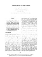
Comparison of median survival in 7 studies using the recursive partitioning analyses (RPA) classes (treatment was WBRT with or without local measures, none of the studies is limited to one particular cancer type)Figure 1
Comparison of median survival in 7 studies using the recursive partitioning analyses (RPA) classes (treatment
was WBRT with or without local measures, none of the studies is limited to one particular cancer type).
0
2
4
6
8
10
12
14
RPA I RPA II RPA III
RTOG 1997
RTOG 2000
Nieder et al. 2000
Lutterbach et al.
2000
Saito et al. 2006
RTOG 2008
Nieder et al. 2008
Rades et al. 2008
Months
Radiation Oncology 2009, 4:10 />Page 5 of 11
(page number not for citation purposes)
3 different values (0, 0.5 or 1) are assigned for each of
these 4 parameters: age (≥ 60; 50–59; <50), KPS (<70; 70–
80; 90–100), number of brain metastases (>3; 2–3; 1),
and extracranial metastases (present; not applicable;
none). Assessment of primary tumor activity or control is
no longer mandated. It was concluded by the authors that
"GPA is the least subjective, most quantitative and easiest
to use of the 4 indices" and that future trials should com-
pare these scores and validate the GPA. One of the
authors' group has embarked on this comparison in 2 dif-
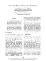
Comparison of median survival in 2 studies using the score index for radiosurgery (SIR) (treatment was WBRT with or without local measures, studies not limited to one particular cancer type)Figure 2
Comparison of median survival in 2 studies using the score index for radiosurgery (SIR) (treatment was WBRT
with or without local measures, studies not limited to one particular cancer type).
0
1
2
3
4
5
6
7
8
9
10
SIR 8-
10
SIR 4-7 SIR 1-3
RTOG 2008
Nieder et al.
2008
Months
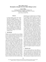
Comparison of median survival in 2 studies using the basic score for brain metastases (BSBM) (treatment was WBRT with or without local measures, studies not limited to one particular cancer type)Figure 3
Comparison of median survival in 2 studies using the basic score for brain metastases (BSBM) (treatment was
WBRT with or without local measures, studies not limited to one particular cancer type).
0
2
4
6
8
10
12
14
BSBM 3 BSBM 2 BSBM 1 BSBM 0
RTOG 2008
Nieder et al.
2008
Months
Radiation Oncology 2009, 4:10 />Page 6 of 11
(page number not for citation purposes)
ferent patient populations, i.e. those managed with WBRT
with or without SRS (comparable to the RTOG study pop-
ulation) [42] and those managed with surgery and WBRT
[35]. Both studies basically relied on the methods used by
the RTOG in their analysis, though with patients treated in
clinical routine outside of randomized trials. Compared
to RTOG's patients treated with WBRT with or without
SRS, the median age, KPS, number of lesions and lesion
volume were similar. Obvious differences existed, how-
ever, regarding controlled primary tumor (47 vs. 67%)
and extracranial metastases (56 vs. 36%). Thus, the cohort
is expected to have inferior survival. Figure 4 shows the
survival results.
Last but not least, Rades et al. developed a new prognostic
index based on 4 parameters (age, KPS, extracranial
metastases at the time of WBRT, interval from tumor diag-
nosis to WBRT) [45]. The major difference from the RPA
classes is the replacement of primary tumor control by
interval from tumor diagnosis to WBRT (not by number
of brain metastases as in the GPA). This index separated
patients into 4 subgroups with significantly different
prognosis and was also validated in one of the authors'
database (unpublished results, Figure 5).
Discussion
As stated on the website of the National Cancer Institute
/>bd_alpha.aspx?CdrID=44246, a prognostic factor is
regarded as a situation or condition, or a characteristic of
a patient, that can be used to estimate the chance of recov-
ery from a disease or the chance of the disease recurring.
Based on such prognostic factors, 6 different prognostic
indices for adult patients with brain metastases from solid
tumors have been developed over the last decade. As dem-
onstrated in Table 1, the 3 most recently published indices
all expanded from the primary 3-tiered RPA system to a 4-
tiered system. The 6 indices are based on a different
number of prognostic factors, i.e. 3–6. Of course, increas-
ing numbers of parameters will lead to less convenience
and ease of administration. None of the groups that devel-
oped these indices included all potential prognostic fac-
tors in their analysis. This is most likely due to the
unavailability of all the information in the databases and
the difficulty in collecting missing data in 1,000 or more
patients treated over many years. As can be seen in Figures
3, 4, 5, the performance of the 4-tiered indices is not tre-
mendously different, although further data are needed to
confirm this finding.
There is agreement in all indices on the importance of per-
formance status and extracranial disease activity. How-
ever, whether both primary tumor and extracranial
metastases should be considered is less clear (2 indices
would not include primary tumor control). Assessment of
extracranial disease status is not trivial. It might require
considerable resources in patients with very limited life
expectancy and therapeutic options. When collecting data
over long time periods, one must expect a shift in diagnos-
tic modalities, i.e. increasing use of magnetic resonance
Comparison of median survival in 2 studies using the graded prognostic assessment (GPA) (treatment was WBRT with or without local measures, studies not limited to one particular cancer type)Figure 4
Comparison of median survival in 2 studies using the graded prognostic assessment (GPA) (treatment was
WBRT with or without local measures, studies not limited to one particular cancer type).
0
2
4
6
8
10
12
14
GPA
3.5-4
GPA 3 GPA
1.5-2.5
GPA 0-1
RTOG 2008
Nieder et al.
2008
Months
Radiation Oncology 2009, 4:10 />Page 7 of 11
(page number not for citation purposes)
imaging of the brain as compared to computed tomogra-
phy (CT) or increasing use of chest CT or even positron
emission tomography (PET). Such a shift will likely result
in no longer assigning patients to the most favourable
prognostic class (stage migration). This might compro-
mise the comparison of the different studies.
Two of the 6 indices did not include age and the ones that
did, used slightly different cut-off values. A minority of stud-
ies (n = 2) included number of brain metastases and only
one each included response to steroids, volume of the largest
lesion in the brain, and time interval to development of
brain metastases, respectively. Other previous reports lend
credence to the examination of each of these factors. In their
multivariate analysis of 334 patients, DiLuna et al. reported
significantly better survival in patients with 1–3 vs 4 or more
brain metastases and in those patients with both limited
number and volume of brain metastases (<5 cc total vol-
ume) [46]. Bhatnagar et al. also reported on the impact of
treatment volume as independent prognostic factor in
patients treated with SRS [47]. In a randomised trial with
544 patients, Priestman et al. found that dose of steroids was
independently associated with survival [48]. Interval to
development of brain metastases appears particularly impor-
tant in patients with primary NSCLC and malignant
melanoma. The multivariate analyses of 3 studies with 292–
686 patients support this observation [23,49,50].
The latter findings lead to the general question on the use-
fulness of lumping together patients with different primary
tumors in these models. Breast cancer poses an interesting
dilemma here, because although tumor type and histology
were not prognostically significant in the RPA, recent data,
especially since the advent of trastuzumab and lapatinib,
suggest that, receptor status and her-2-neu expression
might have prognostic impact, even if this issue is not with-
out controversy (Table 3). The recently suggested prognos-
tic factor lymphopenia falls into the same category [19,51].
Unusual primary tumors and their potential differences in
biology or unique treatment approaches are not well repre-
sented in large pooled analyses. Table 4 provides examples
on analyses of prognostic factors in such groups.
Surrogate markers of disease activity that are easy to meas-
ure and inexpensive, such as lactate dehydrogenase and
other laboratory parameters have repeatedly been shown
to be independent prognostic factors for survival [71-74].
Studies that were not limited to patients with brain metas-
tases suggest that the anorexia-cachexia syndrome, dysp-
nea, pain, and co-morbidity are further candidates for
prospective evaluation [74]. The same holds true for neu-
rofunction class [75,76] and mini mental status examina-
tion results, which was an independent prognostic factor
for survival in a multivariate model that also included KPS
[77]. The current prognostic indices unfortunately do not
incorporate these features.
One of the purposes of prognostic indices is to guide the
choice of treatment in individual patients. In this context,
a prognostic index should be accurate enough to avoid
Comparison of median survival in 2 studies using the index proposed by Rades et alFigure 5
Comparison of median survival in 2 studies using the index proposed by Rades et al. [45](treatment was WBRT
with or without local measures, studies not limited to one particular cancer type, median survival estimated
from the Kaplan-Meier curves in the publication).
0
2
4
6
8
10
12
14
17-18
points
14-16
points
11-13
points
9-10
points
Rades et al.
2008
Nieder et al.
2008
Months
Radiation Oncology 2009, 4:10 />Page 8 of 11
(page number not for citation purposes)
overtreatment in patients that actually have very short sur-
vival. Even more important, one should not withhold
treatment because the index erroneously predicts an unfa-
vorable outcome. These aspects of the indices have not
been thoroughly evaluated, even in the recent GPA analy-
sis [44]. In our analysis of 239 patients, which confirms
that RPA, SIR, BSBM and GPA each split the dataset into
groups with significantly different prognosis, this issue
was addressed [42]. With regard to the outcome of
patients with unfavorable survival, defined as ≤ 2 months
(n = 93), no significant difference between the indices was
observed. Regarding patients with favorable survival,
defined as ≥ 6 months (n = 66), again no significant dif-
ference was observed, although RPA performed worse
than the other indices. Overall, GPA misassigned 6% of
the patients (9 out of 159), compared to 11% with RPA.
Therefore, the available validation data certainly do not
discourage further evaluation of the new GPA. However,
such evaluation should also include comparison with the
2 other scores (Rotterdam and Rades et al.). It is just the
stark reality of the disease process that in all of the scoring
systems, the most favorable prognostic group is very small
(e.g., GPA ≥ 3.5: 9% of RTOG and 7% of our own patients;
RPA class I: 16% of RTOG and 11% of our own patients).
The open questions after publication of 6 prognostic indi-
ces include:
- how should the ideal index perform?
- how many parameters should form the basis of the
ideal index?
- can we lump together patients with breast cancer,
small-cell lung cancer, malignant melanoma etc. or do
we lose potentially important information?
- do we need candidate parameters beyond the ones
examined so far (lactate dehydrogenase, anemia,
weight loss, pain etc.)?
Table 3: Prognostic impact of hormone receptor and HER-2 status in patients with brain metastases from breast cancer
n Prognostic impact of hormone receptor status Prognostic impact of HER-2 status
Claude et al.
51
120 none not examined
Bartsch et al.
52
174 none None
Le Scodan et al.
19
117 receptor negative significantly worse None
Nam et al.
53
126 receptor negative significantly worse HER-2 negative significantly worse
Kirsch et al.
54
95 not examined HER-2 negative significantly worse*
Eichler et al.
55
83 none HER-2 negative significantly worse
^
Melisko et al.
56
112 receptor negative significantly worse none
Harputluoglu et al.
57
144 none none
Park et al.
58
125 none HER-2 positive significantly worse
Church et al.
59
86 not examined HER-2 negative significantly worse*
^
80% of HER-2 overexpressing cases received trastuzumab after diagnosis of brain metastases
* the difference in survival was limited to patients with HER-2 overexpressing cancer treated with trastuzumab after diagnosis of brain metastases
Table 4: Prognostic factors in patients underrepresented in large studies (minimum number of patients n = 20)
Author Population Significant prognostic factors
Ogawa et al.
60
esophageal cancer, n = 36 KPS, aggressive local treatment (multivariate)
Weinberg et al.
61
esophageal cancer, n = 27 no liver metastases, RPA class I (trend, p = 0.1, multivariate)
Khuntia et al.
62
esophageal cancer, n = 27 KPS, aggressive local treatment (multivariate)
Cohen et al.
63
ovarian cancer, n = 72 aggressive local treatment
Cormio et al.
64
ovarian cancer, n = 22 extracranial disease, time to development of brain metastases
Growdon et al.
65
gynaecological cancers, n = 30 extracranial disease, histology, use of chemotherapy (multivariate)
Tremont-Lukats et al.
66
prostate cancer, n = 103 adenocarcinoma vs other histology
Rades et al.
24
unknown primary, n = 101 KPS, extracranial metastases, RPA class
Bartelt and Lutterbach
67
unknown primary, n = 47 KPS, surgical resection status (multivariate)
Ruda et al.
68
unknown primary, n = 33 number of brain metastases (multivariate)
Kim et al.
69
patients ≥ 75 years, SRS treatment, n = 44 single brain metastasis, NSCLC vs other primary
Noel et al.
70
patients ≥ 65 years, SRS treatment, n = 117 KPS (multivariate)
WBRT: whole-brain radiotherapy, KPS: Karnofsky performance status, RPA: recursive partitioning analysis, SRS: stereotactic radiosurgery, NSCLC:
non-small cell lung cancer
Radiation Oncology 2009, 4:10 />Page 9 of 11
(page number not for citation purposes)
- is it justifiable to assign the same point value to dif-
ferent degrees of extracranial disease, e.g., 2 small
asymptomatic lung metastases, 8 large liver metastases
with increased bilirubin, skin metastases already
treated by radiotherapy etc.?
- can international groups collaborate to develop a
consensus score, or maybe even an online tool?
Other aspects of predicting the outcome in patients with
brain metastases that many clinicians might appreciate,
relate to the important issue of neurologic function and
quality of life. In many instances, radiotherapy aims more
on improving deficits and preventing neurologic decline
than prolonging survival, but no attempts have been
made to develop scores that address endpoints other than
overall survival. It appears therefore worthwhile to collect
data on such endpoints, as done, e.g., in the recently com-
pleted randomized trial of radiotherapy with or without
motexafin gadolinium [78], which used time to neuro-
logic progression as primary endpoint. Other opportuni-
ties for future research include examination of prognostic
models that provide estimates on both risk of systemic
cancer progression with death from non-neurologic
causes and risk of death from uncontrolled brain metas-
tases.
Competing interests
The authors declare that they have no competing interests.
Authors' contributions
CN and MM drafted the manuscript and participated in
the design of the study. Both authors read and approved
the final manuscript.
Acknowledgements
None
References
1. Kunthia D, Brown P, Li J, Mehta MP: Whole-brain radiotherapy in
the management of brain metastasis. J Clin Oncol 2006,
24:1295-1304.
2. Langer CJ, Mehta MP: Current management of brain metas-
tases, with a focus on systemic options. J Clin Oncol 2005,
23:6207-6219.
3. Nieder C, Grosu AL, Astner ST, Thamm R, Molls M: Integration of
chemotherapy into current treatment strategies for brain
metastases from solid tumors. Radiat Oncol 2006, 1:19.
4. Aoyama H, Shirato H, Tago M, Nakagawa K, Toyoda T, Hatano K,
Kenjyo M, Oya N, Hirota S, Shioura H, Kunieda E, Inomata T, Hay-
akawa K, Katoh N, Kobashi G: Stereotactic radiosurgery plus
whole-brain radiation therapy vs stereotactic radiosurgery
alone for treatment of brain metastases. A randomized con-
trolled trial. JAMA 2006, 295:2483-2491.
5. Andrews DW, Scott CB, Sperduto PW, Flanders AE, Gaspar LE,
Schell MC, Werner-Wasik M, Demas W, Ryu J, Bahary JP, Souhami L,
Rotman M, Mehta MP, Curran WJ Jr: Whole brain radiation ther-
apy with or without stereotactic radiosurgery boost for
patients with one to three brain metastases: phase III results
of the RTOG 9508 randomised trial. Lancet 2004,
363:1665-1672.
6. Kondziolka D, Patel A, Lunsford LD, Kassam A, Flickinger JC: Stere-
otactic radiosurgery plus whole brain radiotherapy versus
radiotherapy alone for patients with multiple brain metas-
tases. Int J Radiat Oncol Biol Phys 1999, 45:427-434.
7. Patchell RA, Tibbs PA, Regine WF, Dempsey RJ, Mohiuddin M, Kry-
scio RJ, Markesbery WR, Foon KA, Young B: Postoperative radio-
therapy in the treatment of single metastases to the brain: a
randomized trial. JAMA 1998, 80:1485-1489.
8. Noordijk EM, Vecht CJ, Haaxma-Reiche H, Padberg GW, Voormolen
JH, Hoekstra FH, Tans JT, Lambooij N, Metsaars JA, Wattendorf AR:
The choice of treatment of single brain metastasis should be
based on extracranial tumor activity. Int J Radiat Oncol Biol Phys
1994, 29:711-717.
9. Patchell RA, Tibbs PA, Walsh JW, Dempsey RJ, Maruyama Y, Kryscio
RJ, Markesbery WR, Macdonald JS, Young B: A randomized trial of
surgery in the treatment of single metastases to the brain. N
Engl J Med 1990, 322:494-500.
10. Nieder C, Astner ST, Grosu AL, Andratschke NH, Molls M: The role
of postoperative radiotherapy after resection of a single
brain metastasis: combined analysis of 643 patients. Strahlen-
ther Onkol 2007, 183:576-580.
11. Gaspar L, Scott C, Rotman M, Asbell S, Phillips T, Wasserman T,
McKenna WG, Byhardt R: Recursive partitioning analysis (RPA)
of prognostic factors in three Radiation Therapy Oncology
Group (RTOG) brain metastases trials. Int J Radiat Oncol Biol
Phys 1997, 37:745-751.
12. Gaspar LE, Scott C, Murray K, Curran W: Validation of the RTOG
recursive partitioning analysis (RPA) classification for brain
metastases. Int J Radiat Oncol Biol Phys 2000, 47:1001-1006.
13. Lutterbach J, Bartelt S, Stancu E, Guttenberger R: Patients with
brain metastases: hope for recursive partitioning analysis
(RPA) class 3. Radiother Oncol 2002, 63:339-345.
14. Nieder C, Nestle U, Motaref B, Walter K, Niewald M, Schnabel K:
Prognostic factors in brain metastases: should patients be
selected for aggressive treatment according to recursive
partitioning analysis (RPA) classes? Int J Radiat Oncol Biol Phys
2000, 46:297-302.
15. Fleckenstein K, Hof H, Lohr F, Wenz F, Wannenmacher M: Prognos-
tic factors for brain metastases after whole brain radiother-
apy. Data from a single institution. Strahlenther Onkol 2004,
180:268-273.
16. Saito EY, Viani GA, Ferrigno R, Nakamura RA, Novaes PE, Pellizzon
CA, Fogaroli RC, Conte MA, Salvajoli JV: Whole brain radiation
therapy in management of brain metastasis: results and
prognostic factors. Radiat Oncol 2006, 1:20.
17. Mahmoud-Ahmed AS, Suh JH, Lee SY, Crownover RL, Barnett GH:
Results of whole brain radiotherapy in patients with brain
metastases from breast cancer: a retrospective study. Int J
Radiat Oncol Biol Phys 2002, 54:810-817.
18. Viani GA, Castilho MS, Salvajoli JV, Pellizzon AC, Novaes PE, Guima-
rães FS, Conte MA, Fogaroli RC: Whole brain radiotherapy for
brain metastases from breast cancer: estimation of survival
using two stratification systems. BMC Cancer 2007,
7:53.
19. Le Scodan R, Massard C, Mouret-Fourme E, Guinebretierre JM,
Cohen-Solal C, De Lalande B, Moisson P, Breton-Callu C, Gardner M,
Goupil A, Renody N, Floiras JL, Labib A: Brain metastases from
breast carcinoma: validation of the Radiation Therapy
Oncology Group recursive partitioning analysis classification
and proposition of a new prognostic score. Int J Radiat Oncol Biol
Phys 2007, 69:839-845.
20. Kepka L, Cieslak E, Bujko K, Fijuth H, Wierzchowski M: Results of
the whole-brain radiotherapy for patients with brain metas-
tases from lung cancer: the RTOG RPA intra-classes analy-
sis. Acta Oncol 2005, 44:389-398.
21. Videtic GM, Adelstein DJ, Mekhail TM, Rice TW, Stevens GH, Lee SY,
Suh JH: Validation of the RTOG recursive partitioning analy-
sis (RPA) classification for small-cell lung cancer-only brain
metastases. Int J Radiat Oncol Biol Phys 2007, 67:240-243.
22. Gülbas H, Erkal HS, Serin M: The use of recursive partitioning
analysis grouping in patients with brain metastases from
non-small-cell lung cancer. Jpn J Clin Oncol 2006, 36:193-196.
23. Rades D, Schild SE, Lohynska R, Veninga T, Stalpers LJ, Dunst J: Two
radiation regimens and prognostic factors for brain metas-
tases in nonsmall cell lung cancer patients. Cancer 2007,
110:1077-1082.
Radiation Oncology 2009, 4:10 />Page 10 of 11
(page number not for citation purposes)
24. Rades D, Bohlen G, Lohynska R, Veninga T, Stalpers LJ, Schild SE,
Dunst J: Whole-brain radiotherapy with 20 Gy in 5 fractions
for brain metastases in patients with cancer of unknown pri-
mary (CUP). Strahlenther Onkol 2007, 183:631-636.
25. Kanner AA, Suh JH, Siomin VE, Lee SY, Barnett GH, Vogelbaum MA:
Posterior fossa metastases: aggressive treatment improves
survival. Stereotact Funct Neurosurg 2003, 81:18-23.
26. Nam TK, Lee JI, Jung YJ, Im YS, An HY, Nam DH, Park K, Kim JH:
Gamma knife surgery for brain metastases in patients har-
boring four or more lesions: survival and prognostic factors.
J Neurosurg 2005, 102(suppl):147-150.
27. Buchsbaum JC, Suh JH, Lee SY, Chidel MA, Greskovich JF, Barnett
GH: Survival by Radiation Therapy Oncology Group recur-
sive partitioning analysis class and treatment modality in
patients with brain metastases from malignant melanoma.
Cancer 2002, 94:2265-2272.
28. Harrison BE, Johnson JL, Clough RW, Halperin EC: Selection of
patients with melanoma brain metastases for aggressive
treatment. Am J Clin Oncol 2003, 26:354-357.
29. Morris SL, Low SH, A'Hern RP, Eisen TG, Gore ME, Nutting CM, Har-
rington KJ: A prognostic index that predicts outcome follow-
ing palliative whole brain radiotherapy for patients with
metastatic malignant melanoma. Br J Cancer 2004, 91:829-833.
30. Chidel MA, Suh JH, Reddy CA, Chao ST, Lundbeck MF, Barnett GH:
Application of recursive partitioning analysis and evaluation
of the use of whole brain radiation among patients treated
with stereotactic radiosurgery for newly diagnosed brain
metastases. Int J Radiat Oncol Biol Phys 2000, 47:993-999.
31. Agboola O, Benoit B, Cross P, Da Silva V, Esche B, Lesiuk H, Gon-
salves C: Prognostic factors derived from recursive partition-
ing analysis (RPA) of Radiation Therapy Oncology Group
(RTOG) brain metastases trials applied to surgically
resected and irradiated brain metastatic cases. Int J Radiat
Oncol Biol Phys 1998, 42:155-159.
32. Paek SH, Audu PB, Sperling MR, Cho J, Andrews DW: Reevaluation
of surgery for the treatment of brain metastases: review of
208 patients with single or multiple brain metastases treated
at one institution with modern neurosurgical techniques.
Neurosurgery 2005,
56:1021-1034.
33. Tendulkar RD, Liu SW, Barnett GH, Vogelbaum MA, Toms SA, Jin T,
Suh JH: RPA classification has prognostic significance for sur-
gically resected single brain metastasis. Int J Radiat Oncol Biol
Phys 2006, 66:810-817.
34. Rades D, Pluemer A, Veninga T, Dunst J, Schild SE: A boost in addi-
tion to whole-brain radiotherapy improves patient outcome
after resection of 1 or 2 brain metastases in recursive parti-
tioning analysis class 1 and 2 patients. Cancer 2007,
110:1551-1559.
35. Nieder C, Geinitz H, Molls M: Validation of the graded prognos-
tic assessment index for surgically treated patients with
brain metastases. Anticancer Res 2008, 28:3015-3017.
36. Lagerwaard FJ, Levendag PC, Nowak PJ, Eijkenboom WM, Hanssens
PE, Schmitz PI: Identification of prognostic factors in patients
with brain metastases: a review of 1292 patients. Int J Radiat
Oncol Biol Phys 1999, 43:795-803.
37. Weltman E, Salvajoli JV, Brandt RA, de Morais Hanriot R, Prisco FE,
Cruz JC, de Oliveira Borges SR, Wajsbrot DB: Radiosurgery for
brain metastases: A score index for predicting prognosis. Int
J Radiat Oncol Biol Phys 2000, 46:1155-1161.
38. Selek U, Chang EL, Hassenbusch SJ 3rd, Shiu AS, Lang FF, Allen P,
Weinberg J, Sawaya R, Maor MH: Stereotactic radiosurgical
treatment in 103 patients for 153 cerebral melanoma metas-
tases. Int J Radiat Oncol Biol Phys 2004, 59:1097-1106.
39. Goyal S, Prasad D, Harrell F Jr, Matsumoto J, Rich T, Steiner L:
Gamma knife surgery for the treatment of intracranial
metastases from breast cancer. J Neurosurg 2005, 103:218-223.
40. Gaudy-Marqueste C, Regis JM, Muracciole X, Laurans R, Richard MA,
Bonerandi JJ, Grob JJ: Gamma-Knife radiosurgery in the man-
agement of melanoma patients with brain metastases: a
series of 106 patients without whole-brain radiotherapy. Int
J Radiat Oncol Biol Phys 2006, 65:809-816.
41. Akyurek S, Chang EL, Mahajan A, Hassenbusch SJ, Allen PK, Mathews
LA, Shiu AS, Maor MH, Woo SY: Stereotactic radiosurgical
treatment of cerebral metastases arising from breast can-
cer. Am J Clin Oncol 2007, 30:310-314.
42. Nieder C, Molls M: Validation of the graded prognostic assess-
ment index for patients with brain metastases: in regards to
Sperduto et al. (Int J Radiat Oncol Biol Phys 2008;70:510–
514). Int J Radiat Oncol Biol Phys 2008, 72:1619.
43. Lorenzoni J, Devriendt D, Massager N, David P, Ruiz S, Vanderlinden
B, Van Houtte P, Brotchi J, Levivier M: Radiosurgery for treat-
ment of brain metastases: Estimation of patient eligibility
using three stratification systems. Int J Radiat Oncol Biol Phys
2004, 60:218-224.
44. Sperduto PW, Berkey B, Gaspar LE, Mehta M, Curran W: A new
prognostic index and comparison to three other indices for
patients with brain metastases: an analysis of 1,960 patients
in the RTOG database. Int J Radiat Oncol Biol Phys 2008,
70:510-514.
45. Rades D, Dunst J, Schild SE: A new scoring system to predicting
the survival of patients treated with whole-brain radiother-
apy for brain metastases. Strahlenther Onkol 2008, 184:251-255.
46. DiLuna ML, King JT Jr, Knisely JP, Chiang VL: Prognostic factors for
survival after stereotactic radiosurgery vary with the
number of cerebral metastases. Cancer 2007, 109:135-145.
47. Bhatnagar AK, Kondziolka D, Lunsford LD, Flickinger JC: Recursive
partitioning analysis of prognostic factors for patients with
four or more intracranial metastases treated with radiosur-
gery. Technol Cancer Res Treat 2007, 6:153-160.
48. Priestman TJ, Dunn J, Brada M, Rampling R, Baker PG: Final results
of the Royal College of Radiologists' trial comparing two dif-
ferent radiotherapy schedules in the treatment of cerebral
metastases. Clin Oncol (R Coll Radiol) 1996, 8:308-315.
49. Tang SG, Tseng CK, Tsay PK, Chen CH, Chang JW, Pai PC, Hong JH:
Predictors for patterns of brain relapse and overall survival
in patients with non-small cell lung cancer. J Neurooncol 2005,
73:153-161.
50. Fife KM, Colman MH, Stevens GN, Firth IC, Moon D, Shannon KF,
Harman R, Petersen-Schaefer K, Zacest AC, Besser M, Milton GW,
McCarthy WH, Thompson JF: Determinants of outcome in
melanoma patients with cerebral metastases. J Clin Oncol
2004, 22:1293-1300.
51. Claude L, Perol D, Ray-Coquard I, Petit T, Blay JY, Carrie C, Bachelot
T:
Lymphopenia: A new independent prognostic factor for
survival in patients treated with whole brain radiotherapy
for brain metastases from breast carcinoma. Radiother Oncol
2005, 76:334-339.
52. Bartsch R, Fromm S, Rudas M, Wenzel C, Harbauer S, Roessler K,
Kitz K, Steger GG, Weitmann HD, Poetter R, Zielinski CC, Dieck-
mann K: Intensified local treatment and systemic therapy sig-
nificantly increase survival in patients with brain metastases
from advanced breast cancer – A retrospective analysis. Radi-
other Oncol 2006, 80:313-317.
53. Nam BH, Kim SY, Han HS, Kwon Y, Lee KS, Kim TH, Ro J: Breast
cancer subtypes and survival in patients with brain metas-
tases. Breast Cancer Res 2008, 10:R20.
54. Kirsch DG, Ledezma CJ, Mathews CS, Bhan AK, Ancukiewicz M,
Hochberg FH, Loeffler JS: Survival after brain metastases from
breast cancer in the trastuzumab era. J Clin Oncol 2005,
23:2114-2116.
55. Eichler AF, Kuter I, Ryan P, Schapira L, Younger J, Henson JW: Sur-
vival in patients with brain metastases from breast cancer.
Cancer 2008, 112:2359-2367.
56. Melisko ME, Moore DH, Sneed PK, De Franco J, Rugo HS: Brain
metastases in breast cancer: clinical and pathologic charac-
teristics associated with improvements in survival. J Neuroon-
col 2008, 88:359-365.
57. Harputluoglu H, Dizdar O, Aksoy S, Kilickap S, Dede DS, Ozisik Y,
Guler N, Barista I, Gullu I, Hayran M, Selek U, Cengiz M, Zorlu F,
Tekuzman G, Altundag K: Characteristics of breast cancer
patients with central nervous system metastases: a single-
center experience. J Natl Med Assoc 2008, 100:521-526.
58. Park BB, Uhm JE, Cho EY, et al.: Prognostic factor analysis in
patients with brain metastases from breast cancer: how can
we improve the treatment outcomes? Cancer Chemother Phar-
macol 2008. epub.
59. Church DN, Modgil R, Guglani S, Bahl A, Hopkins K, Braybrooke JP,
Blair P, Price CG: Extended survival in women with brain
metastases from HER2 overexpressing breast cancer. Am J
Clin Oncol 2008, 31:250-254.
Publish with BioMed Central and every
scientist can read your work free of charge
"BioMed Central will be the most significant development for
disseminating the results of biomedical research in our lifetime."
Sir Paul Nurse, Cancer Research UK
Your research papers will be:
available free of charge to the entire biomedical community
peer reviewed and published immediately upon acceptance
cited in PubMed and archived on PubMed Central
yours — you keep the copyright
Submit your manuscript here:
/>BioMedcentral
Radiation Oncology 2009, 4:10 />Page 11 of 11
(page number not for citation purposes)
60. Ogawa K, Toita T, Sueyama H, Fuwa N, Kakinohana Y, Kamata M,
Adachi G, Saito A, Yoshii Y, Murayama S: Brain metastases from
esophageal carcinoma: natural history, prognostic factors,
and outcome. Cancer 2002, 94:759-764.
61. Weinberg JS, Suki D, Hanbali F, Cohen ZR, Lenzi R, Sawaya R: Metas-
tasis of esophageal carcinoma to the brain. Cancer 2003,
98:1925-1933.
62. Khuntia D, Sajja R, Chidel MA, Lee SY, Rice TW, Adelstein DJ, Carl-
son TP, Saxton JP, Barnett GH, Suh JH: Factors associated with
improved survival in patients with brain metastases from
esophageal cancer: a retrospective review. Technol Cancer Res
Treat 2003, 2:267-272.
63. Cohen ZR, Suki D, Weinberg JS, Marmor E, Lang FF, Gershenson DM,
Sawaya R: Brain metastases in patients with ovarian carci-
noma: prognostic factors and outcome. J Neurooncol 2004,
66:313-325.
64. Cormio G, Maneo A, Colamaria A, Loverro G, Lissoni A, Selvaggi L:
Surgical resection of solitary brain metastasis from ovarian
carcinoma: an analysis of 22 cases. Gynecol Oncol 2003,
89:116-119.
65. Growdon WB, Lopez-Varela E, Littell R, Oliva E, Seiden M, Krasner
C, Lee H, Fuller A: Extent of extracranial disease is a powerful
predictor of survival in patients with brain metastases from
gynecological cancer. Int J Gynecol Cancer 2008, 18:262-268.
66. Tremont-Lukats IW, Bobustuc G, Lagos GK, Lolas K, Kyritsis AP,
Puduvalli VK: Brain metastasis from prostate carcinoma: The
M. D. Anderson Cancer Center experience. Cancer 2003,
98:363-368.
67. Bartelt S, Lutterbach J: Brain metastases in patients with cancer
of unknown primary. J Neurooncol 2003, 64:249-253.
68. Rudà R, Borgognone M, Benech F, Vasario E, Soffietti R: Brain
metastases from unknown primary tumour: a prospective
study. J Neurol 2001, 248:394-398.
69. Kim SH, Weil RJ, Chao ST, Toms SA, Angelov L, Vogelbaum MA, Suh
JH, Barnett GH: Stereotactic radiosurgical treatment of brain
metastases in older patients. Cancer 2008, 113:
834-840.
70. Noel G, Bollet MA, Noel S, Feuvret L, Boisserie G, Tep B, Delattre
JY, Baillet F, Ambroise Valery C, Cornu P, Mazeron JJ: Linac stere-
otactic radiosurgery: an effective and safe treatment for eld-
erly patients with brain metastases. Int J Radiat Oncol Biol Phys
2005, 63:1555-1561.
71. Chatani M, Matayoshi Y, Masaki N, Inoue T: Radiation therapy for
brain metastases from lung carcinoma. Prospective rand-
omized trial according to the level of lactate dehydrogenase.
Strahlenther Onkol 1994, 170:155-161.
72. Jacot W, Quantin X, Boher JM, Andre F, Moreau L, Gainet M,
Depierre A, Quoix E, Chevalier TL, Pujol JL: Brain metastases at
the time of presentation of non-small cell lung cancer: a
multi-centric AERIO analysis of prognostic factors. Br J Cancer
2001, 84:903-909.
73. Gripp S, Moeller S, Bölke E, Schmitt G, Matuschek C, Asgari S,
Asgharzadeh F, Roth S, Budach W, Franz M, Willers R: Survival pre-
diction in terminally ill cancer patients by clinical estimates,
laboratory tests, and self-rated anxiety and depression. J Clin
Oncol 2007, 25:3313-3320.
74. Hauser CA, Stockler MR, Tattersall MH: Prognostic factors in
patients with recently diagnosed incurable cancer: a system-
atic review. Support Care Cancer 2006, 14:999-1011.
75. Rodrigus P, de Brouwer P, Raaymakers E: Brain metastases and
non-small cell lung cancer. Prognostic factors and correla-
tion with survival after irradiation. Lung Cancer 2001,
32:129-136.
76. Raizer JJ, Hwu WJ, Panageas KS, Wilton A, Baldwin DE, Bailey E, von
Althann C, Lamb LA, Alvarado G, Bilsky MH, Gutin PH: Brain and
leptomeningeal metastases from cutaneous melanoma: sur-
vival outcomes based on clinical features. Neuro Oncol 2008,
10:199-207.
77. Murray KJ, Scott C, Zachariah B, Michalski JM, Demas W, Vora NL,
Whitton A, Movsas B: Importance of the mini-mental status
examination in the treatment of patients with brain metas-
tases: a report from the Radiation Therapy Oncology Group
protocol 91-04. Int J Radiat Oncol Biol Phys 2000, 48:59-64.
78. Mehta MP, Shapiro WR, Phan SC, Gervais R, Carrie C, Chabot P,
Patchell RA, Glantz MJ, Recht L, Langer C, Sur RK, Roa WH, Mahe
MA, Fortin A, Nieder C, Meyers CA, Smith JA, Miller RA, Renschler
MF: Motexafin gadolinium combined with prompt whole
brain radiotherapy prolongs time to neurologic progression
in non-small-cell lung cancer patients with brain metastases:
results of a phase III trial. Int J Radiat Oncol Biol Phys 2009,
73:1069-1076.