Review Cells of the synovium in rheumatoid arthritis pot
Bạn đang xem bản rút gọn của tài liệu. Xem và tải ngay bản đầy đủ của tài liệu tại đây (396.49 KB, 11 trang )
Page 1 of 11
(page number not for citation purposes)
Available online />Abstract
Recent findings have substantiated the importance of T lympho-
cytes to the pathogenesis of rheumatoid arthritis (RA). Here, we
review emerging data regarding genetic predisposition, spon-
taneous animal models of arthritis, and cell-cell interactions that
implicate T cells as driving synovial inflammation and joint
destruction. Information regarding the proinflammatory role of
interleukin-17-producing T cells and the functional state of
regulatory T cells both in animal models and in patients with RA is
also discussed. In light of the overwhelming evidence that
disrupted T-cell homeostasis greatly contributes to joint pathology
in RA, the therapeutic potential of targeting activators of pro-
inflammatory T cells or their products is compelling.
Introduction
Our understanding of how T lymphocytes participate in the
pathogenesis of rheumatoid arthritis (RA) is evolving rapidly
with fundamental new insights into basic T-cell biology and
the orchestration and regulation of immune responses. The
simplistic notion of RA as a homogeneous, clonally driven,
T cell-mediated autoimmune disease is outdated, as is the
notion that the large numbers of T cells in RA synovium may
be irrelevant bystanders. What is replacing these polarized
hypotheses is a more integrated view of T cells as a central
component of organ-focused immune-mediated pathology,
capable of interactions not only with classical cells of the
immune system but with tissue-specific cell populations that
contribute to inflammation and tissue destruction. RA is
emerging as a prototypic disease not only for the study of
such interactions but also for the introduction of novel
biologic therapies that inhibit these processes. This review
will selectively focus on newer and topical aspects of T-cell
biology in RA.
T cells and the genetics of RA
RA is a polygenic disease and its most important loci are in
the major histocompatibility complex (MHC). The concept of
the RA shared epitope, a peptide sequence common to
disease-associated human leukocyte antigen-DR (HLA-DR)
alleles, remains valid, but the precise mechanism of how the
shared epitope predisposes individuals to RA is not yet
established. Multiple possibilities have been proposed, most
of which focus on recognition of antigen by mature T cells
and/or T-cell repertoire differentiation [1]. Recent analyses of
the full range of DRB1 alleles have emphasized that
sequence variations at amino acids 67 to 74 can encode
either susceptibility to or protection from RA and can
influence disease severity as well as susceptibility [2-4]. It
would be attractive to link protection from RA to
immunoregulatory mechanisms, but evidence for such a link
is not yet available.
An important epidemiologic study has linked smoking, the
shared epitope, and seropositive RA [5]. In this Scandinavian
population, the relative risk of seropositive (rheumatoid factor-
positive) RA in individuals who smoked and were
homozygous for the shared epitope was 15.7. In seronegative
RA, neither smoking nor the shared epitope was a risk factor.
Antibodies to citrullinated proteins have become established
as an RA feature that is more specific than rheumatoid factor,
Review
Cells of the synovium in rheumatoid arthritis
T lymphocytes
Steven K Lundy, Sujata Sarkar, Laura A Tesmer and David A Fox
Department of Internal Medicine, Division of Rheumatology and Rheumatic Diseases Core Center, University of Michigan Medical School,
4043 Biomedical Sciences Research Bldg., 109 Zina Pitcher Pl., Ann Arbor, MI 48109-2200, USA
Corresponding author: David A Fox,
Published: 13 February 2007 Arthritis Research & Therapy 2007, 9:202 (doi:10.1186/ar2107)
This article is online at />© 2007 BioMed Central Ltd
APC = antigen-presenting cell; CCP = cyclic citrullinated peptide; CIA = collagen-induced arthritis; CTLA-4 = cytotoxic T lymphocyte antigen-4;
FLS = fibroblast-like synoviocytes; Foxp3 = forkhead box p3; HLA-DR = human leukocyte antigen-DR; ICAM-1 = intercellular adhesion molecule-1;
ICOS = inducible costimulator; IFN-γ = interferon-gamma; IL = interleukin; IL-1Ra = interleukin-1 receptor antagonist; LFA = lymphocyte function-
associated antigen; MHC = major histocompatibility complex; NF-κB = nuclear factor-kappa B; PBMC = peripheral blood mononuclear cell; PD-1
= programmed death-1; PTPN22 = protein tyrosine phosphatase non-receptor type 22; RA = rheumatoid arthritis; RANK = receptor activator of
nuclear factor-kappa B; SNP = single-nucleotide polymorphism; TCR = T-cell receptor; TGF-β = transforming growth factor-beta; TNF = tumor
necrosis factor; Treg = regulatory T cell; ZAP-70 = zeta-associated protein of 70 kDa.
Page 2 of 11
(page number not for citation purposes)
Arthritis Research & Therapy Vol 9 No 1 Lundy et al.
but information about the role of T-cell responses and genetic
factors in this intriguing form of autoimmunity is just beginning
to emerge. Auger and colleagues [6] reported that both
citrullinated and non-citrullinated fibrinogen peptides bound
to a range of HLA-DR molecules, both RA-associated alleles
and non-associated alleles, but that T-cell proliferative
responses were much more common in RA. These data
argue that the shared epitope is not the sole factor governing
development of T-cell autoreactivity to citrullinated proteins.
Nonetheless, production of antibodies to citrullinated
fibrinogen was more common in RA patients who carry
HLA-DRB1*0404, a shared epitope-containing allele.
Analysis of US and Dutch cohorts with RA found clear
linkage of the shared epitope to anti-cyclic citrullinated
peptide (CCP)-positive RA but not to anti-CCP-negative RA
[7]. The presence of anti-CCP antibodies appeared to fully
account for the greater disease severity observed in shared
epitope-positive RA. Based on analysis of a cohort of patients
with recent-onset inflammatory arthritis, the provocative
suggestion has been advanced that the sole role of the
shared epitope is to provide the genetic basis for stimulation
of T-cell help in anti-CCP antibody formation and that it does
not otherwise contribute to the development of RA [8].
Additional studies in cohorts of various ethnicities will help to
further test this concept.
Apart from the MHC, the best-established genetic locus that
influences RA is the gene PTPN22 (protein tyrosine
phosphatase non-receptor type 22), which encodes Lyp, a
tyrosine phosphatase that is expressed in T lymphocytes and
that regulates signal transduction from the T-cell receptor
(TCR) [9,10]. Substitution of tryptophan for arginine at
residue 620 results in a gain-of-function, leading to
decreased TCR signaling and decreased production of
interleukin (IL)-2 [11]. The current understanding is that this
causes a failure to delete autoreactive T cells during thymic
development and/or a decreased formation of regulatory T
(Treg) cells. The combination of the shared epitope and the
RA-associated PTPN22 allele (termed PTPN22*R620W)
was found to predispose to formation of autoantibodies to
type II collagen in early RA, which implies concurrent T-cell
autoreactivity to this cartilage autoantigen [12].
Another set of single-nucleotide polymorphisms (SNPs)
implicated in susceptibility to RA has been found in the gene
encoding programmed death-1 (PD-1), a molecule that
regulates T-cell homeostasis through apoptosis [13-15].
Although different PD-1 SNPs have been identified in RA
patients from distinct ethnic backgrounds and some but not
all SNPs of PD-1 have also been linked to susceptibility
toward systemic lupus erythematosus, these polymorphisms
most likely have in common a defective activity of PD-1,
leading to decreased elimination of autoreactive T cells.
Interestingly, a very recent study showed that PD-1 and its
ligand are overexpressed in synovial cells from patients with
RA and that an alternatively spliced variant of PD-1 leading to
formation of an inhibitory soluble form of the protein was
abundant in serum and synovial fluid of patients with RA [16].
These data suggest that soluble PD-1 may protect
autoreactive T cells from undergoing apoptosis and
corroborate the idea that ineffective PD-1 signaling is an
important contributor to RA susceptibility.
It appears likely that several genes will also be validated as
being linked to RA severity but not to susceptibility. Although
the notion of RA as a Th1 disease as opposed to a Th17
disease is currently in flux (see below), it remains clear that
RA is not a Th2 disease, and it is plausible that the
production or function of Th2 cytokines or both are deficient
in RA. In this context, a report that associates a
hypofunctional allele of the IL-4 receptor with increased
radiographic damage in RA is of particular interest [17].
Given the ability of IL-4 to regulate Th17 responses (as well
as Th1 responses), reduced responsiveness of the IL-4
receptor would be expected to worsen the outcome of RA.
Overall, recent advances in our knowledge concerning the
genetics of RA not only reinforce the importance of T cells in
both susceptibility to and outcome of this disease but also
emphasize the complex and interdependent roles of T cells in
the context of the entire immune response.
T cells in spontaneous animal models of
arthritis
Over the past several decades, many inductive models of
inflammatory polyarthritis, such as collagen-induced arthritis
(CIA) and adjuvant-induced arthritis, have been employed to
study immune responses in arthritis. These animal models of
arthritis have contributed significantly to our understanding of
cellular and molecular events that may be relevant to RA.
Recently, several models of spontaneous arthritis have been
identified due to perturbations in the TCR and alterations of
cytokine regulation. This section will focus on new findings
related to T cells in four of these recently identified models of
spontaneous arthritis: SKG, K/BxN-transgenic, gp130 (IL-6R)
mutant, and IL-1 receptor antagonist (IL-1Ra)-deficient mice.
An intrinsic defect in TCR signaling or alteration of the
cytokine milieu can lead to T cell-dependent arthritis in mice.
Sakaguchi and colleagues [18,19] have generated mice with
a point mutation in the COOH-terminal SH2 domain of zeta-
associated protein of 70 kDa (ZAP-70) which develop
spontaneous arthritis and demonstrate extra-articular
manifestations found in RA, including interstitial pneumonitis,
subcutaneous nodules, and vasculitis. The role of T cells in
the SKG mutant model has been demonstrated by the
predominant infiltration of a Vβ-restricted subset of CD4
+
T cells in the inflamed synovium [20]. Adoptive transfer of
splenic or lymph node ZAP-70 mutant T cells or thymocytes
leads to chronic arthritis in syngeneic, nude, or severe
combined immunodeficient mice [18,20]. As a result of the
mutation, ZAP-70 expression is not altered but ZAP-70 does
Page 3 of 11
(page number not for citation purposes)
not bind normally to the TCR. This probably leads to signaling
abnormalities of the TCR in the thymus, resulting in positive
selection of self-reactive T-cell clones that would otherwise
be eliminated. In addition to providing strong evidence for the
ability of autoreactive T-cell clones to initiate arthritis, this
model is also dependent on the proinflammatory cytokines
IL-6, IL-1β, and tumor necrosis factor (TNF)-α, which are
highly implicated in RA synovial pathology [19]. The
dependence on proinflammatory cytokines in this model was
further substantiated by a report that spontaneous arthritis in
SKG mice did not occur in specific pathogen-free housing
conditions but was inducible with the fungal glucan, zymosan
A, a dectin-1 and toll-like receptor 2 agonist that stimulated
IL-1β and IL-6 production in the model [21,22]. The pattern of
cytokine expression in zymosan-treated SKG mice is highly
correlated with the conditions required to drive a Th17
response; moreover, dependence of this model on IL-17 has
recently been established [23].
Another spontaneous arthritis model that is contributing to
our understanding of the role of T cells in arthritis is the
IL-1Ra-deficient mouse strain [24]. IL-1Ra is an endogenous,
natural inhibitor of IL-1 bioactivity through binding and
blockade of the IL-1 receptor. An important finding was that
arthritis fails to develop in IL-1Ra-deficient mice in the
absence of mature T cells [25]. Transfer of T cells from
IL-1Ra-deficient mice into nude mice resulted in arthritis,
further substantiating the role of T cells in this model [25].
Cytokines, especially IL-17 and TNF-α, also play an important
role in this model of arthritis [25,26].
Recently, IL-6 in conjunction with transforming growth factor-
beta (TGF-β) has been implicated in the generation of Th17
cells. A mutation in the tyrosine residue, at the phosphatase-
binding site, of the gp130 subunit of the IL-6 receptor has
been shown to result in spontaneous arthritis in mice [27,28].
This mutation leads to an increase in receptor signaling
through STAT3 (signal transducer and activator of trans-
cription 3), resulting in both increased IL-7-dependent
proliferation as well as impaired Fas ligand expression and
decreased T-cell apoptosis. Development of arthritis in the
gp130 mutant model is dependent on CD4
+
T cells despite
the finding that mutation of gp130 in nonhematopoietic cells
is sufficient to drive disease. These data suggest that
arthritogenic T cells are usually regulated by nonhemato-
poietic cells through a mechanism that can be overridden by
increased signaling through gp130.
The K/BxN transgenic mouse is another example of
spontaneous arthritis in the mouse, resulting from recognition
of self-antigens and breakdown of tolerance [29]. F1
offspring (K/BxN) of nonobese diabetic mice crossed with
KRN TCR transgenic mice, which have specificity for a
glucose-6-phosphate isomerase peptide in the context of
I-A
g7
, develop spontaneous arthritis. Despite low cell
numbers, there was enrichment of CD4
+
T cells in the
synovial compartment with high levels of expression of the
KRN transgene. Administration of anti-CD4 T-cell antibody
before, but not during or after, the onset of arthritis blocked
arthritis development. This suggests that T cells are important
in only the early pathogenesis of arthritis in K/BxN mice. A
subsequent study demonstrated that passive transfer of serum
from K/BxN arthritic mice resulted in arthritis in various mouse
strains which was dependent on engagement of the innate
immune system [30,31]. These data suggest that the main
pathogenic role of autoreactive T cells in the K/BxN model is
to drive the development of autoantibodies. In contrast,
Schubert et al. [32] have more recently reported a model,
based on immunization with glucose-6-phosphate isomerase,
that is T cell-dependent at both initiating and effector phases
and that establishes disease in DBA/1 mice only.
Recent data from these animal models emphasize that
inflammatory arthritis can be engendered by T-cell auto-
reactivity through pathways that also require participation of
other arms of both the innate and adaptive immune
responses, ranging from production of autoantibodies by B
cells to elaboration of proinflammatory cytokines.
Cell-cell interactions important to T-cell
function in RA
Cell-cell contact is necessary both at the inductive phase of T-
cell activation in RA and at the effector phase, in which T cells
indirectly mediate autoantibody production, joint inflammation,
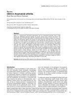
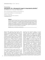
and bone resorption [33]. The schematic diagram in Figure 1
summarizes many of the cell-cell and cell-cytokine interactions
that have been implicated in RA and arthritis animal models. In
the inductive phase, TCR binding to antigen/MHC on antigen-
presenting cells (APCs) is a critical first step for T-cell
activation and might form part of the basis for the importance
of MHC alleles that contain the shared epitope. However, it is
likely that the nature of accessory signals received from APCs
and the local environment during TCR stimulation determines
the type of T-cell response and governs disease progression.
This section will focus on recent advances in our
understanding of accessory interactions between T cells,
APCs, and synovial cells.
Costimulation of naïve T cells through ligation of CD28 by
B7-1 (CD80) or B7-2 (CD86) is perhaps the most important
secondary signal to drive T-cell proliferation and
differentiation [34]. Once activated, the T cell upregulates
expression of cytotoxic T lymphocyte antigen-4 (CTLA-4), an
inhibitory receptor that has a higher affinity for CD80 and
CD86, in order to modulate activation. Use of a CTLA-4-
immunoglobulin fusion protein, which blocks the interaction
of CD28 with B7 ligands, has yielded promising results as a
treatment for RA and demonstrates the importance of this
cell-cell interaction in immune-mediated disease [35].
Other CD28/B7 family interactions have been shown to
mediate important interactions between T cells and other
Available online />cells involved in RA pathogenesis. Inducible costimulator
(ICOS) (CD278) is more highly expressed on activated
T cells found in patients with RA than in healthy individuals
[36]. The ligand for ICOS, CD275, is expressed by
professional APCs and has been shown to be expressed in
synovial tissue [36,37]. Blockade of CD275/CD278 binding
by antibodies was shown to diminish proinflammatory
cytokine production, autoantibody formation, and inflam-
mation in the CIA model [37]. Another CD28 family member,
OX40 (CD134), was shown to be inducible on T cells by
Arthritis Research & Therapy Vol 9 No 1 Lundy et al.
Page 4 of 11
(page number not for citation purposes)
Figure 1
Schematic diagram of the putative interactions of pathogenic Th17 cells in the synovial microenvironment. Induction of T-cell responses in
rheumatoid arthritis (RA) is initiated by T-cell receptor (TCR) interaction with shared epitope major histocompatibility complex class II (MHCII-SE)
and peptide on antigen-presenting cells (APCs) either systemically or in the synovium. Accessory molecules expressed by APCs, including ICAM-1
(intercellular adhesion molecule-1) (CD54), OX40L (CD252), inducible costimulator (ICOS) ligand (CD275), B7-1 (CD80), and B7-2 (CD86),
participate in T-cell activation by binding lymphocyte function-associated antigen (LFA)-1 (CD11a/CD18), OX40 (CD134), ICOS (CD278), and
CD28. Activated fibroblast-like synoviocytes (FLS) may also participate in antigen presentation and have additional accessory molecules such as
LFA-3 (CD58) and ALCAM (activated leukocyte cell adhesion molecule) (CD166) which interact with T cell-expressed CD2 and CD6, respectively.
Cytokines interleukin (IL)-6 and transforming growth factor-beta (TGF-β), most likely derived from activated APCs, signal the T cell to differentiate
into IL-17-producing Th17 cells. IL-17 has independent and synergistic effects with other proinflammatory cytokines (tumor necrosis factor-alpha
[TNF-α] and IL-1β) in the synovium to induce further cytokine release, matrix metalloproteinase production, RANK/RANK ligand (CD265/CD254)
expression, and osteoclastogenesis. CD40L (CD154) interaction with CD40 also leads to activation of synovial monocytes/macrophages
(Mo/Mac), FLS, and B cells. Although present in the synovia of most patients with RA, CD4
+
CD25
hi
regulatory T (Treg) cells are ineffective at
controlling inflammation and may be deactivated by synovial TNF-α. IL-10 is abundant in synovial fluid but its effect on Th17 regulation has yet to
be determined. Expression of accessory molecules on Th17 cells, as denoted in the figure, are speculative and are inferred from expressions found
on non-subdivided T-cell populations in animal models. Further investigation is necessary to directly demonstrate expression of these structures on
the Th17 cell subset in human RA synovium. DC, dendritic cell; RANK, receptor activator of nuclear factor-kappa B.
TNF-α in the IL-1Ra-deficient mouse model, and blockade of
CD134/CD252 interaction protected mice from developing
spontaneous arthritis [25].
The integrin lymphocyte function-associated antigen (LFA)-1
(CD11a/CD18) is expressed on activated T cells and binds
to intercellular adhesion molecule-1 (ICAM-1) (CD54) found
on the surface of many cell types. Previously, it was thought
that the main role of LFA-1/CD54 in inflammation had to do
with lymphocyte homing given the importance of this
adhesion axis in tight binding of lymphocytes to blood vessel
walls and their subsequent extravasation. However, the
cellular translocation of LFA-1 and CD54 to points of contact
between T cells and APCs also suggests an important role in
maintaining cell-cell contact during antigen presentation. In
rodent models of arthritis, disruption of the LFA-1/CD54
interaction has consistently decreased the severity of
inflammation, results that have led to the testing of LFA-
1/CD54 blockade for treatment of RA in clinical trials [38,39].
In addition to antigen presentation mediated by professional
APCs, our laboratory has been studying the ability of
activated fibroblast-like synoviocytes (FLS) to present anti-
gens to T cells. FLS, T cells, and macrophage-like synovio-
cytes are the three most abundant cell types in RA synovia.
Interferon-gamma (IFN-γ)-treated FLS lines from patients with
RA express MHC class II molecules and are able to stimulate
T cells to respond to superantigens in vitro [40]. We have
recently extended these observations by stimulating IL-2
production from T-cell hybridoma lines that are HLA-
DRB1*0401-restricted and arthritogenic peptide-specific
[41]. FLS lines do not express CD80 or CD86 but do
express other molecules that have receptors or ligands on T
cells, including ICAM-1 (CD54), VCAM-1 (vascular cell
adhesion molecule-1) (CD106), CD40, LFA-3 (CD58),
ALCAM (activated leukocyte cell adhesion molecule)
(CD166), and a novel CD6 ligand termed 3A11 [38,42]. At
the cell surface, FLS also express fractalkine, which has
recently been shown to be involved in activation of
CD28
null
CD4
+
‘senescent’ T cells from patients with RA [43].
The relative importance of activated FLS versus professional
APCs in antigen presentation in RA synovium has not been
determined.
The effector functions of arthritogenic T cells are carried out
in the synovial lining and intra-articular space of the joints.
Upon activation, T cells upregulate surface expression of
CD40 ligand (CD154), which stimulates APCs through
interaction with CD40. In B cells, ligation of CD40 in
combination with cytokine activation stimulates antibody
synthesis and isotype switching. Ligation of CD40 also
induces CD80, CD86, and CD54, as well as production of
proinflammatory cytokines, including IL-6, IL-8, MIP-1 (macro-
phage inflammatory protein-1), TNF-α, and IL-12, by APCs
[44,45]. These cytokines are known to participate in joint
inflammation and act reciprocally on T cells to drive
production of other cytokines and surface molecules involved
in the effector phase of joint inflammation.
The population of effector T cells in the joint may not be
limited to antigen-stimulated T cells. Brennan et al. [46] found
that TNF-α production in RA synovia was T cell-dependent
and that synovial T cells from patients with RA were able to
stimulate TNF-α production from peripheral blood monocytes.
Interestingly, using blocking reagents for nuclear factor-kappa
B (NF-κB) and PI3 (phosphoinositol 3) kinase, it was found
that the RA synovial T cells more closely resembled normal
T cells activated by IL-2, IL-6, and TNF-α than T cells
activated through the TCR. These data suggest that these
‘cytokine-activated’ bystander T cells (Tck) in the cytokine
milieu of the joint may become non-specifically activated and
contribute to joint pathology.
Another important effector function of synovial T cells involves
upregulation of receptor activator of NF-κB (RANK) ligand
(CD254) on the cell surface [47]. CD254
+
T cells interact
with synovial monocytes, leading to osteoclast differentiation.
These monocyte-derived osteoclasts then mediate bone
destruction.
T cells require many receptor-ligand interactions to become
activated and to carry out their tissue-destructive role in RA.
Disruption or modification of these cell-cell interactions may
prove to be an effective strategy for treatment of RA. Recent
data regarding the abilities of methotrexate and leflunomide
to interrupt T-cell interactions with FLS and APCs may
partially explain the efficacy of these medications and
highlight the importance of cell-cell contact to the patho-
genesis of RA [48-50].
A novel T-cell subset that secretes IL-17:
relevance to RA
Until recently, T-cell responses were typically classified as
either Th1 or Th2, based on the relative expression levels of
cytokines, especially IFN-γ and IL-4. Although neither Th1 nor
Th2 cytokines are present at high levels in the RA joint, IFN-γ
consistently predominated over IL-4 and RA had been viewed
as a Th1 disease. Recent evidence from mouse models has
questioned the role of Th1 cells in RA and identified a new
T-helper subset, Th17, with effector functions that make it a
prime candidate for mediating joint pathology. Th17 cells are
characterized by production of the highly inflammatory
cytokine IL-17. The first evidence of the inflammatory role of
IL-17 arose 10 years ago, when Fossiez and colleagues [51]
cloned human IL-17 from activated memory T cells and
showed that adding IL-17 to primary cultures of human RA
synovial fibroblasts induced expression of IL-6, IL-8,
prostaglandin E
2
, and G-CSF (granulocyte colony stimulating
factor). Furthermore, IL-17 synergized with TNF-α to induce
high levels of IL-6 and GM-CSF (granulocyte-monocyte
colony stimulating factor). Since then, the effects of IL-17
have been widely studied, resulting in a remarkable list of
Available online />Page 5 of 11
(page number not for citation purposes)
target cell types and downstream inflammatory mediators
relevant to RA. Table 1 summarizes the IL-17-inducible
factors produced by cell types relevant to RA synovium
[51-60]. The downstream activities of these factors
contribute to pathology through recruitment and activation of
inflammatory cells, positive feedback of the IL-17 response,
and destruction of tissue and bone.
One of the reasons that IL-17 can play an important role in
the pathogenesis of multiple autoimmune and inflammatory
diseases is the ubiquitous expression of the IL-17 receptor.
IL-17 directly and indirectly augments both inflammatory
mediator production and joint destruction. Early reports
suggested that IL-17 had little effect on its own and acted
primarily in synergy with IL-1β and TNF-α, but it is now known
that IL-17 can be pathogenic independently of IL-1β and
TNF-α. Although IL-17-induced TNF-α, IL-1β, and IL-6 can
induce cartilage destruction and bone erosion, IL-17 itself
has independent effects on cartilage and bone. IL-17
upregulates CD265 (RANK ligand) expression on chondro-
cytes and osteoblasts and acts on chondrocyte metabolism
by reducing proteoglycan synthesis and enhancing cartilage
degradation [52,53]. IL-17 enhances matrix breakdown,
cartilage proteoglycan depletion, chondrocyte death, and
cartilage and bone erosion in mice even under TNF- or IL-1-
neutralizing conditions [52,53,61]. These results suggest that
treatments designed to block IL-17 would not be redundant
with treatments that block TNF-α or IL-1β and that
combination therapy could be beneficial, especially for
patients who do not respond to TNF blockade. In cultures of
RA synovium, combining TNF blockade with agents that
block IL-1 and IL-17 was more effective at controlling IL-6
production and collagen degradation than blocking TNF-α
alone [62]. Similarly, combination blockade of TNF-α and
IL-17 suppressed ongoing CIA and was more effective than
neutralization of TNF-α alone [53].
Clearly, IL-17 has the ability to induce inflammation and joint
destruction when administered in vitro and in vivo in animal
models, but the question remains: how relevant is it to RA?
IL-17 is found in RA synovial fluid and in the T cell-rich area of
RA synovial tissue [52,53]. In addition, IL-17 is over-
expressed in serum and activated peripheral blood mono-
nuclear cell (PBMC) cultures of patients with RA compared
to healthy controls [45,63]. Experiments in multiple animal
models of arthritis demonstrate a requirement for IL-17 at
both early and late stages for full disease development. Both
the incidence and severity of arthritis were significantly
Arthritis Research & Therapy Vol 9 No 1 Lundy et al.
Page 6 of 11
(page number not for citation purposes)
Table 1
Effector molecules induced by IL-17 from human cells
Molecule produced Cell source Major functional effects Reference
IL-1β FLS, Mono/Mac, Chond/OC Inflammation, fever, synergy with IL-17 [52,53,55,58]
IL-6 FLS, Mono/Mac, Chond/OC Acute-phase reaction, B-cell stimulation, Th17 differentiation [51-53,58]
IL-23 FLS Inflammation, Th 17 cell stimulation [56]
TNF-α Mono/Mac Inflammation, synergy with IL-17 [52,53]
CXCL1 (GRO-α) FLS, Chond/OC Leukocyte recruitment [55,60]
CXCL5 (LIX) Chond/OC Leukocyte recruitment [60]
CXCL8 (IL-8) FLS Leukocyte recruitment [51-53,55]
CCL2 (MCP-1) FLS, Chond/OC Leukocyte recruitment [55,60]
CCL20 (MIP-3α) FLS Leukocyte recruitment [54]
G-CSF FLS Granulopoiesis [51]
GM-CSF FLS Granulopoiesis [51-53]
VEGF FLS, Chond/OC Angiogenesis [57,59]
Cyclooxygenase-2 FLS, Mono/Mac, Chond/OC Inflammation [52,58]
Prostaglandin E
2
FLS, Mono/Mac, Chond/OC Inflammation [51-53]
RANK/RANK ligand Chond/OC Osteoclastogenesis and bone resorption [53]
Nitric oxide Chond/OC Tissue destruction [52,53,58]
Matrix metalloproteinases Mono/Mac, Chond/OC Cartilage and tissue destruction [53,60]
Chond/OC, chondrocytes and osteoclasts; FLS, fibroblast-like synoviocytes; G-CSF, granulocyte colony stimulating factor; GM-CSF, granulocyte-
monocyte colony stimulating factor; GRO, growth-regulated oncogene; IL, interleukin; MCP, monocyte chemotactic protein; MIP-3α, macrophage
inflammatory protein-3 alpha; Mono/Mac, monocytes and macrophages; RANK, receptor activator of nuclear factor-kappa B; TNF-α, tumor
necrosis factor-alpha; VEGF, vascular endothelial growth factor.
reduced in mice deficient in IL-17 or IL-17 receptor during
CIA and streptococcal cell wall arthritis [53,64]. In addition,
spontaneous arthritis in IL-1Ra-deficient mice was completely
blocked in the absence of IL-17 [53]. Several groups have
also demonstrated that administering blocking antibody or
soluble IL-17 receptor during either the induction or effector
phase of experimental arthritis reduced inflammation and joint
destruction [53]. Recent evidence suggests a similar corre-
lation between IL-17 expression and joint damage progression
in patients with RA. Using several different methods of
statistical analysis, a 2-year prospective study of 50 patients
with RA found that synovial membrane mRNA levels of IL-1β,
TNF-α, IL-10, and IL-17 were consistently predictive of
damage progression [65]. Moreover, IL-17 and TNF-α mRNA
levels were synergistic as prognostic factors. This study
provides important clinical corroboration of observations
regarding the role of IL-17 in animal models of arthritis.
These and other studies have built a strong case that IL-17 is
a key suspect in the pathogenesis of RA: it is overexpressed
in RA synovium and blood, it induces and synergizes with
many inflammatory mediators important in joint pathology, and
it is both necessary and sufficient for joint inflammation in
animal models. The Th1-type response mediated by IFN-γ, on
the other hand, may have been falsely implicated. In CIA,
knocking out the IFN-γ receptor or IL-12 actually exacerbated
disease [53,66]. The protective role of IL-12 and IFN-γ is
likely to stem from the ability of Th1 and Th2 cytokines to
inhibit Th17 development. In addition, the same prospective
study that found IL-17 to be predictive of joint damage
progression in patients with RA also found that IFN-γ in the
synovium was predictive of protection [65]. IFN-γ may in fact
play dual roles, supporting inflammation in the early phase of
the disease and inhibiting inflammation later. Administration of
neutralizing anti-IFN-γ antibodies early in an experimental
arthritis model was protective, whereas later administration
exacerbated disease [67]. As evidenced by the IFN-γ
dependence of the proteoglycan-induced arthritis model, the
relative importance of IL-17 or IFN-γ may also depend on the
method used to induce disease [67]. This leaves open the
possibility that RA may not be as dependent on IL-17 as in
mouse models or that distinct subsets of patients with RA
may have different cytokine dependencies. These discrepan-
cies will hopefully be resolved by continuing research into the
role of human IL-17 in RA.
One of the important questions about IL-17 is the nature of
the stimuli that cause IL-17 to be produced. Early studies
found that IL-17 production by CD4
+
effector and memory
T cells was augmented by IL-23, a heterodimeric cytokine
composed of the IL-12 p40 subunit and a unique p19 subunit
[52,53,68]. IL-23 stimulation of activated murine T cells
induced production of IL-17, IL-17F (a close relative of IL-17),
IL-6, TNF-α, and low levels of IFN-γ. In human T cells, IL-23
induced production of IL-17 and low levels of IL-10 and IFN-γ
[69]. Notably, IL-23 has been found in RA synovial fluid and is
produced by FLS [56]. Remarkably, three groups
concurrently identified the combination of TGF-β and IL-6 as
key initiators of Th17 differentiation in murine T-cell cultures
[70-72]. IL-23 did not play a role in Th17 differentiation but
did appear to be important for Th17 survival and expansion.
The newly discovered role of TGF-β in the differentiation of a
highly inflammatory T-cell subset seems paradoxical given
that TGF-β is conventionally regarded as anti-inflammatory.
However, there is also a documented role for TGF-β in
exacerbating inflammatory responses and promoting
autoimmunity. In rat models of arthritis, injection of TGF-β into
the joint results in enhanced neutrophil recruitment, synovial
inflammation, and hyperplasia, whereas injection of blocking
antibody to TGF-β inhibits acute and chronic synovial
inflammation [73]. The divergent effects of TGF-β may
depend on systemic versus local expression and on the
cytokine milieu. For example, in vitro stimulation of naïve
CD4
+
T cells with TGF-β induces a regulatory phenotype, but
when IL-6 is added to these cultures, T-cell differentiation is
skewed toward Th17 instead [72]. Although many of these
observations have not yet been confirmed in humans, they
emphasize the potential importance and mechanisms of
action of IL-6 blocking therapeutic strategies that are
currently under evaluation in several rheumatologic diseases.
Moreover, much of the current understanding of the role of
TGF-β in arthritis and its suitability as a target in any immune-
mediated disease needs to be re-evaluated in the context of
what we now know about IL-17.
In addition to the effects of TGF-β, IL-6, and IL-23, Stockinger
and colleagues [70] found that IL-17 could be upregulated by
IL-1β and TNF-α. These recent reports provide a clear
explanation of how Th17 cells might differentiate and expand
within the joint: TGF-β, IL-6, IL-23, IL-1β, and TNF-α are all
found in RA synovium [52,53,56,73]. Upregulation of IL-17
by IL-6, IL-1β, and TNF-α, all of which are induced by IL-17,
also creates a positive feedback loop. Thus, one can imagine
how initially minor acute inflammation, in the right
microenvironment and cytokine milieu, might escalate and
ultimately lead to self-perpetuating chronic inflammation
through IL-17-dependent pathways.
Treg cells in RA
Treg cells have become a major focus of immunologic
research in the past decade due to their participation in
controlling effector T-cell functions in vitro and to their
potential for regulating autoimmune inflammatory responses
in vivo [74]. Several phenotypically distinct subsets of CD4
+
T cells constitute the Treg cell repertoire, but some markers
such as forkhead box p3 (Foxp3), neuropilin, LAG3
(lymphocyte activation gene 3) (CD223), CD103, and high
surface expression of CD25 have emerged as specific
markers of Treg cells [75-77]. Treg cells also produce high
levels of TGF-β and IL-10 [78]. The precise mechanisms of
suppression mediated by Treg cells are not fully understood.
It is possible that Treg cells suppress immunological
Available online />Page 7 of 11
(page number not for citation purposes)
responses in multiple ways, which may involve negative
signals produced by inhibitory surface molecules, cytotoxic
killing, downregulation of APC function, and/or induction of
other regulatory cells.
Some studies have been performed to evaluate the role of
Treg cells in RA, and there is controversy regarding the
relative number and function of CD4
+
CD25
+
Treg cells in RA
[79,80]. Treg cells have been identified in peripheral blood
and synovial tissue of patients with RA [80-82]. However,
most studies have shown that the CD4
+
CD25
+
Treg cells
from patients with RA have a defect in suppression of TNF-α
and IFN-γ production from CD4
+
T cells or monocytes, even
though they can suppress the proliferation of effector T cells
[81,83]. In other studies, it has been shown that effector
T cells from peripheral blood of patients with RA were
resistant to Treg-mediated suppression [84]. CD4
+
CD25
+
cells express TNF receptor 2, and signaling through this
receptor by TNF-α results in inhibition of suppressive function
and decreased Foxp3 expression [85]. Treatment of RA
patients with anti-TNF-α antibody leads to in vivo expansion
of CD4
+
CD25
+
Treg cells, increased Foxp3 expression, and
restoration of cytokine suppressive function [81,85]. Inter-
estingly, one study showed that PBMCs of MHC-shared
epitope-positive, healthy individuals responded to the
arthritis-associated autoantigen, HCgp39 (human cartilage
glycoprotein of 39 kDa), by producing IL-10 whereas PBMCs
of patients with RA tended to produce proinflammatory
cytokines [86]. IL-10 production was attributed to Treg cells,
which suggests that an important difference between healthy
people and patients with RA is the ability to expand Treg cells
specific for autoantigens.
The role of Treg cells has also been studied in the mouse CIA
model. CD4
+
CD25
+
cells are important in controlling the
pathogenesis of CIA, and depletion of CD25
+
cells with anti-
CD25 antibody led to aggravation of joint inflammation
[87,88]. Adoptive transfer of CD4
+
CD25
+
Treg cells during
the initiation phase of arthritis resulted in decreased severity of
disease, whereas the course of established arthritis remained
unchanged [87,89,90]. Recent studies have demonstrated
the ability to influence Treg development in vivo using oral
collagen, immature dendritic cells, or vasoactive intestinal
peptide as potential therapeutic agents to treat CIA [91-93].
Activation or reactivation of Treg cells in patients holds
promise as a potential treatment for RA, but uncertainty
persists concerning the effectiveness and durability of such
strategies and the best way to mechanistically approach the in
vivo manipulation of Treg cells.
Conclusion
Integration of the many compelling lines of evidence
regarding the roles of T cells in RA remains a challenge for
current and future research. A full description of the genetic
loci that control RA will be essential, including further
analyses in non-Caucasian populations, to provide a more
comprehensive view of this complex disease and increased
predictive and prognostic power. Once the interactions of
T cells with other synovial cell populations are better
understood, proof of their importance may ultimately be
confirmed by application of new biologic therapies to the
treatment of RA. Recent breakthroughs in the delineation of
the novel Th17 subset have come primarily through animal
studies, and data are urgently needed to assess which
principles also apply to the human immune system and
human disease. At the present time, it appears likely that the
Th17 cells and their product, IL-17, will be attractive targets
for therapy of RA and other immune-mediated human
diseases. This therapy may come in the form of neutralizing
inductive cytokines such as IL-6 and IL-23, blocking Th17-
specific costimulatory signals, disruption of IL-17 signaling
cascades, or direct targeting of antigen-specific Th17 cells
for elimination. An interesting question is whether Treg cells
can be mobilized to specifically regulate Th17-driven
pathology. Progress in all of these areas should move us
closer to the goal of re-establishing long-term physiologic
regulation of immune responses in RA, using well-tolerated
and more specifically targeted interventions.
Competing interests
The authors declare that they have no competing interests.
Acknowledgements
SKL was supported by the Edward T and Ellen K Dryer Charitable
Foundation and by the Michigan Technology Transfer Core grant
program. SS was supported by an Arthritis Foundation Post-Doctoral
Fellowship. LAT was supported by a National Institutes of Health (NIH)
Experimental Biology Predoctoral Training grant. DAF was supported
by NIH grant AR38477.
References
1. Fox DA: T lymphocytes in rheumatoid arthritis. In Rheumatoid
Arthritis. Edited by Firestein GS, Panayi GS, Wollheim FA. New
York, NY: Oxford University Press; 2006:77-94.
2. de Vries N, Tijssen H, van Riel PL, van de Putte LB: Reshaping
the shared epitope hypothesis: HLA-associated risk for
rheumatoid arthritis is encoded by amino acid substitutions at
positions 67-74 of the HLA-DRB1 molecule. Arthritis Rheum
2002, 46:921-928.
3. Gourraud PA, Boyer JF, Barnetche T, Abbal M, Cambon-Thomsen
A, Cantagrel A, Constantin A: A new classification of HLA-DRB1
alleles differentiates predisposing and protective alleles for
rheumatoid arthritis structural severity. Arthritis Rheum 2006,
54:593-599.
4. du Montcel ST, Michou L, Petit-Teixeira E, Osorio J, Lemaire I,
Lasbleiz S, Pierlot C, Quillet P, Bardin T, Prum B, et al.: New clas-
sification of HLA-DRB1 alleles supports the shared epitope
hypothesis of rheumatoid arthritis susceptibility. Arthritis
Rheum 2005, 52:1063-1068.
Arthritis Research & Therapy Vol 9 No 1 Lundy et al.
Page 8 of 11
(page number not for citation purposes)
This review is part of a series on
Cells of the synovium in rheumatoid arthritis
edited by Gary Firestein.
Other articles in this series can be found at
/>review-series.asp?series=ar_Cells
5. Padyukov L, Silva C, Stolt P, Alfredsson L, Klareskog L: A gene-
environment interaction between smoking and shared
epitope genes in HLA-DR provides a high risk of seropositive
rheumatoid arthritis. Arthritis Rheum 2004, 50:3085-3092.
6. Auger I, Sebbag M, Vincent C, Balandraud N, Guis S, Nogueira L,
Svensson B, Cantagrel A, Serre G, Roudier J: Influence of HLA-
DR genes on the production of rheumatoid arthritis-specific
autoantibodies to citrullinated fibrinogen. Arthritis Rheum
2005, 52:3424-3432.
7. Huizinga TW, Amos CI, van der Helm-van Mil AH, Chen W, van
Gaalen FA, Jawaheer D, Schreuder GM, Wener M, Breedveld FC,
Ahmad N, et al.: Refining the complex rheumatoid arthritis
phenotype based on specificity of the HLA-DRB1 shared
epitope for antibodies to citrullinated proteins. Arthritis Rheum
2005, 52:3433-3438.
8. van der Helm-van Mil AH, Verpoort KN, Breedveld FC, Huizinga
TW, Toes RE, de Vries RR: The HLA-DRB1 shared epitope
alleles are primarily a risk factor for anti-cyclic citrullinated
peptide antibodies and are not an independent risk factor for
development of rheumatoid arthritis. Arthritis Rheum 2006,
54:1117-1121.
9. van Oene M, Wintle RF, Liu X, Yazdanpanah M, Gu X, Newman B,
Kwan A, Johnson B, Owen J, Greer W, et al.: Association of the
lymphoid tyrosine phosphatase R620W variant with rheuma-
toid arthritis, but not Crohn’s disease, in Canadian popula-
tions. Arthritis Rheum 2005, 52:1993-1998.
10. Pierer M, Kaltenhauser S, Arnold S, Wahle M, Baerwald C,
Hantzschel H, Wagner U: Association of PTPN22 1858 single-
nucleotide polymorphism with rheumatoid arthritis in a
German cohort: higher frequency of the risk allele in male
compared to female patients. Arthritis Res Ther 2006, 8:R75-
R85.
11. Vang T, Congia M, Macis MD, Musumeci L, Orru V, Zavattari P,
Nika K, Tautz L, Tasken K, Cucca F, et al.: Autoimmune-associ-
ated lymphoid tyrosine phosphatase is a gain-of-function
variant. Nat Genet 2005, 37:1317-1319.
12. Burkhardt H, Huffmeier U, Spriewald B, Bohm B, Rau R, Kallert S,
Engstrom A, Holmdahl R, Reis A: Association between protein
tyrosine phosphatase 22 variant R620W in conjunction with
the HLA-DRB1 shared epitope and humoral autoimmunity to
an immunodominant epitope of cartilage-specific type II colla-
gen in early rheumatoid arthritis. Arthritis Rheum 2006, 54:82-
89.
13. Lin S-C, Yen J-H, Tsai J-J, Tsai W-C, Ou T-T, Liu H-W, Chen C-J:
Association of a programmed death 1 gene polymorphism
with the development of rheumatoid arthritis, but not sys-
temic lupus erythematosus. Arthritis Rheum 2004, 50:770-775.
14. Prokunina L, Padyukov L, Bennet A, de Faire U, Wiman B, Prince
J, Alfredsson L, Klareskog L, Alarcon-Riquelme M: Association of
the PD-1.3A allele of the PDCD1 gene in patients with
rheumatoid arthritis negative for rheumatoid factor and the
shared epitope. Arthritis Rheum 2004, 50:1770-1773.
15. Kong EK-P, Prokunina-Olsson L, Wong WH-S, Lau C-S, Chan T-
M, Alarcon-Riquelme M, Lau Y-L: A new haplotype of PDCD1 is
associated with rheumatoid arthritis in Hong Kong Chinese.
Arthritis Rheum 2005, 52:1058-1062.
16. Wan B, Nie H, Liu A, Feng G, He D, Xu R, Zhang Q, Dong C,
Zhang JZ: Aberrant regulation of synovial T cell activation by
soluble costimulatory molecules in rheumatoid arthritis. J
Immunol 2006, 177:8844-8850.
17. Prots I, Skapenko A, Wendler J, Mattyasovszky S, Yone CL,
Spriewald B, Burkhardt H, Rau R, Kalden JR, Lipsky PE, Schulze-
Koops H: Association of the IL4R single-nucleotide polymor-
phism I50V with rapidly erosive rheumatoid arthritis. Arthritis
Rheum 2006, 54:1491-1500.
18. Sakaguchi N, Takahashi T, Hata H, Nomura T, Tagami T, Yamazaki
S, Sakihama T, Matsutani T, Negishi I, Nakatsuru S, et al.: Altered
thymic T-cell selection due to a mutation of the ZAP-70 gene
causes autoimmune arthritis in mice. Nature 2003, 426:454-
460.
19. Hata H, Sakaguchi N, Yoshitomi H, Iwakura Y, Sekikawa K, Azuma
Y, Kanai C, Moriizumi E, Nomura T, Nakamura T, et al.: Distinct
contribution of IL-6, TNF-alpha, IL-1, and IL-10 to T cell-medi-
ated spontaneous autoimmune arthritis in mice. J Clin Invest
2004, 114:582-588.
20. Zhao W, Zhang L, Kobari Y, Misaki Y, Yamamoto K: Analysis of
accumulating clonotypes of T cell in joints of a spontaneous
murine model of rheumatoid arthritis. Cell Mol Immunol 2004,
1:300-303.
21. Yoshitomi H, Sakaguchi N, Kobayashi K, Brown GD, Tagami T,
Sakihama T, Hirota K, Tanaka S, Nomura T, Miki I, et al.: A role for
fungal b-glucans and their receptor Dectin-1 in the induction
of autoimmune arthritis in genetically susceptible mice. J Exp
Med 2005, 201:949-960.
22. Kobayashi K, Suda T, Nan-ya K, Sakaguchi N, Sakaguchi S, Miki I:
Cytokine production profile of splenocytes derived from
zymosan A-treated SKG mice developing arthritis. Inflamm
Res 2006, 55:335-341.
23 Hirota K, Hashimoto M, Yoshitomi H, Tanaka S, Nomura T, Yam-
aguchi T, Iwakura Y, Sakaguchi N, Sakaguchi S: T cell self-reac-
tivity forms a cytokine milieu for spontaneous development of
IL-17+ Th cells that cause autoimmune arthritis. J Exp Med
2007, 204:41-47.
24. Horai R, Saijo S, Tanioka H, Nakae S, Sudo K, Okahara A, Ikuse T,
Asano M, Iwakura Y: Development of chronic inflammatory
arthropathy resembling rheumatoid arthritis in interleukin 1
receptor antagonist-deficient mice. J Exp Med 2000, 191:313-
320.
25. Horai R, Nakajima A, Habiro K, Kotani M, Nakae S, Matsuki T,
Nambu A, Saijo S, Kotaki H, Sudo K, et al.: TNF-alpha is crucial
for the development of autoimmune arthritis in IL-1 receptor
antagonist-deficient mice. J Clin Invest 2004, 114:1603-1611.
26. Nakae S, Saijo S, Horai R, Sudo K, Mori S, Iwakura Y: IL-17 pro-
duction from activated T cells is required for the spontaneous
development of destructive arthritis in mice deficient in IL-1
receptor antagonist. Proc Natl Acad Sci U S A 2003, 100:
5986-5990.
27. Atsumi T, Ishihara K, Kamimura D, Ikushima H, Ohtani T, Hirota S,
Kobayashi H, Park S-J, Saeki Y, Kitamura Y, et al.: A point muta-
tion of Tyr-759 in interleukin 6 family cytokine receptor
subunit gp130 causes autoimmune arthritis. J Exp Med 2002,
196:979-990.
28. Sawa S, Kamimura D, Jin G-H, Morikawa H, Kamon H, Nishihara
M, Ishihara K, Murakami M, Hirano T: Autoimmune arthritis
associated with mutated interleukin (IL)-6 receptor gp130
driven by STAT3/IL-7-dependent homeostatic proliferation of
CD4
+
T cells. J Exp Med 2006, 203:1459-1470.
29. Kouskoff V, Korganow AS, Duchatelle V, Degott C, Benoist C,
Mathis D: Organ-specific disease provoked by systemic
autoimmunity. Cell 1996, 87:811-822.
30. Maccioni M, Zeder-Lutz G, Huang H, Ebel C, Gerber P, Hergueux
J, Marchal P, Duchatelle V, Degott C, van Regenmortel M, et al.:
Arthritogenic monoclonal antibodies from K/BxN mice. J Exp
Med 2002, 195:1071-1077.
31. Kyburz D, Corr M: The KRN mouse model of inflammatory
arthritis. Springer Semin Immunopathol 2003, 25:79-90.
32. Schubert D, Maier B, Morawietz L, Krenn V, Kamradt T: Immu-
nization with glucose-6-phosphate isomerase induces T cell-
dependent peripheral polyarthritis in genetically unaltered
mice. J Immunol 2004, 172:4503-4509.
33. Tran CN, Lundy SK, Fox DA: Synovial biology and T cells in
rheumatoid arthritis. Pathophysiology 2005, 12:183-189.
34. Kotani M, Hirata K, Ogawa S, Habiro K, Ishida Y, Tanuma S, Horai
R, Iwakura Y, Kishimoto H, Abe R: CD28-dependent differentia-
tion into the effector/memory phenotype is essential for
induction of arthritis in interleukin-1 receptor antagonist-defi-
cient mice. Arthritis Rheum 2006, 54:473-481.
35. Malmstrom V, Trollmo C, Klareskog L: Modulating co-stimula-
tion: a rational strategy in the treatment of rheumatoid arthri-
tis? Arthritis Res Ther 2005, 7 Suppl 2:S15-S20.
36. Okamoto T, Saito S, Yamanaka H, Tomatsu T, Kamatani N,
Ogiuchi H, Uchiyama T, Yagi J: Expression and function of the
co-stimulator H4/ICOS on activated T cells of patients with
rheumatoid arthritis. J Rheumatol 2003, 30:1157-1163.
37. Iwai H, Kozono Y, Hirose S, Akiba H, Yagita H, Okumura K,
Kohsaka H, Miyasaka N, Azuma M: Amelioration of collagen-
induced arthritis by blockade of inducible costimulator-B7
homologous protein costimulation. J Immunol 2002, 169:
4332-4339.
38. Agarwal SK, Brenner MB: Role of adhesion molecules in syn-
ovial inflammation. Curr Opin Rheumatol 2006, 18:268-276.
39. Schulze-Koops H, Lipsky PE, Kavanaugh AF, Davis LS: Persis-
tent reduction in IL-6 mRNA in peripheral blood mononuclear
cells of patients with rheumatoid arthritis after treatment with
Available online />Page 9 of 11
(page number not for citation purposes)
a monoclonal antibody to CD54 (ICAM-1). Clin Exp Immunol
1996, 106:190-196.
40. Tsai C, Diaz LA Jr., Singer NG, Li LL, Kirsch AH, Mitra R, Nickoloff
BJ, Crofford LJ, Fox DA: Responsiveness of human T lympho-
cytes to bacterial superantigens presented by cultured
rheumatoid arthritis synoviocytes. Arthritis Rheum 1996, 39:
125-136.
41 Tran CN, Davis MJ, Tesmer LA, Endres JL, Motyl CD, Smuda C,
Somers EC, Chung KC, Urquhart AG, Lundy SK, Kovats S, Fox
DA: Presentation of arthritogenic peptide to antigen-specific T
cells by fibroblast-like synoviocytes. Arthritis Rheum, in press.
42. Saifullah MK, Fox DA, Sarkar S, Abidi SM, Endres J, Piktel J,
Haqqi TM, Singer NG: Expression and characterization of a
novel CD6 ligand in cells derived from joint and epithelial
tissues. J Immunol 2004, 173:6125-6133.
43. Sawai H, Park YW, Roberson J, Imai T, Goronzy JJ, Weyand CM:
T cell costimulation by fractalkine-expressing synoviocytes in
rheumatoid arthritis. Arthritis Rheum 2005, 52:1392-1401.
44. Aarvak T, Natvig JB: Cell-cell interactions in synovitis: antigen
presenting cells and T cell interaction in rheumatoid arthritis.
Arthritis Res 2001, 3:13-17.
45. Cho ML, Yoon CH, Hwang SY, Park MK, Min SY, Lee SH, Park
SH, Kim HY: Effector function of type II collagen-stimulated T
cells from rheumatoid arthritis patients: cross-talk between T
cells and synovial fibroblasts. Arthritis Rheum 2004, 50:776-
784.
46. Brennan FM, Hayes AL, Ciesielski CJ, Green P, Foxwell BM, Feld-
mann M: Evidence that rheumatoid arthritis synovial T cells
are similar to cytokine-activated T cells: involvement of phos-
phatidylinositol 3-kinase and nuclear factor kappaB pathways
in tumor necrosis factor alpha production in rheumatoid
arthritis. Arthritis Rheum 2002, 46:31-41.
47. Kotake S, Nanke Y, Mogi M, Kawamoto M, Furuya T, Yago T,
Kobashigawa T, Togari A, Kamatani N: IFN-gamma-producing
human T cells directly induce osteoclastogenesis from
human monocytes via the expression of RANKL. Eur J
Immunol 2005, 35:3353-3363.
48. Johnston A, Gudjonsson JE, Sigmundsdottir H, Ludviksson BR,
Valdimarsson H: The anti-inflammatory action of methotrexate
is not mediated by lymphocyte apoptosis, but by the suppres-
sion of activation and adhesion molecules. Clin Immunol
2005, 114:154-163.
49. Miranda-Carus ME, Balsa A, Benito-Miguel M, Perez de Ayala C,
Martin-Mola E: IL-15 and the initiation of cell contact-depen-
dent synovial fibroblast-T lymphocyte cross-talk in rheuma-
toid arthritis: effect of methotrexate. J Immunol 2004, 173:
1463-1476.
50. Zeyda M, Poglitsch M, Geyeregger R, Smolen JS, Zlabinger GJ,
Horl WH, Waldhausl W, Stulnig TM, Saemann MD: Disruption of
the interaction of T cells with antigen-presenting cells by the
active leflunomide metabolite teriflunomide: involvement of
impaired integrin activation and immunologic synapse forma-
tion. Arthritis Rheum 2005, 52:2730-2739.
51. Fossiez F, Djossou O, Chomarat P, Flores-Romo L, Ait-Yahia S,
Maat C, Pin JJ, Garrone P, Garcia E, Saeland S, et al.: T cell
interleukin-17 induces stromal cells to produce proinflamma-
tory and hematopoietic cytokines. J Exp Med 1996, 183:2593-
2603.
52. Stamp LK, James MJ, Cleland LG: Interleukin-17: the missing
link between T-cell accumulation and effector cell actions in
rheumatoid arthritis? Immunol Cell Biol 2004, 82:1-9.
53. Lubberts E, Koenders MI, van den Berg WB: The role of T-cell
interleukin-17 in conducting destructive arthritis: lessons
from animal models. Arthritis Res Ther 2005, 7:29-37.
54. Chabaud M, Page G, Miossec P: Enhancing effect of IL-1, IL-17,
and TNF-alpha on macrophage inflammatory protein-3alpha
production in rheumatoid arthritis: regulation by soluble
receptors and Th2 cytokines. J Immunol 2001, 167:6015-6020.
55. Honorati MC, Bovara M, Cattini L, Piacentini A, Facchini A: Con-
tribution of interleukin 17 to human cartilage degradation and
synovial inflammation in osteoarthritis. Osteoarthritis Cartilage
2002, 10:799-807.
56. Kim HR, Cho ML, Kim KW, Juhn JY, Hwang SY, Yoon CH, Park
SH, Lee SH, Kim HY: Up-regulation of IL-23p19 expression in
rheumatoid arthritis synovial fibroblasts by IL-17 through PI3-
kinase-, NF-{kappa}B- and p38 MAPK-dependent signalling
pathways. Rheumatology (Oxford). 2007, 46:57-64.
57. Honorati MC, Cattani L, Facchini A: IL-17, IL-1b and TNF-a stim-
ulate VEGF production by dedifferentiated chondrocytes.
Osteoarthritis Cartilage 2004, 12:683-691.
58. Shalom-Barak T, Quach J, Lotz M: Interleukin-17-induced gene
expression in articular chondrocytes is associated with activa-
tion of mitogen-activated protein kinases and NFkB. J Biol
Chem 1998, 273:27467-27473.
59. Ryu S, Lee JH, Kim SI: IL-17 increased the production of vascu-
lar endothelial growth factor in rheumatoid arthritis synovio-
cytes. Clin Rheum 2006, 25:16-20.
60. Gaffen SL, Kramer JM, Yu JJ, Shen F: The IL-17 cytokine family.
Vitam Horm 2006, 74:255-282.
61. Koenders MI, Lubberts E, van de Loo FA, Oppers-Walgreen B,
van den Bersselaar L, Helsen MM, Kolls JK, Di Padova FE,
Joosten LA, van den Berg WB: Interleukin-17 acts indepen-
dently of TNF-alpha under arthritic conditions. J Immunol
2006, 176:6262-6269.
62. Chabaud M, Miossec P: The combination of tumor necrosis
factor alpha blockade with interleukin-1 and interleukin-17
blockade is more effective for controlling synovial inflamma-
tion and bone resorption in an ex vivo model. Arthritis Rheum
2001, 44:1293-1303.
63. Kim KW, Cho ML, Park MK, Yoon CH, Park SH, Lee SH, Kim HY:
Increased interleukin-17 production via a phosphoinositide 3-
kinase/Akt and nuclear factor kappaB-dependent pathway in
patients with rheumatoid arthritis. Arthritis Res Ther 2005, 7:
R139-R148.
64. Nakae S, Nambu A, Sudo K, Iwakura Y: Suppression of immune
induction of collagen-induced arthritis in IL-17-deficient mice.
J Immunol 2003, 171:6173-6177.
65. Kirkham BW, Lassere MN, Edmonds JP, Juhasz KM, Bird PA, Lee
CS, Shnier R, Portek IJ: Synovial membrane cytokine expres-
sion is predictive of joint damage progression in rheumatoid
arthritis: a two-year prospective study (the DAMAGE study
cohort). Arthritis Rheum 2006, 54:1122-1131.
66. Vermeire K, Heremans H, Vandeputte M, Huang S, Billiau A,
Matthys P: Accelerated collagen-induced arthritis in IFN-
gamma receptor-deficient mice. J Immunol 1997, 158:5507-
5513.
67. Rosloniec EF, Latham K, Guedez YB: Paradoxical roles of IFN-
gamma in models of Th1-mediated autoimmunity. Arthritis
Res 2002, 4:333-336.
68. Langrish CL, Chen Y, Blumenschein WM, Mattson J, Basham B,
Sedgwick JD, McClanahan T, Kastelein RA, Cua DJ: IL-23 drives
a pathogenic T cell population that induces autoimmune
inflammation. J Exp Med 2005, 201:233-240.
69. van Eijnden S, Goriely S, De Wit D, Willems F, Goldman M: IL-23
up-regulates IL-10 and induces IL-17 synthesis by polyclon-
ally activated naïve T cells in human. Eur J Immunol 2005, 35:
469-475.
70. Veldhoen M, Hocking RJ, Atkins CJ, Locksley RM, Stockinger B:
TGFbeta in the context of an inflammatory cytokine milieu
supports de novo differentiation of IL-17-producing T cells.
Immunity 2006, 24:179-189.
71. Mangan PR, Harrington LE, O’Quinn DB, Helms WS, Bullard DC,
Elson CO, Hatton RD, Wahl SM, Schoeb TR, et al.: Transform-
ing growth factor-beta induces development of the T(H)17
lineage. Nature 2006, 441:231-234.
72. Bettelli E, Carrier Y, Gao W, Korn T, Strom TB, Oukka M, Weiner
HL, Kuchroo VK. Reciprocal developmental pathways for the
generation of pathogenic effector TH17 and regulatory T cells.
Nature 2006, 441:235-238.
73. Li MO, Wan YY, Sanjabi S, Robertson AK, Flavell RA: Transform-
ing growth factor-beta regulation of immune responses. Annu
Rev Immunol 2006, 24:99-146.
74. Leipe J, Skapenko A, Lipsky PE, Schulze-Koops H: Regulatory T
cells in rheumatoid arthritis. Arthritis Res Ther 2005, 7:93-99.
75. Piccirillo CA, Thornton AM: Cornerstone of peripheral toler-
ance: naturally occurring CD4
+
CD25
+
regulatory T cells.
Trends Immunol 2004, 25:374-380.
76. Bruder D, Probst-Kepper M, Westendorf AM, Geffers R, Beissert
S, Loser K, von Boehmer H, Buer J, Hansen W: Neuropilin-1: a
surface marker of regulatory T cells. Eur J Immunol 2004, 34:
623-630.
77. Huang CT, Workman CJ, Flies D, Pan X, Marson AL, Zhou G,
Hipkiss EL, Ravi S, Kowalski J, Levitsky HI, et al.: Role of LAG-3
in regulatory T cells. Immunity 2004, 21:503-513.
Arthritis Research & Therapy Vol 9 No 1 Lundy et al.
Page 10 of 11
(page number not for citation purposes)
78. Bluestone JA, Tang Q: How do CD4
+
CD25
+
regulatory T cells
control autoimmunity? Curr Opin Immunol 2005, 17:638-642.
79. Lawson CA, Brown AK, Bejarano V, Douglas SH, Burgoyne CH,
Greenstein AS, Boylston AW, Emery P, Ponchel F, Isaacs JD:
Early rheumatoid arthritis is associated with a deficit in the
CD4
+
CD25
high
regulatory T cell population in peripheral blood.
Rheumatology (Oxford) 2006, 45:1210-1217.
80. Mottonen M, Heikkinen J, Mustonen L, Isomaki P, Luukkainen R,
Lassila O: CD4
+
CD25
+
T cells with the phenotypic and func-
tional characteristics of regulatory T cells are enriched in the
synovial fluid of patients with rheumatoid arthritis. Clin Exp
Immunol 2005, 140:360-367.
81. Ehrenstein MR, Evans JG, Singh A, Moore S, Warnes G, Isenberg
DA, Mauri C: Compromised function of regulatory T cells in
rheumatoid arthritis and reversal by anti-TNFalpha therapy. J
Exp Med 2004, 200:277-285.
82. Cao D, Borjesson O, Larsson P, Rudin A, Gunnarsson I,
Klareskog L, Malmstrom V, Trollmo C: FOXP3 identifies regula-
tory CD25bright CD4
+
T cells in rheumatic joints. Scand J
Immunol 2006, 63:444-452.
83. Cao D, van Vollenhoven R, Klareskog L, Trollmo C, Malmstrom V:
CD25
bright
CD4
+
regulatory T cells are enriched in inflamed
joints of patients with chronic rheumatic disease. Arthritis Res
Ther 2004, 6:R335-R346.
84. van Amelsfort JM, Jacobs KM, Bijlsma JW, Lafeber FP, Taams LS:
CD4(+)CD25(+) regulatory T cells in rheumatoid arthritis: dif-
ferences in the presence, phenotype, and function between
peripheral blood and synovial fluid. Arthritis Rheum 2004, 50:
2775-2785.
85. Valencia X, Stephens G, Goldbach-Mansky R, Wilson M, Shevach
EM, Lipsky PE. TNF downmodulates the function of human
CD4
+
CD25
hi
T-regulatory cells. Blood 2006, 108:253-261.
86. van Bilsen JH, van Dongen H, Lard LR, van der Voort EI, Elferink
DG, Bakker AM, Miltenburg AM, Huizinga TW, de Vries RR, Toes
RE: Functional regulatory immune responses against human
cartilage glycoprotein-39 in health vs. proinflammatory
responses in rheumatoid arthritis. Proc Natl Acad Sci U S A
2004, 101:17180-17185.
87. Frey O, Petrow PK, Gajda M, Siegmund K, Huehn J, Scheffold A,
Hamann A, Radbruch A, Brauer R: The role of regulatory T cells
in antigen-induced arthritis: aggravation of arthritis after
depletion and amelioration after transfer of CD4
+
CD25
+
T
cells. Arthritis Res Ther 2005, 7:R291-R301.
88. Morgan ME, Sutmuller RP, Witteveen HJ, van Duivenvoorde LM,
Zanelli E, Melief CJ, Snijders A, Offringa R, de Vries RR, Toes RE:
CD25
+
cell depletion hastens the onset of severe disease in
collagen-induced arthritis. Arthritis Rheum 2003, 48:1452-
1460.
89. Morgan ME, Flierman R, van Duivenvoorde LM, Witteveen HJ, van
Ewijk W, van Laar JM, de Vries RR, Toes RE: Effective treatment
of collagen-induced arthritis by adoptive transfer of CD25
+
regulatory T cells. Arthritis Rheum 2005, 52:2212-2221.
90. Kelchtermans H, De Klerck B, Mitera T, Van Balen M, Bullens D,
Billiau A, Leclercq G, Matthys P: Defective CD4
+
CD25
+
regula-
tory T cell functioning in collagen-induced arthritis: an impor-
tant factor in pathogenesis, counter-regulated by endogenous
IFN-gamma. Arthritis Res Ther 2005, 7:R402-R415.
91. Charbonnier LM, van Duivenvoorde LM, Apparailly F, Cantos C,
Han WG, Noel D, Duperray C, Huizinga TW, Toes RE, Jorgensen
C, et al.: Immature dendritic cells suppress collagen-induced
arthritis by in vivo expansion of CD49b
+
regulatory T cells. J
Immunol 2006, 177:3806-3813.
92. Chorny A, Gonzalez-Rey E, Ganea D, Delgado M: Vasoactive
intestinal peptide generates CD4
+
CD25
+
regulatory T cells in
vivo: therapeutic applications in autoimmunity and transplan-
tation. Ann N Y Acad Sci 2006, 1070:190-195.
93. Min SY, Hwang SY, Park KS, Lee JS, Lee KE, Kim KW, Jung YO,
Koh HJ, Do JH, Kim H, et al.: Induction of IL-10-producing
CD4
+
CD25
+
T cells in animal model of collagen-induced
arthritis by oral administration of type II collagen. Arthritis Res
Ther 2004, 6:R213-R219.
Available online />Page 11 of 11
(page number not for citation purposes)