Báo cáo y học: "Biology of adult mesenchymal stem cells: regulation of niche, self-renewal and differentiation" potx
Bạn đang xem bản rút gọn của tài liệu. Xem và tải ngay bản đầy đủ của tài liệu tại đây (665.13 KB, 10 trang )
Page 1 of 10
(page number not for citation purposes)
Available online />Abstract
Recent advances in understanding the cellular and molecular
signaling pathways and global transcriptional regulators of adult
mesenchymal stem cells have provided new insights into their
biology and potential clinical applications, particularly for tissue
repair and regeneration. This review focuses on these advances,
specifically in the context of self-renewal and regulation of lineage-
specific differentiation of mesenchymal stem cells. In addition we
review recent research on the concept of stem cell niche, and its
relevance to adult mesenchymal stem cells.
Introduction
Since the seminal identification of mesenchymal stem cells
(MSCs) as colony-forming unit-fibroblasts (CFU-Fs) by
Friedenstein and colleagues in 1970 [1] and the first detailed
description of the tri-lineage potential of MSCs by Pittenger
and colleagues [2], our understanding of these unique cells
has taken great strides forward. MSCs have great appeal for
tissue engineering and therapeutic applications because of
their general multipotentiality and relative ease of isolation
from numerous tissues. This review highlights recent
discoveries in the areas of MSC self-renewal, differentiation,
and niche biology, and presents molecular signaling and
mechanistic models of MSC development.
MSC markers
Plastic-adherent multipotent cells, capable of differentiating
into bone, cartilage and fat cells (among others), can be
isolated from many adult tissue types. However, even if
isolated by density-gradient fractionation, they remain a
heterogeneous mixture of cells with varying proliferation and
differentiation potentials. Although acceptable for cell-based
therapeutic applications, a rigorous understanding of the
MSC requires a better definition of what an MSC is. Many
attempts have been made to develop a cell-surface antigen
profile for the better purification and identification of MSCs.
Particularly important is whether MSCs isolated from different
tissues are identifiable by the same immunophenotype. Table 1
provides information on 16 surface proteins reported in
various studies. Most of the studies focused on MSCs from
human and mouse bone marrow, but some examined MSCs
from other organs. There is a surprisingly small amount of
variation between populations, even among cells isolated
from different sources. It is also noteworthy that the mouse
bone marrow-derived multipotent adult progenitor cell
(MAPC) subpopulation [3], reported to have more differen-
tiation potential than the MSC population as a whole, does
not express specific, known surface markers.
Negative markers
There is a consensus that MSCs do not express CD11b (an
immune cell marker), glycophorin-A (an erythroid lineage
marker), or CD45 (a marker of all hematopoietic cells). CD34,
the primitive hematopoietic stem cell (HSC) marker, is rarely
expressed in human MSCs, although it is positive in mice.
CD31 (expressed on endothelial and hematopoietic cells)
and CD117 (a hematopoietic stem/progenitor cell marker)
are almost always absent from human and mouse MSCs.
Currently, the thorn in the side of the MSC biologist is the
lack of a definitive positive marker for MSCs; there is a myriad
Review
Mesenchymal stromal cells
Biology of adult mesenchymal stem cells:
regulation of niche, self-renewal and differentiation
Catherine M Kolf*, Elizabeth Cho* and Rocky S Tuan
Cartilage Biology and Orthopaedics Branch, National Institute of Arthritis, and Musculoskeletal and Skin Diseases, National Institutes of Health,
Department of Health and Human Services, 50 South Drive, Bethesda, MD 20892, USA
*These authors contributed equally to this work
Corresponding author: Rocky S Tuan,
Published: 19 February 2007 Arthritis Research & Therapy 2007, 9:204 (doi:10.1186/ar2116)
This article is online at />© 2007 BioMed Central Ltd
αSMA = α-smooth muscle actin; bHLH = basic helix–loop–helix; BMP = bone morphogenetic protein; CFU-F = colony-forming unit-fibroblast;
ECM = extracellular matrix; FGF = fibroblast growth factor; GDF = growth and differentiation factor; HAT = histone acetyltransferase; HGF =
hepatocyte growth factor; HSC = hematopoietic stem cell; LIF = leukemia inhibitory factor; MAPK = mitogen-activated protein kinase; MSC =
mesenchymal stem cell; MSK = mitogen- and stress-activated protein kinase; PCAF = p300/CBP-associated factor; PDGF = platelet-derived
growth factor; PPAR = peroxisome proliferator-activated receptor; TAZ = transcriptional coactivator with PDZ-binding motif; TGF-β = transforming
growth factor-β; TIP = tension-induced/-inhibited protein; TNF-α = tumor necrosis factor-α; Wnt = mammalian homologue of Drosophila wingless.
Page 2 of 10
(page number not for citation purposes)
Arthritis Research & Therapy Vol 9 No 1 Kolf et al.
of reported positive markers, with each research group using
a different subset of markers. Without a definitive marker, in
vivo studies on cell lineage and niche are difficult. Only the
most characterized and promising markers with the highest
specificities are described below.
Positive markers
Stro-1 is by far the best-known MSC marker. The cell
population negative for Stro-1 is not capable of forming
colonies (that is, it does not contain CFU-Fs) [4]. Negative
selection against glycophorin-A, together with selection of
strongly Stro-1-positive cells, enriches CFU-Fs in harvested
bone marrow cells to a frequency of 1 in 10 [5]. Stro-1-
positive cells can become HSC-supporting fibroblasts,
smooth muscle cells, adipocytes, osteoblasts, and
chondrocytes [6], which is consistent with the functional role
of MSCs. In addition, expression of Stro-1 distinguishes
between two cultured populations of MSCs that have
different homing and HSC-supportive capacities [7].
However, Stro-1 is unlikely to be a general MSC marker, for
three reasons: first, there is no known mouse counterpart of
Stro-1; second, Stro-1 expression is not exclusive to MSCs;
and third, its expression in MSCs is gradually lost during
culture expansion [5], limiting the use of Stro-1 labeling to the
isolation of MSCs and/or their identification during early
passages. Because the exact function of the Stro-1 antigen is
unknown, it is unclear whether loss of Stro-1 expression
alone has functional consequences for MSC stemness.
Application of Stro-1 as an MSC marker is therefore best
done in conjunction with other markers (see below).
CD106, or VCAM-1 (vascular cell adhesion molecule-1), is
expressed on blood vessel endothelial and adjacent cells,
consistent with a perivascular location of MSCs (see the ‘MSC
niche’ section below). It is likely to be functional in MSCs
because it is involved in cell adhesion, chemotaxis, and signal
transduction, and has been implicated in rheumatoid arthritis [8].
CD106 singles out 1.4% of Stro-1-positive cells, increasing the
CFU-F frequency to 1 in 3, which are all high Stro-1-expressing
cells and are the only Stro-1-positive cells that form colonies
and show stem cell characteristics such as multipotentiality,
expression of telomerase, and high proliferation in vitro [5].
Taken together, these data suggest that Stro-1 and CD106
combine to make a good human MSC marker.
Table 1
Surface antigens commonly identified during isolation of mesenchymal stem cells (MSCs)
Number of populations reported with specified antigen levels
b
Human MSCs
c
Murine MSCs
c
Marker type Surface antigen
a
+ +/– – + +/– – References
Positive Stro-1 7 1 2 0 0 0 4-7,66,82-84
CD13 5 0 0 1 0 1 2,12,84-87,89-90
CD29 5 0 0 11 0 0 2,12,63,84-87,90
CD44 11 0 1 10 1 0 2,63,82,84-87,90-91
CD73 5 0 0 0 0 0 2,10,83-85
CD105 7 0 0 1 0 0 2,10,12,83-87
CD106 4 0 2 4 1 0 2,5,83-84,86-89
Negative CD11b 0 0 3 0 1 5 2,82,86-88,90
CD31 0 3 10 0 0 6 2,82,84-91
CD34 1 1 10 5 6 3 2,12,63,82,84-89,91
CD45 0 0 11 0 0 6 2,82,84-91
CD117 0 2 3 1 1 13 2,63,82,87-90
Variable Sca-1 0 0 0 6 5 4 63,87-88,90
CD10 6 0 5 0 1 0 82,85-87,89
CD90 11 1 1 2 4 10 2,12,63,82,84-85,87-91
Flk-1 2 1 1 0 0 5 82,88-89
a
Antigen chosen if tested in at least 4 MSC populations from the 19 papers reviewed;
b
number of MSC populations (isolated from various tissues
from human or mouse) reported in these studies to be mostly positive (+), somewhat positive (+/–), or negative (–);
c
MSCs isolated primarily from
bone marrow but also from fat, skin, thymus, kidney, muscle, liver, lung, and placenta.
Page 3 of 10
(page number not for citation purposes)
CD73, or lymphocyte-vascular adhesion protein 2, is a 5′-
nucleotidase [9]. Although also expressed on many other cell
types, two monoclonal antibodies (SH-3 and SH-4) against
CD73 were developed with specificity for mesenchymal
tissue-derived cells [10]. These antibodies do not react with
HSCs, osteoblasts, or osteocytes, all of which could
potentially contaminate plastic-adherent MSC cultures. The
persistence of CD73 expression throughout culture also
supports its utility as an MSC marker.
Other markers
Many other surface antigens are often expressed on MSCs,
but they are not highlighted above because of their lack of
consistent expression or specificity or because of insufficient
data. These include: CD271/NGFR [11], CD105, CD90/Thy-1,
CD44, CD29, CD13, Flk-1/CD309, Sca-1, and CD10. (See
Table 1 for further details.)
We recommend Stro-1, CD73, and CD106 as the most
useful markers, although their functions remain to be deter-
mined. Cell migration, cytoskeletal response, and signaling
pathway stimulation assays currently used to analyze other
MSC membrane proteins may prove to be helpful in studying
these markers [12].
MSC self-renewal and maintenance
Self-renewal refers to the biological pathways and mecha-
nisms that preserve the undifferentiated stem state. Genomic
arrays have been used to identify putative molecular signa-
tures that maintain the stem cell state, including that of MSCs
[13]. Candidate gene approaches have also been successful
in understanding how MSCs self-renew (Figure 1).
Leukemia inhibitory factor (LIF) [14,15], fibroblast growth
factors (FGFs) [16,17], and mammalian homologues of
Drosophila wingless (Wnts) [18,19], among other growth
factors and cytokines, have been implicated in MSC ‘stem-
ness’ maintenance. These factors have drawn particular focus
because of their demonstrated role in the self-renewal of
other stem cell types, in the maintenance of undifferentiated
embryonic mesenchymal tissue, and/or in dedifferentiation
programs, including tumorigenesis.
LIF, a pleiotropic cytokine, maintains the stem state of MSCs
[14] and other stem cells [15]. LIF also activates and
represses osteoblast and osteoclast activities [20]. The
bipotency of LIF suggests that the cellular environment and
the developmental stage of the target cell influence its
differential responses to LIF. Mechanisms of LIF action in
MSC self-renewal are unknown but may involve paracrine
crosstalk with neighboring cells [21].
FGF2 maintains the stem state of MSCs from a variety of
species by prolonging their viability in culture [16], sometimes
in a cell-autonomous fashion [17]. This is reminiscent of the
maintenance of undifferentiated limb bud by an FGF4, FGF8,
and FGF10 feedforward loop between the apical ectodermal
ridge and underlying mesenchyme [22]. Extensive genetic
mapping has established causal links between FGF/FGF-
receptor allelic mutations and a spectrum of human cranio-
synostoses and achondrodysplastic syndromes [23], reca-
pitulated in animal models [22]. Target genes of FGF involved
in maintaining MSC stemness are not known. It is plausible
that an autocrine regulatory loop may underlie FGF self-
renewal function, as during vertebrate limb development [23].
Evidence from our laboratory suggests that Wnts may also
regulate MSC maintenance [19], as they do in the self-
renewal of hematopoietic, neural, intestinal, and skin stem
cells [18]. Wnt3a treatment increases adult MSC proliferation
while inhibiting their osteogenic differentiation [19]. However,
discerning the exact involvement of Wnts is complicated by
their pleiotropic effects. Examples of canonical Wnt functions
include the promotion of long-term culture expansion of stem
cells, increased in vivo reconstitution of hematopoietic
lineages, and Wnt3a-specific maintenance of skin and
intestinal stem cell populations [18]. Because stem cells may
share signaling mechanisms with cancer cells that arise from
deregulated differentiation programs, the sustained β-catenin
expression observed in some colon carcinomas [24] suggests
a downstream involvement of β-catenin in Wnt regulation of
MSC self-renewal.
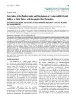
Available online />Figure 1
Mesenchymal stem cell self-renewal and cytodifferentiation.
Extracellular signaling factors, including growth factors and cytokines,
demonstrated to promote and/or maintain mesenchymal stem cell
(MSC) self-renewal, in vitro. Gene markers characteristic of MSC self-
renewal include oct-4, sox-2, and rex-1. LIF, leukemia inhibitory factor;
EGF, epidermal growth factor; HGF, hepatocyte growth factor; PDGF,
platelet-derived growth factor; FGF, fibroblast growth factor; CFU-F,
colony forming unit-fibroblast; c, chondroblast; o, osteoblast;
a, adipoblast; m, myoblast; cm, cardio-myoblast; t, tenoblast.
MSCs from a variety of mammalian species also express the
embryonic stem cell gene markers oct-4, sox-2, and rex-1,
among others [25]. Recent chromatin immunoprecipitation on
chromatin immunoprecipitation array studies suggest that
some Polycomb chromatin-associated proteins are involved
globally in maintaining the repression of differentiation genes
[26]. Thus, Polycomb proteins may indirectly maintain oct-4,
sox-2, and rex-1 activation in MSCs; alternatively, Trithorax
proteins, which complement Polycomb proteins [27] by
maintaining the activation of homeotic genes, may directly
regulate the expression of oct-4, sox-2, and rex-1.
Biochemical studies linking stemness gene expression with
chromatin-associated proteins will be an interesting future
avenue of research.
Several other exciting areas of MSC biology that are beyond
the scope of this review have recently begun to be explored.
These areas concern the regulation of other cell types by
MSCs, including MSCs as trophic mediators [28] and the
immunomodulatory effects of MSCs [29].
MSC differentiation
The identification of specific signaling networks and ‘master’
regulatory genes that govern unique MSC differentiation
lineages remains a challenge. The ability to modulate
biological effectors to maintain a desired differentiation
program, or possibly to prevent spurious differentiation of
MSCs, is needed for effective clinical application, as in tissue
engineering and regeneration. Some of the recently
discovered lineage-restrictive molecular regulators and their
mechanisms of action will be reviewed here.
Chondrogenesis
Chondrogenic differentiation of MSCs in vitro mimics that of
cartilage development in vivo. Expression markers associated
with chondrogenesis have been positively characterized in
MSC-derived chondrocytes, including transcription factors
(sox-9, scleraxis) and extracellular matrix (ECM) genes
(collagen types II and IX, aggrecan, biglycan, decorin, and
cartilage oligomeric matrix protein) [30,31]. However, the
specific signaling pathways that induce the expression of
these benchmark chondrogenic genes remain generally
unknown. Naturally occurring human mutations and molecular
genetic studies have identified several instructive signaling
molecules, including various transforming growth factor-β
(TGF-β) [32], bone morphogenetic protein (BMP), growth
and differentiation factor (GDF) [33] and Wnt [34] ligands.
Recombinant proteins and/or adenoviral infection of MSCs
with TGF-β1 and TGF-β3, BMP-2, BMP-4, BMP-6 [35],
BMP-12 [36], BMP-13 [37], and GDF-5 have been shown to
rapidly induce chondrogenesis of MSCs from a variety of
mesodermal tissue sources (reviewed in [31]). Upon receptor
binding, TGF-βs and BMPs signal through specific intra-
cellular Smad proteins and major mitogen-activated protein
kinase (MAPK) cascades, providing levels of specificity that
are actively being investigated in MSC differentiation contexts
[32,38]. Recent studies into mechanisms of crosstalk
between downstream MAPK signaling and Smad effectors
have revealed that MAPK substrates include chromatin histone
acetyltransferases (HATs) [39]. HATs in turn are directly
recruited by Smads and enhance Smad transactivation
capability [40]. For example, the p38 MAPK substrate MSK
phosphorylates p300-PCAF HATs [39], thereby enhancing
their direct binding to and formation of a Smad2/4–HAT
complex. This may be a general model of how the two major
signaling mediators of the TGF-β and BMP ligands converge
synergistically to transactivate target genes of chondro-
genesis, with a specificity probably dependent, in part, on the
unique combinatorial crosstalk between R-Smads and MAPK
pathways.
Wnts have an important bipotent modulatory function in
chondrogenesis. In murine C3H10T1/2 cells, canonical
Wnt3a enhances BMP-2-induced chondrogenesis [41,42].
Wnt3a in turn regulates bmp2 expression [43], suggesting a
feedforward regulatory loop during chondrogenesis. In human
MSCs, transient upregulation of Wnt7a also enhances
chondrogenesis through various TGF-β1–MAPK signaling
pathways, but sustained Wnt7a expression is chondro-
inhibitory [44]. A recent study in ATDC5 cells revealed that
Wnt1 inhibits chondrogenesis through the upregulation of
the important mesodermal basic helix–loop–helix (bHLH)
transcription factor, Twist 1 [45], perhaps involving negative
sequestration of chondrostimulatory factors or direct
repression of target genes. Further investigations should
focus on the crosstalk between pathways, such as those of
TGF-βs and Wnts.
Osteogenesis
BMPs, in particular BMP-2 and BMP-6, strongly promote
osteogenesis in MSCs [33,46]. BMP-2 induces the p300-
mediated acetylation of Runx2, a master osteogenic gene,
which results in enhanced Runx2 transactivating capability.
The acetylation is specific to histone deacetylases 4 and 5,
which, by deacetylating Runx2, promote its subsequent
degradation by Smurf1 and Smurf2, and E3 ubiquitin ligases
[47]. Interestingly, the cytokine TNF-α, which is associated
with inflammation-mediated bone degradation, also down-
regulates Runx2 protein levels through increased degradation
mediated by Smurf1 and Smurf2. Transgenic TNF-α mice
also showed increased levels of Smurf1 and Smurf2,
concurrent with decreased Runx2 protein levels [48]. These
findings suggest that therapeutic approaches to MSC-based
bone tissue engineering, centered on BMPs, Runx2, and
histone deacetyltransferases, may enhance existing TNF-α-
based immunotherapy of bone diseases.
Wnts have an important modulatory function in osteogenesis.
Knockout and dosage compensation in Wnt-pathway-related
transgenic animals provide the strongest proof that high
levels of endogenous Wnts promote osteogenesis, whereas
low levels inhibit osteogenesis [49]. In C3H10T1/2 and
Arthritis Research & Therapy Vol 9 No 1 Kolf et al.
Page 4 of 10
(page number not for citation purposes)
murine osteoprogenitor cells, canonical Wnt signaling up-
regulates runx2. Chromatin immunoprecipitation and promoter
mutational analyses showed that β-catenin/LEF (lymphoid
enhancer binding factor)/TCF1 (T-cell factor 1) occupy a
cognate binding site in the proximal runx2 promoter and may
therefore directly regulate runx2 expression [50]. However, in
human MSCs, canonical Wnts decrease osteogenesis [19].
Independently, these observations suggest a mechanistic
model of MSC osteogenesis involving crosstalk between
BMPs and canonical Wnts that converges on Runx2 (Figure 2).
In 293T cells, tbx5, a critical T-box gene involved in human
Holt–Oram syndrome and also implicated in osteogenesis,
was shown to interact directly with the chromatin coregulator
TAZ (transcriptional coactivator with PDZ-binding motif),
resulting in enhanced Tbx-5 activation of the osteogenic
FGF10 target gene. By recruiting HATs, TAZ mediates the
opening of chromatin, thereby increasing Tbx-5 transcriptional
activity [51], which may also occur during MSC osteogenesis.
The exciting new discoveries of transcriptional mechanisms
driving the balance of bone formation and loss around a
global osteogenic gene, runx2, and a specific osteogenic
homeobox gene, tbx5, represent two strong models of
transcriptional regulation of osteogenesis, and potentially
other MSC lineage differentiation programs.
Adipogenesis
The nuclear hormone receptor peroxisome proliferator-
activated receptor γ (PPARγ) is a critical adipogenic regulator
promoting MSC adipogenesis while repressing osteogenesis
[52]. The binding of PPARγ to various ligands, including long-
chain fatty acids and thiazolidinedione compounds, induces
the transactivation and transrepression of PPARγ. The bipotent
coregulator TAZ was recently discovered to function as a
coactivator of Runx2 and as a corepressor of PPARγ, thus
promoting osteogenesis while blocking adipogenesis [53].
Mechanistically, the converse, in which a coactivator of adipo-
genic genes corepresses osteogenic genes, is also possible.
Available online />Page 5 of 10
(page number not for citation purposes)
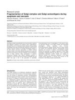
Figure 2
Molecular regulation of mesenchymal stem cell cytodifferentiation programs. Extracellular molecular signaling and mechanical inducers of
differentiation transduce effects through putative receptors, channels, and/or other cell-surface-associated mechanisms. Downstream crosstalk of
signaling pathways, including that between distinct mitogen-activated protein kinases (MAPKs) and R-Smads, provides a level of specificity that
gives rise to unique lineages, such as chondrocytes and osteoblasts. Specificity of lineage differentiation can also result from the recruitment of
master transcriptional switches with binary regulation of cell fate, such as TAZ (transcriptional coactivator with PDZ-binding motif). Depending on
potentially unique multiprotein complexes that it may form in response to specific upstream signaling, TAZ promotes osteogenesis and inhibits
adipogenesis. Furthermore, coregulator subtypes can be invoked, such as tension-induced/-inhibited proteins (TIPs), which regulate adipogenesis
and myogenesis. Specific molecular induction/regulation of cardiomyogenic and tenogenic-specific development are as yet largely unknown, with
the exception of those depicted. Broken lines, unknown or putative; solid lines, as in published data;
*
, juxtaposing cell; GDF, growth and
differentiation factor; TGF, transforming growth factor; BMP, bone morphogenetic protein; FA, fatty acid; βcat, β-catenin; PPAR, peroxisome
proliferator-activated receptor; MSK, mitogen- and stress-activated protein kinase; PCAF, p300/CBP-associated factor; Ac, acetyl; c,
chondroblast; o, osteoblast; a, adipoblast; m, myoblast; cm, cardiomyoblast; t, tenoblast.
This type of cellular efficiency is plausible, given that both
lineages may be derived from a common MSC.
Interestingly, another example of interplay between trans-
criptional cofactors of adipogenesis involves stretch-related
mechano-induction. Mouse embryonic lung mesenchymal
cells form myocytes under stretch induction but form
adipocytes if uninduced. Stretch/non-stretch mechano-
stimulation activates specific isoforms of tension-induced/-
inhibited proteins (TIPs) [54], chromatin-modifying proteins
with intrinsic HAT activity that have other distinctive domains
such as nuclear receptor-interacting motifs. TIP-1 is
expressed under non-stretch conditions and promotes adipo-
genesis, whereas TIP-3 promotes myogenesis. TIP-1 also
provides a potential mechanistic endpoint for cytoplasmic
RhoA-mediated induction of adipogenesis; that is, round
formation of cells, associated with lack of cell tension,
induces RhoA signaling, which promotes adipogenesis [55].
Together, these findings suggest a molecular model that
potentially links mechanical induction, cell morphology,
cytoskeletal signaling, and transcriptional response in the
induction of MSC adipogenesis.
Myogenesis
Most investigations of myogenesis in adult stem cells are
based on a small population of skeletal muscle-derived stem
cells, or satellite cells. A recent study showed the highly
successful induction of myogenesis from adult stromal
MSCs, after transfection with activated Notch 1 [56];
however, the mechanisms of action remain unknown. Other
investigations, largely focused on cardiomyogenesis, showed
the importance of cell–cell contact in stimulating cardio-
myogenesis by using co-cultured MSCs and cardiomyocytes,
and the stimulation of MSC cardiomyogenesis in a rat intra-
myocardial infarct model by Jagged 1, a Notch ligand [57].
Other animal cardiac and vascular injury models and human
clinical trials are being actively investigated to explore the
potential regeneration of cardiac tissue.
Tenogenesis
GDF proteins, members of the TGF-β superfamily, promote
the formation of tendons in vivo [58]. In addition to culture
medium specifications, differentiation of MSCs into tenocytes
in vitro requires mechanical loading [59], which is critical to
tendon fiber alignment during development. The identity of
specific differentiation gene markers to track the tenogenesis
of MSCs remains unknown. Expression of scleraxis, which
encodes a bHLH transcription factor, is detectable in vivo in
a somitic tendon progenitor compartment, and remains
expressed through mature tendon development. However,
other mesenchymal tissues destined to form axial skeleton,
chondrocytes [60], and ligament [61] are also scleraxis-
positive, indicating the need for additional, more
discriminating markers to follow tenogenesis. Recently, it was
shown that R-Smad8 specifically transduced BMP-2
signaling in murine C3H10T1/2 cells to form tenocytes rather
than osteoblasts [62]. The activation domain of R-Smad8
may be uniquely regulated or used to form distinct trans-
criptional complexes specific for tenogenic differentiation.
MSC niche
In analyzing the differentiation of stem cells, it is critical to
consider the influence of their tissue of origin. MSCs are now
routinely isolated from the bone marrow of many mammalian
model organisms, as well as from other tissues of meso-
dermal origin such as adipose, muscle, bone, and tendon.
Recently, multipotent cells have also been isolated from many
other tissue types of non-mesodermal origin. Specifically, a
recent study reported plastic-adherent MSC-like colonies
derived from the brain, spleen, liver, kidney, lung, bone
marrow, muscle, thymus, and pancreas of mice [63], all with
similar morphologies and immunophenotypes after several
passages. In another study, murine MSCs were obtained
from freshly isolated cells of the heart, liver, kidney, thymus,
ovary, dermis, and lung on the basis of a CD45
–
/CD31
–
/
Sca-1
+
/Thy-1
+
phenotype [64], raising the question of what
the common in vivo microenvironment of the MSC might be.
Is there an MSC niche that is common to all of these tissues,
or do MSCs function autonomously, in a manner that is
independent of their environment?
Since Schofield first introduced the concept of a stem cell
‘niche’ in 1978 [65], the idea has gained wide support,
particularly in recent years. In brief, the niche encompasses
all of the elements immediately surrounding the stem cells
when they are in their naïve state, including the non-stem
cells that might be in direct contact with them as well as ECM
and soluble molecules found in that locale. All of these act
together to maintain the stem cells in their undifferentiated
state. It is then assumed that certain cues must find their way
into the niche to signal to the stem cells that their
differentiation potential is needed for the regeneration or
repopulation of a tissue.
Cellular components
Two recent studies suggested a perivascular nature of the
MSC niche (Figure 3), on the basis of the expression of α-
smooth muscle actin (αSMA) in MSCs isolated from all tissue
types tested [63] and the immunohistochemical localization
of CD45
–
/CD31
–
/Sca-1
+
/Thy-1
+
cells to perivascular sites
[64]. In support of this, MSCs were found, with the use of the
markers Stro-1 and CD146, lining blood vessels in human
bone marrow and dental pulp [66]. These cells also
expressed αSMA and some even expressed 3G5, a pericyte-
associated cell-surface marker. Some researchers have
hypothesized that pericytes are in fact MSCs, because they
can differentiate into osteoblasts, chondrocytes, and
adipocytes [67]. Localization of MSCs to perivascular niches
throughout the body gives them easy access to all tissues
and lends credence to the notion that MSCs are integral to
the healing of many different tissues (see the ‘Homing and
wound healing’ section below). Experiments in vivo that
Arthritis Research & Therapy Vol 9 No 1 Kolf et al.
Page 6 of 10
(page number not for citation purposes)
perturb this perivascular environment are needed to validate
this theory.
The transmembrane cell adhesion proteins, cadherins,
function in cell–cell adhesion, migration, differentiation, and
polarity, including in MSCs [44], and are known to interact
with Wnts, which are important in MSC biology, as described
above. They are also implicated in the biology of other stem
cell niches [68]. Their role in the MSC niche is an unexplored
territory and is crucial to an understanding of the molecular
basis of the interactions between the MSC and its neighbors.
Soluble components
That the bone marrow milieu is hypoxic in nature is of
particular relevance. Comparison of human MSCs cultured
in hypoxic versus normoxic conditions (2% and 20% oxygen)
showed that their proliferative capacity was better
maintained in the former [69]. In addition, hypoxia at least
doubled the number of CFU-Fs present while enhancing the
expression of oct-4 and rex-1, genes expressed by
embryonic stem cells and thought to be pivotal in
maintaining ‘stemness’. These data suggest that hypoxia
enhances not only the proliferative capacity but also the
plasticity of MSCs. The mechanism of action of hypoxia on
MSCs is currently unknown, although oct-4 upregulation by
the transcription factor HIF-2α (hypoxia-induced factor-2α)
is possible [70].
The role of secreted proteins in the MSC niche is not
understood. Many studies have used conditioned media and
Transwell set-ups to analyze the effects of proteins secreted
by various cell types on MSCs without direct cellular contact
(see, for example, [71,72]). So far, we know of no studies that
identify the effective proteins or that present a cell type
whose secreted factors exhibit a ‘niche effect’ on MSCs. In
other words, the cell types studied have either had no effect
on MSCs or they have induced differentiation instead. Finding
one or more soluble proteins that inhibit MSC differentiation
while allowing proliferation would be ideal for mimicking the
niche and expanding MSCs ex vivo.
Extracellular matrix components
Again, no specific matrix components have been identified
that help to maintain MSCs in their naïve state, as a niche
matrix would do. However, there is evidence that ECM alone
can regulate MSC differentiation, with potential applications
for tissue engineering. For example, ECM left by osteoblasts
on titanium scaffolds after decellularization increased
osteogenesis markers, such as alkaline phosphatase and
calcium deposition, in MSCs [73]. Our recent observations
also suggest that ECM deposited by microvascular
endothelial cells enhances MSC endotheliogenesis (T Lozito
and RS Tuan, unpublished data). Designing artificial matrices
that can mimic the tissue microenvironment in vivo and
regulate the appropriate differentiation of stem cells is a
promising approach to therapeutic applications. Molecular
information on ECM–MSC interactions, most probably
involving integrins, which have already been implicated in
niche biology in other systems (see, for example, [74]), is
clearly needed.
Available online />Page 7 of 10
(page number not for citation purposes)
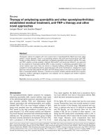
Figure 3
Mesenchymal stem cell niche. Mesenchymal stem cells (MSCs) are shown in their putative perivascular niche (BV, blood vessel), interacting with
(1) various other differentiated cells (DC
1
, DC
2
, etc.) by means of cell-adhesion molecules, such as cadherins, (2) extracellular matrix (ECM)
deposited by the niche cells mediated by integrin receptors, and (3) signaling molecules, which may include autocrine, paracrine, and endocrine
factors. Another variable is O
2
tension, with hypoxia associated with MSCs in the bone marrow niche.
Homing and wound healing
Another stem cell niche-related phenomenon is the homing of
stem cells to sites of injury and subsequent wound healing.
Although some tissue repair may be accomplished by the
division of indigenous differentiated cells, such cells are most
frequently post-mitotic. Thus, signaling to progenitor/stem
cells to home to the site of injury and differentiate into the
required cell type is required. To understand the niche, it is
important to analyze not only what keeps stem cells in their
niche but also what signals them to emigrate from it.
Even in healthy animals, MSCs are capable of homing to
tissues other than the bone marrow, such as lung and
muscles [75]. Interestingly, the capacity of an MSC for
homing seems to be related in part to its expression of Stro-1
(see the ‘MSC markers’ section above) [7]. Whereas Stro-1-
negative cells were better able to aid in the engraftment and
survival of HSCs, Stro-1-positive cells were more capable of
homing and engrafting to most of the tissues studied. Exciting
new work in vitro shows that MSC migration is regulated by
stromal-derived factor-1/CXCR4 and hepatocyte growth
factor/c-Met complexes, and involves matrix metalloproteinases
[76]. In vivo expression profiles of the responsible factors will
shed light on when, where, and how MSCs migrate. What is
known is that injury alters the patterns of migration and
differentiation of exogenously added MSCs. In the mouse,
irradiation of both the whole animal and specific sites caused
injected MSCs to engraft to more organs and in higher
numbers than in unconditioned mice [75].
In addition, it seems that mature cells that have been injured
are able to secrete not only homing signals but also
differentiation signals. Rat bone marrow-derived MSCs, for
example, begin myogenesis in response to conditioned
medium from damaged but not undamaged skeletal muscle
[77]. Other studies in vitro suggest that some uninjured cells
can also induce differentiation when direct contact is allowed.
Our preliminary results show that direct co-culturing with
osteoblasts enhances the osteogenesis of MSCs (CM Kolf, L
Song and RS Tuan, unpublished data). Liver cells also seem
to be capable of inducing hepatogenesis [78]. However, it is
important to note that mature cells do not always induce
MSC differentiation along their own lineage. Direct contact
with chondrocytes induces osteogenesis but not
chondrogenesis [72]. Clearly, the environment of an MSC is
a critical defining factor of its identity.
Conclusion
Adult MSCs are a potentially powerful candidate cell type for
regenerative medicine as well as for the study of cellular
differentiation. A key requirement for both fields is the
identification of MSCs in vivo. In mouse, genetic markers and
pulse–chase techniques can be used to label stem cells [79].
In other systems, asymmetric division has been shown to be
integral to stem cell self-renewal. This unique property of
stem cells has been exploited to identify mouse muscle
satellite cells [80] and could possibly be used to identify
MSCs in vivo and to study their division. Once the true MSC
population is identified, global characterization using gene
arrays and surface antigen profiling may be achieved. The
roles of each component of the MSC system should then be
functionally analyzed. Critical challenges include identifying
the signaling factors that promote the self-renewal of MSCs,
as well as elucidating the master transcriptional regulatory
switches and the crosstalk between the signaling pathways
that mediate exclusive lineage differentiation in MSCs. Future
investigations should incorporate combinatorial knockdown
approaches using inducible and stable expression systems to
address redundancy in signaling functions, for example within
the TGF-β and Wnt families. The identification of specific cell-
surface receptors activated by signaling molecules, such as
TGF-βs (BMPs) and Wnts, during self-renewal and
cytodifferentiation is also crucial to understanding the link
between extracellular and intracellular signaling networks.
Finally, alterations in the MSC niche will help to determine the
intrinsic and extrinsic specificity of MSC regulators. In an
elegant model experiment, quiescent muscle and liver stem
cells of aged mice were rejuvenated when exposed to the
circulating blood of younger animals [81]. That an extrinsic
change can enhance stem cell functions presents hope for
harnessing the healing powers of adult stem cells in the future.
Competing interests
The authors declare that they have no competing interests.
Acknowledgment
This work was supported by the Intramural Research Program of
NIAMS, NIH (Z01 AR41131).
References
1. Friedenstein AJ, Chailakhjan RK, Lalykina KS: The development
of fibroblast colonies in monolayer cultures of guinea-pig
bone marrow and spleen cells. Cell Tissue Kinet 1970, 3:393-
403.
2. Pittenger MF, Mackay AM, Beck SC, Jaiswal RK, Douglas R,
Mosca JD, Moorman MA, Simonetti DW, Craig S, Marshak DR:
Multilineage potential of adult human mesenchymal stem
cells. Science 1999, 284:143-147.
3. Jiang Y, Jahagirdar BN, Reinhardt RL, Schwartz RE, Keene CD,
Ortiz-Gonzalez XR, Reyes M, Lenvik T, Lund T, Blackstad M, et al.:
Pluripotency of mesenchymal stem cells derived from adult
marrow. Nature 2002, 418:41-49.
4. Simmons PJ, Torok-Storb B: Identification of stromal cell pre-
cursors in human bone marrow by a novel monoclonal anti-
body, STRO-1. Blood 1991, 78:55-62.
5. Gronthos S, Zannettino AC, Hay SJ, Shi S, Graves SE, Kortesidis
A, Simmons PJ: Molecular and cellular characterisation of
highly purified stromal stem cells derived from human bone
marrow. J Cell Sci 2003, 116:1827-1835.
Arthritis Research & Therapy Vol 9 No 1 Kolf et al.
Page 8 of 10
(page number not for citation purposes)
This review is part of a series on
Mesenchymal stromal cells
edited by Steffen Gay.
Other articles in this series can be found at
/>review-series.asp?series=ar_Mesenchymal
6. Dennis JE, Carbillet JP, Caplan AI, Charbord P: The STRO-1+
marrow cell population is multipotential. Cells Tissues Organs
2002, 170:73-82.
7. Bensidhoum M, Chapel A, Francois S, Demarquay C, Mazurier C,
Fouillard L, Bouchet S, Bertho JM, Gourmelon P, Aigueperse J, et
al.: Homing of in vitro expanded Stro-1– or Stro-1+ human
mesenchymal stem cells into the NOD/SCID mouse and their
role in supporting human CD34 cell engraftment. Blood 2004,
103:3313-3319.
8. Carter RA, Wicks IP: Vascular cell adhesion molecule 1
(CD106): a multifaceted regulator of joint inflammation. Arthri-
tis Rheum 2001, 44:985-994.
9. PROW: CD73
[ />10. Haynesworth SE, Baber MA, Caplan AI: Cell surface antigens
on human marrow-derived mesenchymal cells are detected
by monoclonal antibodies. Bone 1992, 13:69-80.
11. Quirici N, Soligo D, Bossolasco P, Servida F, Lumini C, Deliliers
GL: Isolation of bone marrow mesenchymal stem cells by
anti-nerve growth factor receptor antibodies. Exp Hematol
2002, 30:783-791.
12. Honczarenko M, Le Y, Swierkowski M, Ghiran I, Glodek AM, Sil-
berstein LE: Human bone marrow stromal cells express a dis-
tinct set of biologically functional chemokine receptors. Stem
Cells 2006, 24:1030-1041.
13. Song L, Webb NE, Song Y, Tuan RS: Identification and func-
tional analysis of candidate genes regulating mesenchymal
stem cell self-renewal and multipotency. Stem Cells 2006, 24:
1707-1718.
14. Jiang Y, Vaessen B, Lenvik T, Blackstad M, Reyes M, Verfaillie
CM: Multipotent progenitor cells can be isolated from postna-
tal murine bone marrow, muscle, and brain. Exp Hematol
2002, 30:896-904.
15. Metcalf D: The unsolved enigmas of leukemia inhibitory factor.
Stem Cells 2003, 21:5-14.
16. Tsutsumi S, Shimazu A, Miyazaki K, Pan H, Koike C, Yoshida E,
Takagishi K, Kato Y: Retention of multilineage differentiation
potential of mesenchymal cells during proliferation in
response to FGF. Biochem Biophys Res Commun 2001, 288:
413-419.
17. Zaragosi LE, Ailhaud G, Dani C: Autocrine FGF2 signaling is
critical for self-renewal of human multipotent adipose-derived
stem cells. Stem Cells 2006, 24:2412-2419.
18. Kleber M, Sommer L: Wnt signaling and the regulation of stem
cell function. Curr Opin Cell Biol 2004, 16:681-687.
19. Boland GM, Perkins G, Hall DJ, Tuan RS: Wnt 3a promotes pro-
liferation and suppresses osteogenic differentiation of adult
human mesenchymal stem cells. J Cell Biochem 2004, 93:
1210-1230.
20. Heymann D, Rousselle AV: gp130 Cytokine family and bone
cells. Cytokine 2000, 12:1455-1468.
21. Schwartz J, Van de Pavert S, Clarke I, Rao A, Ray D, Vrana K:
Paracrine interactions within the pituitary gland. Ann NY Acad
Sci 1998, 839:239-243.
22. Niswander L: Interplay between the molecular signals that
control vertebrate limb development. Int J Dev Biol 2002, 46:
877-881.
23. Marie PJ, Coffin JD, Hurley MM: FGF and FGFR signaling in
chondrodysplasias and craniosynostosis. J Cell Biochem
2005, 96:888-896.
24. Bienz M: The subcellular destinations of APC proteins. Nat Rev
Mol Cell Biol 2002, 3:328-338.
25. Izadpanah R, Trygg C, Patel B, Kriedt C, Dufour J, Gimble JM,
Bunnell BA: Biologic properties of mesenchymal stem cells
derived from bone marrow and adipose tissue. J Cell Biochem
2006, 99:1285-1597.
26. Boyer LA, Plath K, Zeitlinger J, Brambrink T, Medeiros LA, Lee TI,
Levine SS, Wernig M, Tajonar A, Ray MK, et al.: Polycomb com-
plexes repress developmental regulators in murine embry-
onic stem cells. Nature 2006, 441:349-353.
27. Ringrose L, Paro R: Epigenetic regulation of cellular memory
by the Polycomb and Trithorax group proteins. Annu Rev
Genet 2004, 38:413-443.
28. Caplan AI, Dennis JE: Mesenchymal stem cells as trophic
mediators. J Cell Biochem 2006, 98:1076-1084.
29. Chen X, Armstrong MA, Li G: Mesenchymal stem cells in
immunoregulation. Immunol Cell Biol 2006, 84:413-421.
30. Baksh D, Song L, Tuan RS: Adult mesenchymal stem cells:
characterization, differentiation, and application in cell and
gene therapy. J Cell Mol Med 2004, 8:301-316.
31. Tuan RS, Boland G, Tuli R: Adult mesenchymal stem cells and
cell-based tissue engineering. Arthritis Res Ther 2003, 5:32-45.
32. Massague J, Blain SW, Lo RS: TGF
ββ
signaling in growth
control, cancer, and heritable disorders. Cell 2000, 103:295-
309.
33. Chen D, Zhao M, Mundy GR: Bone morphogenetic proteins.
Growth Factors 2004, 22:233-241.
34. Hartmann C: A Wnt canon orchestrating osteoblastogenesis.
Trends Cell Biol 2006, 16:151-158.
35. Boskey AL, Paschalis EP, Binderman I, Doty SB: BMP-6 acceler-
ates both chondrogenesis and mineral maturation in differen-
tiating chick limb-bud mesenchymal cell cultures. J Cell
Biochem 2002, 84:509-519.
36. Gooch KJ, Blunk T, Courter DL, Sieminski AL, Vunjak-Novakovic
G, Freed LE: Bone morphogenetic proteins-2, -12, and -13
modulate in vitro development of engineered cartilage. Tissue
Eng 2002, 8:591-601.
37. Nochi H, Sung JH, Lou J, Adkisson HD, Maloney WJ, Hruska KA:
Adenovirus mediated BMP-13 gene transfer induces chondro-
genic differentiation of murine mesenchymal progenitor cells.
J Bone Miner Res 2004, 19:111-122.
38. Goumans MJ, Mummery C: Functional analysis of the TGF
ββ
receptor/Smad pathway through gene ablation in mice. Int J
Dev Biol 2000, 44:253-265.
39. Abecassis L, Rogier E, Vazquez A, Atfi A, Bourgeade MF: Evi-
dence for a role of MSK1 in transforming growth factor-
ββ
-
mediated responses through p38
αα
and Smad signaling
pathways. J Biol Chem 2004, 279:30474-30479.
40. Kahata K, Hayashi M, Asaka M, Hellman U, Kitagawa H, Yanagi-
sawa J, Kato S, Imamura T, Miyazono K: Regulation of trans-
forming growth factor-
ββ
and bone morphogenetic protein
signalling by transcriptional coactivator GCN5. Genes Cells
2004, 9:143-151.
41. Fischer L, Boland G, Tuan RS: Wnt-3A enhances bone morpho-
genetic protein-2-mediated chondrogenesis of murine
C3H10T1/2 mesenchymal cells. J Biol Chem 2002, 277:
30870-30878.
42. Fischer L, Boland G, Tuan RS: Wnt signaling during BMP-2
stimulation of mesenchymal chondrogenesis. J Cell Biochem
2002, 84:816-831.
43. Kengaku M, Capdevila J, Rodriguez-Esteban C, De La Pena J,
Johnson RL, Belmonte JC, Tabin CJ: Distinct WNT pathways
regulating AER formation and dorsoventral polarity in the
chick limb bud. Science 1998, 280:1274-1277.
44. Tuli R, Tuli S, Nandi S, Huang X, Manner PA, Hozack WJ, Daniel-
son KG, Hall DJ, Tuan RS: Transforming growth factor-
ββ
-medi-
ated chondrogenesis of human mesenchymal progenitor
cells involves N-cadherin and mitogen-activated protein
kinase and Wnt signaling cross-talk. J Biol Chem 2003, 278:
41227-41236.
45. Reinhold MI, Kapadia RM, Liao Z, Naski MC: The Wnt-inducible
transcription factor Twist1 inhibits chondrogenesis. J Biol
Chem 2006, 281:1381-1388.
46. Friedman MS, Long MW, Hankenson KD: Osteogenic differenti-
ation of human mesenchymal stem cells is regulated by bone
morphogenetic protein-6. J Cell Biochem 2006, 98:538-554.
47. Jeon EJ, Lee KY, Choi NS, Lee MH, Kim HN, Jin YH, Ryoo HM,
Choi JY, Yoshida M, Nishino N, et al.: Bone morphogenetic
protein-2 stimulates Runx2 acetylation. J Biol Chem 2006,
281:16502-16511.
48. Kaneki H, Guo R, Chen D, Yao Z, Schwarz EM, Zhang YE, Boyce
BF, Xing L: Tumor necrosis factor promotes Runx2 degrada-
tion through up-regulation of Smurf1 and Smurf2 in
osteoblasts. J Biol Chem 2006, 281:4326-4333.
49. Gaspar C, Fodde R: APC dosage effects in tumorigenesis and
stem cell differentiation. Int J Dev Biol 2004, 48:377-386.
50. Gaur T, Lengner CJ, Hovhannisyan H, Bhat RA, Bodine PV, Komm
BS, Javed A, van Wijnen AJ, Stein JL, Stein GS, et al.: Canonical
WNT signaling promotes osteogenesis by directly stimulating
Runx2 gene expression. J Biol Chem 2005, 280:33132-33140.
51. Murakami M, Nakagawa M, Olson EN, Nakagawa O: A WW
domain protein is a critical coactivator for TBX5, a transcrip-
tion factor implicated in Holt–Oram syndrome. Proc Natl Acad
Sci USA 2005, 102:18034-18039.
Available online />Page 9 of 10
(page number not for citation purposes)
52. Nuttall ME, Gimble JM: Controlling the balance between
osteoblastogenesis and adipogenesis and the consequent
therapeutic implications. Curr Opin Pharmacol 2004, 4:290-
294.
53. Hong JH, Hwang ES, McManus MT, Amsterdam A, Tian Y,
Kalmukova R, Mueller E, Benjamin T, Spiegelman BM, Sharp PA,
et al.: TAZ, a transcriptional modulator of mesenchymal stem
cell differentiation. Science 2005, 309:1074-1078.
54. Jakkaraju S, Zhe X, Pan D, Choudhury R, Schuger L: TIPs are
tension-responsive proteins involved in myogenic versus adi-
pogenic differentiation. Dev Cell 2005, 9:39-49.
55. McBeath R, Pirone DM, Nelson CM, Bhadriraju K, Chen CS: Cell
shape, cytoskeletal tension, and RhoA regulate stem cell
lineage commitment. Dev Cell 2004, 6:483-495.
56. Dezawa M, Ishikawa H, Itokazu Y, Yoshihara T, Hoshino M,
Takeda S, Ide C, Nabeshima Y: Bone marrow stromal cells gen-
erate muscle cells and repair muscle degeneration. Science
2005, 309:314-317.
57. Li H, Yu B, Zhang Y, Pan Z, Xu W, Li H: Jagged1 protein
enhances the differentiation of mesenchymal stem cells into
cardiomyocytes. Biochem Biophys Res Commun 2006, 341:
320-325.
58. Wolfman NM, Hattersley G, Cox K, Celeste AJ, Nelson R, Yamaji
N, Dube JL, DiBlasio-Smith E, Nove J, Song JJ, et al.: Ectopic
induction of tendon and ligament in rats by growth and differ-
entiation factors 5, 6, and 7, members of the TGF-
ββ
gene
family. J Clin Invest 1997, 100:321-330.
59. Altman GH, Horan RL, Martin I, Farhadi J, Stark PR, Volloch V,
Richmond JC, Vunjak-Novakovic G, Kaplan DL: Cell differentia-
tion by mechanical stress. FASEB J 2002, 16:270-272.
60. Brown D, Wagner D, Li X, Richardson JA, Olson EN: Dual role of
the basic helix-loop-helix transcription factor scleraxis in
mesoderm formation and chondrogenesis during mouse
embryogenesis. Development 1999, 126:4317-4329.
61. Schweitzer R, Chyung JH, Murtaugh LC, Brent AE, Rosen V,
Olson EN, Lassar A, Tabin CJ: Analysis of the tendon cell fate
using Scleraxis, a specific marker for tendons and ligaments.
Development 2001, 128:3855-3866.
62. Hoffmann A, Pelled G, Turgeman G, Eberle P, Zilberman Y, Shinar
H, Keinan-Adamsky K, Winkel A, Shahab S, Navon G, et al.: Neo-
tendon formation induced by manipulation of the Smad8 sig-
nalling pathway in mesenchymal stem cells. J Clin Invest
2006, 116:940-952.
63. da Silva Meirelles L, Chagastelles PC, Nardi NB: Mesenchymal
stem cells reside in virtually all post-natal organs and tissues.
J Cell Sci 2006, 119:2204-2213.
64. Blashki D, Short B, Bertoncello I, Simmons PJ, Brouard N: Identi-
fication of stromal MSC candidates from multiple adult
mouse tissues. In Int Soc Stem Cell Res 4th Annual Meeting
2006: 206.
65. Schofield R: The relationship between the spleen colony-
forming cell and the haemopoietic stem cell. Blood Cells
1978, 4:7-25.
66. Shi S, Gronthos S: Perivascular niche of postnatal mesenchy-
mal stem cells in human bone marrow and dental pulp. J
Bone Miner Res 2003, 18:696-704.
67. Doherty MJ, Canfield AE: Gene expression during vascular per-
icyte differentiation. Crit Rev Eukaryot Gene Expr 1999, 9:1-17.
68. Nelson WJ, Nusse R: Convergence of Wnt,
ββ
-catenin, and cad-
herin pathways. Science 2004, 303:1483-1487.
69. Grayson WL, Zhao F, Izadpanah R, Bunnell B, Ma T: Effects of
hypoxia on human mesenchymal stem cell expansion and
plasticity in 3D constructs. J Cell Physiol 2006, 207:331-339.
70. Covello KL, Kehler J, Yu H, Gordan JD, Arsham AM, Hu CJ,
Labosky PA, Simon MC, Keith B: HIF-2
αα
regulates Oct-4:
effects of hypoxia on stem cell function, embryonic develop-
ment, and tumor growth. Genes Dev 2006, 20:557-570.
71. Kaigler D, Krebsbach PH, West ER, Horger K, Huang YC,
Mooney DJ: Endothelial cell modulation of bone marrow
stromal cell osteogenic potential. FASEB J 2005, 19:665-667.
72. Gerstenfeld LC, Barnes GL, Shea CM, Einhorn TA: Osteogenic
differentiation is selectively promoted by morphogenetic
signals from chondrocytes and synergized by a nutrient rich
growth environment. Connect Tissue Res 2003, 44(Suppl 1):
85-91.
73. Datta N, Holtorf HL, Sikavitsas VI, Jansen JA, Mikos AG: Effect of
bone extracellular matrix synthesized in vitro on the
osteoblastic differentiation of marrow stromal cells. Biomateri-
als 2005, 26:971-977.
74. Campos LS:
ββ
1 integrins and neural stem cells: making sense
of the extracellular environment. BioEssays 2005, 27:698-707.
75. Francois S, Bensidhoum M, Mouiseddine M, Mazurier C, Allenet
B, Semont A, Frick J, Sache A, Bouchet S, Thierry D, et al.: Local
irradiation not only induces homing of human mesenchymal
stem cells at exposed sites but promotes their widespread
engraftment to multiple organs: a study of their quantitative
distribution after irradiation damage. Stem Cells 2006, 24:
1020-1029.
76. Son BR, Marquez-Curtis LA, Kucia M, Wysoczynski M, Turner AR,
Ratajczak J, Ratajczak MZ, Janowska-Wieczorek A: Migration of
bone marrow and cord blood mesenchymal stem cells in vitro
is regulated by stromal-derived factor-1-CXCR4 and hepato-
cyte growth factor-c-met axes and involves matrix metallopro-
teinases. Stem Cells 2006, 24:1254-1264.
77. Santa Maria L, Rojas CV, Minguell JJ: Signals from damaged but
not undamaged skeletal muscle induce myogenic differentia-
tion of rat bone-marrow-derived mesenchymal stem cells. Exp
Cell Res 2004, 300:418-426.
78. Lange C, Bassler P, Lioznov MV, Bruns H, Kluth D, Zander AR,
Fiegel HC: Liver-specific gene expression in mesenchymal
stem cells is induced by liver cells. World J Gastroenterol
2005, 11:4497-4504.
79. Tumbar T, Guasch G, Greco V, Blanpain C, Lowry WE, Rendl M,
Fuchs E: Defining the epithelial stem cell niche in skin.
Science 2004, 303:359-363.
80. Conboy IM, Rando TA: The regulation of Notch signaling con-
trols satellite cell activation and cell fate determination in
postnatal myogenesis. Dev Cell 2002, 3:397-409.
81. Conboy IM, Conboy MJ, Wagers AJ, Girma ER, Weissman IL,
Rando TA: Rejuvenation of aged progenitor cells by exposure
to a young systemic environment. Nature 2005, 433:760-764.
82. Colter DC, Sekiya I, Prockop DJ: Identification of a subpopula-
tion of rapidly self-renewing and multipotential adult stem
cells in colonies of human marrow stromal cells. Proc Natl
Acad Sci USA 2001, 98:7841-7845.
83. Tuli R, Tuli S, Nandi S, Wang ML, Alexander PG, Haleem-Smith H,
Hozack WJ, Manner PA, Danielson KG, Tuan RS: Characteriza-
tion of multipotential mesenchymal progenitor cells derived
from human trabecular bone. Stem Cells 2003, 21:681-693.
84. Zuk PA, Zhu M, Ashjian P, De Ugarte DA, Huang JI, Mizuno H,
Alfonso ZC, Fraser JK, Benhaim P, Hedrick MH: Human adipose
tissue is a source of multipotent stem cells. Mol Biol Cell
2002, 13:4279-4295.
85. Etheridge SL, Spencer GJ, Heath DJ, Genever PG: Expression
profiling and functional analysis of wnt signaling mechanisms
in mesenchymal stem cells. Stem Cells 2004, 22:849-860.
86. Gronthos S, Franklin DM, Leddy HA, Robey PG, Storms RW,
Gimble JM: Surface protein characterization of human adipose
tissue-derived stromal cells. J Cell Physiol 2001, 189:54-63.
87. Baddoo M, Hill K, Wilkinson R, Gaupp D, Hughes C, Kopen GC,
Phinney DG: Characterization of mesenchymal stem cells iso-
lated from murine bone marrow by negative selection. J Cell
Biochem 2003, 89:1235-1249.
88. Peister A, Mellad JA, Larson BL, Hall BM, Gibson LF, Prockop DJ:
Adult stem cells from bone marrow (MSCs) isolated from dif-
ferent strains of inbred mice vary in surface epitopes, rates of
proliferation, and differentiation potential. Blood 2004, 103:
1662-1668.
89. Reyes M, Lund T, Lenvik T, Aguiar D, Koodie L, Verfaillie CM:
Purification and ex vivo expansion of postnatal human
marrow mesodermal progenitor cells. Blood 2001, 98:2615-
2625.
90. da Silva Meirelles L, Nardi NB: Murine marrow-derived mes-
enchymal stem cell: isolation, in vitro expansion, and charac-
terization. Br J Haematol 2003, 123:702-711.
91. Musina RA, Bekchanova ES, Sukhikh GT: Comparison of mes-
enchymal stem cells obtained from different human tissues.
Bull Exp Biol Med 2005, 139:504-509.
Arthritis Research & Therapy Vol 9 No 1 Kolf et al.
Page 10 of 10
(page number not for citation purposes)