Báo cáo y học: " T cells and post menopausal osteoporosis in murine models" doc
Bạn đang xem bản rút gọn của tài liệu. Xem và tải ngay bản đầy đủ của tài liệu tại đây (231.75 KB, 4 trang )
Page 1 of 4
(page number not for citation purposes)
Available online />Abstract
Estrogen deficiency is one of the most frequent causes of
osteoporosis in women and a possible cause of bone loss in men.
But the mechanism involved remains largely unknown. Estrogen
deficiency leads to an increase in the immune function, which
culminates in an increased production of tumor necrosis factor
(TNF) by activated T cells. TNF increases osteoclast formation and
bone resorption both directly and by augmenting the sensitivity of
maturing osteoclasts to the essential osteoclastogenic factor
RANKL (the RANK ligand). Increased T cell production of TNF is
induced by estrogen deficiency via a complex mechanism
mediated by antigen presenting cells and the cytokines IFNγ, IL-7
and transforming growth factor-β. The experimental evidence that
suggests that estrogen prevents bone loss by regulating T cell
function and the interactions between immune cells and bone is
reviewed here.
Estrogen deficiency is the most frequent cause of bone loss
in humans. Bone loss results both from decreased ovarian
production of sex steroids and the increase in follicle
stimulating hormone (FSH) production induced by estrogen
deficiency. FSH is now known to directly stimulate the
production of tumor necrosis factor (TNF), a potent
osteoclastogenic cytokine from bone marrow granulocytes
and macrophages [1,2]. While FSH is likely to play a relevant
role in the mechanism by which natural and surgical
menopause lead to bone loss, this article will focus on the
direct (FSH independent) mechanisms by which estrogen
deficiency causes bone loss.
Estrogen deficiency is experimentally induced by ovariectomy
(ovx). The main effect of ovx is a marked stimulation of bone
resorption, which is caused primarily by increased osteoclast
(OC) formation, but estrogen deficiency also increases OC
lifespan due to reduced apoptosis [3]. The net bone loss
caused by increased OC number and life span is limited in
part by a compensatory augmentation of bone formation
within each remodeling unit. This event is a consequence of
stimulated osteoblastogenesis fueled by an expansion of the
pool of early mesenchymal progenitors, and by increased
commitment of such pluripotent precursors toward the
osteoblastic lineage [4]. In spite of stimulated osteoblasto-
genesis, the net increase in bone formation is inadequate to
compensate for enhanced bone resorption because of an
augmentation in osteoblast (OB) apoptosis, a phenomenon
also induced by estrogen deficiency [5]. An additional event
triggered by estrogen withdrawal that limits the magnitude of
the compensatory elevation in bone formation is the
increased production of inflammatory cytokines such as IL-7
and TNF, which limit the functional activity of mature OBs
[6,7]. Increased bone resorption, trabecular thinning and
perforation, and a loss of connection between the remaining
trabeculae are the dominant features of the initial phase of
rapid bone loss that follows the onset of estrogen deficiency
[8]. This acute phase is followed by a long-lasting period of
slower bone loss where the dominant microarchitectural
change is trabecular thinning. This phase is due, in part, to
impaired osteoblastic activity secondary to increased OB
apoptosis [9].
OC formation is induced by the cytokines ‘receptor activator
of NF-κB ligand’ (RANKL) and macrophage colony stimu-
lating factor (M-CSF). These factors are produced primarily
by bone marrow (BM) stromal cells and OBs [10], and
activated T cells [11]. RANKL binds to RANK, a receptor
expressed on OCs and OC precursors, and to osteo-
protegerin, a soluble decoy receptor produced by numerous
hematopoietic cells. Thus, osteoprotegerin, by sequestering
RANKL and preventing its binding to RANK, functions as an
anti-osteoclastogenic cytokine. M-CSF induces the
proliferation of OC precursors, the differentiation of more
mature OCs, and increases the survival of mature OCs.
RANKL promotes the differentiation of OC precursors from
an early stage of maturation into fully mature multinucleated
OCs and activates mature OCs.
Commentary
T cells and post menopausal osteoporosis in murine models
Roberto Pacifici
Division of Endocrinology, Metabolism and Lipids, Department of Medicine, Emory University School of Medicine, Atlanta, GA 30322, USA
Corresponding author: Roberto Pacifici,
Published: 5 March 2007 Arthritis Research & Therapy 2007, 9:102 (doi:10.1186/ar2126)
This article is online at />© 2007 BioMed Central Ltd
BM = bone marrow; ERE = estrogen responsive element; FSH = follicle stimulating hormone; IFN = interferon; IL = interleukin; M-CSF =
macrophage colony stimulating factor; OB = osteoblast; OC = osteoclast; ovx = ovariectomy; RANK = receptor activator of NF-κB; RANKL =
RANKL ligand; TGF = transforming growth factor; TNF, tumor necrosis factor.
Page 2 of 4
(page number not for citation purposes)
Arthritis Research & Therapy Vol 9 No 2 Pacifici
Additional cytokines, either produced by or regulating T cells,
are responsible for the upregulation of OC formation
observed in a variety of conditions, such as inflammation, and
estrogen deficiency. One such factor is TNF, a cytokine that
enhances OC formation by upregulating the stromal cell
production of RANKL and M-CSF [12] and by augmenting
the responsiveness of OC precursors to RANKL [13].
Furthermore, TNF stimulates OC activity and inhibits osteo-
blastogenesis [12], thus further driving an imbalance between
bone formation and bone resorption. The relevance of TNF
has been demonstrated in multiple animal models. For
example, ovx fails to induce bone loss in mice lacking TNF or
its type 1 receptor [14]. Likewise, transgenic mice insensitive
to TNF due to the overexpression of a soluble TNF receptor
[15], and mice treated with the TNF inhibitor TNF binding
protein [16] are protected from ovx-induced bone loss.
The presence of increased levels of TNF in the BM of ovx
animals and in the conditioned media of peripheral blood
cells of postmenopausal women is well documented [17].
However, the cells responsible for this phenomenon had not
been conclusively identified. Recent studies on highly purified
BM cells have revealed that ovx increases the production of
TNF by T cells, but not by monocytes [13], and that earlier
identification of TNF production by monocytes was likely due
to T cell contamination of monocytes purified by adherence.
Thus, the ovx-induced increase in TNF levels is likely to be
due to T cell TNF production. Attesting to the relevance of
T cells in estrogen deficiency induced bone loss in vivo,
measurements of trabecular bone by peripheral quantitative
computed tomography and µ-computed tomography revealed
that athymic T cell deficient nude mice are completely
protected against the trabecular bone loss induced by ovx
[13,14,18]. T cells are key inducers of bone-wasting because
ovx increases T cell TNF production to a level sufficient to
augment RANKL-induced osteoclastogenesis [13]. T cell
produced TNF may further augment bone loss by stimulating
T cell RANKL production. The specific relevance of T cell
TNF production in vivo was demonstrated by the finding that
while reconstitution of nude recipient mice with T cells from
wild-type mice restores the capacity of ovx to induce bone
loss, reconstitution with T cells from TNF deficient mice does
not [14].
Ovx upregulates T cell TNF production by increasing the
number of TNF producing T cells without altering the amount
of TNF produced by each T cell [14]. Ovx causes an
expansion of the T cell pool in the BM by increasing T cell
activation, a phenomenon that results in increased T cell
proliferation and life span. Ovx increases T cell activation by
enhancing antigen presentation by BM macrophages [19]
and dendritic cells. This phenomenon is a result of the ability
of estrogen deficiency to upregulate the expression of major
histocompatibility complex II and the costimulatory molecule
CD80. Although the mechanism of T cell activation elicited by
estrogen deficiency is similar to that triggered by infections,
the intensity of the events that follow estrogen withdrawal is
significantly less severe and this process should be
envisioned as a partial increase in T cell autoreactivity to self-
peptides resulting in a modest expansion in the pool of
effector CD4
+
cells.
The physiological inducer of major histocompatibility complex
II expression is IFNγ, an inflammatory cytokine produced by
helper T cells. Ovx increases T cell production of IFNγ through
complex mechanisms that remain largely unknown. The
relevance of IFNγ is shown by the failure of mice lacking the
IFNγ receptor (IFNγR-/-) and IFNγ (IFNγ-/- mice) to sustain
bone loss in response to ovx [19,20].
A mechanism by which estrogen deficiency upregulates the
production of IFNγ is through repression of transforming
growth factor (TGF)β production [18]. The production of
TGFβ by bone and BM cells is directly stimulated by estrogen
through binding of the activated estrogen receptor on a
estrogen responsive element (ERE) element on the TGFβ
promoter [21]. Thus, estrogen withdrawal leads to increased
production of TGFβ in the BM. TGFβ receptors are
expressed in T cells and TGFβ signaling in T cells leads to
powerful repression of T cell activation and of their
production of IFNγ. Thus, TGFβ blocks T cell activation both
directly and by decreasing antigen presentation via
diminished production of IFNγ.
Studies with a transgenic mouse that expresses a dominant
negative form of the TGFβ receptor in T cells have allowed the
significance of the repressive effects of this cytokine on T cell
function in the bone loss associated with estrogen deficiency
to be established [18]. This strain, known as CD4dnTGFβRII,
is severely osteopenic due to increased bone resorption. More
importantly, mice with T cell-specific blockade of TGFβ
signaling are completely insensitive to the bone sparing effect
of estrogen [18]. This phenotype results from a failure of
estrogen to repress IFNγ production which, in turn, leads to
increased T cell activation and T cell TNF production.
As a proof of principle, a somatic gene therapy approach was
used to induce the overexpression of TGFβ1 in ovx mice.
These experiments confirmed that elevation of the systemic
levels of TGFβ prevents the bone loss and the increase in
bone turnover induced by ovx [18].
Another mechanism by which estrogen regulates IFNγ
production is through IL-7, a potent lymphopoietic cytokine
and inducer of bone destruction in vivo [22]. IL-7 is produced
primarily by bone marrow stromal cells and OBs, but the
mechanism by which ovx increases IL-7 production and the
exact source of this cytokine remain to be determined. The
BM levels of IL-7 are significantly elevated following ovx
[6,23,24], and in vivo IL-7 blockade, using neutralizing anti-
bodies, is effective in preventing ovx induced bone destruc-
tion [6] by suppressing T cell expansion and T cell IFNγ
Page 3 of 4
(page number not for citation purposes)
production [23]. Indeed, the elevated BM levels of IL-7
contribute to the expansion of the T cell population in
peripheral lymphoid organs through several mechanisms.
Firstly, IL-7 directly stimulates T cell proliferation by lowering
tolerance to weak self antigens. Secondly, IL-7 increases
antigen presentation by upregulating the production of IFNγ.
Thirdly, IL-7 and TGFβ inversely regulate the production of
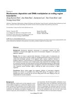
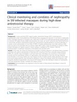
each other [25,26]. The factors that regulate T cell function
and contribute to ovx induced bone loss are shown in
Figure 1.
T cells differentiate in the thymus, an organ that undergoes
progressive structural and functional declines with age,
coinciding with increased circulating sex-steroid levels at
puberty [27]. However, the thymus continues to generate
new T cells even into old age. In fact, active lymphocytic
thymic tissue has been documented in adults up to 107 years
of age [28]. Under severe T cell depletion secondary to HIV
infection, chemotherapy or BM transplant, an increase in
thymic output (known as thymic rebound) becomes critical for
long-term restoration of T cell homeostasis. For example,
middle aged women treated with autologous BM transplants
develop thymic hypertrophy and a resurgence of thymic T cell
output that contributes to the restoration of a wide T cell
repertoire [29], although the intensity of thymic rebound
declines with age.
Restoration of thymic function after castration occurs in
young [30] as well as in very old rodents [31]. Similarly, ovx
increases the thymic export of naïve T cells [23]. Indeed,
stimulated thymic T cell output accounts for approximately
50% of the increase in the number of T cells in the periphery,
while the remaining 50% is due to enhanced peripheral
expansion. Similarly, thymectomy decreases by approximately
50% the bone loss induced by ovx, thus demonstrating that
the thymus plays a previously unrecognized causal effect in
ovx-induced bone loss in mice. The remaining bone loss is a
consequence of the peripheral expansion of naïve and
memory T cells [23]. This finding, which awaits confirmation
in humans, suggests that estrogen deficiency-induced thymic
rebound may be responsible for the exaggerated bone loss in
young women undergoing surgical menopause or for the
rapid bone loss characteristic of women in their first five to
seven years after natural menopause. Indeed, an age-related
decrease in estrogen deficiency-induced thymic rebound
could mitigate the stimulatory effects of sex steroid
deprivation and explain why the rate of bone loss in
postmenopausal women diminishes as aging progresses.
Conclusions
Remarkable progress has been made in elucidating the
crosstalk between the immune system and bone and in
uncovering the mechanism by which sex-steroids, infection
and inflammation lead to bone loss by disregulating T
lymphocyte function in animal models. If the findings in
experimental animals are confirmed in humans, it will,
perhaps, be appropriate to classify osteoporosis as an
inflammatory, or even an autoimmune condition and certainly
new therapeutic ‘immune’ targets will emerge.
Competing interests
The author declares that they have no competing interests.
References
1. Sun L, Peng Y, Sharrow AC, Iqbal J, Zhang Z, Papachristou DJ,
Zaidi S, Zhu LL, Yaroslavskiy BB, Zhou H, et al.: FSH directly
regulates bone mass. Cell 2006, 125:247-260.
2. Iqbal J, Sun L, Kumar TR, Blair HC, Zaidi M: Follicle-stimulating
hormone stimulates TNF production from immune cells to
enhance osteoblast and osteoclast formation. Proc Natl Acad
Sci USA 2006, 103:14925-14930.
3. Hughes DE, Dai A, Tiffee JC, Li HH, Mundy GR, Boyce BF: Estro-
gen promotes apoptosis of murine osteoclasts mediated by
TGF-beta. Nat Med 1996, 2:1132-1136.
4. Jilka RL, Takahashi K, Munshi M, Williams DC, Roberson PK,
Manolagas SC: Loss of estrogen upregulates osteoblastogen-
esis in the murine bone marrow. Evidence for autonomy from
factors released during bone resorption. J Clin Invest 1998,
101:1942-1950.
5. Kousteni S, Bellido T, Plotkin LI, O’Brien CA, Bodenner DL, Han
L, Han K, DiGregorio GB, Katzenellenbogen JA, Katzenellenbogen
BS, et al.: Nongenotropic, sex-nonspecific signaling through
the estrogen or androgen receptors: dissociation from tran-
scriptional activity. Cell 2001, 104:719-730.
6. Weitzmann MN, Roggia C, Toraldo G, Weitzmann L, Pacifici R:
Increased production of IL-7 uncouples bone formation from
bone resorption during estrogen deficiency. J Clin Invest
2002, 110:1643-1650.
7. Gilbert L, He X, Farmer P, Boden S, Kozlowski M, Rubin J, Nanes
MS: Inhibition of osteoblast differentiation by tumor necrosis
factor-alpha. Endocrinology 2000, 141:3956-3964.
8. Eriksen EF, Hodgson SF, Eastell R, Cedel SL, O’Fallon WM,
Riggs BL: Cancellous bone remodeling in type I (post-
Available online />Figure 1
Factors that regulate T cell function and contribute to ovx induced
bone loss. Ag, antigen; BMM, bone marrow macrophages; DC,
dendritic cells; IFN, interferon; IL, interleukin; OC, osteoclast; ovx,
ovariectomy; RANK, receptor activator of NF-κB; RANKL, RANKL
ligand; TGF, transforming growth factor; TNF, tumor necrosis factor.
menopausal) osteoporosis: quantitative assessment of rates
of formation, resorption, bone loss at tissue and cellular
levels. J Bone Miner Res 1990, 5:311-319.
9. Riggs BL, Parfitt AM: Drugs used to treat osteoporosis: the
critical need for a uniform nomenclature based on their action
on bone remodeling. J Bone Miner Res 2005, 20:177-184.
10. Khosla S: Minireview: the OPG/RANKL/RANK system.
Endocrinology 2001, 142:5050-5055.
11. Kong YY, Yoshida H, Sarosi I, Tan HL, Timms E, Capparelli C,
Morony S, Oliveira-dos-Santos AJ, Van G, Itie A, et al.: OPGL is a
key regulator of osteoclastogenesis, lymphocyte development
and lymph-node organogenesis. Nature 1999, 397:315-323.
12. Nanes MS: Tumor necrosis factor-alpha: molecular and cellu-
lar mechanisms in skeletal pathology. Gene 2003, 321:1-15.
13. Cenci S, Weitzmann MN, Roggia C, Namba N, Novack D,
Woodring J, Pacifici R: Estrogen deficiency induces bone loss
by enhancing T-cell production of TNF-alpha. J Clin Invest
2000, 106:1229-1237.
14. Roggia C, Gao Y, Cenci S, Weitzmann MN, Toraldo G, Isaia G,
Pacifici R: Up-regulation of TNF-producing T cells in the bone
marrow: A key mechanism by which estrogen deficiency
induces bone loss in vivo. Proc Natl Acad Sci USA 2001, 98:
13960-13965.
15. Ammann P, Rizzoli R, Bonjour JP, Bourrin S, Meyer JM, Vassalli P,
Garcia I: Transgenic mice expressing soluble tumor necrosis
factor-receptor are protected against bone loss caused by
estrogen deficiency. J Clin Invest 1997, 99:1699-1703.
16. Kimble R, Bain S, Pacifici R: The functional block of TNF but not
of IL-6 prevents bone loss in ovariectomized mice. J Bone Min
Res 1997, 12:935-941.
17. Weitzmann MN, Pacifici R: Estrogen deficiency and bone loss:
an inflammatory tale. J Clin Invest 2006, 116:1186-1194.
18. Gao Y, Qian WP, Dark K, Toraldo G, Lin AS, Guldberg RE, Flavell
RA, Weitzmann MN, Pacifici R: Estrogen prevents bone loss
through transforming growth factor beta signaling in T cells.
Proc Natl Acad Sci USA 2004, 101:16618-16623.
19. Cenci S, Toraldo G, Weitzmann MN, Roggia C, Gao Y, Qian WP,
Sierra O, Pacifici R: Estrogen deficiency induces bone loss by
increasing T cell proliferation and lifespan through IFN-
gamma-induced class II transactivator. Proc Natl Acad Sci
USA 2003, 100:10405-10410.
20. Gao Y, Grassi F, Ryan MR, Terauchi M, Page K, Yang X, Weitz-
mann MN, Pacifici R: IFN-gamma stimulates osteoclast forma-
tion and bone loss in vivo via antigen-driven T cell activation. J
Clin Invest 2007, 117:122-132.
21. Yang NN, Venugopalan M, Hardikar S, Glasebrook A: Identifica-
tion of an estrogen response element activated by metabo-
lites of 17b-estradiol and raloxifene. Science 1996, 273:
1222-1225.
22. Miyaura C, Onoe Y, Inada M, Maki K, Ikuta K, Ito M, Suda T:
Increased B-lymphopoiesis by interleukin 7 induces bone
loss in mice with intact ovarian function: similarity to estrogen
deficiency. Proc Natl Acad Sci USA 1997, 19:9360-9365.
23. Ryan MR, Shepherd R, Leavey JK, Gao Y, Grassi F, Schnell FJ,
Qian WP, Kersh GJ, Weitzmann MN, Pacifici R: An IL-7-depen-
dent rebound in thymic T cell output contributes to the bone
loss induced by estrogen deficiency. Proc Natl Acad Sci USA
2005, 102:16735-16740.
24. Lindberg MK, Svensson J, Venken K, Chavoshi T, Andersson N,
Moverare Skrtic S, Isaksson O, Vanderschueren D, Carlsten H,
Ohlsson C: Liver-derived IGF-I is permissive for ovariectomy-
induced trabecular bone loss. Bone 2006, 38:85-92.
25. Huang M, Sharma S, Zhu LX, Keane MP, Luo J, Zhang L, Burdick
MD, Lin YQ, Dohadwala M, Gardner B, et al.: IL-7 inhibits fibrob-
last TGF-beta production and signaling in pulmonary fibrosis.
J Clin Invest 2002, 109:931-937.
26. Dubinett SM, Huang M, Dhanani S, Economou JS, Wang J, Lee P,
Sharma S, Dougherty GJ, McBride WH: Down-regulation of
murine fibrosarcoma transforming growth factor-beta 1
expression by interleukin 7. J Natl Cancer Inst 1995, 87:593-
597.
27. Haynes BF, Sempowski GD, Wells AF, Hale LP: The human
thymus during aging. Immunol Res 2000, 22:253-261.
28. Steinmann GG, Klaus B, Muller-Hermelink HK: The involution of
the ageing human thymic epithelium is independent of
puberty. A morphometric study. Scand J Immunol 1985, 22:
563-575.
29. Hakim FT, Memon SA, Cepeda R, Jones EC, Chow CK, Kasten-
Sportes C, Odom J, Vance BA, Christensen BL, Mackall CL, et
al.: Age-dependent incidence, time course, and consequences
of thymic renewal in adults. J Clin Invest 2005, 115:930-939.
30. Roden AC, Moser MT, Tri SD, Mercader M, Kuntz SM, Dong H,
Hurwitz AA, McKean DJ, Celis E, Leibovich BC, et al.: Augmenta-
tion of T cell levels and responses induced by androgen
deprivation. J Immunol 2004, 173:6098-6108.
31. Sutherland JS, Goldberg GL, Hammett MV, Uldrich AP, Berzins
SP, Heng TS, Blazar BR, Millar JL, Malin MA, Chidgey AP, et al.:
Activation of thymic regeneration in mice and humans follow-
ing androgen blockade. J Immunol 2005, 175:2741-2753.
Arthritis Research & Therapy Vol 9 No 2 Pacifici
Page 4 of 4
(page number not for citation purposes)