Báo cáo y học: "Morphological characterization of intra-articular HMGB1 expression during the course of collagen-induced arthritis" docx
Bạn đang xem bản rút gọn của tài liệu. Xem và tải ngay bản đầy đủ của tài liệu tại đây (3.38 MB, 10 trang )
Open Access
Available online />Page 1 of 10
(page number not for citation purposes)
Vol 9 No 2
Research article
Morphological characterization of intra-articular HMGB1
expression during the course of collagen-induced arthritis
Karin Palmblad
1,2
, Erik Sundberg
1,2
, Margarita Diez
3
, Riikka Söderling
2
, Ann-Charlotte Aveberger
2
,
Ulf Andersson
1,2
and Helena Erlandsson Harris
2
1
Department of Woman and Child Health, Karolinska Institutet, Astrid Lindgren Children's Hospital, SE-171 76 Stockholm, Sweden
2
Department of Medicine, Rheumatology Research Unit, Center of Molecular Medicine L8:04,, Karolinska Institutet, SE-171 76 Stockholm, Sweden
3
Department of Clinical Neuroscience, Neuroimmunology Unit, Center of Molecular Medicine L8:04, Karolinska Institutet, SE-171 76 Stockholm,
Sweden
Corresponding author: Karin Palmblad,
Received: 18 Nov 2006 Revisions requested: 21 Dec 2006 Revisions received: 9 Feb 2007 Accepted: 30 Mar 2007 Published: 30 Mar 2007
Arthritis Research & Therapy 2007, 9:R35 (doi:10.1186/ar2155)
This article is online at: />© 2007 Palmblad et al.; licensee BioMed Central Ltd.
This is an open access article distributed under the terms of the Creative Commons Attribution License ( />),
which permits unrestricted use, distribution, and reproduction in any medium, provided the original work is properly cited.
Abstract
High-mobility group chromosomal box protein 1 (HMGB1) is a
structural nuclear protein that promotes inflammation when
present extracellularly. Aberrant, extracellular HMGB1
expression has been demonstrated in human and experimental
synovitis. The aim of the present study was to elucidate the
temporal and spatial expression of HMGB1 compared to that of
the central mediators tumor necrosis factor (TNF) and
interleukin-1-beta (IL-1β) during the course of collagen-induced
arthritis. Thus, Dark Agouti rats were immunized with
homologous type II collagen and synovial tissue specimens
were obtained at various time points prior to and during the
course of clinical arthritis. Local cytokine responses were
assessed by immunohistochemistry and by in situ hybridization.
We demonstrate a distinct nuclear expression of HMGB1 at
early disease-preceding time points. Preceding clinical onset by
a few days, cytoplasmic HMGB1 expression was evident in
synoviocytes within the non-proliferative lining layer.
Pronounced cytoplasmic and additional extracellular HMGB1
expression coincided with the progression of clinical disease. In
advanced arthritis, the number of cells with cytoplasmic
HMGB1 expression was quantitatively comparable to that of
cells expressing TNF and IL-1β. Interestingly, although HMGB1
was abundantly expressed throughout the inflamed synovium at
a protein level, upregulation of HMGB1 mRNA was restricted
mainly to areas of cartilage and bone destruction. In conclusion,
these new findings implicate a role for HMGB1 in both inducing
and perpetuating inflammatory events of significant importance
in the destructive processes in chronic arthritis.
Introduction
Rheumatoid arthritis (RA) is characterized by chronic inflam-
mation of multiple joints which leads to the marked destruction
of cartilage and bone. Although the etiology of RA is still
unknown, evidence is accumulating that, once initiated, the
inflammatory process in the synovial tissue is dominated by
activated monocytes/macrophages and fibroblasts. Cytokines
derived from these cell types are abundantly expressed, and it
is now commonly accepted that tumor necrosis factor (TNF)
and interleukin-1 (IL-1) are pivotal mediators in the pathogen-
esis of RA, providing validated targets for successful therapy
[1,2].
High-mobility group chromosomal box protein 1 (HMGB1),
previously called HMG-1 or amphoterin, is an abundant
nuclear component in all eukaryons [3]. Although widely stud-
ied as a DNA-binding protein, HMGB1 has recently been
shown to possess important extracellular functions as well.
Outside the cell, HMGB1 plays a critical role as a pro-inflam-
matory cytokine that mediates lipopolysaccharide (LPS)
lethality, acute lung injury, and smooth muscle cell migration
and induces the release of TNF and IL-1 from macrophages
and dendritic cells [4-7]. In both experimental septic shock
and acute lung injury, treatment targeting HMGB1 ameliorated
inflammation and improved survival. HMGB1 translocation to
the extracellular milieu can occur via two separate
CIA = collagen-induced arthritis; HMGB1 = high-mobility group chromosomal box protein 1; LPS = lipopolysaccharide; Ig = immunoglobulin; IL-1β
= interleukin-1-beta; PBS = phosphate-buffered saline; p.i. = post-immunization; RA = rheumatoid arthritis; TNF = tumor necrosis factor.
Arthritis Research & Therapy Vol 9 No 2 Palmblad et al.
Page 2 of 10
(page number not for citation purposes)
mechanisms. Through a regulated process, stimulated inflam-
matory cells may actively secrete HMGB1 [8-11]. In addition,
HMGB1 can be passively released during disintegration of
necrotic cells. In apoptotic cells, which do not trigger inflam-
mation, HMGB1 is tightly bound to the chromatin, preventing
extracellular release. Necrotic Hmgb1
-/-
cells mediate a mini-
mal inflammatory response, thus implying that HMGB1 is a
critical factor connecting unprogrammed/necrotic cell death
to inflammation [12].
Recent evidence implicates a role for HMGB1 in the patho-
genesis of arthritis (reviewed in [13]). We and others have
demonstrated local overexpression of cytoplasmic and extra-
cellular HMGB1 in synovial biopsy specimens in RA and
experimental arthritis [14,15]. Intra-articular injection of
HMGB1 in mice induces arthritis, and treatment with HMGB1
antagonist attenuates collagen-induced arthritis (CIA) in rats
and mice [16,17].
CIA is a widely used animal model that mimics the joint inflam-
mation evident in human RA. The Dark Agouti rat is particularly
susceptible to CIA, presenting an erosive chronic relapsing
disease in 100% of immunized animals when induced with
homologous collagen type II emulsified with Freund's incom-
plete adjuvant [18]. This model was used in the present study
to elucidate characteristics of HMGB1 expression in compar-
ison with the well-characterized cytokines TNF and IL-1β in the
initiation and progression of arthritis. Local cytokine responses
were determined at the protein level by means of recently
developed immunohistochemical techniques that enable dis-
crimination of the localization of HMGB1 in cellular compart-
ments. In addition, HMGB1 mRNA expression was
determined using in situ hybridization techniques.
Materials and methods
Induction and evaluation of experimental arthritis
Male Dark Agouti rats weighing 220 to 230 g were bred and
kept at the animal unit at Karolinska Hospital in Stockholm,
Sweden. The light/darkness cycle was 12 hours, and the rats
were fed standard rodent chow and water ad libitum. The
health status of the animals was monitored according to
guidelines of the Swedish Veterinary Board (SVA), and the
animals were reported to be free of screened pathogens. The
Stockholm North Ethical Committee, Sweden, approved all of
the procedures during the experiments. On day 0, 28 rats
were immunized intradermally in the base of the tail with rat
type II collagen emulsified with Freund's incomplete adjuvant
(Difco, Detroit, MI, USA) as previously described [18]. With
this protocol, chronic polyarthritis is expected to develop in
100% of the animals and clinical onset occurs at approxi-
mately day 15 after immunization. The paws of the rats were
monitored daily for visual inflammatory signs such as erythema
and swelling by means of a previously described scoring sys-
tem [19]. Arthritis was graded semiquantitatively on a scale of
0 to 4 for each paw. An arthritis index that expressed a cumu-
lative score for all paws (maximum possible value = 16) was
calculated for each animal.
Preparation of samples for immunohistochemical
analysis
Thirty-two animals were included in this longitudinal trial. Four
unimmunized animals were sacrificed at day 0 as normal con-
trols. Three early time points (3, 6, and 10 days post-immuni-
zation [p.i.]), the time point of expected onset (day 15 p.i.), the
time point for expected maximal clinical severity of arthritis (day
21 p.i.), the time point for transition to a chronic phase of dis-
ease (day 28 p.i.), and a late time point (day 38 p.i.) were
selected, and four animals per time point were sacrificed. To
examine and compare local histology, rats were perfused in
vivo with paraformaldehyde solution; paws were then dis-
sected and decalcified using a modification of a protocol pre-
viously described [20]. Briefly, animals were deeply
anesthetized by intraperitoneal injection of a mixture of equal
volumes of Hypnorm
®
(fentanyl citrate 0.315 mg/ml and flu-
anisone 10 mg/ml; Janssen Pharmaceutica N.V., Beerse, Bel-
gium) and Dormicum
®
(midazolam 1 mg/ml; Roche,
Stockholm, Sweden), diluted 1:2 in sterile water, in which 800
μl per 200 g of animal's body weight was given. Central intra-
arterial perfusion with phosphate-buffered saline (PBS) pre-
ceded perfusion with the fixative, which consisted of 4% (wt/
vol) paraformaldehyde (Merck, Darmstadt, Germany) in 0.2 M
Sörensen phosphate buffer, pH 7.2, containing 0.2% picric
acid (Riedel-de Haën, Seelze, Germany). Ankle joints were
dissected and immersed in the same fixative overnight at room
temperature and thereafter thoroughly washed in PBS twice
daily for three to four days until clear of picric acid. The joint
specimens were then subjected to demineralization in a 4%
(wt/vol) EDTA (ethylenediaminetetraacetic acid) (Sigma-
Aldrich, St. Louis, MO, USA) solution containing 0.2 M sodium
cacodylate (Sigma-Aldrich), pH 7.3, for approximately four
weeks, followed by eight days in 20% (wt/vol) sucrose
(Sigma-Aldrich) in 0.1 M Sörensen phosphate buffer, pH 7.2,
containing 0.01% (wt/vol) sodium azide (Sigma-Aldrich). The
ankle joints were then cut in saggital sections of 7 to 8 μm in
thickness by means of a Leica Cryostat (Leica, Wetzlar, Ger-
many). The sections were mounted directly on Superfrost
slides (Novakemi AB, Stockholm, Sweden), air-dried at room
temperature, and subsequently stored at -70°C until used for
staining. Because the arthritic lesions were symmetrical and
scoring in the hind paws was equal, only one paw per rat was
studied.
Immunohistochemical stainings
To detect expressions of HMGB1, TNF, and IL-1β, sections
were stained according to immunohistochemical methods pre-
viously described by us [21]. The primary antibodies used
were a peptide affinity-purified polyclonal rabbit anti-HMGB1
antibody (cat. no. 556528; BD Pharmingen, San Diego, CA,
USA), a polyclonal ligand affinity-purified rabbit anti-rat TNF
(8–14; U-CyTech biosciences, Utrecht University, Utrecht,
Available online />Page 3 of 10
(page number not for citation purposes)
The Netherlands), and a polyclonal ligand affinity-purified goat
anti-rat IL-1β (AF-501-NA; R&D Systems, Inc., Minneapolis,
MN, USA). The HMGB1 antibody was used at a final concen-
tration of 1 μg/ml, and the TNF and IL-1β antibodies were
used at a final concentration of 2 μg/ml.
In each assay, controls for specificity of cytokine stainings
based on parallel staining studies omitting the primary anti-
body or using primary isotype-matched immunoglobulin (Ig) of
irrelevant antigen specificity at the same concentration as the
cytokine-detecting antibodies were included. The irrelevant
control antibodies used in the present study were fractioned
rabbit Ig (no. XO936; DakoCytomation, Glostrup, Denmark)
and goat anti-human IL-2 (AF-202; R&D Systems, Inc.). The
specificities of extracellular and intracellular cytokine immuno-
reactivities were verified by their complete inhibition in block-
ing experiments with preabsorption of the cytokine-specific
antibody with recombinant target cytokine prior to staining. In
addition, a morphology of HMGB1 expression similar to the
stainings demonstrated in this report using the BD Pharmin-
gen anti-HMGB1 antibody was obtained using a polyclonal
peptide affinity-purified rabbit anti-HMGB1 antibody pur-
chased from Innovagen AB (Lund, Sweden). These two anti-
body preparations recognize separate epitopes of the
HMGB1 molecule.
Evaluation of the stained sections
By means of a Polyvar II microscope (Reichert-Jung, now part
of Leica Microsystems Nussloch GmbH, Nussloch, Germany)
connected to a 3-CCD (charge-coupled device) color camera
(DXC-750P; Sony Corporation, Tokyo, Japan), slides were
evaluated by two independent observers blinded to the iden-
tity of the specimens. All animals were studied in at least three
separate staining experiments for each given cytokine. The rel-
ative frequencies of positively stained cells in the articular tis-
sue were estimated and assigned an expression score on a
scale of 0 to 4: 0, negative cells; 0.5, less than 1%; 1, 1% to
5%; 2, 5% to 20%; 3, 20% to 50%; and 4, more than 50%
positively stained cells.
Immunofluorescence two-color staining
To determine the phenotype of the HMGB1-releasing cells,
we performed a two-color staining of HMGB1 and ED1 (Sero-
tec Ltd, Oxford, UK), a surface membrane antigen expressed
on rat macrophages, monocytes, and dendritic cells, by means
of a modified staining protocol. Briefly, PBS supplemented
with 0.1% (wt/vol) saponin (Riedel-de Haën) was used in all
subsequent washes and incubation steps. Endogenous biotin
was blocked with avidin for 30 minutes and with biotin for an
additional 15 minutes (avidin/biotin blocking kit; Vector Labo-
ratories, Burlingame, CA, USA), both substituted with 0.1%
saponin. Sections were then incubated overnight with a mix-
ture of primary antibodies directed against HMGB1 and ED1,
supplemented with 0.1% Aurion BSA-c (acetylated bovine
serum albumin) (10%) (Scandinavian Medical Services, Hels-
ingborg, Sweden) to reduce background staining due to non-
specific binding sites. HMGB1 staining was developed with a
secondary biotin-labeled Fab
2
-fragmented donkey anti-rabbit
antibody (Jackson ImmunoResearch Laboratories, Inc., West
Grove, PA, USA) diluted 1:1,000, followed by the streptavidin-
conjugated fluorophore Oregon green at 2 μg/ml; both incu-
bations were performed for 30 minutes. Subsequently, after
another blocking with avidin for 30 minutes and biotin for an
additional 15 minutes, the surface antigen ED1 staining was
developed with biotin-labeled Fab
2
-fragmented donkey anti-
mouse antibody (Jackson ImmunoResearch Laboratories, Inc.)
diluted 1:1,000 for 30 minutes, followed by a 30-minute incu-
bation with streptavidin-conjugated Alexa 546 (red fluoro-
phore) coupled to avidin diluted 1:400 in PBS-saponin. Slides
were air-dried and then mounted with PBS-buffered glycerol.
Slides were examined with a Polyvar 2 UV microscope (Leica
Microsystems Nussloch GmbH) equipped with a 200-W mer-
cury lamp.
In situ hybridization
A 50-base pair oligonucleotide probe for HMGB1 (TCTTCT-
TCCTCCTCTTCCTCATCCTCTTCATCCTC-
CTCGTCGTCTTCCTC) and a random probe having no
similarities to known sequences (GenBank, National Institutes
of Health, Bethesda, MD, USA) were synthesized (DNA Tech-
nology A/S, Århus, Denmark). In situ hybridization was per-
formed as previously described [22]. Briefly, oligonucleotide
probes were labeled with
33
P-dATP (DuPont-New England
Nuclear, now part of PerkinElmer Life and Analytical Sciences,
Inc., Waltham, MA, USA) at the 3' end by means of terminal
deoxynucleotidyltransferase (Amersham, now part of GE
Healthcare, Little Chalfont, Buckinghamshire, UK) and purified
through QIAquick spin columns (Qiagen GmbH, Hilden, Ger-
many). Sections were hybridized overnight at 42°C in humidi-
fied boxes with 0.5 ng of labeled probe (1 to 4 × 10
6
cpm/l)
per slide in a hybridization cocktail and rinsed 5 × 15 minutes
in saline sodium citrate at 60°C. As a control, an excess
(×100) of cold probe was added to the hybridization cocktail.
Tissue sections were dehydrated, air-dried, dipped in NTB2
nuclear track photographic emulsion (Eastman Kodak,
Rochester, NY, USA), and exposed for 7 to 14 days at 8°C.
Dipped slides were developed for 4 minutes in D19 (Eastman
Kodak), fixed in Unifix (Eastman Kodak) for 7 minutes, and
rinsed in tap water for 20 minutes. After air-drying, sections
were counterstained with eosin-hematoxylin and mounted.
Results
Cytokine expression before onset of arthritis
Immunohistochemical stainings were performed to study the
spatial and temporal cytoplasmic expression of the novel
cytokine HMGB1 compared to those of IL-1β and TNF in syn-
ovial tissue specimens at different time points after immuniza-
tion with type II collagen. In synovial sections from animals
sacrificed before the onset of disease (before day 15 p.i.), the
Arthritis Research & Therapy Vol 9 No 2 Palmblad et al.
Page 4 of 10
(page number not for citation purposes)
synovial tissue appeared (as expected) non-proliferative, con-
taining only a few cell layers.
Joint tissue specimens at these disease-preceding time points
revealed a strict nuclear cellular localization of detectable
HMGB1 in virtually all cells in the synovial membrane (Figures
1a and 2a). Although cytoplasmic HMGB1 expression was
scarce at early disease-preceding time points, more evident
signs of extranuclear deposition of HMGB1 appeared at day
10 p.i. (Figure 2b), thus preceding the time for clinical disease
onset by five days. This extranuclear HMGB1 appeared as a
general cytoplasmic staining in a large portion of cells in the
lining layer. At this time point, the synovial membrane remained
unproliferative with an appearance indistinguishable from
unimmunized animals.
Scattered TNF- and IL-1β-expressing cells could even be rec-
ognized in the superficial cell layer of the synovial lining in
unimmunized animals (Figure 1b,c). Sections from days 3 to
15 p.i. displayed a rather congruent picture. One difference
was evident between expressions of these two cytokines.
Chondrocytes expressing IL-1β could be detected at all time
points with a more prominent expression in the superficial
articular cartilage layer, whereas the cartilage remained nega-
tive for TNF expression throughout the study (Figures 1 and 3;
Table 1). The chondrocyte staining pattern of IL-1β resembled
that of HMGB1 (Figures 1a,b and 3a,b).
Cytokine expression after onset of arthritis
At the time point of clinical disease onset (day 15 p.i.), the first
signs of cell infiltration were noted and the synovial membrane
increased in thickness. Although the arthritis index varied
within the group of four animals studied per time point, the
estimated expression scores of the cytokine expression
appeared to be very similar within the group with reproducible
results in at least three staining experiments. A substantial
number of the first infiltrating inflammatory cells expressed
HMGB1 in their cytoplasm, which was apparently more pro-
nounced than the expressions of TNF or IL-1β.
However, a more evident presence of all three studied
cytokines coincided with the progression of clinical disease
(Table 1). Accordingly, maximal cytokine expression was
recorded from day 21 p.i. onward, corresponding to maximal
paw swelling, cell infiltration, and manifestation of erosive
changes in cartilage and bone (Figure 3). At these time points,
the number of TNF-expressing cells dominated quantitatively
throughout the synovial tissue, the most abundant expression
being within the lining layer. Both spatial and quantitative
aspects of extranuclear HMGB1 expression were similar to
those of IL-1β, in which most of the expression was evident
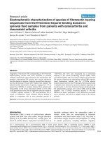
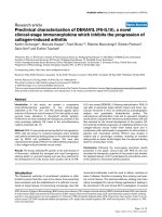
Figure 1
Synovial cytokine expression at an early disease-preceding time pointSynovial cytokine expression at an early disease-preceding time point. Representative micrographs illustrating immunohistochemical staining of cry-
ocut synovial tissue for expressions of high-mobility group chromosomal box protein 1 (HMGB1) (a), interleukin-1-beta (IL-1β) (b), and tumor necro-
sis factor (TNF) (c) three days after immunization. A thin, non-proliferative synovia is evident at this disease-preceding time point. TNF- and IL-1β-
expressing cells are located in superficial parts of the synovial lining layer. HMGB1 expression was restricted to cell nuclei at this early time point. (d)
A representative section is stained with irrelevant control antibody. Original magnification ×125.
Available online />Page 5 of 10
(page number not for citation purposes)
within sublining and pannus regions (Table 1). The most
prominent staining of cytoplasmic HMGB1 and of IL-1β was
located in erosive parts of synovial tissue close to cartilage
and bone undergoing destruction. As opposed to expression
of TNF, those of both cytoplasmic HMGB1 and IL-1β were
lower within the lining layer. Cytoplasmic HMGB1 could be
demonstrated in many macrophage-like cells, and the nuclear
HMGB1 staining in a subset of these cells was clearly
reduced or absent. Two-color staining revealed that a substan-
tial number of cells with cytoplasmic HMGB1 expression were
also positive for ED1, a marker for rat macrophages and den-
dritic cells (Figure 4). An extracellular presence of HMGB1
was indicated by a brownish immunoreactivity that encom-
passed cells displaying cytoplasmic HMGB1 staining in the
inflamed synovium (Figure 2a).
Scattered cells were stained for all studied cytokines and were
distributed in the interstitial tissue, perivascularly, and within
the vessel endothelium. A quantitative difference was also
evident in that almost all vessel endothelium cells stained pos-
itively for TNF, whereas several vessels remained unstained for
IL-1β and a nuclear staining pattern dominated for HMGB1,
although endothelium cells with cytoplasmic HMGB1 could
also be visualized (Table 1). The expressions of all three stud-
ied pro-inflammatory cytokines were still prominent at days 28
and 38 p.i., when (in clinical terms) a transition of the acute
inflammation to a chronic phase occurred.
HMGB1 mRNA expression
A low mRNA expression was detected in most cells at all time
points, even in paw sections of healthy unimmunized animals.
Because HMGB1 is an abundantly displayed protein in all cell
nuclei (where it binds to DNA-regulating transcription [23]),
these findings were expected. The abundant extranuclear
HMGB1 protein expression throughout the inflamed synovium
in advanced arthritis, however, was not accompanied by an
overall upregulation of HMGB1 mRNA. In contrast, upregula-
tion of HMGB1 mRNA was restricted mainly to synovial tissue
adjacent to areas with cartilage and bone destruction, where
the expression was pronounced with cells expressing numer-
ous grains (Figure 5; Table 2). No detectable labeling
appeared after hybridization with cold probe or random probe.
Discussion
In the present report, we provide evidence that, in rats with
CIA, the number of synovial cells expressing cytoplasmic
HMGB1 may be quantitatively comparable with the number of
cells expressing the well-characterized cytokines TNF and IL-
1β. HMGB1 is released as a late mediator during acute inflam-
mation and plays a crucial role in the pathogenesis of systemic
inflammation in sepsis after the early mediator response has
resolved [4,24]. We thus anticipated analogous results in the
present study of chronic inflammation, with synovial expres-
sion of TNF and IL-1β preceding that of HMGB1. However, we
did not observe a distinct, sequential order of appearance of
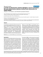
Figure 2
High-mobility group chromosomal box protein 1 (HMGB1) expression at different time points after immunizationHigh-mobility group chromosomal box protein 1 (HMGB1) expression
at different time points after immunization. Representative micrographs
illustrating immunohistochemical staining of HMGB1. (a) In the non-
proliferative synovial membrane of an unimmunized animal, a nuclear
HMGB1 deposition is evident. (b) In addition to the nuclear expression,
cytoplasmic HMGB1 staining appears in a large portion of cells in the
synovial membrane 10 days after immunization, a time point preceding
the expected clinical disease onset by 5 days. (c) An arthritic lesion, 28
days after immunization, in which an additional extracellular presence of
HMGB1 is indicated by a brownish extracellular immunoreactivity sur-
rounding cells displaying cytoplasmic HMGB1 staining. Original magni-
fication ×500.
Arthritis Research & Therapy Vol 9 No 2 Palmblad et al.
Page 6 of 10
(page number not for citation purposes)
the three cytokines studied. At early disease-preceding time
points, cellular HMGB1 expression was almost exclusively
restricted to the nuclear compartment. Interestingly, a more
evident extranuclear staining pattern for HMGB1 was noted in
resident cells in the synovium even a few days before the onset
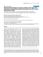
Figure 3
The number of cells expressing extranuclear high-mobility group chromosomal box protein 1 (HMGB1) is quantitatively comparable to the number of cells expressing tumor necrosis factor (TNF) and interleukin-1-beta (IL-1β) in arthritic jointsThe number of cells expressing extranuclear high-mobility group chromosomal box protein 1 (HMGB1) is quantitatively comparable to the number of
cells expressing tumor necrosis factor (TNF) and interleukin-1-beta (IL-1β) in arthritic joints. Representative micrographs illustrating immunohisto-
chemical staining of synovial tissue from an arthritic animal at day 21 after immunization. Sequential cryocut sections were analyzed for expressions
of HMGB1 (a), IL-1β (b), and TNF (c). Abundant expressions were demonstrated for all three cytokines. (d) A section stained with an irrelevant iso-
type-matched control. Original magnification ×125.
Table 1
Expression of extranuclear HMGB1 compared to those of TNF and IL-1β
Lining Sublining Destructive zone Cartilage Vessels
Days p.i. M.A.I. HMGB1 TNF IL-1β HMGB1 TNF IL-1β HMGB1 TNF IL-1β HMGB1 TNF IL-1β HMGB1 TNF IL-1β
1 1111
0 0 0 0.50.5 0 00 - - - 1 01 0 00
3 0 0 0.50.5 0 00 - - - 1 01 0 00
6 0 0 0.50.5 0 00 - - - 1 01 0 00
10 0 0.5 0.5 0.5 0.5 0 0 - - - 1 0 1 0 0 0
15 3.2 ± 1.8 1 1 1 1 0.5 0 - - - 1 0 1 0 0.5 0
21 9.8 ± 3.3 2 4 2 3 3 3 3 3 3 2 0 2 1 3 1
28 11.0 ± 2.3 2 4 2 3 3 3 3 3 3 2 0 2 1 3 1
38 11.8 ± 2.6 2 4 2 3 3 3 3 3 3 2 0 2 1 3 1
The relative frequencies of positively stained cells in the articular tissue were estimated by immunohistochemistry and assigned an expression
score on a scale of 0 to 4: 0, negative cells; 0.5, less than 0.5%; 1, 0.5% to 5%; 2, 5% to 20%; 3, 20% to 50%; and 4, more than 50% positively
stained cells. For clinical evaluation, a mean arthritis index (M.A.I.) was calculated for the group of four animals studied per time point and
expressed as the mean ± standard deviation. The destructive zone did not appear until day 21 p.i. and was defined as synovial tissue adjacent to
areas of cartilage and bone destruction. HMGB1, high-mobility group chromosomal box protein 1; IL-1β, interleukin-1-beta; p.i., post-
immunization; TNF, tumor necrosis factor.
Available online />Page 7 of 10
(page number not for citation purposes)
of clinical disease. In addition, at the time point of arthritis
onset, a substantial number of the infiltrating inflammatory cells
expressed HMGB1 in their cytoplasm, apparently more than
cells expressing TNF or IL-1β. Thus, in the context of chronic
inflammation, HMGB1 may be considered an early mediator.
This is in concordance with the demonstration of HMGB1 as
an early mediator of inflammation following acute, local organ
injury in liver ischemia reperfusion [25] as well as in post-
ischemic brain injury [26].
All three macrophage-derived cytokines studied in this report
were abundantly detected in synovial tissues with established
arthritis. However, some clear differences were also discerni-
ble. A distinct TNF expression was observed in the lining layer
as well as in sublining areas in synovitis, whereas HMGB1 and
IL-1β expressions were most often restricted to the sublining
areas. HMGB1 and IL-1β were abundantly displayed in
chondrocytes, especially in those located superficially in the
articular cartilage, whereas no TNF was detectable in
chondrocytes at any time point. The discrepancy between TNF
and IL-1β expressions in chondrocytes was unexpected.
Although the destructive effects of IL-1 on cartilage and bone
are well recognized, the biological implication that chondro-
cytes also express IL-1β remains unclear. The similarities
between IL-1β and HMGB1 expressions in synovitis are note-
worthy. Both cytokines lack a signal peptide [27,28], and it
was recently shown that both are secreted by myeloid cells
through a non-classical pathway involving regulated exocyto-
sis of secretory lysosomes [8].
Rheumatoid synovium is characterized by excessive growth
and invasion into adjacent tissues, including bone and carti-
lage. In many ways, it behaves and appears like a locally inva-
sive tumor in the joints. Extracellular HMGB1 is known to bind
to several components of the plasminogen activation system
and to enhance the activity of tissue plasminogen activator
[29] and matrix metalloproteinases 2 and 9 [30]. Degradation
of extracellular matrix proteins is an important step in cell
migration processes. The HMGB1-promoted increase of
extracellular protease activity might enable cells to migrate and
invade, analogous to the migratory response elicited in smooth
muscle cells [31]. Because HMGB1 initiates endothelial
growth as well as endothelial cell migration and sprouting, it
has also been identified as an angiogenetic switch molecule
[32]. Synovial angiogenesis is thought to be a critical compo-
nent in RA pathogenesis, contributing to pannus proliferation,
infiltration of inflammatory leukocytes, as well as osteophyte
formation [33]. In the present study, we demonstrate that cyto-
plasmic and extracellular HMGB1 appears early in the devel-
opment of arthritis. We speculate that HMGB1 might be a
major contributor to pannus formation in chronic arthritis.
Surprisingly, although our immunohistochemical analyses
demonstrate the abundant presence of extranuclear HMGB1
throughout the inflamed synovium at a protein level, assess-
ment with in situ hybridization reveals that the predominant
upregulation of HMGB1 mRNA is restricted to synovial tissue
adjacent to areas with cartilage and bone destruction.
Figure 4
A substantial portion of cells expressing cytoplasmic high-mobility group chromosomal box protein 1 (HMGB1) are also ED1-positiveA substantial portion of cells expressing cytoplasmic high-mobility
group chromosomal box protein 1 (HMGB1) are also ED1-positive. (a)
Micrograph illustrating a high magnification of inflamed synovial tissue
stained with hematoxylin (arrows). (b) The intranuclear HMGB1 stain-
ing (Oregon green) of resident cells is evident. A substantial portion of
cells express extranuclear HMGB1 (arrows). (c) A substantial portion
of cells expressing extranuclear HMGB1 were also ED1-positive (Red
Alexa 546) (arrows). ED1 is a surface membrane antigen expressed on
rat macrophages, monocytes, and dendritic cells. Original magnifica-
tion ×800.
Arthritis Research & Therapy Vol 9 No 2 Palmblad et al.
Page 8 of 10
(page number not for citation purposes)
HMGB1 is an abundant nuclear protein. Most cells contain
approximately 1 × 10
6
HMGB1 molecules in their nuclei [34],
which may be translocated actively to the cytoplasm upon
stimulation or passively by necrotic cells. De novo synthesis is
thus not required for extranuclear expression. We speculate
that the upregulated mRNA expression in the destructive zone
may be of quantitative importance. It was recently demon-
strated that osteoblasts themselves express HMGB1 as well
as its signaling receptor RAGE (receptor for advanced
glycation end products) and are capable of releasing HMGB1
[35]. Thus, HMGB1 represents a functional link between bone
and immune cells. The precise role of HMGB1 in bone home-
ostasis and tissue destruction remains to be elucidated.
Similar to other cytokines, HMGB1 has differential tissue-spe-
cific activities. In addition to its potent pro-inflammatory capac-
ities, HMGB1 has been accredited a role in tissue repair. As a
signal of tissue damage, HMGB1 mediates tissue regenera-
tion by inducing mesoangioblast migration and proliferation
[36]. Synovial tissue has a strong capacity to regenerate, and
not surprisingly mesenchymal stem cells have been isolated
from synovium [37]. Recently, it was demonstrated that
HMGB1 can induce myocardial regeneration after infarction;
injection of HMGB1 into mouse hearts after ischemic damage
resulted in the formation of new myocytes by inducing cardiac
stem cell proliferation and differentiation [38]. Inflammation is
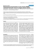
Figure 5
High-mobility group chromosomal box protein 1 (HMGB1) mRNA is predominantly upregulated in areas of tissue destructionHigh-mobility group chromosomal box protein 1 (HMGB1) mRNA is predominantly upregulated in areas of tissue destruction. Micrographs illustrat-
ing intra-articular HMGB1 mRNA expression in advanced arthritis assessed by in situ hybridization. (a) A pronounced upregulation of HMGB1
mRNA is evident in synovial tissue close to cartilage and bone undergoing destruction, detailed in (b).
Table 2
HMGB1 expression at a protein level compared to HMGB1 mRNA expression
Lining Sublining Destructive zone Cartilage Vessels
Days p.i. M.A.I. HMGB1
protein
HMGB1
mRNA
HMGB1
protein
HMGB1
mRNA
HMGB1
protein
HMGB1
mRNA
HMGB1
protein
HMGB1
mRNA
HMGB1
protein
HMGB1
mRNA
0 0 00.500.5 - - 10.500.5
3 0 00.500.5 - - 10.500.5
6 0 00.500.5 - - 10.500.5
10 0 0.5 0.5 0.5 0.5 - - 1 0.5 0 0.5
15 3.2 ± 1.8 1 0.5 1 0.5 - - 1 0.5 0 0.5
219.8 ± 3.32131342111
2811.0 ± 2.32131342111
3811.8 ± 2.62131342111
Most cells expressed a low HMGB1 mRNA labeling at all time points. To be able to compare the relative frequencies of cells with HMGB1 protein
expression to those of cells with upregulated HMGB1 mRNA expression, an estimation of expression scores similar to that used in Table 1 was
used for in situ results; cells with numerous grains were regarded as positive. HMGB1, high-mobility group chromosomal box protein 1; M.A.I.,
mean arthritis index; p.i., post-immunization.
Available online />Page 9 of 10
(page number not for citation purposes)
the common driving force leading to cartilage, bone, and soft
tissue destruction in chronic arthritis. Many factors involved in
the regulation of normal tissue, in particular cartilage and bone,
are dysregulated in arthritic diseases (reviewed in [39]). The
persistence of synovial inflammation and its structural reorgan-
ization can be considered a remodeling process with abnormal
tissue responses such as cartilage calcification and ankylosis
that contribute to disease progression and loss of joint func-
tion. HMGB1 is a comprehensive cytokine that is able to
orchestrate the regulation of both inflammation and tissue
regeneration to promote wound healing, depending on differ-
ent factors such as dose, spatio-temporal expression, target
cells, and possibly even post-translational modifications of the
secreted protein.
Conclusion
We have demonstrated that extranuclear HMGB1 appears
early in disease progression and is abundantly expressed in
advanced arthritis. This suggests that, in chronic arthritis,
HMGB1 may be considered an early mediator involved in both
induction and perpetuation of inflammatory processes. The
progressive destruction of cartilage and subchondral bone
represents a major unsolved consequence of chronic arthritis.
The marked presence of HMGB1 mRNA in the microenviron-
ment of bone and cartilage destruction likely represents
another functional link between inflammation and tissue
destruction. Blockade of extracellular HMGB1 may offer a
novel therapeutic alternative for the treatment of RA.
Competing interests
The authors declare that they have no competing interests.
Authors' contributions
KP helped conceive of the study, shared responsibility for
study design coordination and was responsible for most of the
experiments, data analysis and drafting of the manuscript.
HEH helped conceive of the study and shared responsibility
for study design coordination and drafting of the manuscript.
ES assisted with the immunization, scoring, and collection of
tissue specimens and contributed to the interpretation and
discussion of data. RS assisted with the immunization, scor-
ing, and collection of tissue specimens. MD assisted with and
shared her knowledge of the in situ hybridization technique. A-
CA assisted with the two-color staining. UA contributed to the
interpretation and discussion of data. All authors read and
approved of the final manuscript.
Acknowledgements
The authors thank Robert A Harris for linguistic advice. This work was
supported by grants from the Swedish Society for Medical Research,
Åke Wiberg's Foundation, the Swedish Association against Rheuma-
tism, the Swedish National Cancer Foundation, the Freemason Lodge
Barnhuset in Stockholm, and the Swedish Research Council.
References
1. Feldmann M: Development of anti-TNF therapy for rheumatoid
arthritis. Nat Rev Immunol 2002, 2:364-371.
2. Dinarello CA: Biological basis for interleukin-1 in disease.
Blood 1996, 87:2095-2147.
3. Einck L, Bustin M: The intracellular distribution and function of
the high mobility group chromosomal proteins. Exp Cell Res
1985, 156:295-310.
4. Wang H, Bloom O, Zhang M, Vishnubhakat JM, Ombrellino M, Che
J, Frazier A, Yang H, Ivanova S, Borovikova L, et al.: HMG-1 as a
late mediator of endotoxin lethality in mice. Science 1999,
285:248-251.
5. Abraham E, Arcaroli J, Carmody A, Wang H, Tracey KJ: HMG-1 as
a mediator of acute lung inflammation. J Immunol 2000,
165:2950-2954.
6. Andersson U, Wang H, Palmblad K, Aveberger A-C, Bloom O,
Erlandsson-Harris H, Janson A, Kokkola R, Yang H, Tracey KJ:
HMG-1 stimulates proinflammatory cytokine synthesis in
human monocytes. J Exp Med 2000, 192:565-570.
7. Yang D, Chen Q, Yang H, Tracey KJ, Bustin M, Oppenheim JJ:
High mobility group box-1 (HMGB1) protein induces the
migration and activation of human dendritic cells and acts as
an alarmin. J Leukoc Biol 2007, 81:59-66.
8. Gardella S, Andrei C, Ferrera D, Lotti LV, Torrisi MR, Bianchi ME,
Rubartelli A: The nuclear protein HMGB1 is secreted by mono-
cytes via a non-classical, vesicle-mediated secretory pathway.
EMBO Rep 2002, 3:995-1001.
9. Dumitriu IE, Baruah P, Valentinis B, Voll RE, Herrmann M, Nawroth
PP, Arnold B, Bianchi ME, Manfredi AA, Rovere-Querini P:
Release of high mobility group box 1 by dendritic cells con-
trols T cell activation via the receptor for advanced glycation
end products. J Immunol 2005, 174:7506-7515.
10. Semino C, Angelini G, Poggi A, Rubartelli A: NK/iDC interaction
results in IL-18 secretion by DCs at the synaptic cleft followed
by NK cell activation and release of the DC maturation factor
HMGB1. Blood 2005, 106:609-616.
11. Bonaldi T, Talamo F, Scaffidi P, Ferrera D, Porto A, Bachi A, Rubar-
telli A, Agresti A, Bianchi ME: Monocytic cells hyperacetylate
chromatin protein HMGB1 to redirect it towards secretion.
Embo J 2003, 22:5551-5560.
12. Scaffidi P, Misteli T, Bianchi ME: Release of chromatin protein
HMGB1 by necrotic cells triggers inflammation. Nature 2002,
418:191-195.
13. Jiang W, Pisetsky DS: Mechanisms of disease: the role of high-
mobility group protein 1 in the pathogenesis of inflammatory
arthritis. Nat Clin Pract Rheumatol 2007, 3:52-58.
14. Kokkola R, Sundberg E, Ulfgren AK, Palmblad K, Li J, Wang H,
Ulloa L, Yang H, Yan XJ, Furie R, et al.: High mobility group box
chromosomal protein 1: a novel proinflammatory mediator in
synovitis. Arthritis Rheum 2002, 46:2598-2603.
15. Taniguchi N, Kawahara K, Yone K, Hashiguchi T, Yamakuchi M,
Goto M, Inoue K, Yamada S, Ijiri K, Matsunaga S, et al.: High
mobility group box chromosomal protein 1 plays a role in the
pathogenesis of rheumatoid arthritis as a novel cytokine.
Arthritis Rheum 2003, 48:971-981.
16. Pullerits R, Jonsson I-M, Verdrengh M, Bokarewa M, Andersson U,
Erlandsson Harris H, Tarkowski A: High mobility group chromo-
somal protein 1, a DNA binding cytokine, induces arthritis.
Arthritis Rheum 2003, 48:1693-1700.
17. Kokkola R, Li J, Sundberg E, Aveberger A-C, Palmblad K, Yang H,
Tracey K, Andersson U, Erlandsson Harris H: Successful therapy
in collagen-induced arthritis in mice and rats targeting extra-
cellular HMGB1 activity. Arthritis Rheum 2003, 48:2052-2058.
18. Larsson P, Kleinau S, Holmdahl R, Klareskog L: Homologous type
II collagen-induced arthritis in rats. Characterization of the dis-
ease and demonstration of clinically distinct forms of arthritis
in two strains of rats after immunization with the same colla-
gen preparation. Arthritis Rheum 1990, 33:693-701.
19. Åkerlund K, Erlandsson-Harris H, Tracey KJ, Wang H, Fehniger T,
Klareskog L, Andersson J, Andersson U: Anti-inflammatory
effects of a new tumour necrosis factor-alpha (TNF-α) inhibi-
tor (CNI-1493) in collagen-induced arthritis (CIA) in rats. Clin
Exp Immunol 1999, 115:32-41.
20. Ahmed M, Bjurholm A, Schultzberg M, Theodorsson E, Kreicbergs
A: Increased levels of substance P and calcitonin gene-related
peptide in rat adjuvant arthritis. A combined immunohisto-
Arthritis Research & Therapy Vol 9 No 2 Palmblad et al.
Page 10 of 10
(page number not for citation purposes)
chemical and radioimmunoassay analysis. Arthritis Rheum
1995, 38:699-709.
21. Palmblad K, Harris-Erlandsson H, Tracey KJ, Andersson U:
Dynamics of early synovial cytokine expression in rodent col-
lagen-induced arthritis: a therapeutic study using a macro-
phage deactivating compound. Am J Pathol 2001,
158:491-500.
22. Diez M, Koistinaho J, Dearmond SJ, Groth D, Prusiner SB, Hökfelt
T: Marked decrease of neuropeptide Y Y2 receptor binding
sites in the hippocampus in murine prion disease. Proc Natl
Acad Sci USA 1997, 94:13267-13272.
23. Bianchi ME: Significant (re)location: how to use chromatin
and/or abundant proteins as messages of life and death.
Trends Cell Biol 2004, 14:287-293.
24. Yang H, Ochani M, Li J, Qiang X, Tanovic M, Harris HE, Susarla
SM, Ulloa L, Wang H, DiRaimo R, et al.: Reversing established
sepsis with antagonists of endogenous high-mobility group
box 1. Proc Natl Acad Sci USA 2004, 101:296-301.
25. Tsung A, Sahai R, Tanaka H, Nakao A, Fink MP, Lotze MT, Yang H,
Li J, Tracey KJ, Geller DA, et al.: The nuclear factor HMGB1
mediates hepatic injury after murine liver ischemia-reper-
fusion. J Exp Med 2005, 201:1135-1143.
26. Kim JB, Sig Choi J, Yu YM, Nam K, Piao CS, Kim SW, Lee MH, Han
PL, Park JS, Lee JK: HMGB1, a novel cytokine-like mediator
linking acute neuronal death and delayed neuroinflammation
in the postischemic brain. J Neurosci 2006, 26:6413-6421.
27. Stevenson FT, Torrano F, Locksley RM, Lovett DH: Interleukin 1:
the patterns of translation and intracellular distribution sup-
port alternative secretory mechanisms. J Cell Physiol 1992,
152:223-231.
28. Ferrari S, Ronfani L, Calogero S, Bianchi ME: The mouse gene
coding for High Mobility Group 1 protein (HMG1). J Biol Chem
1994, 269:28803-28808.
29. Parkkinen J, Raulo E, Merenmies J, Nolo R, Kajander EO, Baumann
M, Rauvala H: Amphoterin, the 30-kDa family of HMG1-type
polypeptides. Enhanced expression in transformed cells, lead-
ing edge localization, and interactions with plasminogen
activation. J Biol Chem 1993, 268:19726-19738.
30. Taguchi A, Blood DC, del Toro G, Canet A, Lee DC, Qu W, Tanji
N, Lu Y, Lalla E, Fu C,
et al.: Blockade of RAGE-amphoterin sig-
nalling suppresses tumour growth and metastases. Nature
2000, 405:354-359.
31. Degryse B, Bonaldi T, Scaffidi P, Muller S, Resnati M, Sanvito F,
Arrigoni G, Bianchi ME: The high mobility group (HMG) boxes
of the nuclear protein HMG1 induce chemotaxis and cytoskel-
eton reorganization in rat smooth muscle cells. J Cell Biol
2001, 152:1197-1206.
32. Schlueter C, Weber H, Meyer B, Rogalla P, Roser K, Hauke S,
Bullerdiek J: Angiogenetic signaling through hypoxia: HMGB1:
an angiogenetic switch molecule. Am J Pathol 2005,
166:1259-1263.
33. Walsh DA: Angiogenesis and arthritis. Rheumatology (Oxford)
1999, 38:103-112.
34. Gabrielli F, Hancock R, Faber AJ: Characterisation of a chroma-
tin fraction bearing pulse-labelled RNA. 2. Quantification of
histones and high-mobility-group proteins. Eur J Biochem
1981, 120:363-369.
35. Charoonpatrapong K, Shah R, Robling AG, Alvarez M, Clapp DW,
Chen S, Kopp RP, Pavalko FM, Yu J, Bidwell JP: HMGB1 expres-
sion and release by bone cells. J Cell Physiol 2006,
207:480-490.
36. Palumbo R, Sampaolesi M, De Marchis F, Tonlorenzi R, Colombetti
S, Mondino A, Cossu G, Bianchi ME: Extracellular HMGB1, a sig-
nal of tissue damage, induces mesoangioblast migration and
proliferation. J Cell Biol 2004, 164:441-449.
37. De Bari C, Dell'Accio F, Tylzanowski P, Luyten FP: Multipotent
mesenchymal stem cells from adult human synovial
membrane. Arthritis Rheum 2001, 44:1928-1942.
38. Limana F, Germani A, Zacheo A, Kajstura J, Di Carlo A, Borsellino
G, Leoni O, Palumbo R, Battistini L, Rastaldo R, et al.: Exogenous
high-mobility group box 1 protein induces myocardial regen-
eration after infarction via enhanced cardiac C-Kit
+
cell prolif-
eration and differentiation. Circ Res 2005, 97:e73-83.
39. Walsh NC, Crotti TN, Goldring SR, Gravallese EM: Rheumatic
diseases: the effects of inflammation on bone. Immunol Rev
2005, 208:228-251.