Báo cáo y học: "Treatment of posttraumatic and focal osteoarthritic cartilage defects of the knee with autologous polymer-based three-dimensional chondrocyte grafts: 2-year clinical results" doc
Bạn đang xem bản rút gọn của tài liệu. Xem và tải ngay bản đầy đủ của tài liệu tại đây (665.61 KB, 11 trang )
Open Access
Available online />Page 1 of 11
(page number not for citation purposes)
Vol 9 No 2
Research article
Treatment of posttraumatic and focal osteoarthritic cartilage
defects of the knee with autologous polymer-based
three-dimensional chondrocyte grafts: 2-year clinical results
Christian Ossendorf
1
, Christian Kaps
2
, Peter C Kreuz
1
, Gerd R Burmester
2
, Michael Sittinger
2
and
Christoph Erggelet
1
1
Department of Traumotology and Orthopaedic Surgery, University of Freiburg, Hugstetter Strasse 55, 79106 Freiburg, Germany
2
Department of Rheumatology, Charité Campus Mitte, Charité – Universitätsmedizin Berlin, Charitéplatz 1, 10117 Berlin, Germany
Corresponding author: Christian Kaps,
Received: 21 Nov 2006 Revisions requested: 31 Jan 2007 Revisions received: 26 Mar 2007 Accepted: 23 Apr 2007 Published: 23 Apr 2007
Arthritis Research & Therapy 2007, 9:R41 (doi:10.1186/ar2180)
This article is online at: />© 2007 Ossendorf et al.; licensee BioMed Central Ltd.
This is an open access article distributed under the terms of the Creative Commons Attribution License ( />),
which permits unrestricted use, distribution, and reproduction in any medium, provided the original work is properly cited.
Abstract
Autologous chondrocyte implantation (ACI) is an effective
clinical procedure for the regeneration of articular cartilage
defects. BioSeed
®
-C is a second-generation ACI tissue
engineering cartilage graft that is based on autologous
chondrocytes embedded in a three-dimensional bioresorbable
two-component gel-polymer scaffold. In the present prospective
study, we evaluated the short-term to mid-term efficacy of
BioSeed-C for the arthrotomic and arthroscopic treatment of
posttraumatic and degenerative cartilage defects in a group of
patients suffering from chronic posttraumatic and/or
degenerative cartilage lesions of the knee. Clinical outcome was
assessed in 40 patients with a 2-year clinical follow-up before
implantation and at 3, 6, 12, and 24 months after implantation
by using the modified Cincinnati Knee Rating System, the
Lysholm score, the Knee injury and Osteoarthritis Outcome
Score, and the current health assessment form (SF-36) of the
International Knee Documentation Committee, as well as
histological analysis of second-look biopsies. Significant
improvement (p < 0.05) in the evaluated scores was observed
at 1 and/or 2 years after implantation of BioSeed-C, and
histological staining of the biopsies showed good integration of
the graft and formation of a cartilaginous repair tissue. The Knee
injury and Osteoarthritis Outcome Score showed significant
improvement in the subclasses pain, other symptoms, and knee-
related quality of life 2 years after implantation of BioSeed-C in
focal osteoarthritic defects. The results suggest that implanting
BioSeed-C is an effective treatment option for the regeneration
of posttraumatic and/or osteoarthritic defects of the knee.
Introduction
Cartilage has a low intrinsic regenerative and reparative
capacity. Cartilage defects may be accompanied by pain,
immobility, stiffness, and loss of quality of life, and can poten-
tially lead to severe osteoarthritis in the long term. Because
chondral lesions of the knee occur frequently and are a great
health problem, several efforts were made to develop tech-
niques for restoration of the cartilage surface and regeneration
of the cartilage [1]. These common repair techniques com-
prise debridement, bone marrow-stimulating techniques, oste-
ochondral grafting, and autologous chondrocyte implantation
(ACI) [2-5]. Some of these techniques may be useful only for
small defects [6], whereas others merely provide limited dura-
bility of the repair tissue [7,8]. Using the cell-based approach
of ACI, such disadvantages were not reported [9,10].
Since the clinical introduction of ACI by Brittberg and col-
leagues [2], more than 15,000 patients worldwide have been
treated with ACI [11] and a variety of clinical studies have doc-
umented the clinical effectiveness of implanting autologous
culture-expanded chondrocytes for the regeneration of carti-
lage [9,12-14]. ACI involves the use of a periosteal flap or a
collagen sheet [15], which is fixed to the surrounding cartilage
and creates a reservoir for the injection of the autologous
chondrocyte cell suspension. The use of ACI may therefore be
delicate or even impossible in some regions of the knee. In
ACI, the fixation of the periosteal flap or collagen sheets
ACI = autologous chondrocyte implantation; IKDC = International Knee Documentation Committee.
Arthritis Research & Therapy Vol 9 No 2 Ossendorf et al.
Page 2 of 11
(page number not for citation purposes)
covering the chondrocyte suspension may be insecure, espe-
cially in degenerative defects lacking an intact cartilage rim. In
addition, periosteal hypertrophy, ablation, uneven cell distribu-
tion, and loss of cells into the joint cavity may be potential
sources of complications [16,17] resulting in repetition of sur-
gery in up to 25 to 36% of the patients [15,18].
Recently, to overcome the intrinsic technical disadvantages of
ACI, cartilage tissue engineering grafts were developed that
use the regenerative potential of autologous chondrocytes
with three-dimensional scaffolds to stabilize the graft. Mean-
while, clinical results show the safety and effectiveness of
hyaluronan-based [19,20] and collagen-based autologous
chondrocyte grafts for the repair of cartilage defects [21,22].
More advanced cartilage tissue engineering grafts ensure the
even distribution of a high number of vital chondrocytes, medi-
ate initial biomechanical stability, promote chondrocyte differ-
entiation and the formation of cartilage matrix, inhibit
chondrocyte proliferation, and allow easy handling of the graft
by the surgeon [23]. The cartilage tissue engineering graft Bio-
Seed
®
-C combines autologous chondrocytes with the tissue
development-promoting properties of gel-like matrices in an
initially mechanically stable bioresorbable polymer scaffold
[24]. Polymer-based cartilage tissue engineering grafts for the
regeneration of articular cartilage defects have been shown to
facilitate development toward hyaline cartilage in vitro [25].
Three-dimensional assembly of chondrocytes in fibrin and pol-
ymer-based scaffolds initiates the redifferentiation of dediffer-
entiated culture-expanded chondrocytes, whereas matrix
formation and tissue maturation occur in vivo after implanta-
tion of the graft [26]. Preclinical evaluation in the large-animal
horse model showed the formation of a hyaline-like cartilage
matrix as well as firm bonding of the graft to the adjacent
healthy cartilage and to the subchondral bone tissue [27]. In
BioSeed-C, the chondrocytes are immobilized in and pro-
tected by the fibrin-polymer matrix; additional cover materials
or a healthy cartilage rim surrounding the defect are therefore
not mandatory, and arthroscopical implantation and secure fix-
ation are feasible [28].
The aim of this prospective study was to evaluate ACI using
BioSeed-C, which is based on a bioresorbable two-compo-
nent gel-polymer scaffold, for the treatment of posttraumatic,
mild degenerative, and osteoarthritic defects of the knee. Mag-
netic resonance imaging (MRI) and histological analyses of the
cartilage repair tissue as well as the clinical evaluation of a
series of 40 patients with a 2-year clinical follow-up document
the effectiveness of BioSeed-C for the treatment of cartilage
defects.
Materials and methods
Patients
This ongoing prospective observational case report study was
designed to investigate the effectiveness of BioSeed-C for the
treatment of posttraumatic and degenerative cartilage defects
of the knee. Candidates for inclusion were patients suffering
from posttraumatic, mild degenerative, or osteoarthritic clini-
cally significant, symptomatic defects of the articular cartilage
of the knee. Patients gave their consent to participate.
From December 2001 to October 2002, 79 patients with
chondral defects of the knee joint were treated with BioSeed-
C. By November 2004, 40 out of 79 patients had reached a
follow-up of 2 years. In this interim report, the clinical data of
these 40 patients with 52 chondral defects and a minimum fol-
low-up period of 2 years as available by November 2004 are
presented. Patients' characteristics are given in Table 1.
In general, the average age of patients (18 females, 22 males;
mean body mass index 25, range 19 to 34) was 36 years
(range 17 to 64 years). The mean defect size of the first lesion
was 4.6 cm
2
(range 2 to 15 cm
2
). All defects (first lesion) were
classified as Outerbridge class IV [29]. The defects (first
lesion) were situated on the medial femoral condyle (n = 27),
on the lateral femoral condyle (n = 3), on the patella (n = 6),
on the trochlea (n = 3), or on the tibia (n = 1). Previous surgical
procedures were meniscectomies (n = 20), anterior cruciate
ligament/collateral ligament reconstructions (n = 13), lateral
releases (n = 2), abrasion arthroplasty (n = 7), drilling or micro-
fracture (n = 13), shaving (n = 23), high tibial osteotomy (n =
8), or ACI (n = 3). When implanting BioSeed-C, 24 concomi-
tant surgical procedures such as anterior cruciate ligament
reconstruction (n = 10), high tibial osteotomy (n = 10), drilling/
microfracture (n = 2), patella realignment surgery (n = 1), and
medial capsular shift (n = 1) were performed.
To assess the degree of osteoarthritic degeneration of the
defects, the Jaeger-Wirth score and the Kellgren-Lawrence
score were applied. Thirteen of the patients had osteoarthritic
cartilage defects showing a Jaeger-Wirth score of 3 [30,31];
27 patients had posttraumatic and/or mild degenerative
defects showing a Jaeger-Wirth score of 1 to 2, or had no
signs of osteoarthritis.
Radiographs of the respective knee of 30 of the patients with
osteoarthritic symptoms were taken preoperatively. Osteoar-
thritic degenerations were evaluated with the Kellgren-Law-
rence scoring system [32] by two independent observers. The
observer was blinded to the procedure. A Kellgren-Lawrence
score of 2 or more defines osteoarthritis in a particular joint
and was found in 22 patients with a clinical follow-up of 2
years.
Clinical examinations were performed at 3, 6, 12, and 24
months.
Implantation of BioSeed-C
Autologous chondrocytes were isolated from approximately
250 mg of the patient's healthy cartilage that was harvested
Available online />Page 3 of 11
(page number not for citation purposes)
arthroscopically from a less weight-bearing area of the knee.
For autologous chondrocyte cultivation, 100 ml of whole blood
was collected with a conventional blood sampling system
(Sarstedt AG, Nümbrecht, Germany). Chondrocytes were
expanded in vitro and subsequently 20 million cells were rear-
ranged three-dimensionally in fibrin and a polymer-based scaf-
fold of polyglycolic/polylactic acid (polyglactin, vicryl) and
polydioxanone. After careful debridement of the defective car-
tilage down to the subchondral bone, the tissue engineering
graft (2 cm × 3 cm × 0.2 cm thick) was fitted to the size of the
defect, implanted arthrotomically or arthroscopically at the
defect site. The decision when to do arthroscopic implantation
and when to perform open procedure was made on the basis
of the location and size of the defects. Arthroscopic implanta-
tion was performed when defects were located on the medial/
lateral condyle and when only one graft was sufficient to cover
the defect.
As reported previously [28], for secure fixation the graft was
armed on the corners with resorbable threads forming loops
secured by three-fold knots that tightened pulley slings and
served as anchors. On every corner of the defect, a k-wire was
drilled transosseously with an inside-out technique. Then the
pulley slings were pulled through the femoral bone by the
guide wire and the knots were guided into the femoral bone,
securely anchoring the graft.
Follow-up treatment of patients after transplantation of
BioSeed-C
On the day after surgery, the rehabilitation program started
with continuous passive motion and subsequently allowed
mobilization and partial loading with 15% of body weight as
well as isometric tension exercises for 6 weeks. At 7 to 12
weeks after surgery, patients gradually increased the loading
and performed specific strengthening exercises, active physi-
otherapy, and, if appropriate, ergometric training at a gentle
level. Crutches were used to take weight off the operated
knee. From week 13 onward, patients increased weight bear-
ing and performed muscular and coordination exercises up to
full weight bearing. Gentle exertion (such as cycling or jog-
ging) was allowed after 6 months, and more strenuous activi-
ties and contact sports (such as tennis or football) after 12
months. The postoperative treatment plan was not mandatory
and was drawn up specifically for each patient. Patient compli-
ance was not monitored.
Evaluation of clinical results
For evaluation of clinical results after transplantation of Bio-
Seed-C, the modified Cincinnati Knee Rating System [33], the
Lysholm score [34], the Knee injury and Osteoarthritis Out-
come Score [35], and the International Knee Documentation
Committee (IKDC) Knee Examination Form [36], with empha-
sis on the current health assessment form (SF-36), were
applied and documented the clinical situation before trans-
plantation of the graft and at 3, 6, 12, and 24 months after
transplantation.
Table 1
Patients' characteristics
Characteristic Group 1 (Jaeger-Wirth score < 3) Group 2 (Jaeger-Wirth score = 3)
Sex 13 female, 14 male 5 female, 8 male
Age (years) 34 (range 17–47) 38 (range 25–64)
Height (cm) 175 (range 160–189) 174 (range 164–181)
Weight (kg) 76.85 (range 54–100) 76.25 (range 60–102)
Body mass index 25 (range 19–34) 25 (range 21–31)
Defect size, 1st lesion (cm
2
) 4.2 (range 2–6) 5 (range 2–15)
Cartilage grade, 1st lesion, Outerbridge IV (n = 27) IV (n = 13)
Localization, 1st lesion Medial femoral condyle (n = 19), lateral femoral condyle
(n = 2), patella (n = 4), trochlea (n = 1), tibia (n = 1)
Medial femoral condyle (n = 8), lateral femoral condyle (n
= 1), patella (n = 2), trochlea (n = 2)
2nd lesion n = 6 n = 6
Defect size, 2nd lesion (cm
2
) 2.5 (range 1–4) 3 (range 2–4)
Cartilage grade, 2nd lesion, Outerbridge IV (n = 6) II (n = 1), IV (n = 5)
Localization, 2nd lesion Patella (n = 4), trochlea (n = 2) Medial femoral condyle (n = 2), lateral femoral condyle (n
= 1), trochlea (n = 3),
Previous surgical procedures High tibial osteotomy (n = 6), shaving (n = 15), abrasion
arthroplasty (n = 4), microfracture/drilling (n = 8), ACI (n
= 1), meniscectomy (n = 10), anterior cruciate ligament/
collateral ligament reconstruction (n = 7), lateral release
(n = 2)
High tibial osteotomy (n = 2), shaving (n = 8), abrasion
arthroplasty (n = 4), microfracture/drilling (n = 4), ACI (n
= 2), meniscectomy (n = 10), anterior cruciate ligament/
collateral ligament reconstruction (n = 6)
ACI, autologous chondrocyte implantation.
Arthritis Research & Therapy Vol 9 No 2 Ossendorf et al.
Page 4 of 11
(page number not for citation purposes)
Radiological and histological evaluation of cartilage
repair
At 6 and 12 months after transplantation, repair and resurfac-
ing of cartilage defects were evaluated by MRI (Philips Magn-
etron, Philips, Hamburg, Germany); 14 of the 79 patients had
second-look arthroscopy for investigative and diagnostic pur-
poses. After patient's consent had been obtained, biopsies of
the repair tissue (n = 4) were harvested 9 to 12 months after
transplantation of BioSeed-C, for investigative purposes. Par-
affin sections were stained with alcian blue and nuclear fast
red or with hematoxylin and eosin.
Statistical analysis
For statistical analysis, the non-parametric Mann-Whitney rank
sum test was applied; differences were considered significant
at p < 0.05. All comparisons were performed between scor-
ings at the individual points in time of the follow-up period
against the preoperative scores.
Results
Postoperative radiological and histological evaluation of
repair tissue formation
BioSeed-C was implanted arthrotomically or arthroscopically
and fixed by transosseous anchor knots. Intra-operatively, no
loosening, ablation, or derangement of the transplant occurred
(Figure 1a). Postoperatively, no clinical signs of knee joint
infection or persistent allergic reactions were evident. Neither
knee joint extension deficiency nor flexion deficiency could be
observed, and no knee joint effusion occurred.
Of the 79 patients, 14 underwent second-look arthroscopy as
a result of symptoms such as persistent grinding, catching,
pain, or swelling. The implanted grafts completely filled the
defects and formed a tough hyaline-like cartilage (Figure 1b).
MRI analysis at 6 months (Figure 1c) and 12 months (Figure
1d) after implantation showed good defect filling. The grafts
were well integrated into the surrounding tissue and displayed
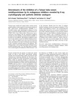
Figure 1
Arthroscopic and magnetic resonance imaging evaluation of cartilage defects treated with autologous chondrocyte grafts (BioSeed
®
-C)Arthroscopic and magnetic resonance imaging evaluation of cartilage defects treated with autologous chondrocyte grafts (BioSeed
®
-C). (a) Intra-
operative situation of a cartilage defect situated at the femoral condyle covered with transosseously fixed BioSeed-C (20 mm × 30 mm). Note that
the healthy cartilage rim is partly intact. (b) At 9 months after surgery, second-look arthroscopy showed the formation of a cartilage repair tissue of a
tough condition (asterisk). Magnetic resonance imaging (MRI) at 6 months (c) and 12 months (d) after implantation of BioSeed-C shows transos-
seous drilling holes (white asterisks) due to fixation of the graft. The repair tissue covers the defect (white triangles) and gives a slightly altered MRI
signal.
Available online />Page 5 of 11
(page number not for citation purposes)
good connection to the articular cartilage as well as to the
underlying subchondral bone. No transplant loosening,
debonding, ablation of the transplant, or articular constriction
was observed. The newly formed cartilage showed a visible
contrast in color to the surrounding cartilage, and transos-
seous drill holes were still evident. Subchondral edemas were
not observed. Osseous healing after drilling did not differ from
bone regeneration after osteosynthesis or reconstruction of
the anterior cruciate ligament, for example.
Five patients underwent repeat surgery comprising synovecto-
mies (n = 2), debridement (n = 1), total knee arthroplasty (n =
1), and removal of the graft (n = 1) in another hospital.
Histological analysis of biopsies after 9 to 12 months from
implantation of BioSeed-C showed that one repair tissue
appeared as a mixed tissue of hyaline-like and fibrous carti-
lage, whereas three biopsies documented the development
toward a hyaline repair tissue (Figure 2). The mixed repair tis-
sue (Figure 2a,b) showed good bonding of the engineered
cartilage to the underlying bone with progressive remodeling
of the subchondral bone tissue (asterisk in Figure 2c) and
areas of fibrocartilage (black triangle in Figure 2c) and hyaline-
like cartilage (white triangle in Figure 2c). The mixed repair tis-
sue was rich in evenly distributed viable cells and was charac-
terized by a proteoglycan-rich extracellular matrix (Figure 2d).
Specimens with hyaline-like repair tissue (Figure 2e–j)
showed intense staining of proteoglycans (Figure 2e) and
good integration with the subchondral bone (Figure 2f). The
chondrocytes were viable, round-shaped within lacunae, and
showed a columnar distribution with some clustering (Figure
2g,i,j). The surface of the repair tissue appeared smooth and
showed the typical articular cartilage surface-related tissue
with a gradual decrease of proteoglycans within the extracel-
lular matrix (Figure 2h). There were no signs of abnormal calci-
fication or formation of a fibrous connective tissue within the
repair tissue, and neither necrosis of the tissue nor apoptosis
of chondrocytes was evident.
Clinical evaluation of surgical results 2 years after
transplantation of BioSeed-C
To assess the impact of osteoarthritic degeneration of the car-
tilage on the functional outcome, two subgroups were consid-
ered, namely those with a Jaeger-Wirth score of 3,
representing patients with osteoarthritic degeneration, and
those with a score less than 3, comprising patients with post-
traumatic and mild degenerative defects.
According to the modified Cincinnati Knee Rating System
(Figure 3), statistically significant improvements (p < 0.05)
Figure 2
Histological analysis of second-look biopsy tissue from patients treated with BioSeed
®
-CHistological analysis of second-look biopsy tissue from patients treated
with BioSeed
®
-C. At 9 to 12 months after implantation, second-look
biopsy tissue was stained for proteoglycans with alcian blue. One
patient's biopsy tissue showed the formation of mixed repair tissue (a-
d) with areas of fibrocartilage ((c), black triangle) and hyaline-like carti-
lage ((c), white triangle) and a firm bonding to the subchondral bone
that was undergoing remodeling ((c), asterisk). Biopsy tissue from three
patients (e-j) shows the formation of a hyaline-like cartilaginous repair
tissue with intense staining of proteoglycans by alcian blue (e-h), good
integration with the underlying bone tissue (f), viable, round cells within
lacunae (g) and a smooth surface (h). Chondrocytes showed a colum-
nar distribution and some clustering (g-j). Hematoxylin/eosin staining (i,
j) of biopsy tissue of two patients confirmed the presence of viable
chondrocytes and the absence of abnormal calcification, apoptosis,
necrosis or formation of a fibrous repair tissue.
Arthritis Research & Therapy Vol 9 No 2 Ossendorf et al.
Page 6 of 11
(page number not for citation purposes)
were observed as early as 6 months after implantation of Bio-
Seed-C, independently of the degree of osteoarthritic degen-
eration of the cartilage. Rating by physicians yielded
statistically significant improvement at 6 months and 2 years
after implantation. Interestingly, the improvement at 6 months
after implantation of BioSeed-C was observed only in patients
with osteoarthritic degenerations. After 2 years, the median
Cincinnati Knee Rating System score increased from 4.0 to
6.0 postoperatively.
In comparison with the preoperative scores, the Lysholm score
(Figure 4) improved significantly (p < 0.007) in both groups of
patients with osteoarthritic degeneration or with posttraumatic
and/or mild degenerative defects as early as 3 months to up to
2 years after implantation of BioSeed-C. In comparison with
the preoperative status, the median Lysholm score increased
from 46.0 to 81.0 in patients with posttraumatic and/or mild
degenerative defects and from 47.0 to 78.5 in patients with
osteoarthritic degeneration, 2 years after implantation.
The Knee Injury and Osteoarthritis Outcome Score describes
the patient's view about his knee and associated problems
(Additional file 1). At 2 years of follow-up, the patient's status
had improved significantly (p < 0.05) compared with the his or
her preoperative situation, showing an increase in the mean
scores of the subclasses pain (64.3 to 78.2), symptoms (68.2
to 78.9), activities of daily living (67.6 to 80.6), sports (25.8 to
45.7), and knee-related quality of life (26.9 to 52.9). Patients
with posttraumatic and/or mild degenerative defects (Jaeger-
Wirth score < 3) showed a significant improvement in only the
subclass knee-related quality of life (28.4 to 52.9) 2 years after
implantation, whereas patients with osteoarthritic
degeneration showed improvement in pain (61.4 to 80.1),
symptoms (64.0 to 78.4), and quality of life (23.6 to 52.8).
Figure 3
Clinical outcome after 2 years evaluated by the Modified Cincinnati Knee Rating SystemClinical outcome after 2 years evaluated by the Modified Cincinnati Knee Rating System. The score from this system is shown for the entire patient
cohort compared with patients with posttraumatic and mild degenerative defects (Jaeger-Wirth score < 3) and patients with osteoarthritic defects
(Jaeger-Wirth score = 3). The preoperative and follow-up times are as indicated. Scores are presented as medians; the ends of the boxes define the
25th and 75th centiles, and error bars the 10th and 90th centiles. Where indicated (asterisks), differences were statistically significant (p < 0.05)
compared with the preoperative situation.
Available online />Page 7 of 11
(page number not for citation purposes)
The health of patients was evaluated with the IKDC SF-36 cur-
rent health assessment form (Additional file 2). Implantation of
BioSeed-C resulted in a statistically significant (p < 0.05)
increase in the mean scores after 6 months to 2 years in the
subclasses physical functioning (42.8 to 64.6), role limitations
due to physical health (25.7 to 53.6), bodily pain (38.9 to
61.6), general health problems (62.0 to 70.6), and social func-
tioning (59.5 to 77.5) in all outcome measures compared with
the patients' preoperative status. Evaluating the outcome
measures according to the impact of the degree of osteoar-
thritic degeneration of the knee showed an improvement in the
mean scores of the subclasses physical functioning (44.8 to
62.6), bodily pain (39.0 to 61.3), and social functioning (61.9
to 78.8) after implantation of BioSeed-C in posttraumatic and/
or mild degenerative cartilage defects with a Jaeger-Wirth
score of less than 3. Patients with osteoarthritic degeneration
of the knee cartilage (Jaeger-Wirth score = 3) reported a sig-
nificant impairment related to social functioning at 3 months
after implantation of the cartilage transplant and showed a
continual improvement in social functioning status from 6
months to 2 years. In addition, these patients showed a signif-
icant increase in the mean scores of the IKDC SF-36 sub-
classes physical functioning (38.8 to 68.3), role limitations due
to physical health (10.4 to 52.1), and bodily pain (38.8 to
62.2) 2 years after implantation of BioSeed-C.
Clinical evaluation of surgical results 2 years after
implantation of BioSeed-C in defects of patients with
radiographically confirmed osteoarthritis
Radiographs of the degenerated knee of 30 patients with
osteoarthritic symptoms were taken preoperatively. Applying
the Kellgren-Lawrence score showed that 22 of the 30
patients with a clinical follow-up of 2 years had osteoarthritis,
having obtained a Kellgren-Lawrence score of 2 or more. The
clinical outcome after 2 years after implantation of BioSeed-C
in osteoarthritic focal defects was evaluated with the modified
Cincinnati Knee Rating System and the Lysholm score (Figure
5). After 2 years, patients' (p < 0.0001) and physicians' (p =
0.0074) ratings showed a significant improvement in the
median scores (patient 4.0 to 7.0, physician 5.0 to 7.0) of the
modified Cincinnati Knee Rating System in comparison with
the preoperative situation.
The Lysholm score showed significant improvement (p <
0.0001) in the median scores from 53.0 preoperatively to 79.5
after 2 years after implantation of BioSeed-C in focal osteoar-
thritic defects of the knee.
Discussion
In the present study, we showed the benefit and reliability of
the use of the autologous gel-polymer-based cartilage tissue
engineering graft BioSeed-C for the treatment of full-thickness
cartilage defects of the knee. The evaluation of the clinical
outcome 2 years after implantation demonstrated that Bio-
Seed-C is well suited for the treatment of patients with
Figure 4
Clinical outcome after 2 years evaluated by the Lysholm scoreClinical outcome after 2 years evaluated by the Lysholm score. The
score is given for the entire patient cohort compared with patients with
posttraumatic and mild degenerative defects (Jaeger-Wirth score < 3)
and patients with osteoarthritic defects (Jaeger-Wirth score = 3). The
preoperative and follow-up times are as indicated. Scores are pre-
sented as medians; the ends of the boxes define the 25th and 75th
centiles, and error bars the 10th and 90th centiles. Where indicated
(asterisks), differences were statistically significant (p < 0.007) com-
pared with the preoperative situation.
Arthritis Research & Therapy Vol 9 No 2 Ossendorf et al.
Page 8 of 11
(page number not for citation purposes)
posttraumatic and mild degenerative defects as well as for the
treatment of focal osteoarthritic defects.
The implantation of first-generation tissue engineering grafts
such as ACI has been shown to be suitable for the regenera-
tion of posttraumatic defects [12,37]. However, second-gen-
eration cartilage tissue engineering grafts using a variety of
matrices to support the autologous chondrocytes were
recently considered to be technically more attractive. For
instance, Bartlett and colleagues reported the use of a colla-
gen-based scaffold seeded with autologous chondrocytes for
the treatment of 47 symptomatic chondral defects. After 1
year, the Cincinnati Knee Rating System score increased by
19.6, and 36.4% of the biopsies showed hyaline-like cartilage
or a mixed repair tissue with fibrocartilage. Similar outcomes
were obtained for defects treated with 'classical' ACI with a
porcine-based collagen membrane covering the defects [21].
In a prospective study, 5 years after transplantation of cell-
seeded collagen grafts, 8 of 11 patients rated the function of
their knee better than before, and the clinical evaluation
showed significant improvement in the Meyers score, the
Lysholm-Gillquist score and the International Cartilage Repair
Society score [22]. In a multicenter retrospective cohort study
with Hyalograft C, a graft of autologous chondrocytes embed-
ded in a derivative of hyaluronic acid, 91.5% of 141 patients
with a follow-up from 2 to 5 years improved according to the
IKDC subjective evaluation, and second-look biopsies showed
hyaline-like cartilage [19]. The use of second-generation carti-
lage grafts based on collagen or hyaluronan matrices is
therefore suggested to be as effective as ACI, both clinically
and histologically.
Here we introduced the use of a new second-generation car-
tilage graft based on a biocompatible and bioresorbable two-
component gel-polymer scaffold. The BioSeed-C concept of
embedding autologous chondrocytes in a gel-like matrix dis-
tributed in a porous three-dimensional textile polymer structure
goes back to more than 10 years of cartilage tissue
engineering research [24,25]. Gel-like matrices such as fibrin
allow the even distribution of a large number of vital chondro-
cytes within the graft and promote chondrocyte differentiation
as well as the formation of a cartilaginous repair tissue, while
the polymer scaffold mediates initial biomechanical stability
and allows easy handling of the graft by the surgeon [23,38].
The arrangement of chondrocytes in three-dimensional scaf-
folds permits the arthroscopic implantation of cells, ensures
secure fixation of the graft even in posttraumatic or degenera-
tive defects without intact surrounding cartilage, and avoids
the loss of cells into the joint cavity even after implantation in
defects without an intact surrounding cartilage rim [28]. Drob-
nic and colleagues have shown that the transosseous fixation
technique provides excellent stability of the polymer-based
graft with high endpoint fixation strength and no detachment
after continuous passive motion with loading in the initial post-
operative period [39]. Mechanical testing of the scaffold used
Figure 5
Clinical outcome after 2 years of implantation of BioSeed-C in osteoar-thritic defectsClinical outcome after 2 years of implantation of BioSeed-C in osteoar-
thritic defects. The Modified Cincinnati Knee Rating System score and
Lysholm score are given for 22 patients with radiologically confirmed
osteoarthritic defects at 2 years after implantation of BioSeed-C. Oste-
oarthritis was defined according to a Kellgren-Lawrence score of 2 or
more. Scores are presented as medians; the ends of the boxes define
the 25th and 75th centiles, and error bars the 10th and 90th centiles.
Where indicated (asterisks), differences were statistically significant
(**p = 0.0074, ***p < 0.0001) compared with the preoperative
situation.
Available online />Page 9 of 11
(page number not for citation purposes)
in this study showed that the graft withstands a maximal tensile
load of up to 15 N when fixed transosseously or by chondral
suture, whereas gel-like matrices or collagen membranes rup-
tured on being loaded with up to 10 N [40]. The capability of
such polymer-based grafts to form an adequate cartilaginous
repair tissue has been shown preclinically in several animal
studies with cryopreserved and non-cryopreserved chondro-
cytes [41,42]. In addition, in a large-animal model system with
Haflinger horses, polymer-based cartilage grafts have been
shown to develop a cartilaginous repair tissue that is well inte-
grated into the surrounding cartilage and is firmly bonded to
the subchondral bone [27]. The bioresorbable scaffold mate-
rial is composed of a copolymer of polyglactin (vicryl) and poly-
dioxanone, shows good biocompatibility, and is frequently
used clinically as suture material. In a rabbit dural tissue reac-
tion study, the absorbable polyglactin and polydioxanone
material guided tissue development with complete resolution
of the inflammatory reaction during absorption and without any
morphological sequelae [43]. Additionally, in cartilage regen-
eration, various in vitro and animal studies have shown that the
scaffold supports cartilaginous tissue development with no
signs of necrosis, apoptosis, or abnormal tissue reaction
[26,27,38,44].
In this case series we demonstrated the benefit and reliability
of the gel-polymer-based chondrocyte graft BioSeed-C for the
treatment of posttraumatic and degenerative large full-thick-
ness cartilage lesions of the knee. Histological analysis of the
biopsies after implantation of BioSeed-C showed good
formation of a cartilaginous repair tissue, and significant
improvements in the clinical scores used could be ascer-
tained, implying improvements in activities of daily living, ability
to work, and in sports. However, despite these encouraging
results one must take into account the fact that randomized
clinical trials and long follow-up periods may offer more wide-
spread information about the clinical effectiveness of a given
cartilage repair approach [13,45-47]. ACI will therefore not be
given an unrestricted recommendation for the treatment of full-
thickness cartilage lesions of the knee. Nevertheless, patient
status at 2 years of follow-up was reported as an important
indicator for future outcome [10], because most of the compli-
cations of ACI occur during this period. In addition, major
improvements in clinical scores, clinical evaluation, and sub-
jective patient satisfaction were found during this time; for
example, patients who did not return to sports within 2 years
did not return later. The features identified as an indicator of a
worse prognosis, namely multiple surgical procedures, higher
age, and large defects, correspond to findings published by
others [21].
With the gel-polymer-based three-dimensional cartilage
grafts, 18% of the patients in this study underwent second-
look arthroscopy as a result of grinding, catching, pain, or
swelling of the knee. This is consistent with other studies
reporting rates of revision surgery between 0% [48] and 25%
[18]. Instead, 2 of 79 patients treated with BioSeed-C showed
a failure of the graft, which represents a lower rate of graft fail-
ure than earlier findings, in which rates of failure in ACI with
other implants between 5% [9] to 13% [18] were described.
Repeat operations using the 'classical' ACI procedure as
described by Peterson and Brittberg were mainly caused by
problems associated with the periosteal flap [9,17,49]. This
disadvantage of the original ACI technique could not occur in
patients treated with BioSeed-C. Another advantage of the
BioSeed technique is the reduced operating time. Further-
more, the procedure is less invasive because there is no need
to harvest periosteum from the tibia. The complication rate is
lower because there is no possibility of periosteal hypertrophy,
which is a common complication of ACI [15]. Furthermore, the
BioSeed-C procedure can be performed arthroscopically,
which may be associated with faster recovery after surgery
and with cosmetically better results. However, it should be
taken into consideration that performing ACI arthroscopically
is technically demanding and the use of specially designed
instruments is essential.
After 2 years of follow-up, mean scores increased significantly,
between 20 and 35% depending on the score analyzed. This
indicates a significant decrease in the patient's pain and knee
instabilities during activity. Intriguingly, Cincinnati score
improvement at 6 months after implantation of BioSeed-C
could be observed only in patients with osteoarthritic degen-
erations. In addition, patients suffering from osteoarthritic
degenerations showed an improved Knee injury and Osteoar-
thritis Outcome Score in pain, symptoms, and quality of life,
whereas scores for patients whose cartilage defects resulted
from posttraumatic causes increased only in the quality of life
section. According to the impact of the degree of osteoar-
thritic degeneration, patients with osteoarthritis of the knee
reported impairment in four subclasses of the SF-36 score.
Obviously, tissue regeneration, improvement in clinical scores,
and improvement in patient's quality of life are achieved after
implantation of polymer-based autologous cartilage grafts
even in osteoarthritic conditions.
Currently, ACI is considered not to be indicated for osteoar-
thritic patients. In spite of this, many young patients suffer from
early stages of osteoarthritis or display deformities predispos-
ing to osteoarthritis that are idiopathic or follow trauma. These
patients lack good treatment options and are too young for
total joint replacement. This is particularly true for those having
an active lifestyle that includes sports or demanding recrea-
tional activities. Most of the patients of the present study suf-
fered preoperatively from pain or dysfunction of the knee joint.
They frequently underwent several failed cartilage repair pro-
cedures, and subsequently had to endure massive restrictions
of quality of life, ability to work, and sporting activities. Thus,
we consider the outcome of this study as a promising result for
the treatment of large cartilage lesions of the knee, particularly
for this challenging patient cohort with difficult cartilage condi-
Arthritis Research & Therapy Vol 9 No 2 Ossendorf et al.
Page 10 of 11
(page number not for citation purposes)
tions and in need of a variety of concomitant surgery proce-
dures such as anterior cruciate ligament reconstruction or
high tibial osteotomy. Besides, as a first step, it would be a
beneficial effort to postpone total joint replacement for a dec-
ade. Recently, the effectiveness of second-generation carti-
lage grafts has been shown for the treatment of osteoarthritic
knees. Hollander and colleagues reported the use of a hyaluro-
nan-based second-generation cartilage tissue engineering
graft for the treatment of osteoarthritic knees [50]. Histological
and biochemical analyses of second-look biopsies docu-
mented the regeneration of cartilage as early as about 1 year
after transplantation in 10 of 23 patients and showed that
osteoarthritis did not inhibit the regeneration progress.
Conclusion
The present study supports the use of the three-dimensional
autologous cartilage graft, BioSeed-C, for the treatment of
posttraumatic and osteoarthritic cartilage defects of the knee.
Clinical evaluation 2 years after implantation showed that the
treatment of posttraumatic and osteoarthritic defects with Bio-
Seed-C leads to an improvement in the patient's condition as
documented by the significant improvement in reliable clinical
outcome scores. Further long-term studies with more patients
are needed to prove the effectiveness of tissue engineering
cartilage grafts to postpone total joint replacement in
osteoarthritis.
Competing interests
CK is an employee of TransTissue Technologies GmbH. Tran-
sTissue Technologies GmbH is a subsidiary of BioTissue
Technologies GmbH, which produces and distributes Bio-
Seed
®
-C. MS works as a consultant for TransTissue Technol-
ogies GmbH. CE works as a consultant for BioTissue
Technologies GmbH. All other authors declare that they have
no competing interests.
Authors' contributions
CO and CK performed the data evaluation and drafted the
manuscript. PCK participated in the patient data collection.
GRB and MS partly conceived the study and participated in
the study design. CE conceived the study, participated in its
design and coordination, performed the surgical procedures,
and was involved in the patient data collection and interpreta-
tion. All authors read and approved the final manuscript.
Additional files
References
1. Smith GD, Knutsen G, Richardson JB: A clinical review of carti-
lage repair techniques. J Bone Joint Surg Br 2005, 87:445-449.
2. Brittberg M, Lindahl A, Nilsson A, Ohlsson C, Isaksson O, Peter-
son L: Treatment of deep cartilage defects in the knee with
autologous chondrocyte transplantation. N Engl J Med 1994,
331:889-895.
3. Hubbard MJ: Articular debridement versus washout for degen-
eration of the medial femoral condyle. A five-year study. J
Bone Joint Surg Br 1996, 78:217-219.
4. Matsusue Y, Yamamuro T, Hama H: Arthroscopic multiple oste-
ochondral transplantation to the chondral defect in the knee
associated with anterior cruciate ligament disruption. Arthros-
copy 1993, 9:318-321.
5. Steadman JR, Rodkey WG, Rodrigo JJ: Microfracture: surgical
technique and rehabilitation to treat chondral defects. Clin
Orthop Relat Res 2001, (391 Suppl):S362-S369.
6. Curl WW, Krome J, Gordon ES, Rushing J, Smith BP, Poehling
GG: Cartilage injuries: a review of 31,516 knee arthroscopies.
Arthroscopy 1997, 13:456-460.
7. Nehrer S, Spector M, Minas T: Histologic analysis of tissue after
failed cartilage repair procedures. Clin Orthop Relat Res 1999,
365:149-162.
8. Kreuz PC, Steinwachs MR, Erggelet C, Krause SJ, Konrad G, Uhl
M, Sudkamp N: Results after microfracture of full-thickness
chondral defects in different compartments in the knee. Oste-
oarthritis Cartilage 2006, 29:29.
9. Peterson L, Minas T, Brittberg M, Nilsson A, Sjogren-Jansson E,
Lindahl A: Two- to 9-year outcome after autologous chondro-
cyte transplantation of the knee. Clin Orthop Relat Res 2000,
374:212-234.
10. Peterson L, Brittberg M, Kiviranta I, Akerlund EL, Lindahl A: Autol-
ogous chondrocyte transplantation. Biomechanics and long-
term durability. Am J Sports Med 2002, 30:2-12.
11. Minas T: Autologous chondrocyte implantation in the arthritic
knee. Orthopedics 2003, 26:
945-947.
12. Browne JE, Anderson AF, Arciero R, Mandelbaum B, Moseley JB
Jr, Micheli LJ, Fu F, Erggelet C: Clinical outcome of autologous
chondrocyte implantation at 5 years in US subjects. Clin
Orthop Relat Res 2005, 436:237-245.
13. Knutsen G, Engebretsen L, Ludvigsen TC, Drogset JO, Grontvedt
T, Solheim E, Strand T, Roberts S, Isaksen V, Johansen O: Autol-
ogous chondrocyte implantation compared with microfracture
in the knee. A randomized trial. J Bone Joint Surg Am 2004, 86-
A:455-464.
14. Henderson I, Francisco R, Oakes B, Cameron J: Autologous
chondrocyte implantation for treatment of focal chondral
defects of the knee – a clinical, arthroscopic, MRI and histo-
logic evaluation at 2 years. Knee 2005, 12:209-216.
15. Gooding CR, Bartlett W, Bentley G, Skinner JA, Carrington R,
Flanagan A: A prospective, randomised study comparing two
techniques of autologous chondrocyte implantation for osteo-
chondral defects in the knee: periosteum covered versus type
I/III collagen covered. Knee 2006, 13:203-210.
16. Driesang IM, Hunziker EB: Delamination rates of tissue flaps
used in articular cartilage repair. J Orthop Res 2000,
18:909-911.
17. Micheli LJ, Browne JE, Erggelet C, Fu F, Mandelbaum B, Moseley
JB, Zurakowski D: Autologous chondrocyte implantation of the
knee: multicenter experience and minimum 3-year follow-up.
Clin J Sport Med 2001, 11:223-228.
The following Additional files are available online:
Additional file 1
An EPS file showing the clinical outcome after 2 years
evaluated by the Knee injury and Osteoarthritis Outcome
Score (KOOS).
See />supplementary/ar2180-S1.eps
Additional file 2
An EPS file showing the clinical outcome after 2 years,
evaluated by the International Knee Documentation
Committee (IKDC) SF-36 score.
See />supplementary/ar2180-S2.eps
Available online />Page 11 of 11
(page number not for citation purposes)
18. Minas T: Autologous chondrocyte implantation for focal chon-
dral defects of the knee. Clin Orthop Relat Res 2001,
391(Suppl):S349-S361.
19. Marcacci M, Berruto M, Brocchetta D, Delcogliano A, Ghinelli D,
Gobbi A, Kon E, Pederzini L, Rosa D, Sacchetti GL, et al.: Articular
cartilage engineering with Hyalograft C: 3-year clinical results.
Clin Orthop Relat Res 2005, 435:96-105.
20. Nehrer S, Domayer S, Dorotka R, Schatz K, Bindreiter U, Kotz R:
Three-year clinical outcome after chondrocyte transplantation
using a hyaluronan matrix for cartilage repair. Eur J Radiol
2006, 57:3-8.
21. Bartlett W, Skinner JA, Gooding CR, Carrington RW, Flanagan
AM, Briggs TW, Bentley G: Autologous chondrocyte implanta-
tion versus matrix-induced autologous chondrocyte implanta-
tion for osteochondral defects of the knee: a prospective,
randomised study. J Bone Joint Surg Br 2005, 87:640-645.
22. Behrens P, Bitter T, Kurz B, Russlies M: Matrix-associated autol-
ogous chondrocyte transplantation/implantation (MACT/
MACI) – 5-year follow-up. Knee 2006, 13:194-202.
23. Sittinger M, Hutmacher DW, Risbud MV: Current strategies for
cell delivery in cartilage and bone regeneration. Curr Opin
Biotechnol 2004, 15:411-418.
24. Sittinger M, Bujia J, Minuth WW, Hammer C, Burmester GR: Engi-
neering of cartilage tissue using bioresorbable polymer carri-
ers in perfusion culture. Biomaterials 1994, 15:451-456.
25. Bujia J, Sittinger M, Minuth WW, Hammer C, Burmester G, Kasten-
bauer E: Engineering of cartilage tissue using bioresorbable
polymer fleeces and perfusion culture. Acta Otolaryngol 1995,
115:307-310.
26. Kaps C, Fuchs S, Endres M, Vetterlein S, Krenn V, Perka C, Sit-
tinger M: Molecular characterization of tissue-engineered
articular chondrocyte transplants based on resorbable poly-
mer fleece. Orthopade 2004, 33:76-85.
27. Barnewitz D, Endres M, Kruger I, Becker A, Zimmermann J, Wilke
I, Ringe J, Sittinger M, Kaps C: Treatment of articular cartilage
defects in horses with polymer-based cartilage tissue engi-
neering grafts. Biomaterials 2006,
27:2882-2889.
28. Erggelet C, Sittinger M, Lahm A: The arthroscopic implantation
of autologous chondrocytes for the treatment of full-thickness
cartilage defects of the knee joint. Arthroscopy 2003,
19:108-110.
29. Outerbridge RE: The etiology of chondromalacia patellae. J
Bone Joint Surg Br 1961, 43-B:752-757.
30. Jaeger M, Wirth CJ: Praxis der Orthopädie 1st edition. Stuttgart:
Thieme; 1986.
31. Scheller G, Sobau C, Bulow JU: Arthroscopic partial lateral
meniscectomy in an otherwise normal knee: clinical, func-
tional, and radiographic results of a long-term follow-up study.
Arthroscopy 2001, 17:946-952.
32. Kellgren JH, Lawrence JS: Radiological assessment of osteo-
arthrosis. Ann Rheum Dis 1957, 16:494-502.
33. Barber-Westin SD, Noyes FR, McCloskey JW: Rigorous statisti-
cal reliability, validity, and responsiveness testing of the
Cincinnati knee rating system in 350 subjects with uninjured,
injured, or anterior cruciate ligament-reconstructed knees.
Am J Sports Med 1999, 27:402-416.
34. Lysholm J, Gillquist J: Evaluation of knee ligament surgery
results with special emphasis on use of a scoring scale. Am J
Sports Med 1982, 10:150-154.
35. Roos EM, Roos HP, Lohmander LS, Ekdahl C, Beynnon BD: Knee
Injury and Osteoarthritis Outcome Score (KOOS) – develop-
ment of a self-administered outcome measure. J Orthop
Sports Phys Ther 1998, 28:88-96.
36. Irrgang JJ, Anderson AF, Boland AL, Harner CD, Kurosaka M,
Neyret P, Richmond JC, Shelborne KD: Development and valida-
tion of the international knee documentation committee sub-
jective knee form. Am J Sports Med 2001, 29:600-613.
37. Fu FH, Zurakowski D, Browne JE, Mandelbaum B, Erggelet C,
Moseley JB Jr, Anderson AF, Micheli LJ: Autologous chondrocyte
implantation versus debridement for treatment of full-thick-
ness chondral defects of the knee: an observational cohort
study with 3-year follow-up. Am J Sports Med 2005,
33:1658-1666.
38. Kaps C, Frauenschuh S, Endres M, Ringe J, Haisch A, Lauber J,
Buer J, Krenn V, Haupl T, Burmester GR, Sittinger M: Gene
expression profiling of human articular cartilage grafts gener-
ated by tissue engineering. Biomaterials
2006, 27:3617-3630.
39. Drobnic M, Radosavljevic D, Ravnik D, Pavlovcic V, Hribernik M:
Comparison of four techniques for the fixation of a collagen
scaffold in the human cadaveric knee. Osteoarthritis Cartilage
2006, 14:337-344.
40. Knecht S, Erggelet C, Endres M, Sittinger M, Kaps C, Stussi E:
Mechanical testing of fixation techniques for scaffold-based
tissue-engineered grafts. J Biomed Mater Res B Appl Biomater
2007, 22:. Epub 2007 Feb 22
41. Perka C, Sittinger M, Schultz O, Spitzer RS, Schlenzka D, Burm-
ester GR: Tissue engineered cartilage repair using cryopre-
served and noncryopreserved chondrocytes. Clin Orthop Relat
Res 2000, 378:245-254.
42. Perka C, Schultz O, Sittinger M, Zippel H: Chondrocyte trans-
plantation in PGLA/polydioxanone fleece. Orthopade 2000,
29:112-119.
43. Barbolt TA, Odin M, Leger M, Kangas L, Hoiste J, Liu SH: Biocom-
patibility evaluation of dura mater substitutes in an animal
model. Neurol Res 2001, 23:813-820.
44. Rotter N, Aigner J, Naumann A, Planck H, Hammer C, Burmester
G, Sittinger M: Cartilage reconstruction in head and neck sur-
gery: comparison of resorbable polymer scaffolds for tissue
engineering of human septal cartilage. J Biomed Mater Res
1998, 42:347-356.
45. Horas U, Schnettler R, Pelinkovic D, Herr G, Aigner T: Osteochon-
dral transplantation versus autogenous chondrocyte trans-
plantation. A prospective comparative clinical study. Chirurg
2000, 71:1090-1097.
46. Bentley G, Biant LC, Carrington RW, Akmal M, Goldberg A, Wil-
liams AM, Skinner JA, Pringle J: A prospective, randomised com-
parison of autologous chondrocyte implantation versus
mosaicplasty for osteochondral defects in the knee. J Bone
Joint Surg Br 2003, 85:223-230.
47. Horas U, Pelinkovic D, Herr G, Aigner T, Schnettler R: Autologous
chondrocyte implantation and osteochondral cylinder trans-
plantation in cartilage repair of the knee joint. A prospective,
comparative trial. J Bone Joint Surg Am 2003, 85-A:185-192.
48. Loehnert J, Ruhnau K, Gossen A, Bernsmann K, Wiese M: Autol-
oge Chondrozytentransplantation (ACT) im Kniegelenk. Erste
klinische Ergebnisse. Arthroskopie 1999,
12:34-42.
49. Minas T, Peterson L: Advanced techniques in autologous
chondrocyte transplantation. Clin Sports Med 1999, 18:13-44.
50. Hollander AP, Dickinson SC, Sims TJ, Brun P, Cortivo R, Kon E,
Marcacci M, Zanasi S, Borrione A, De Luca C, et al.: Maturation
of tissue engineered cartilage implanted in injured and oste-
oarthritic human knees. Tissue Eng 2006, 12:1787-1798.