Báo cáo y học: "Bonding of articular cartilage using a combination of biochemical degradation and surface cross-linking" pdf
Bạn đang xem bản rút gọn của tài liệu. Xem và tải ngay bản đầy đủ của tài liệu tại đây (1.02 MB, 11 trang )
Open Access
Available online />Page 1 of 11
(page number not for citation purposes)
Vol 9 No 3
Research article
Bonding of articular cartilage using a combination of biochemical
degradation and surface cross-linking
Carsten Englert
1
, Torsten Blunk
2
, Rainer Müller
3
, Sabine Schulze von Glasser
1
, Julia Baumer
2
,
Johann Fierlbeck
4
, Iris M Heid
5,6
, Michael Nerlich
1
and Joachim Hammer
4
1
Department of Trauma Surgery, University Medical Centre Regensburg, Franz-Josef-Strauss-Allee, 93053 Regensburg, Germany
2
Department of Pharmaceutical Technology, University of Regensburg, Universitätsstrasse, 93053 Regensburg, Germany
3
Institute of Physical and Theoretical Chemistry, University of Regensburg, Universitätsstrasse, 93053 Regensburg, Germany
4
Mechanical Engineering Faculty, University of Applied Sciences, Galgenbergstrasse, 93053 Regensburg, Germany
5
GSF-National Research Centre, Institute of Epidemiology, Ingolstädter Landstrasse, 85674 Neuherberg, Germany
6
Institute of Medical Informatics, Biometry, and Epidemiology, Ludwig-Maximilians-University, Munich, Germany
Corresponding author: Carsten Englert,
Received: 22 Jan 2007 Revisions requested: 10 Apr 2007 Revisions received: 30 Apr 2007 Accepted: 15 May 2007 Published: 15 May 2007
Arthritis Research & Therapy 2007, 9:R47 (doi:10.1186/ar2202)
This article is online at: />© 2007 Englert et al.; licensee BioMed Central Ltd.
This is an open access article distributed under the terms of the Creative Commons Attribution License ( />),
which permits unrestricted use, distribution, and reproduction in any medium, provided the original work is properly cited.
Abstract
After trauma, articular cartilage often does not heal due to
incomplete bonding of the fractured surfaces. In this study we
investigated the ability of chemical cross-linkers to facilitate
bonding of articular cartilage, either alone or in combination with
a pre-treatment with surface-degrading agents. Articular
cartilage blocks were harvested from the femoropatellar groove
of bovine calves. Two cartilage blocks, either after pre-treatment
or without, were assembled in a custom-designed chamber in
partial apposition and subjected to cross-linking treatment.
Subsequently, bonding of cartilage was measured as adhesive
strength, that is, the maximum force at rupture of bonded
cartilage blocks divided by the overlap area. In a first approach,
bonding was investigated after treatment with cross-linking
reagents only, employing glutaraldehyde, 1-ethyl-3-
diaminopropyl-carbodiimide (EDC)/N-hydroxysuccinimide
(NHS), genipin, or transglutaminase. Experiments were
conducted with or without compression of the opposing
surfaces. Compression during cross-linking strongly enhanced
bonding, especially when applying EDC/NHS and
glutaraldehyde. Therefore, all further experiments were
performed under compressive conditions. Combinations of
each of the four cross-linking agents with the degrading pre-
treatments, pepsin, trypsin, and guanidine, led to distinct
improvements in bonding compared to the use of cross-linkers
alone. The highest values of adhesive strength were achieved
employing combinations of pepsin or guanidine with EDC/NHS,
and guanidine with glutaraldehyde. The release of extracellular
matrix components, that is, glycosaminoglycans and total
collagen, from cartilage blocks after pre-treatment was
measured, but could not be directly correlated to the determined
adhesive strength. Cytotoxicity was determined for all
substances employed, that is, surface degrading agents and
cross-linkers, using the resazurin assay. Taking the favourable
cell vitality after treatment with pepsin and EDC/NHS and the
cytotoxic effects of guanidine and glutaraldehyde into account,
the combination of pepsin and EDC/NHS appeared to be the
most advantageous treatment in this study. In conclusion,
bonding of articular cartilage blocks was achieved by chemical
fixation of their surface components using cross-linking
reagents. Application of compressive forces and prior
modulation of surface structures enhanced cartilage bonding
significantly. Enzymatic treatment in combination with cross-
linkers may represent a promising addition to current techniques
for articular cartilage repair.
Introduction
After trauma, articular cartilage often does not heal due to
incomplete bonding of the fractured surfaces. The pathophys-
iological mechanism of articular cartilage integration has been
intensively investigated in vitro, showing that integration
depends on collagen metabolism [1,2], collagen cross-linking
[3], cell vitality [4], and hormonal stimulation [5]. Inhibiting fac-
tors have also been described, such as synovial fluid compo-
nents, which may inhibit the integrative repair by binding to the
cracked surface [6], cytokines, which abolish the anabolic
EDC = 1-ethyl-3-diaminopropyl-carbodiimide; G = geometry (of cartilage blocks); GAG = glycosaminoglycan; NHS = N-hydroxysuccinimide; PBS =
phosphate-buffered saline; RFU = relative fluorescence unit.
Arthritis Research & Therapy Vol 9 No 3 Englert et al.
Page 2 of 11
(page number not for citation purposes)
steroid hormone effect [5], and the synovial fluid flow, which
might act at the interface in joint motion to keep the surfaces
apart [7].
Based on these findings, therapeutic options for articular car-
tilage integration have been investigated. Collagen cross-link-
ing has been stimulated over time in vitro [8] or articular
cartilage surfaces have been degraded in order to stimulate
repair in vitro [9-12] as well as in vivo [13]. Enzymes that were
employed for articular cartilage degradation included trypsin
[5,11,13], chondroitinase ABC [9], and hyaluronidase with
subsequent collagenase treatment [12], resulting in enhanced
integrative cartilage repair in vitro. Physical swelling of surface
structures by guanidine was also reported to stimulate the
integrative repair process [10].
Cartilage can be considered as a composite material consist-
ing of a collagen network and other extracellular matrix compo-
nents, mainly glycosaminoglycans (GAGs). Collagen and its
derivatives have been cross-linked for tissue engineering or
biomaterial purposes [14,15]. Glutaraldehyde is the most
extensively used reagent for cross-linking primary amino
groups, mainly exposed by collagen [16,17]. However, it has
been reported to elicit cytotoxic effects [18,19]. In proteogly-
cans, amino groups are mainly acetylated and, therefore, not
subjected to glutaraldehyde cross-linking. Water-soluble car-
bodiimides activate carboxylic groups of proteins such as col-
lagen, which results in the formation of amide-type cross-links
without any residual reactive groups [20,21]. In addition, car-
bodiimides were found to cross-link hyaluronic acid molecules
by forming ester bonds between hydroxyl and carboxyl groups
[22]. The carbodiimide method has been shown to be superior
to glutaraldehyde in terms of cyto- and biocompatibility
[19,23]. Another favourable cross-linker for primary amino
groups is the naturally occurring reagent genipin, which has
been reported to be significantly less cytotoxic than glutaralde-
hyde [23,24]. Transglutaminase, an enzyme in mammalian
chondrocytes whose expression is strongly correlated with
cell differentiation, has also been used as a collagen cross-
linking reagent [25] and has been introduced for articular car-
tilage gluing [26]. Taken together, glutaraldehyde, carbodiim-
ides, genipin, and transglutaminase all cross-link functional
groups of extracellular matrix components. Such reagents
may, therefore, also be used to cross-link exposed functional
groups on a fractured surface of articular cartilage after trauma
or transplantation.
The objective of this study was to investigate the initiation of
immediate bonding of articular cartilage blocks by means of
combining cartilage degradation and cross-linking reagents. In
addition, it was investigated whether a considerable compres-
sion of the cartilage blocks was necessary to achieve bonding.
For all combinations, that is, compression of cartilage blocks
and application of surface degrading and cross-linking rea-
gents, specific emphasis was put on the achievable adhesive
strength of the bonding interface according to the integration
model established by Reindel and colleagues [4].
Materials and methods
Cartilage preparation, compression, and bonding
Within one day after sacrificing of 8- to 12-week-old bovine
calves, osteochondral fragments were harvested from the fem-
oropatellar groove, using a reciprocating saw (Stryker Instru-
ments, Kalamazoo, MI, USA). Blocks of 10 mm × 10 mm in
length and 20 mm in height were harvested (Figure 1a). A
sledge microtome (Microm HM440E, Neuss, Germany) was
used to cut the osteochondral fragments into cartilage slices
of two precisely defined thicknesses, that is, 0.25 mm (geom-
etry one (G1)) and 0.3 mm (geometry two (G2)) (Figure 1b).
In all cases, the two top slices were discarded and only the fol-
lowing two underlying slices were used for experiments. These
slices were cut into rectangles of 8 mm × 2.5 mm (Figure 1c).
During the entire preparation procedure the specimens were
kept moist and free of blood by copious irrigation with cooled
PBS.
The integration specimens were assembled by positioning two
cartilage rectangles in partial apposition, creating a defined
overlap area of 4 mm × 2.5 mm for bonding. The precise
assembly was guaranteed by a custom-made chamber and an
additional fixation stamp (Figure 1d) [5]. After fixation (without
cartilage blocks) a gap of exactly 0.5 mm between stamp sur-
face and chamber bottom remained. Thus, when the cartilage
samples were inserted into the chamber and fixed by the
stamp, for two G1 cartilage blocks almost no compression
was acting during bonding, whereas for G2 blocks a defined
compressive strain of 17% of the total thickness was applied
(Figure 1e). To determine the creep modulus for both
geometries (G1 and G2), the custom-made chamber and the
stamp were modified and connected to the test rig (Hegewald
and Peschke, Nossen, Germany). Samples were compressed
by the stamp and the resulting force relaxation behaviour was
analysed by recording the load over time.
Cross-linking
For bonding, the cartilage blocks within the chamber were
placed in a 24-well culture plate and each sample was sub-
jected to one of the following cross-linking agents for 2 h at
room temperature (750 μl per sample): glutaraldehyde (Roth,
Germany) at a concentration of 20 mg/ml, buffered in PBS; 1-
ethyl-3-diaminopropyl-carbodiimide (EDC) and N-hydroxysuc-
cinimide (NHS) (Fluka, Neu-Ulm, Germany) at concentrations
of 20 mg/ml and 5 mg/ml, respectively, in morpholi-
noethanesulfonic acid buffered solution (pH 5.5); genipin
(WAKO Chemicals, Germany) at a concentration of 5 mg/ml
in PBS; transglutaminase (Ajinomoto Foods, Hamburg, Ger-
many) at a final concentration of 60 U per gram dry weight of
cartilage block in 0.01 M acetic acid, adjusted to pH 6 (trans-
glutaminase was applied according to the protocol by Chen
and colleagues [27], with the exception that cartilage blocks
Available online />Page 3 of 11
(page number not for citation purposes)
were incubated in treatment solution for 2 h in contrast to the
described protocol with a treatment duration of 12 h). See
also Figure 2 for chemical reaction schemes.
Surface degradation
For improved cross-linking, additional surface degradation
treatments prior to cross-linking were applied. In all cases, the
tissue samples, that is, the single cartilage pieces, were incu-
bated in a 100 μl solution. After surface degradation, all sam-
ples were washed three times with PBS before being inserted
into the chamber described above for bonding experiments.
The following three solutions were used for degradation:
trypsin (Gibco, Eggenstein, Germany) at a concentration of
0.5 μg/ml in PBS, pH 7.4 for 30 minutes at room temperature
[6]; pepsin from hog stomach with 3,348 U/mg (Sigma-Fluka,
Steinheim Germany), used at 0.5 mg/ml in PBS for 30 min-
utes, pH 7.4 at room temperature; guanidinium hydrochloride
(Sigma, Steinheim Germany) at a concentration of 4 mol/l for
10 minutes at 10°C in a solution that was prepared with
sodium acetate and adjusted to pH 6.0 by hydrochloric acid
[10].
Biomechanical testing
The adhesive shear strength after cross-linking was investi-
gated under uniaxial tensile loading, as first described by Rein-
del and colleagues [4]. All experiments were performed until
rupture. Prior to mechanical testing the integrated interface
area of each sample was determined by optical microscopy
using the imaging software analySIS 3.1 (SZX12, Olympus,
Hamburg, Germany). The samples were carefully removed
from the incubation chambers and mounted into the fixings of
the test rig (Hegewald and Peschke). Particular care was
taken to exclude any influence resulting from misalignment in
the orientation of the load axis to the neutral fibre of the inter-
face area by using a biaxial positioning device with an accu-
racy of 0.01 mm. Both custom-designed fixings were
equipped with a small vacuum drill hole for accurate adjust-
ment. The final fixing of the samples was achieved by spring-
loaded jaws. The gauge length (that is, free distance between
the fixings) was 7 mm in all cases.
All tests were run at an extension rate of 0.5 mm/minute. The
displacement was continuously measured as the increase in
distance between the two fixings by means of a linear variable
differential transformer with an accuracy of 0.01 mm (HBM,
Inc., Marlborough, MA, USA; WA/10 mm). The load was
recorded using a 100 N load cell, which was limited to 5 N
effective range (HBM, Inc.; H2/100 N). The accuracy was in
the order of 0.01 N. The displacement and the load signal
were digitized using a data acquisition card (PCI-MIO-16E-4,
National Instruments, Munich, Germany), yielding an accuracy
of 0.08 N for the load signal and 0.06 mm for the strain signal.
The sampling rate of the data was 10 Hz.
Adhesive strength was determined as the maximum shear
force at rupture divided by the measured overlap area. Sam-
ples that failed to adhere, which became obvious during
removal from the culture chamber or during placement into
clamps, were assigned an adhesive strength of 0 kPa.
Determination of glycosaminoglycan and collagen
content
To assess the effects of the surface degradation treatment, the
extracellular matrix content of cartilage blocks was analysed
after being subjected to the respective agents. Additionally,
the supernatant was analysed for released extracellular matrix
components. Before analysis, cartilage samples were
digested with 1 ml of a papainase solution (3.2 U/ml in buffer)
for 18 h at 60°C. Sulfated GAG content was determined
spectrophotometrically at 525 nm after the reaction with
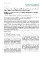
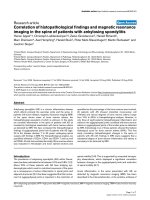
Figure 1
Preparation of the articular cartilage blocks and assembly during bond-ing experimentsPreparation of the articular cartilage blocks and assembly during bond-
ing experiments. (a) Osteochondral fragments were harvested from the
femoropatellar groove of bovine calves. Blocks of 10 × 10 × 20 mm
3
were harvested. (b) These blocks were cut into cartilage slices of two
precisely defined thicknesses, 0.25 and 0.3 mm, which were desig-
nated geometry 1 and 2 (G1 and G2) (in the diagram, only one thick-
ness is shown). (c) These slices were cut into rectangles of 8 mm × 2.5
mm. (d) The integration specimens were assembled by positioning two
cartilage rectangles in partial apposition, creating a defined overlap
area of 4 mm × 2.5 mm for bonding. (e) When cartilage samples (either
two G1 or two G2 blocks) were inserted into the chamber and fixed by
the stamp, different compressive strains were applied.
Arthritis Research & Therapy Vol 9 No 3 Englert et al.
Page 4 of 11
(page number not for citation purposes)
dimethylmethylene blue dye, using bovine chondroitin sulfate
as standard [28]. Total collagen content was determined by
measuring the amount of hydroxyproline according to [29],
with some modifications. Digested sample (100 μl) was hydro-
lyzed with 100 μl 12 N hydrochloric acid for 16 h at 105°C.
After hydrolysis, hydrochloric acid was evaporated. The dry
samples were dissolved in 500 μl of bidistilled water. In a
microtiter plate 100 μl of each sample were oxidized by 50 μl
of a 0.05 M solution of chloramine T in a citrate buffer, pH 6,
for 20 minutes. Afterwards, 50 μl of a 15% (mass/mass)
dimethylaminobenzaldehyde solution in 4 mol perchloric acid
in 70% isopropanol/water (mass/mass) was added and, after
shaking, the plate was incubated for 30 minutes at 60°C. The
plate was cooled down to room temperature and the absorb-
ance of the samples was immediately measured at 557 nm
using a microplate reader (CS-9301 PC, Shimadzu, Duisburg,
Germany).
Histology
Sample pairs were fixed in 2% glutaraldehyde and 4% formal-
dehyde in 0.1 M phosphate buffer, pH 7.3, for 30 minutes, and
again fixed for 60 minutes in 4% formaldehyde, washed in
buffer, embedded in Tissue Tek and frozen. Cryostat sections
were cut perpendicular to the height of the articular cartilage
block to a thickness of 5 μm and stained with toluidine blue for
GAGs [5].
Determination of cytotoxicity
The relative cytotoxicity of degrading and cross-linking rea-
gents was tested by a resazurin reduction test obtained from
Serotec Limited (Düsseldorf, Germany), which was used
according to the manufacturer's instructions; a 10% resazurin
solution was employed [30]. The oxidized, blue, non-fluores-
cent resazurin is reduced to a pink fluorescent dye in the
medium by metabolic activity. For all tested degrading and
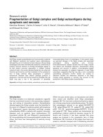
Figure 2
Schematic collagen cross-linking reactions for the employed reagentsSchematic collagen cross-linking reactions for the employed reagents. (a) Glutaraldehyde covalently binds to amino groups, but can also bind to
other glutaraldehyde molecules. (b) 1-Ethyl-3-diaminopropyl-carbodiimide (EDC) and N-hydroxysuccinimide (NHS) catalyses covalent bindings
between carboxylic acid and amino groups; thus, cross-linking between collagen structures is possible. Furthermore, other extracellular matrix com-
ponents containing carboxyl groups, such as glycosaminoglycans, can also be cross-linked. (c) Genipin reacts in a similar manner as glutaraldehyde,
but can only bind to one other genipin molecule. (d) Transglutaminase is a highly specific enzyme catalysing collagen cross-linking between lysine
and glutamine in collagen structures with the release of ammonia.
Available online />Page 5 of 11
(page number not for citation purposes)
cross-linking reagents, the same concentrations as stated in
the cross-linking and surface degradation sections were used
(see above).
Statistical analysis
The statistical analysis was carried out using SPSS 12.0 G
(SPSS, Munich, Germany). Of the free analysed parameters,
adhesive strength (kPa), GAG content (μg), collagen content
(μg), and relative fluorescence unit (RFU), the Kolmogorov-
Smirnov test showed evidence against normal distribution for
adhesive strength. Thus, the non-parametric Kruskal Wallis
test for overall testing and the Mann-Whitney-U test for pair-
wise testing was applied to test for differences in adhesive
strength between groups. For analysis of the GAG and colla-
gen content of the cartilage blocks or cytotoxicity (RFU) of the
applied agents, which were normally distributed, overall differ-
ence in the groups was assessed by analysis of variance
(ANOVA) followed by post hoc comparisons made by Tukey's
test. Throughout, statistical significance was accepted for p <
0.05.
Results
Adhesive strength of the bonding area
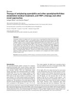
At first, cartilage blocks of the two different geometries were
fixed in partial apposition in the custom-made chamber. The
relaxation behaviour of the G1 and G2 cartilage blocks is
shown in Figure 3a. Due to the precise positioning of the G1
cartilage blocks in the chamber, immediately after mechanical
fixation an instantaneous load drop to almost 0 N was
observed. The remaining compressive load, in the order of 0.1
N, was attributed to swelling of the cartilage. In contrast, for
the oversized G2 cartilage blocks, a compressive load of 5 N
resulted from mechanical fixing by the stamp. With increasing
incubation time and force relaxation, the load decreased and
approached 1 N after approximately 400 s.
To compare the two geometries G1 and G2 with regard to
cartilage bonding, the cross-linking agents were applied with-
out prior surface degradation. The Kruskal Wallis test showed
significant difference in adhesive strength (kPa) between the
groups, which motivated us to perform pairwise testing. Com-
pression to 83% of initial thickness during incubation (G2)
resulted in strongly enhanced bonding after treatment with
glutaraldehyde and EDC/NHS compared to no compression
(Figure 3b). Using transglutaminase, no bonding occurred
without prior surface degradation for either of the two
geometries. Based on these results, the following experiments
investigating the effects of the different combinations of
degrading and cross-linking agents were conducted using G2
cartilage blocks. It should be noted that in experiments with
neither surface degradation nor cross-linking or in experiments
with surface degradation only, no bonding, for either G1 or
G2, was achieved at all.
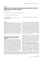
The four cross-linking reagents glutaraldehyde, genipin, EDC/
NHS, and transglutaminase were each combined with the pre-
treatments trypsin, pepsin, and guanidine; the resulting bond-
ing quality measured as adhesive strength is shown in Figure
4. The Kruskal Wallis test showed significant differences in
adhesive strength (kPa) between the groups, prompting us to
perform pairwise testing. With glutaraldehyde, only guanidine
pre-treatment led to a significant increase of adhesive strength
(55 kPa compared to 20 kPa for the group with no pre-treat-
ment; Figure 4a). In combination with EDC/NHS, pepsin or
Figure 3
Stress-relaxation curves for the two sample geometries and bonding dependence on compressionStress-relaxation curves for the two sample geometries and bonding
dependence on compression. (a) Stress-relaxation curves for the sam-
ple geometries G1 and G2 (see Figure 1e) were determined in a stand-
ardised creep modulus set-up. Samples were compressed by a stamp
and the resulting force relaxation behaviour was analysed by recording
the load over time. (b) G1 (almost no compression) or G2 (compres-
sion) cartilage blocks were subjected to different cross-linkers (without
degrading pre-treatment). Adhesive strength as a measure of bonding
was determined immediately after cross-linking. Bars represent the
mean with standard error of the mean of at least 16 samples derived
from 4 independent experiments, each with at least 4 replicates per
group. P values in the graph are from pairwise comparisons using the
Mann Whitney-U test. EDC, 1-ethyl-3-diaminopropyl-carbodiimide;
NHS, N-hydroxysuccinimide.
Arthritis Research & Therapy Vol 9 No 3 Englert et al.
Page 6 of 11
(page number not for citation purposes)
guanidine pre-treatment increased the adhesive strength to 65
kPa, exhibiting the highest values seen among all combina-
tions of pre-treatment and cross-linking reagents in this study
(30 kPa for no pre-treatment; Figure 4b). With genipin, all
three pre-treatments led to a significantly increased adhesive
strength, with the highest values detected with guanidine;
however, mean values were all below those for EDC/NHS and
glutaraldehyde (Figure 4c). For transglutaminase cross-linking,
pre-treatment with guanidine was necessary to induce notice-
able bonding. Overall, transglutaminase clearly resulted in the
lowest values for adhesive strength compared to all other
cross-linkers (Figure 4d).
Effects of surface degradation on glycosaminoglycan
and collagen content
To determine the effects of the degrading agents on extracel-
lular matrix content, the GAG and total collagen content were
determined in cartilage samples and the respective superna-
Figure 4
Bonding of cartilage blocks after treatment with surface-degrading reagents and subsequent cross-linkingBonding of cartilage blocks after treatment with surface-degrading reagents and subsequent cross-linking. Adhesive strength as a measure of bond-
ing was determined immediately after cross-linking with (a) glutaraldehyde, (b) 1-Ethyl-3-diaminopropyl-carbodiimide (EDC)/N-hydroxysuccinimide
(NHS), (c) genipin, or (d) transglutaminase. Before cross-linking, cartilage blocks were pre-treated with either trypsin, pepsin, or guanidine, or blocks
were cross-linked without pre-treatment ('no pre-treatment'). In the control group, neither pre-treatment nor cross-linking were performed. Bars rep-
resent the mean with standard error of the mean of 20 samples derived from 4 independent experiments, each with 5 replicates per group. P values
are from Mann Whitney-U test for pairwise comparisons.
Available online />Page 7 of 11
(page number not for citation purposes)
tants after treatment. The ANOVA test showed significant dif-
ferences between the groups (p < 0.001). Trypsin treatment
strongly decreased the GAG content of cartilage samples to
31% of the control group (Figure 5a); whereas untreated con-
trol blocks had a GAG content of 4.2% per wet weight, trypsin
reduced the GAG content to 1.3%. Guanidine treatment
resulted in a reduction in GAG content to 78% of that of the
control group, whereas only small amounts of GAG were
released from the cartilage samples treated with pepsin (Fig-
ure 5a). The GAG release from cartilage samples was con-
firmed by analysis of the corresponding supernatants (Figure
5b) and staining of histological cross-sections of cartilage
blocks with toluidine blue for GAG (Figure 5c). The collagen
content of untreated control blocks was 12.8% per wet
weight. For all three treatments, trypsin, pepsin, or guanidine,
no significant reduction in collagen content in the cartilage
samples was detected.
Relative cytotoxicity
The resazurin cytotoxicity test performed on cartilage samples
revealed distinct differences for the reagents employed in this
study (p value from ANOVA, p < 0.001). Among the degrading
reagents, pepsin had only small cytotoxic effects whereas
guanidine exhibited strong effects in comparison to the non-
treated control group (Figure 6a). In the cross-linking reagent
group, EDC/NHS showed almost no effect, whereas reduced
metabolic activity to less than 50% within 2 h was detected for
glutaraldehyde and genipin (Figure 6b).
Discussion
In this study, the effects of compression of the cartilage inter-
face, surface degradation, and biochemical cross-linking on
articular cartilage bonding were investigated. Specific empha-
sis was put on the resulting mechanical stability of the bonded
interface due to molecular bridging of opposing surface struc-
tures. This immediate repair technique might provide one fur-
ther option for the therapeutic treatment of articular cartilage
wounds.
Compression
In the absence of treatment with cross-linking agents, neither
just laying the cartilage blocks together nor compressing them
(to 83% of initial thickness) had any effect on bonding
between them. Although the cross-linking reagents EDC/NHS
and glutaraldehyde led to measurable bonding of cartilage
blocks in the absence of compressive strain, large increases in
adhesive strength were achieved by additional compressive
load during the bonding procedure. Therefore, all experiments
investigating combinations of cross-linking reagents with a
pre-treatment with surface degrading agents were carried out
under compressive load conditions.
Cross-linking
EDC/NHS can non-specifically catalyse covalent binding of
the amino or carboxyl groups of collagen; furthermore, carbox-
Figure 5
Glycosaminoglycan (GAG) release from cartilage blocks determined after treatment with surface-degrading agentsGlycosaminoglycan (GAG) release from cartilage blocks determined
after treatment with surface-degrading agents. Cartilage blocks were
subjected to pre-treatment with trypsin, pepsin, or guanidine, as indi-
cated for the bonding experiments; the samples in the control group
were incubated in PBS buffer. (a) Subsequently, the GAG content
within the cartilage blocks was determined. (b) Additionally, the amount
of GAG released into the medium (per cartilage block) was measured.
Nine samples were measured per group. Bars represent the mean with
standard error of the mean. P values are from post hoc Tukey test for
pairwise comparisons. (c) Additionally, histological cross-sections of
cartilage blocks were stained for GAGs.
Arthritis Research & Therapy Vol 9 No 3 Englert et al.
Page 8 of 11
(page number not for citation purposes)
ylic groups of GAGs may also be involved. In our study, EDC/
NHS was the best cross-linking reagent with regard to bond-
ing of articular cartilage blocks in all investigations, that is,
when comparing the cross-linkers alone and in combination
with degrading pre-treatment. The combinations of EDC/NHS
with pepsin or guanidine pre-treatment led to the highest
adhesive strengths detected in this study. To date, cell-based
articular cartilage repair in vitro has been correlated with cell
metabolism [2], collagen deposition [1] and collagen cross-
linking [3,31]. However, the impressive results yielded with
EDC/NHS, the only cross-linker in this study able to addition-
ally catalyse binding of GAGs, may support the idea that mol-
ecules other than those involved in the collagen network can
also contribute to the integrative process in vitro.
With regard to cytoxicity, even after exposure for two hours,
EDC/NHS elicited no significant effects, whereas glutaralde-
hyde and genipin compromised cell vitality considerably. In
previous investigations, EDC/NHS has also been observed to
be advantageous in this respect, compared to glutaraldehyde
[32]. Furthermore, the presented adhesive strength data
resulted from a total incubation with cross-linking reagents for
two hours. In additional experiments in which guanidine or
pepsin pre-treated samples were exposed to EDC/NHS for
only 30 or 10 minutes, an adhesive strength between 58 and
56 kPa was observed, that is, there was no significant differ-
ence to the prolonged treatment of two hours. An exposure for
only 10 minutes would further reduce the risk of any cytotoxic
effects.
Glutaraldehyde and genipin cross-link amino groups of pro-
teins; glutaraldehyde molecules can cross-link to each other
and the chain building properties may have beneficial effects
in comparison to genipin, which can only cross-link in pairs
[33]. In this study, glutaraldehyde alone yielded higher adhe-
sive strengths than genipin alone and also had slight advan-
tages in combination with pre-treatments. With regard to
cytotoxicity, a better tolerance for genipin in comparison to
glutaraldehyde has been shown in other investigations utilising
3T3 mouse fibroblasts [34], human osteoblasts [35] and a
subcutaneous chamber in mice [36]. In our study, both agents
exhibited similarly strong cytotoxic effects.
Transglutaminase, a naturally occurring enzyme in articular
cartilage, catalyses a specific collagen cross-linking reaction
between lysine and glutamine residues. This enzyme has been
previously introduced, combined with compressive load, to
enhance integrative bonding of articular cartilage wounds
[26]. In our investigation, bonding was detectable only in com-
bination with guanidine pre-treatment; however, compared to
the other cross-linking options investigated in this study, trans-
glutaminase resulted in rather weak bonding. The protocol
employed in this study was initially described by Chen and col-
leagues [27] for cross-linking collagen matrices and may not
be well transferable to articular cartilage. The reduced reaction
time in this study (2 h) compared to that reported previously
(12 h) may also have contributed to the reduced effect. Never-
theless, transglutaminase may still play an important role in in
vitro and in vivo integrative repair. Transglutaminase has been
previously shown to be biocompatible [37], which was also
Figure 6
Cytotoxicity of degrading and cross-linking agentsCytotoxicity of degrading and cross-linking agents. The relative cytotox-
icity of (a) degrading and (b) cross-linking agents was determined
using the resazurin assay (expressed as relative fluorescence units).
Cartilage blocks were treated with the respective reagents for either
one or two hours. The controls were incubated in PBS. P values are
from Tukey test for pairwise comparisons. EDC, 1-ethyl-3-diaminopro-
pyl-carbodiimide; NHS, N-hydroxysuccinimide.
Available online />Page 9 of 11
(page number not for citation purposes)
found in this study, with no significant differences to the con-
trol. This enzyme, with its specific catalysing mechanism, may
be especially beneficial in an ongoing integrative repair proc-
ess in which newly synthesised collagen fibrils are present, in
contrast to a static experimental setting such as used in this
study.
For many years, soft tissue adhesives like fibrin [38] have been
used for cartilage repair or as an additive in autologous
chondrocyte transplantation [39,40]. They have been found to
be supportive in chondrocyte transplantation or to seal the
periosteum flap to the cartilage in vitro, but in vivo fibrin glue
did not provide enough mechanical strength to hold the peri-
osteum flap in place [41]. As a further alternative, several syn-
thetic materials have been employed as glues for soft tissues,
for example, aminopropyltrimethoxysilane-methylenebisacryla-
mide siloxane or n-butylcyanoacrylate [42]. In general, the
bonding mechanisms of these polymeric substances differ
from those of the chemical reagents used in the present work.
The polymers penetrate the soft tissue to a certain extent and
adhesion is achieved through an interpenetrating network that
is irremovable and may impair tissue development at the
integration site. In contrast, the chemical cross-linker induces
formation of covalent bonds on the surface of the soft tissue.
In our opinion, EDC/NHS may be beneficial compared to pol-
ymer glue and other chemical cross-linkers (glutaraldehyde
and genipin) due to its pure catalysing function. EDC/NHS will
not be incorporated into the cartilage and can be easily
removed and the scar tissue can be remodelled by cell and
extracellular matrix turnover.
Degradation
Degradation or swelling of articular cartilage surfaces have
been reported to be beneficial in cell-based integrative repair
in vitro [6,9-12]. In our study, bonding between cartilage
blocks did not occur by merely treating the blocks with trypsin,
pepsin, or guanidine (even under compressive conditions).
However, pre-treatment with the endopeptidases pepsin or
trypsin before cross-linking led to distinct improvements in
bonding compared to the use of cross-linkers alone. This was
particularly the case for the combination of pepsin with EDC/
NHS, for which high values for adhesive strength were
achieved. With regard to cytoxicity, pepsin led to no significant
effects, whereas trypsin treatment compromised cell vitality
considerably.
Pre-treatment with guanidine led to the highest adhesive
strengths in combination with all cross-linkers compared to the
endopeptidase pre-treatments. Unfortunately, guanidine elic-
ited the strongest cytotoxic effects of all reagents in the study.
It is noteworthy that the achieved mechanical bonding is com-
parable to previous studies employing a similar model. Reindel
and colleagues [4] first reported a mechanical adhesive
strength of 34 kPa in integrative experiments. Subsequently,
studies including degradation with trypsin followed by
cultivation reported enhanced adhesive strengths up to 100
kPa [6]. In the present study, adhesive strengths up to 65 kPa
were achieved by guanidine or pepsin pre-treatment and
EDC/NHS cross-linking.
Previously, it was assumed that degrading surface treatment
led to cell proliferation or stimulation of cell metabolism [12].
The observation from our investigations that cross-linking rea-
gents lead to significantly stronger bonding of cartilage blocks
after degradation or swelling pre-treatment implies another
hypothesis. The accessibility of functional groups is enhanced
by both treatments and, therefore, may have led to a better
bonding in our study and better integrative repair in previously
described studies. It remains to be clarified on which compo-
nents of the extracellular matrix these functional groups are
located. The endopeptidase trypsin was clearly the most effec-
tive at releasing GAGs from the cartilage blocks, whereas
pepsin released only a minor fraction of the GAGs. The treat-
ment of cartilage blocks with guanidine prior to biochemical
cross-linking leads, in theory, primarily to reduction of non-cov-
alent bonding between molecules of the extracellular matrix.
The tertiary structure of matrix molecules, especially GAGs,
becomes more open after hydrogen bonds are broken. Addi-
tionally, elevated water uptake may occur due to more acces-
sible functional groups or a loosened collagen network with
increased pore diameter (swelling). Nevertheless, in our study,
guanidine also led to the release of a significant fraction of
GAGs from the cartilage blocks. As trypsin led to the lowest
adhesive strength values of all surface-degrading agents in
this study, the bonding observed can not be directly correlated
with the amounts of GAGs released. On the contrary, the large
amount of GAGs released by trypsin may have compromised
the cartilage structure. For total collagen, no significant
release was detected for all the pre-treatments. However, only
small changes in the structure of the cartilage surface, which
are triggered by the pre-treatment agents, but which are not
detectable by the assays employed in this study, may be nec-
essary to elicit distinctly improved responses to the cross-link-
ers.
Clarification of the mechanisms involved appears to be a
worthwhile subject for further investigation. Future studies
should also address the fact that immature and mature carti-
lage differ in extracellular matrix content, structure and
mechanical properties [43-45]. In aging, cartilage undergoes
structural changes that affect the susceptibility to degradation
[46-48]. It is also known that integrative bonding is influenced
by the developmental stage of articular cartilage [4]. There-
fore, in future studies, the introduced treatment may have to be
adjusted to adult cartilage. Furthermore, it has to be noted that
for clinical applications special care should be taken to limit
any treatment with degrading and cross-linking agents to the
area close to the cartilage wound surface. In addition, cell cul-
ture experiments after bonding should assure the long-term
viability of the treated cartilage.
Arthritis Research & Therapy Vol 9 No 3 Englert et al.
Page 10 of 11
(page number not for citation purposes)
Conclusion
This study clearly demonstrates that immediate bonding of
articular cartilage blocks can be achieved by means of chemi-
cal cross-linking. Adhesive strength was superior under
compressive conditions compared to no compression. In gen-
eral, pre-treatment with surface-degrading enzymes or swell-
ing by guanidine salt led to distinct enhancement of cartilage
bonding after chemical cross-linking. Taking both the
observed bonding and the cell vitality after treatment into
account, the combination of pepsin pre-treatment and cross-
linking with EDC/NHS appears to be the most favourable with
regard to this study. The presented work suggests that a com-
bination of selected surface-degrading agents and chemical
cross-linkers is a promising option for enhancing bonding of
opposed surfaces in cartilage repair.
Competing interests
The authors declare that they have no competing interests.
Authors' contributions
CE conceived of the study and its design, participated in the
bonding experiments, and helped to draft the manuscript. TB
participated in the design of the study and drafted the manu-
script. RM participated in the design of the study, specifically
with regard to chemical cross-linking. SSvG carried out the
bonding experiments and the cytotoxicity assay. JB carried out
the analysis of the extracellular matrix content. JF carried out
the analysis of the relaxation behaviour and participated in the
bonding experiments. IH helped to perform the statistical anal-
ysis. MN and JH participated in the design and coordination of
the study. All authors read and approved the final manuscript.
Acknowledgements
The authors thank Richard Kujat, PhD, and Tom Böttner for laboratory
assistance. The authors thank, in particular, Metzgerei Stierstorfer, Wen-
zenbach, for the kind supply of bovine joints two hours after sacrificing.
One of the authors (CE) is grateful to the University Medical Centre
Regensburg for financial support (ReForM A).
References
1. DiMicco MA, Sah RL: Integrative cartilage repair: adhesive
strength is correlated with collagen deposition. J Orthop Res
2001, 19:1105-1112.
2. DiMicco MA, Waters SN, Akeson WH, Sah RL: Integrative artic-
ular cartilage repair: dependence on developmental stage and
collagen metabolism. Osteoarthritis Cartilage 2002,
10:218-225.
3. Ahsan T, Lottman LM, Harwood F, Amiel D, Sah RL: Integrative
cartilage repair: inhibition by beta-aminopropionitrile. J Orthop
Res 1999, 17:850-857.
4. Reindel ES, Ayroso AM, Chen AC, Chun DM, Schinagl RM, Sah
RL: Integrative repair of articular cartilage in vitro: adhesive
strength of the interface region. J Orthop Res 1995,
13:751-760.
5. Englert C, Blunk T, Fierlbeck J, Kaiser J, Stosiek W, Angele P,
Hammer J, Straub R: Steroid hormones strongly support artic-
ular cartilage integration in the absence of interleukin-1β.
Arthritis Rheum 2006, 54:3890-3897.
6. Englert C, McGowan KB, Klein TJ, Giurea A, Schumacher BL, Sah
RL: Inhibition of integrative cartilage repair by proteoglycan 4
in synovial fluid. Arthritis Rheum 2005, 52:1091-1099.
7. Ahsan T, Sah RL: Biomechanics of integrative cartilage repair.
Osteoarthritis Cartilage 1999, 7:29-40.
8. McGowan KB, Sah RL: Treatment of cartilage with beta-amino-
propionitrile accelerates subsequent collagen maturation and
modulates integrative repair. J Orthop Res 2005, 23:594-601.
9. Lee MC, Sung KL, Kurtis MS, Akeson WH, Sah RL: Adhesive
force of chondrocytes to cartilage. Effects of chondroitinase
ABC. Clin Orthop 2000, 370:286-294.
10. Giurea A, DiMicco MA, Akeson WH, Sah RL: Development-asso-
ciated differences in integrative cartilage repair: roles of bio-
synthesis and matrix. J Orthop Res 2002, 20:1274-1281.
11. Obradovic B, Martin I, Padera RF, Treppo S, Freed LE, Vunjak-
Novakovic G: Integration of engineered cartilage. J Orthop Res
2001, 19:1089-1097.
12. van de Breevaart BJ, In der Maur CD, Bos PK, Feenstra L, Verhaar
JA, Weinans H, van Osch GJ: Improved cartilage integration
and interfacial strength after enzymatic treatment in a carti-
lage transplantation model. Arthritis Res Ther 2004,
6:R469-R476.
13. Hunziker EB, Rosenberg LC: Repair of partial-thickness defects
in articular cartilage: cell recruitment from the synovial
membrane. J Bone Joint Surg Am 1996, 78:721-733.
14. Angele P, Abke J, Kujat R, Faltermeier H, Schumann D, Nerlich M,
Kinner B, Englert C, Ruszczak Z, Mehrl R, et al.: Influence of dif-
ferent collagen species on physico-chemical properties of
crosslinked collagen matrices. Biomaterials 2004,
25:2831-2841.
15. Bigi A, Burghammer M, Falconi R, Koch MH, Panzavolta S, Riekel
C: Twisted plywood pattern of collagen fibrils in teleost scales:
an X-ray diffraction investigation. J Struct Biol 2001,
136:137-143.
16. Ruijgrok JM, de Wijn JR, Boon ME: Glutaraldehyde crosslinking
of collagen: effects of time, temperature, concentration and
presoaking as measured by shrinkage temperature. Clin
Mater 1994, 17:23-27.
17. Simionescu A, Simionescu D, Deac R: Lysine-enhanced glutar-
aldehyde crosslinking of collagenous biomaterials. J Biomed
Mater Res 1991, 25:1495-1505.
18. van Luyn MJ, van Wachem PB, Damink LO, Dijkstra PJ, Feijen J,
Nieuwenhuis P: Relations between in vitro cytotoxicity and
crosslinked dermal sheep collagens. J Biomed Mater Res
1992, 26:1091-1110.
19. van Wachem PB, van Luyn MJ, Olde Damink LH, Dijkstra PJ, Feijen
J, Nieuwenhuis P: Biocompatibility and tissue regenerating
capacity of crosslinked dermal sheep collagen. J Biomed
Mater Res 1994, 28:353-363.
20. Olde Damink LH, Dijkstra PJ, van Luyn MJ, van Wachem PB, Nieu-
wenhuis P, Feijen J: In vitro degradation of dermal sheep colla-
gen cross-linked using a water-soluble carbodiimide.
Biomaterials 1996, 17:679-684.
21. Olde Damink LH, Dijkstra PJ, van Luyn MJ, van Wachem PB, Nieu-
wenhuis P, Feijen J: Cross-linking of dermal sheep collagen
using a water-soluble carbodiimide. Biomaterials 1996,
17:765-773.
22. Tomihata K, Ikada Y: Crosslinking of hyaluronic acid with water-
soluble carbodiimide. J Biomed Mater Res 1997, 37:243-251.
23. Chang Y, Tsai CC, Liang HC, Sung HW: Reconstruction of the
right ventricular outflow tract with a bovine jugular vein graft
fixed with a naturally occurring crosslinking agent (genipin) in
a canine model. J Thorac Cardiovasc Surg 2001,
122:1208-1218.
24. Sung HW, Huang RN, Huang LL, Tsai CC: In vitro evaluation of
cytotoxicity of a naturally occurring cross-linking reagent for
biological tissue fixation. J Biomater Sci Polym Ed 1999,
10:63-78.
25. Chau DY, Collighan RJ, Verderio EA, Addy VL, Griffin M: The cel-
lular response to transglutaminase-cross-linked collagen.
Biomaterials 2005, 26:6518-6529.
26. Jurgensen K, Aeschlimann D, Cavin V, Genge M, Hunziker EB: A
new biological glue for cartilage-cartilage interfaces: tissue
transglutaminase. J Bone Joint Surg Am 1997, 79:185-193.
27. Chen RN, Ho HO, Sheu MT: Characterization of collagen matri-
ces crosslinked using microbial transglutaminase. Biomateri-
als 2005, 26:4229-4235.
28. Farndale RW, Buttle DJ, Barrett AJ: Improved quantitation and
discrimination of sulphated glycosaminoglycans by use of
Available online />Page 11 of 11
(page number not for citation purposes)
dimethylmethylene blue. Biochim Biophys Acta 1986,
883:173-177.
29. Woessner JF: The determination of hydroxyproline in tissue
and protein samples containing small proportions of this
imino acid. Arch Biochem Biophys 1961, 93:440-447.
30. O'Brien J, Wilson I, Orton T, Pognan F: Investigation of the
Alamar Blue (resazurin) fluorescent dye for the assessment of
mammalian cell cytotoxicity. Eur J Biochem 2000,
267:5421-5426.
31. Ahsan T, Harwood F, McGowan KB, Amiel D, Sah RL: Kinetics of
collagen crosslinking in adult bovine articular cartilage. Oste-
oarthritis Cartilage 2005, 13:709-715.
32. Sung HW, Huang DM, Chang WH, Huang RN, Hsu JC: Evalua-
tion of gelatin hydrogel crosslinked with various crosslinking
agents as bioadhesives: in vitro study. J Biomed Mater Res
1999, 46:520-530.
33. Sung HW, Chang Y, Liang IL, Chang WH, Chen YC: Fixation of
biological tissues with a naturally occurring crosslinking
agent: fixation rate and effects of pH, temperature, and initial
fixative concentration. J Biomed Mater Res 2000, 52:77-87.
34. Sung HW, Liang IL, Chen CN, Huang RN, Liang HF: Stability of a
biological tissue fixed with a naturally occurring crosslinking
agent (genipin). J Biomed Mater Res 2001, 55:538-546.
35. Gough JE, Scotchford CA, Downes S: Cytotoxicity of glutaralde-
hyde crosslinked collagen/poly(vinyl alcohol) films is by the
mechanism of apoptosis. J Biomed Mater Res 2002,
61:121-130.
36. Huang LL, Sung HW, Tsai CC, Huang DM: Biocompatibility
study of a biological tissue fixed with a naturally occurring
crosslinking reagent. J Biomed Mater Res 1998, 42:568-576.
37. Chau DY, Collighan RJ, Verderio EA, Addy VL, Griffin M: The cel-
lular response to transglutaminase-cross-linked collagen.
Biomaterials 2005, 26:6518-6529.
38. Kaplonyi G, Zimmerman I, Frenyo AD, Farkas T, Nemes G: The use
of fibrin adhesive in the repair of chondral and osteochondral
injuries. Injury 1988,
19:267-272.
39. Homminga GN, Bulstra SK, Bouwmeester PS, van der Linden AJ:
Perichondral grafting for cartilage lesions of the knee. J Bone
Joint Surg Br 1990, 72:1003-1007.
40. Homminga GN, Buma P, Koot HW, van der Kraan PM, van den
Berg WB: Chondrocyte behavior in fibrin glue in vitro. Acta
Orthop Scand 1993, 64:441-445.
41. van Susante JL, Buma P, Schuman L, Homminga GN, van den
Berg WB, Veth RP: Resurfacing potential of heterologous
chondrocytes suspended in fibrin glue in large full-thickness
defects of femoral articular cartilage: an experimental study in
the goat. Biomaterials 1999, 20:1167-1175.
42. Wilson DJ, Chenery DH, Bowring HK, Wilson K, Turner R,
Maughan J, West PJ, Ansell CW: Physical and biological prop-
erties of a novel siloxane adhesive for soft tissue applications.
J Biomater Sci Polym Ed 2005, 16:449-472.
43. Williamson AK, Chen AC, Sah RL: Compressive properties and
function-composition relationships of developing bovine artic-
ular cartilage. J Orthop Res 2001, 19:1113-1121.
44. Williamson AK, Chen AC, Masuda K, Thonar EJ, Sah RL: Tensile
mechanical properties of bovine articular cartilage: variations
with growth and relationships to collagen network
components. J Orthop Res 2003, 21:872-880.
45. Williamson AK, Masuda K, Thonar EJ, Sah RL: Growth of imma-
ture articular cartilage in vitro: correlated variation in tensile
biomechanical and collagen network properties. Tissue Eng
2003, 9:625-634.
46. DeGroot J, Verzijl N, Bank RA, Lafeber FP, Bijlsma JW, TeKoppele
JM: Age-related decrease in proteoglycan synthesis of human
articular chondrocytes: the role of nonenzymatic glycation.
Arthritis Rheum 1999, 42:1003-1009.
47. DeGroot J, Verzijl N, Budde M, Bijlsma JW, Lafeber FP, TeKoppele
JM: Accumulation of advanced glycation end products
decreases collagen turnover by bovine chondrocytes. Exp Cell
Res 2001, 266:303-310.
48. Chen AC, Temple MM, Ng DM, Verzijl N, DeGroot J, TeKoppele
JM, Sah RL: Induction of advanced glycation end products and
alterations of the tensile properties of articular cartilage.
Arthritis Rheum
2002, 46:3212-3217.