Báo cáo y học: "Expert agreement confirms that negative changes in hand and foot radiographs are a surrogate for repair in patients with rheumatoid arthritis" pot
Bạn đang xem bản rút gọn của tài liệu. Xem và tải ngay bản đầy đủ của tài liệu tại đây (148.84 KB, 9 trang )
Open Access
Available online />Page 1 of 9
(page number not for citation purposes)
Vol 9 No 4
Research article
Expert agreement confirms that negative changes in hand and
foot radiographs are a surrogate for repair in patients with
rheumatoid arthritis
Désirée van der Heijde
1
, Robert Landewé
2
, Annelies Boonen
2
, Steve Einstein
3
, Gertraud Herborn
4
,
Rolf Rau
4
, Siegfried Wassenberg
4
, Barbara N Weissman
5
, Carl S Winalski
6
and John T Sharp
7
1
Department of Rheumatology, Leiden University Medical Center, PO Box 9600, Leiden 2300 RC, The Netherlands
2
Department of Rheumatology, University Hospital Maastricht, and CAPHRI Research Institute, PO Box 5800, Maastricht 6202 AZ, The Netherlands
3
BioImaging Technologies, 826 Newtown-Yardley Road, Newtown, PA 18940, USA
4
Department of Rheumatology, Evangelisches Fachkrankenhaus Ratingen, Rosenstrasse 2, Ratingen D-40882, Germany
5
Department of Radiology, Brigham & Women's Hospital, 75 Francis Street, Boston, MA 02115, USA
6
Department of Radiology, The Cleveland Clinic Foundation, 9500 Euclid Avenue, Cleveland, OH 44195, USA
7
Department of Rheumatology, University of Washington, 8387 NE Sunamee Place, Bainbridge Island, WA 98110, USA
Corresponding author: Désirée van der Heijde,
Received: 17 May 2006 Revisions requested: 6 Jul 2006 Revisions received: 25 Apr 2007 Accepted: 2 Jul 2007 Published: 2 Jul 2007
Arthritis Research & Therapy 2007, 9:R62 (doi:10.1186/ar2220)
This article is online at: />© 2007 van der Heijde et al.; licensee BioMed Central Ltd.
This is an open access article distributed under the terms of the Creative Commons Attribution License ( />),
which permits unrestricted use, distribution, and reproduction in any medium, provided the original work is properly cited.
Abstract
The objective of the present study was to test the hypothesis
that experts recognize repair of erosions and, if so, to determine
which, if any, morphologic features permitted them to recognize
the repair. We also tested whether scoring by a standard
method detected repair. Seven experienced readers of
radiographs in rheumatoid arthritis were presented with 64 sets
of single joints-of-interest at two time points, randomized and
blinded for the correct sequence. The readers assessed which
joint was better, and recorded whether any of six specific
features were seen. Two independent readers, experienced in
scoring by the van der Heijde-modified Sharp method who were
not on the expert panel, then scored the complete films that
included the joint-of-interest. The panel agreed very well on
which of two joints was better, and, even though they did not
know the true sequence, the panel accurately assigned a
sequence slightly better than chance alone (58%) but worse
than their agreement on which image was 'better or worse'
(78%). The readers therefore indirectly assigned repair by
choosing the second film as the best. Putative repair features
were seen in cases of both repair and progression, and were not
discriminatory. Similar results were obtained when the experts
were presented with the entire hand or foot containing the joint-
of-interest. In the third repair exercise, two independent readers
who scored whole hands and feet using a standard method
found a mean negative score in 22/60 joints-of-interest. All 22
joints were also scored as repair by the panel. Repair was
detected reliably by a majority of the panel on viewing paired
images based on a better/worse decision and assigning
sequence in a set of images that were blinded for sequence by
an independent project manager. In this test set of images,
repair was manifested by a reduction in the size of erosion in
many cases. Size was one feature that aided the experts to
detect repair but cannot be the only one; the experts had to find
other features to determine whether a smaller erosion was the
first in a sequence of radiographs in a patient with progressive
damage or was the second film in a patient exhibiting repair. The
change in size of erosion was also picked up by independent
readers applying the van der Heijde-modified Sharp scoring
method and was reflected in their scores.
Introduction
Damage of bone and cartilage caused by rheumatoid arthritis
is visualized on radiographs as erosions and joint space nar-
rowing. The focus of assessment until recently was on pro-
gression of damage. The first evidence that drug therapy might
influence the course of rheumatoid arthritis appeared more
than 30 years ago after the development of a method for scor-
ing these abnormalities [1]. A decade and a half ago additional
data became available to validate the term 'disease modifying
antirheumatic drugs' when sulphasalazine was shown to slow
TNF = tumour necrosis factor.
Arthritis Research & Therapy Vol 9 No 4 van der Heijde et al.
Page 2 of 9
(page number not for citation purposes)
radiographic progression [2]. Around the turn of the century it
became obvious that radiographic progression could be
stopped completely by current therapy in a large proportion of
patients followed for 1–5 years, and it was appreciated that a
number of patients had lower erosion scores in follow-up films
[3,4]. During the same time period scattered reports were
appearing of repair of erosions, many with equivocal support-
ing evidence [5-8]. Although a few studies presented images
that were convincing, no studies have been performed to elim-
inate reader bias or to examine whether there are specific
structural features that permit recognition of repair when the
reader is unaware of the true sequence of films. The observa-
tion of negative scores, together with credible reports that
healing might be real, posed the question of whether negative
scores were detecting real structural improvement or artefact
and, if real, whether the observations were clinically relevant or
were trivial
There are additional reasons why these reports of healing and
the negative scores drew attention. Of particular importance,
therapy with TNFα blockers – which are more potent than pre-
viously employed therapies – resulted in lower levels of dis-
ease activity than were previously seen. As inflammation is a
major driver of damage, an absence of inflammation a priori is
a prerequisite for halting progression, and for making possible
reversal of damage. In addition, data have been presented
suggesting that TNFα blockers inhibit radiographic progres-
sion even in the presence of some persistent inflammation [9].
Presenting radiographic data by probability plots also revealed
much more clearly the number of patients with negative scores
by presenting the individual data of all patients, and this pro-
vided new insight into the potential magnitude of repair [10].
There is currently one trial in which a relatively large number of
patients with negative erosion scores supports repair occur-
ring on a group level [4]. Although the appraisal of repair on a
group level is a relatively simple statistical matter, translating
repair from the group level to an individual patient is not
straightforward. The null hypothesis that there is no change
from baseline within the group can be rejected if the mean
change is below zero and the entire 95% confidence limit is
below zero, which occurred in the TEMPO trial [11]. In con-
trast, a negative change score in an individual patient can be
composed of 'true repair', of measurement error or of an image
artefact such as rotation hiding a tangential erosion. The inter-
relationship of these three components is unknown and differ-
ent in each patient. This argument is also pertinent in
evaluation progression scores.
In a preliminary study within the OMERACT working group on
repair we investigated whether a group of experts would agree
on the presence of repair in a set of individual joints [12,13].
The conclusions were that repair indeed exists according to
the majority judgements of the panel, but when the time
sequence was blinded it was not possible for expert readers
to distinguish cases with progression from cases with repair
based on specific features considered relevant to repair, such
as sclerosis, recortication, and filling-in of erosions. In that
study the experts demonstrated good agreement on which
image showed the least damage, but not on whether the best
image was the first or second in time; in other words, whether
the case was one of progression or repair. A few explanations
for this observation were possible. First, there were quite dif-
ferent levels of experience among the readers, raising the pos-
sibility that the readers were not sufficiently experienced to
recognize the features of repair. It was also possible that the
images used in that study did not have a sufficient number of
features of repair, or that the repair features were not clearly
defined for technical reasons. Third, only single joints were
presented to the readers, which might have hampered the cor-
rect ordering of the images into cases of progression or repair
as change in other joints was not available to help in the deci-
sion. Most importantly, we were still not informed about the
relation between cases depicted as repair by experts and neg-
ative scores obtained with traditional scoring methods such as
the modified Sharp method [2,14].
We therefore embarked on three new exercises. First, we
repeated the exercise with the single joints using a completely
new set of images employing a larger number of images of bet-
ter quality. In addition, we held a training session using cases
not employed in the subsequent exercises that demonstrated
repair as collected by one expert in the group (RR). In the sec-
ond of the new exercises, we presented to the experts the
entire hand or foot that included the joint-of-interest from the
single joint exercise. This allowed us to test whether the pres-
entation of the entire hand or foot improved the judgement of
the correct time order of the films, thus indirectly labelling a
case as progression or repair. Finally, we presented the entire
hand and feet images to two independent readers who were
unaware of the purpose of the reading when they scored the
images by the van der Heijde-modified Sharp method. This
third exercise tested the ability of readers to identify cases that
included joints exhibiting repair and to link the scores of indi-
vidual joints to those labelled as progression or repair by the
experts [2].
Methods
Eight experts, all experienced readers of rheumatoid arthritis
radiographs, met for 3 days at Bio-Imaging Technologies
(Newtown, NJ, USA).
The meeting started with a thorough training session discuss-
ing many examples of joints and features showing repair.
These features were filling-in of erosions, recortication, sclero-
sis, remodelling, reconstitution of a normal joint, and trabecu-
lation. The definitions were refined as compared with the
previous exercise, and trabeculation was added as a feature
that can help in distinguishing progression from repair. Filling-
in, although clearly a reduction in the size of erosions, was
Available online />Page 3 of 9
(page number not for citation purposes)
thought by some to have additional information. Because
recortication implies that the reader has concluded that the
case is one of healing, in conducting the exercise readers
recorded cortication of erosions and noted whether this was
better or worse in the paired individual joints without regard to
whether the reader had an opinion as to whether the pair
showed progression or repair. It was also recognized that
reconstitution of normal structure required a prior judgement
as to whether repair was present.
Two exercises were performed thereafter on two consecutive
days. The third exercise was performed separately by two
readers not involved in the first two exercises.
Images for all three exercises were selected by one of the
experts (JTS), who did not participate in the assessments,
from a large set of radiographs available in digitized form from
several data sources. Sixty-four image sets were selected,
knowing the time sequence; approximately equal numbers of
cases with repair, progression and equivocal or no change
were included
Exercise I
Cropped images of hands or feet containing the joint-of-inter-
est with one or two adjacent joints to allow the reader to eval-
uate a change in radiographic positioning were selected.
Images from two time points were paired, randomized, and
blinded for sequence, and were presented to each reader
independently on a reading station consisting of high-resolu-
tion monitors linked to a computerized data recording system.
Readers were asked to choose the film that was worse or to
declare no change, to choose which erosion was larger or to
choose no change, and to choose which film was first in
sequence or state unable to judge. In addition, readers
recorded the presence of specific features of repair in one or
both images. In the analysis, agreement was defined as con-
currence of at least five of seven readers and was assessed for
the better/worse/no change, the erosion size, and sequence
decisions. Subsequently, the judgement of the individual panel
member as to which joint was worse combined with that mem-
ber's assignment of sequence provided an inferred choice of
progression or repair, and was compared with the true
sequence of the films in order to determine the accuracy of the
assignment of progression or repair. The reader's assignment
of progression was therefore a combination of the reader's
choice of the better image with the choice of first in time or the
combination of the worse image and the second in time;
assignment of repair occurred with the choice of the better
image and the second in time or with the choice of the worse
image and the first in time (see Table 1).
All observations of individual readers were pooled and the
specific features were related to the progression and repair
assessment. In total, seven readers provided 448 judgements
of sets of two films. Of these 448 observations, 397 were con-
sidered to show change (repair or progression). These 397
observations were the basis for further testing the perform-
ance of single features of repair for detecting repair, defined
as an appropriate decision about which film was the better in
relation to the true sequence (least damage on the true sec-
ond film). Odds ratios for the specific features for detecting
repair in comparison with progression were calculated, as well
as the sensitivity, the positive predictive value, the specificity,
and the positive and negative likelihood ratios of these
features.
One-third of the cases were selected as stable in the opinion
of the selector (JTS) who is known as conservative in assess-
ing change. In order to check the robustness of the main
results, the analyses were repeated excluding such cases. The
results confirmed and strengthened the conclusions reached
by the primary analyses (data not shown).
Table 1
Study decision tree
Reader judged image A Combined with true sequence of image A
b
Conclusion in analysis
b
Better
a
First time point Progression
Better Second time point Repair
Better and first time point First time point Reader recognized progression
Better and first time point Second time point Reader failed to recognize repair
Better and second time point Second time point Reader recognized repair
Better and second time point First time point Reader failed to recognize progression
The true sequence was unknown to the reader.
a
A similar decision tree constructed for a reader judging image A as worse exchanges repair and progression in the conclusion column.
b
In Exercises I and II, when the reader made a judgement as to whether a pair of images represented progression or repair that decision was
called direct assignment. In the exercises when the analyst interpreted a reader's combined responses on better/worse and the sequence of
images, this is called an inferred or an indirect assignment.
Arthritis Research & Therapy Vol 9 No 4 van der Heijde et al.
Page 4 of 9
(page number not for citation purposes)
Exercise II
During the third day the same readers conducted the second
exercise, in which they were presented with the entire hand or
foot image that included the joints presented in the single joint
experiment. The images were again randomized and recoded
so they were not presented in the same sequence as for the
first exercise, and the readers were not informed as to which
joints had been presented in the earlier exercise, although
some changes may have been sufficiently distinctive to enable
the readers to remember from the exercise performed the day
before.
Initially the experts were asked to judge the entire image as to
which image was better, and whether the difference was due
to progression or repair; in other words to make a direct judge-
ment as opposed to the inferred judgement in Exercise I. The
joint-of-interest in the first exercise was then indicated to the
readers, and they repeated their review and chose which joint
was better, and which film was first in time, to make possible
a second inferred assignment of repair or progression. Panel
agreement (at least five out of seven) was determined for the
progression/repair judgement based on the whole hand/foot
direct assignment and for the better/worse/no change judge-
ment of the joint of interest. Judgement of the whole hand
assignment of progression or repair was compared with the
single joint inferred assignment of progression or repair.
The judgements of the single joints of Exercise II were then
compared with the judgements of the single joints of Exercise
I. Inter-reader agreement for Exercise II was assessed for each
reader pair for both the single joint inferred assignment of
repair and progression and the whole hand/foot direct assign-
ment. All analyses on agreement were carried out with
unweighted kappa statistics.
Exercise III
Complete sets of hands and feet were available for 60 cases
included in the first and second exercise, and these sets were
presented with a blinded time sequence to two readers expe-
rienced in scoring rheumatoid arthritis films for trials but not
involved in any of the exercises or discussions on repair. Read-
ers were not aware of the fact that these images were part of
a study to assess repair.
Films were scored by the van der Heijde-modified Sharp
method [2]. Total scores were calculated (sum of erosions and
joint space narrowing of hands and feet) for both readers.
Average scores of the two readers were used for further anal-
ysis to mimic the situation in scoring clinical trials. Readers'
scores for the joints-of-interest and for the total score were
compared with the panel judgement.
Results
Exercise I
The readers agreed on which film was better, worse or no
change in 77% of the cases. Agreement was similar for ero-
sion size (78%) and better than for the correct sequence
(58%). The readers therefore agreed on which individual joints
showed the least damage, and their single joint inferred
assignment of progression or repair was slightly greater than
expected by chance alone. Taking all of the assignments of all
readers separately, a reader assigned 'no change' to a pair of
films 51 times – indicating that the readers were more willing
than the project manager who selected the cases to assign a
better or worse status than no change. An inferred assignment
of repair was made 254 times, and progression was assigned
143 times, which gives us the prior probability of repair (64%).
Table 2 presents the number of observations (all observations
were pooled) in which single features of repair were scored as
being present. Only the 397 cases in which the readers
scored change were taken into account in this analysis. Fea-
tures are ordered by decreasing prevalence and sensitivity to
detect repair. The most frequently observed feature was filling-
in of erosions (337/397), followed by cortication (276/397),
sclerosis (217/397), remodelling (129/397), trabeculation
(119/397), and reconstitution of a normal joint (78/397). The
odds ratios for filling-in of erosions, cortication, sclerosis, and
remodelling suggest a more frequent recognition of these fea-
tures in repair cases. Despite odds ratios > 1, the discrimina-
tory capacity of a single repair feature in distinguishing
between repair and progression was very low, as deduced
from the positive predictive values in comparison with the prior
probability of repair in this set (64%), and from the likelihood
ratios. For example, the highest odds ratio (2.7) is for filling-in
of erosions, which is equivalent to reduction in the size of ero-
sions. In those cases in which 'filling-in of erosion' was consid-
ered present, however, only 67% of the cases were given a
single joint inferred assignment of repair, as compared with the
prior probability of 64%. This is reflected by a very low positive
likelihood ratio of 1.1 and a rather high negative likelihood ratio
of 0.50. In contrast to the first five listed features (filling-in, cor-
tication, sclerosis, remodelling, and trabeculation), reconstitu-
tion of the normal structure was recorded more frequently in
progression cases. Specificity was less than 0.8 in all cases.
Sensitivity was less than 0.6 for four of the six features: filling-
in of erosions performed badly because of lack of specificity;
sclerosis, remodelling, trabeculation, and reconstitution failed
because of lack of sensitivity.
The contribution of specific features to detect repair was also
checked to determine whether detecting the feature was
dependent on the true sequence in which the images were
presented to the reader. Overall, such an effect could not be
demonstrated (data not shown).
Available online />Page 5 of 9
(page number not for citation purposes)
Exercise II
For this exercise we calculated the kappa statistic (κ) for each
reader-pair. The mean (standard deviation) κ value, computed
across all possible reader-pairs, was 0.52 (0.10) for the
inferred progression/repair/no change assignment, based on
a better/worse decision. The mean (standard deviation) κ
value for a whole hand/foot direct assignment of progression/
repair/no change decision, however, was significantly lower
(0.34 (0.09)); the paired t value for the difference between
indirect and direct assignments was -6.3 (P < 0.001)). This
finding is again compatible with readers agreeing on which
film is better (size of erosions), but agreeing less well on
whether such a difference is due to repair or progression.
Implicitly, this finding also underscores that there are no typical
features regularly recognized by all readers pointing to repair,
confirming what was shown in Exercise I.
Offering the entire hand or foot resulted in an agreement (≥ 5/
7 readers agreed) on an inferred progression/repair assign-
ment in 53/64 (88%) patients. Agreement on a direct whole
hand/foot repair/progression/no change assignment occurred
in 42/64 (66%) patients. In the single joint experiment, these
figures for inferred assignment were 77% and 58%,
respectively.
In the whole hand direct assignment, the panel judged only
nine cases as 'repair' (Table 3). All these cases were assigned
repair by inferred assignment in the same exercise. If the
inferred assignment (28 cases of repair) was considered the
gold standard, however, the panel missed 19 of these cases
in their whole hand/foot direct assignment: eight of the missed
cases were judged as progression and 11 cases did not reach
a majority agreement.
For each reader we compared the direct assignment of repair
with the inferred assignment, using the inferred assignment as
the gold standard because this only involves one decision
about better/worse. All experts' scores were remarkably simi-
lar with respect to assigning progression or repair, except for
one reader. A typical example of results of a single reader is
presented in Table 4. The percentages of correctly classified
cases (agreement between direct whole hand/foot image and
inferred assignment) ranged for all readers from 70% to 75%.
The percentages of cases falsely classified as having repair
ranged from 1.5% to 5%, and those of cases falsely classified
as no repair ranged from 22% to 25% for six of the seven read-
ers. The remaining reader scored repair much more frequently
than the other readers, but classified 11% falsely as repair and
14% falsely as progression. The positive predictive value of a
direct repair assignment ranged from 80% to 96% and the
negative predictive value from 56% to 69%, with the reader
scoring more repair as having the highest negative predictive
value and the lowest positive predictive value. These data are
compatible with a conclusion that experts underperform with
respect to repair if they do not know the true time order. They
indirectly see repair because they see change, but they do not
directly recognize it as such.
After the judgement of the whole hand or foot, without knowl-
edge of the joint-of-interest, we unblinded the joint-of-interest
Table 2
Results of specific repair features in Exercise I
a
Single repair feature First film is
better
b
(progression)
(n = 143, 36%)
Second film is
better (repair)
(n = 254, 64%)
Odds ratio to
detect repair
True positive
rate (sensitivity)
False positive
rate
(1 – specificity)
Positive
likelihood ratio
Negative
likelihood ratio
Filling-in of erosions 112 (33%)* 225 (67%)** 2.2 0.89 0.78 1.1 0.50
Cortication 84 (30%) 192 (70%) 2.2 0.76 0.59 1.3 0.58
Sclerosis 68 (31%) 149 (69%) 1.6 0.59 0.47 1.1 0.78
Remodelling 32 (24%) 97 (76%) 2.1 0.38 0.22 1.7 0.79
Trabeculation 41 (37%) 78 (63%) 1.1 0.20 0.21 1.0 1.0
Reconstitution of a
normal joint
41 (53%) 37 (47%) 0.43 0.15 0.29 0.5 1.2
Any of the above
features of repair
130 (36%) 234 (64%) 1.2 0.92 0.91 1.0 0.89
a
Test performance in Exercise I of putative features of repair in relation to progression and repair as indicated by inferred assignment (first film is
better versus second film is better). Of the total number of 397 observations, 143 (36%) were judged as showing progression and 254 (64%) as
showing repair without taking into account specific features of repair. Adding information on features of repair only marginally influences the
discrimination between progression and repair.
b
Designation of first or second film based on the true sequence. Numbers indicate the numbers of observations in which a given single repair
feature was recorded as present. Percentages indicate the positive predictive value of a specific repair feature for a progression* or repair**
classification. For example, 'filling-in' was observed 325 times: 103 times (32%) in cases with progression and 222 times (68%) in cases of repair.
The positive likelihood ratio is the quotient of the true positive rate divided by the false positive rate (for example, 'filling-in' in cases of repair) and
the false positive rate (for example, 'filling-in' in cases of progression). The negative likelihood ratio is the quotient of the false negative rate (no
'filling-in' in cases of repair) divided by the true negative rate (no 'filling-in' in cases of progression). In order to be of diagnostic value, the positive
likelihood ratio should be high (for example, > 4) and the negative likelihood ratio should be low (for example, < 0.3).
Arthritis Research & Therapy Vol 9 No 4 van der Heijde et al.
Page 6 of 9
(page number not for citation purposes)
and again performed an indirect assignment of repair; we then
compared these results with the results of the indirect assign-
ment of the single joint experiment (Exercise I), both at the level
of the individual experts and at the level of the panel. Absolute
intra-reader agreement varied from 74% to 87% (κ = 0.52–
0.74, indicating moderate to good agreement). Panel agree-
ment (majority decision of at least five out of seven readers)
with regard to the first and the second assignment was 85%
(κ = 0.69).
Exercise III
The complete sets of hand and foot films of the 60 patients
that included 64 joints-of-interest (four patients had more than
one joint of interest) presented to the experts in Exercises I and
II were scored by the van der Heijde-modified Sharp method;
two experienced readers gave a mean negative score to 22 of
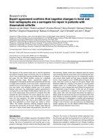
these joints-of-interest. Figure 1 shows that all cases with a
negative joint-of-interest score by these readers were con-
firmed by a majority (≥ 5/7) of the experts as repair in Exercise
I. In 15 of these 22 joints-of-interest with a negative change
score there was complete panel agreement.
Twenty-three patients were given a mean negative change
score on the overall score for both the hands and feet. Seven-
teen of these patients were judged to show repair by the
experts viewing only the joint-of-interest in Exercise I; in the
remaining six cases, the two independent readers did not
score repair in the joint-of-interest, which was in agreement
with the judgement of the expert panel in all cases. In seven
cases there was agreement between both independent read-
ers and the experts that the joint-of-interest showed repair, but
the score for both hands and feet demonstrated progression.
In every case, repair judgement was based on improvement in
erosions, and not on improvement in joint space narrowing. In
contrast, progression in joints-of-interest appeared to be the
consequence both of progression in erosions and of joint
space narrowing.
Discussion
The logic incorporated in the study design in Exercises I and II,
illustrated in Table 1, was critical to the analysis and to the
conclusions reached. If repair is a reality and alters the bone
structure in a distinctive and recognizable way, when experts
view two images of the same joint or of the entire hands or feet
Table 3
Assignment of progression or repair based on direct assignment versus inferred assignment in Exercise II
a
Whole hand direct assignment of
progression/repair
Single joint inferred assignment of progression/repair
Total First film is better
(progression)
Second film is better
(repair)
Both films are similar
(no change)
No majority agreement
obtained
Progression 28 18 8 0 2
Repair 9 0 9 0 0
No change 5 0 0 4 1
No majority agreement obtained 22 3 11 0 8
Total 64 21 28 4 11
a
Based on agreement by five of seven readers.
Table 4
Typical example of the direct versus the inferred assignment of one of the readers
Reader's direct judgement (direct
assignment)
Reader's better/worse interpretation in combination with true time order
of X-rays (inferred assignment):
Totals
Compatible with progression or
no change
Compatible with repair
Progression/no change 21 15 36
Repair 1 27 28
Totals 22 42 64
The inferred assignment is considered the gold standard. Direct assignment underestimates the true prevalence of repair. Percentage correctly
classified, 75%; false direct assignment of repair, 1.5%; false direct assignment of progression, 23%; positive predictive value, 96%; negative
predictive value, 58%.
Available online />Page 7 of 9
(page number not for citation purposes)
that are randomly ordered for sequence they should be able to
tell which image includes the repaired bone, provided that the
technical factors in capturing the image are identical. It is
entirely possible in the early stages of developing an erosion
that, if the inflammatory process is completely halted, the ero-
sion might heal and leave no structural changes indicative of
the healing process. These cases would not be recognized by
an expert under the circumstances of Exercises I or II because
of the randomization and blinding of the sequence, but would
be detected by the standard scoring procedures. In cases that
were characterized by morphologic features of repair, combin-
ing the individual's judgement regarding better/worse with the
true sequence allowed the analyst to infer whether repair or
progression of the erosion has occurred in the interval
between the two images. It is critical to mention here that a
conclusion of repair or progression can be reached irrespec-
tive of the reader's opinion. In other words, readers' bias has
been eliminated.
Results from both Exercises I and II show that readers agree
quite well on which of two images of single joints is the better,
and on which shows the smaller erosion. Agreement was
improved when the cases selected as stable or questionable
and those selected as probable but not definite repair or pro-
gression were omitted. This indicated that readers were able
to accurately recognize a single feature or a combination of
features that was interpreted as repair in many cases. Assign-
ment of the correct sequence was significantly greater than by
chance alone when these cases were omitted from the analy-
sis. The analysis that included all the cases more closely
reflects reality in clinical studies since there will always be
cases that are equivocal. The analysis that excluded these
cases indicates that where there is a clear-cut difference
between images at two time points at intervals of 6 months to
5 years, the panel's agreement as to which is better and which
is the first image in the interval is better than chance alone –
indicating that there are single features or combination of fea-
tures that are recognizable and indicate repair.
Assignment of the correct sequence is only marginally
improved when the experts view the entire hand or foot film.
The association of most of the signs of repair in sets of single
joint images with repair is hardly better than expected by
chance alone. When blinded to sequence, experts frequently
and inadvertently adjudicated signs of repair to sets of images
that actually show progression. Based on these results, single
features of repair should not be included in radiographic scor-
ing methods. The relatively low kappa values for intra-reader
and inter-reader agreement indicate that basing the
assignment of repair on the judgement of only one expert is not
reliable. The panel also performed considerably better, how-
ever, in the test-retest situation if a judgement of repair was
based on a majority decision of at least five out of seven read-
ers. Although the decision to use concurrence by five or more
of the seven readers as a definition of 'agreement' was an a pri-
ori one, the analysis indicates that conclusions based on 5/7
agreement in this analysis are conservative.
In Exercise III two independent readers that regularly score
films of hands and feet according to the van der Heijde-modi-
fied Sharp method, without knowledge of time sequence,
provided negative change scores in individual joints that were
judged as repair by the expert panel in Exercises I and II. In all
cases, this was due to improvement in erosions, not to
improvement in joint space narrowing. The picture, however, is
more complicated. Among patients with a positive change in
total Sharp scores, we found cases of negative erosion scores
in joints-of-interest that were confirmed as repair by the expert
panel. There are two possible interpretations for this observa-
tion: overall progression of damage does not preclude repair
in single joints, or technical factors create apparent improve-
ment (that is, improvement is not real). For example, a change
in radiographic positioning of the joint, a different dynamic
range between the two films, a change in soft tissue during the
interval and possibly other factors may produce spurious
changes.
What clearly emerges from these findings is that experts quite
regularly agree on which image is better. Based on this study,
if we assume that the image is an accurate representation of
the true damage, repair is a reality and this observation con-
firms and extends our previous findings. In another study by
Rau and colleagues, 74 joints out of 1,292 joints showed
repair phenomena [7]. The authors also found that, in the
group of patients with repair phenomena in single joints, an
increase and decrease in the score occurred in different joints
in the same patient at the same time.
Figure 1
Agreement between negative van der Heijde-modified Sharp scores and the expert panel judgement on repairAgreement between negative van der Heijde-modified Sharp scores
and the expert panel judgement on repair. Comparison of the number of
cases with a negative mean score of the van der Heijde-modified Sharp
score by two readers for the joint-of-interest (total n = 22) with the
numbers of experts (total n = 7) assessing the joint as showing repair.
Arthritis Research & Therapy Vol 9 No 4 van der Heijde et al.
Page 8 of 9
(page number not for citation purposes)
The net change score not only reflects numbers of joints, but
also the magnitude of change per joint. If, in an individual
patient, the joints with negative change scores (repair) out-
weigh the joints with positive change scores (progression), the
total Sharp change score, which is the sum of all individual
joint scores, will become negative. It is likely that the magni-
tude of change per joint is higher in cases of progression as
compared with repair, since repair is usually subtle and may be
limited in extent, whereas individual joint progression can be
extensive and can easily involve two or more scoring units.
Moreover, repair in the individual patient is constrained by the
number of joints with damage, and probably also by the level
of damage in those joints. In fact, data from animal studies
clearly indicate that once the matrix is resorbed the rate and
extent of depositing new matrix is limited, which in turn limits
the extent of reconstruction of bone. In contrast, progression
can occur in both damaged and undamaged joints. Scoring
methods therefore cannot capture every individual demon-
strating repair in one or more joints; it is also true that scoring
cannot capture every case of progression. It also is considered
very probable that healing may occur in the minimally damaged
joint without leaving any trace of prior damage or distinctive
features in the reconstituted bone. Under these circum-
stances, even though only the presence/absence of the ero-
sion indicates that repair has occurred, the healing process
would be reflected in the score.
The net change score also reflects the measurement error and
anticipation bias. The former includes the true measurement
error, which includes reading error and error invoked by
changes in radiographic positioning and exposure. Anticipa-
tion bias may arise when readers are influenced by the status
of other joints in the same image and inaccurately score one
or more individual joints; for example, a questionable new ero-
sion becomes much more definite to the reader if several other
joints show clear-cut progression.
In the individual patient it is impossible to judge how and to
what extent measurement error and anticipation bias contrib-
ute to the score, negative or positive. The more negative a
score that includes scores of 44 or more joints for a patient,
the greater the likelihood that repair has occurred at least in
some joints. If in a group of patients the effect of negative
scores outweighs the effect of positive scores, the group
change will become negative. This balance incorporates the
number of patients as well as magnitude of change. Since it
can be expected that, in terms of magnitude, positive change
will outweigh negative change, the number of patients with a
negative score has to be higher than the number of patients
with a positive score before the group change will become
negative. We therefore conclude that a negative change for an
entire group of patients, for example a treatment arm in a ther-
apeutic trial, may be a very conservative estimate of the exist-
ence of repair in single joints. A firm conclusion therefore
seems justified: the more negative a group change, the higher
the total number of single joints with a negative joint score, and
the probability that true repair has contributed to these nega-
tive scores is greater. These arguments clearly demonstrate
how difficult it is to translate negative group change to repair
in a single joint.
In analysing the data of the present study we have seriously
considered whether traditional scoring methods are sufficient
to pick up joints with repair, or whether specific features of
repair should be incorporated in the scoring method to
improve detection of repair. Provided recognition of features of
repair were highly reproducible, incorporating them in a scor-
ing system would improve recognition of repair, particularly in
those cases in which both repair and progression is observed.
Based on the poor performance of the specific features as
indicated by the very low likelihood ratios, however, it would
not be advantageous to include them in the scoring methods
at present. But this should not be considered a closed issue;
a more standardized radiographic technique to reduce imag-
ing artefact and more training of readers might improve sensi-
tivity and reliability of detecting repair.
The present study has shown that a reduction in the score
reflects repair, and, although we are unable to assess how
many cases of repair could not be captured by scoring, as
stated above, the current state of the art does not suggest that
recording presence of features of repair would significantly
improve their capture.
Conclusion
The results of the three exercises combined lead to the follow-
ing conclusions. Repair does exist; a majority of a panel of
experts judged the follow-up image to be better when pre-
sented with single joints from each time point, the pair having
been selected for illustrating repair, even though the images
were blinded as to sequence and were randomly ordered and
mixed with cases of progression and equivocal or no change
when presented to the readers. Furthermore when the panel
was shown the entire hand or foot film in a separate session
that included the joints selected as demonstrating repair, the
panel again selected the second in the true order as improved.
Recording the presence of specific features of repair was not
consistent or sensitive enough to recommend incorporation in
scoring methods. In the present study the most frequently
recorded feature indicating repair was a reduction in the size
of existing erosions. This 'negative progression' was also
picked up by readers applying a standard scoring method who
were not aware that they were seeing repair because the time
order was concealed.
Competing interests
The authors declare that they have no competing interests.
Available online />Page 9 of 9
(page number not for citation purposes)
Authors' contributions
DvdH, RL, SE, and JTS participated in the design of the study.
DvdH, AB, GH, RR, SW, BNW, and CSW scored joints-of-
interest. JTS selected the images for the exercises. RL con-
ducted the statistical analyses. DvdH, RL, and JTS interpreted
the results. DvdH, JTS, and RL drafted the manuscript. All
authors read and approved the final manuscript.
Acknowledgements
The study was supported by unrestricted grants from Abbott, Amgen,
and Centocor to the OMERACT working group on repair and by contrib-
uted services from BioImaging Technologies.
References
1. Sigler J, Bluhm G, Duncan H, Sharp J, Ensign D, McCrum W: Gold
salts in the treatment of rheumatoid arthritis. A double-blind
study. Ann Intern Med 1974, 80:21-26.
2. van der Heijde DM, van Riel PL, Nuver Zwart IH, Gribnau FW, van
de Putte LB: Effects of hydroxychloroquine and sulphasalazine
on progression of joint damage in rheumatoid arthritis. Lancet
1989, 1:1036-1038.
3. Lipsky PE, van der Heijde DM, St Clair EW, Furst DE, Breedveld
FC, Kalden JR, Smolen JS, Weisman M, Emery P, Feldmann M, et
al.: Infliximab and methotrexate in the treatment of rheumatoid
arthritis. Anti-Tumor Necrosis Factor Trial in Rheumatoid
Arthritis with Concomitant Therapy Study Group. N Engl J Med
2000, 343:1594-1602.
4. Klareskog L, van der Heijde D, de Jager JP, Gough A, Kalden J,
Malaise M, Martin Mola E, Pavelka K, Sany J, Settas J, et al.: Ther-
apeutic effect of the combination of etanercept and meth-
otrexate compared with each treatment alone in patients with
rheumatoid arthritis: double-blind randomised controlled trial.
Lancet 2004, 363:675-681.
5. Weinblatt M, Trentham DE, Fraser PE, Holdsworth DE, Falchuk
KR, Weissman BN, Coblyn JS: Long-term prospective trial of
low dose methotrexate in rheumatoid arthritis. Arthritis and
Rheum 1988, 31:167-175.
6. Moeser PJ, Baer AN: Healing of joint erosions in rheumatoid
arthritis [letter]. Arthritis Rheum 1990, 33:151-152.
7. Rau R, Wassenberg S, Herborn G, Perschel WT, Freitag G: Iden-
tification of radiologic healing phenomena in patients with
rheumatoid arthritis. J Rheumatol 2001, 28:2608-2615.
8. Rau R, Herborn G: Healing phenomena of erosive changes in
rheumatoid arthritis patients undergoing disease-modifying
antirheumatic drug therapy. Arthritis Rheum 1996, 39:162-168.
9. Landewé R, van der Heijde D, Klareskog L, van Vollenhoven R,
Fatenejad S: Disconnect between inflammation and joint
destruction after treatment with etanercept plus methotrexate.
Results from the TEMPO trial. Arthritis Rheum 2006,
54:3119-3125.
10. Landewé R, van der Heijde D: Radiographic progression
depicted by probability plots: presenting data with optimal use
of individual values. Arthritis Rheum 2004, 50:699-706.
11. van der Heijde D, Landewé R: Imaging: do erosions heal? Ann
Rheum Dis 2003, 62(Suppl 2):ii10-ii12.
12. Sharp JT, van der Heijde D, Boers M, Boonen A, Bruynesteyn K,
Emery P, Genant HK, Herborn G, Jurik A, Lassere M, et al.: Repair
of erosions in rheumatoid arthritis does occur. Results from 2
studies by the OMERACT Subcommittee on Healing of
Erosions. J Rheumatol 2003, 30:1102-1107.
13. van der Heijde D, Sharp JT, Rau R, Strand V: OMERACT work-
shop: repair of structural damage in rheumatoid arthritis. J
Rheumatol 2003, 30:1108-1109.
14. Sharp JT, Young DY, Bluhm GB, Brook A, Brower AC, Corbett M,
Decker JL, Genant HK, Gofton JP, Goodman N, et al.: How many
joints in the hands and wrists should be included in a score of
radiologic abnormalities used to assess rheumatoid arthritis?
Arthritis Rheum 1985, 28:1326-1335.