Báo cáo y học: "Celastrus aculeatus Merr. suppresses the induction and progression of autoimmune arthritis by modulating immune response to heat-shock protein 65" pptx
Bạn đang xem bản rút gọn của tài liệu. Xem và tải ngay bản đầy đủ của tài liệu tại đây (937.01 KB, 10 trang )
Open Access
Available online />Page 1 of 10
(page number not for citation purposes)
Vol 9 No 4
Research article
Celastrus aculeatus Merr. suppresses the induction and
progression of autoimmune arthritis by modulating immune
response to heat-shock protein 65
Li Tong
1
and Kamal D Moudgil
1,2
1
Department of Microbiology and Immunology, Department of Medicine, University of Maryland School of Medicine, Baltimore, MD 21201, USA
2
Division of Rheumatology, Department of Medicine, University of Maryland School of Medicine, Baltimore, MD 21201, USA
Corresponding author: Kamal D Moudgil,
Received: 4 Mar 2007 Revisions requested: 1 May 2007 Revisions received: 15 Jun 2007 Accepted: 23 Jul 2007 Published: 23 Jul 2007
Arthritis Research & Therapy 2007, 9:R70 (doi:10.1186/ar2268)
This article is online at: />© 2007 Tong and Moudgil.; licensee BioMed Central Ltd.
This is an open access article distributed under the terms of the Creative Commons Attribution License ( />),
which permits unrestricted use, distribution, and reproduction in any medium, provided the original work is properly cited.
Abstract
Complementary and alternative medicine products are
increasingly being used for the treatment of autoimmune
diseases. However, the mechanisms of action of these agents
are not fully defined. Using the rat adjuvant arthritis (AA) model
of human rheumatoid arthritis, we determined whether the
ethanol extract of Celastrus aculeatus Merr. (Celastrus), a
Chinese herb, can down-modulate the severity of AA, and also
examined the Celastrus-induced changes in immune responses
to the disease-related antigen mycobacterial heat-shock protein
65 (Bhsp65). AA was induced in the Lewis (LEW; RT.1
l
) rat by
immunization subcutaneously with heat-killed M. tuberculosis
H37Ra (Mtb). Celastrus was fed to LEW rats by gavage daily,
beginning either before Mtb challenge (preventive regimen) or
after the onset of AA (therapeutic regimen). An additional group
of rats was given methotrexate for comparison. All rats were
graded regularly for the signs of arthritis. In parallel, the draining
lymph node cells of Celastrus-treated rats were tested for
proliferative and cytokine responses, whereas their sera were
tested for the inflammatory mediator nitric oxide. Celastrus
feeding suppressed both the induction as well as the
progression of AA, and the latter effect was comparable to that
of methotrexate. Celastrus treatment induced relative deviation
of the cytokine response to anti-inflammatory type and
enhanced the production of anti-Bhsp65 antibodies, which are
known to be protective against AA. Celastrus feeding also
reduced the levels of nitric oxide. On the basis of our results, we
suggest further systematic exploration of Celastrus as an
adjunct therapeutic modality for rheumatoid arthritis.
Introduction
Rheumatoid arthritis (RA) is a chronic debilitating autoimmune
disorder that affects about 2.1 million Americans [1-5]. The
drugs commonly in use for the treatment of RA include gluco-
corticoids (for example, cortisone and prednisone), non-steroi-
dal anti-inflammatory drugs (NSAIDS; for example, ibuprofen
and naproxen), disease-modifying anti-rheumatic drugs
(DMARDs; for example, methotrexate (MTX) and leflunomide),
and biological response modifiers (for example, tumor necro-
sis factor-αblocking agents) [6,7]. However, besides their high
cost, the prolonged use of many of these drugs is associated
with severe adverse reactions and toxicity, including some risk
of infections in subsets of patients being treated with biologi-
cal response modifiers [6,7]. As a result, alternative treatments
based on natural plant products and herbal mixtures belonging
to the realm of complementary and alternative medicine (CAM)
are becoming increasingly popular in the US and other coun-
tries [2-5,8]. However, there is skepticism about CAM prod-
ucts in the minds of both the public as well as the scientific
community, mostly because the mechanisms of action of many
of these products are poorly defined, or not at all. Thus, there
is a need to systematically study and define the mechanisms
underlying the activity of CAM products that have been used
for the treatment of rheumatic diseases in folk medicine
around the world for centuries.
Celastrus aculeatus Merr. (Celastrus) [9-15] is a Chinese
medicine that belongs to the family Celastraceae and the
genus Celastrus. The roots, stem, and leaves of Celastrus
have been used in folk remedies in China for centuries to treat
AA = adjuvant arthritis; Bhsp65 = mycobacterial hsp65; CAM = complementary and alternative medicine; COX = cyclooxygenase; ELISA = enzyme-
linked immunosorbent assay; HEL = hen eggwhite lysozyme; hsp65 = heat-shock protein 65; IFN = interferon; IL = interleukin; iNOS = inducible
nitric oxide synthase; KLH = keyhole limpet hemocyanin; LEW = Lewis; LNC = lymph node cell; Mtb = M. tuberculosis H37Ra; MTX = methotrexate;
NF-κB = nuclear factor kappa-B; NO = nitric oxide; RA = rheumatoid arthritis; s.c. = subcutaneous; TWHF = Tripterygium wilfordii Hook F.
Arthritis Research & Therapy Vol 9 No 4 Tong and Moudgil
Page 2 of 10
(page number not for citation purposes)
RA, osteoarthritis, lower back pain, and so on. Celastrus and
some of its defined constituents possess anti-inflammatory,
anti-oxidant, and anti-cancer properties [9-15]. However, the
mechanisms underlying the anti-arthritic activity of Celastrus
have not been fully examined. Considering that RA is an
autoimmune disease resulting from a dysregulated immune
system [6,7,16,17], it is imperative to examine the immunolog-
ical basis of Celastrus or any other new potential anti-arthritic
therapeutic agent under consideration.
Animal models of RA have contributed significantly both to our
understanding of the pathogenesis of autoimmune arthritis as
well as to the testing of new therapeutic agents of natural or
synthetic origin [18-21]. As a pre-requisite to unraveling the
mechanisms underlying the beneficial effects of Celastrus in
RA, we set out to first validate the anti-arthritic activity of
Celastrus under controlled experimental conditions using a
well established model of RA, adjuvant-induced arthritis (AA),
which can be induced in the Lewis (LEW; RT.1
l
) rat by subcu-
taneous (s.c.) immunization with heat-killed M. tuberculosis
H37Ra (Mtb) [18,22-26]. Thereafter, we examined in LEW
rats with AA the effects of Celastrus on the T cell and antibody
responses to the disease-related antigen mycobacterial heat-
shock protein 65 (Bhsp65) [18,22-25], which also is the tar-
get of T cell and antibody response in RA patients [27,28].
Our results show that Celastrus can induce protection against
arthritis both in the preventive as well as in the therapeutic set-
ting, and that this beneficial anti-arthritic effect of Celastrus is
attributable in part to modulation both of the immune response
to the disease-related antigen Bhsp65 [22-25] and one of the
mediators of inflammation and tissue damage, nitric oxide
(NO) [29,30].
Materials and methods
Rats
Inbred male Lewis (LEW/SsNHsd; LEW; RT.1
1
) rats (5 to 6
weeks old, 130 to 160 g) were procured from Harlan Sprague
Dawley (Indianapolis, IN, USA), and then maintained in the
vivarium facility of the University of Maryland School of Medi-
cine (UMB). All procedures performed on these animals were
in accordance with the guidelines of the institutional animal
care and use committee (IACUC).
Adjuvant/antigen
Mtb was obtained from Difco Laboratories (Detroit, MI, USA).
Bhsp65 was prepared from BL21 (DE3) pLysS cells (Nova-
gen, Madison, WI, USA) transformed by the vector pET23b-
GroEL2 (Colorado State University, Fort Collins, CO, USA)
[31]. Synthetic peptide 177–191 of Bhsp65 (B177) and other
Bhsp65 peptides were obtained from Global Peptide Serv-
ices (Fort Collins, CO, USA). Purified protein derivative was
purchased from Mycos Research (Fort Collins, CO, USA),
whereas hen egg white lysozyme (HEL) and keyhole limpet
hemocyanin (KLH) were obtained from Sigma-Aldrich (St.
Louis, MO, USA).
Induction and evaluation of adjuvant arthritis
LEW rats were immunized s.c. at the base of the tail with 200
μl (1 mg/rat) of Mtb in mineral oil, and then observed regularly
for clinical signs of arthritis like erythema, swelling and indura-
tion [24,32]. The severity of arthritis in each paw was graded
on a scale from 0 to 4. The maximum arthritic score for each
paw was 4, and the total arthritis score per rat was 16.
For histological assessment of arthritis, hind paws of rats were
harvested, fixed for 3 days in a solution containing 10% forma-
lin, HCl, and H
2
O (10:2:88, v/v), and then embedded in paraf-
fin. Serial paraffin sections (7 μm; Leica RM2135, Leica
Instruments, Germany) were stained with hematoxylin and
eosin, and then examined and graded under the microscope
for histopathological changes in the joints [18], including
inflammatory cell infiltrate, synovial hyperplasia, cartilage dam-
age and bone erosion [33,34]. Each of these parameters was
graded on a scale from 0 to 3 as follows: 0 = absent; 1 = mild;
2 = moderate; and 3 = severe [33,34]. For each rat, a histo-
logical section of either the left or the right hind paw was exam-
ined and the results are presented as median (interquartile
range).
Preparation and characterization of the ethanol extract
of Celastrus
The roots and stems of Celastrus aculeatus Merr. were col-
lected in the Guangdong province of China, and their identity
was confirmed by Dr Ye Hua-gu, a plant taxonomist at South
China Institute of Botany, the Chinese Academy of Sciences,
Guangzhou. The dried roots and stems were minced with a
grinder and then the powder was extracted for 2 h with 75%
ethanol. The ethanol extract was collected, and the procedure
was repeated twice. The final ethanol extract was condensed
with a rotary evaporator, and the concentrated extract was
dried. The presence of three of the major groups of compo-
nents of Celastrus, namely triterpenes (for example, celastrol,
celasdin C), flavonoids (for example, epiafzelechin), and ses-
quiterpenes (for example, orbiculin F) [9-15] was confirmed by
HPLC and LC/MS analysis (data not shown). However, to
assess the anti-arthritic activity of the natural mixture of the
constituents in the ethanol extract, rats were fed with unfrac-
tionated crude extract. For this reason, the amount of Celas-
trus extract fed per rat was relatively high. The LD50 for the
Celastrus extract was found to be 55.7 g/kg. After performing
pilot experiments on the modulation of AA with different doses
of Celastrus ranging from 0.5 to 3 g/kg, the two doses finally
selected for use in this study corresponded to the LD50 dose
as follows: 1.5 g/kg (1/37 of LD50) and 3 g/kg (1/18.5 of
LD50).
Available online />Page 3 of 10
(page number not for citation purposes)
Feeding of Celastrus to LEW rats
Prevention regimen
Naïve LEW rats were fed Celastrus (experimental group; 1.5
or 3 g/kg body weight) or the vehicle (water; control group)
using a gavage needle (FNC-16-3, Kant Scientific Corpora-
tion, Torrington, CT, USA) once daily for 4 days prior to s.c.
immunization with Mtb and then continued uninterrupted for
the entire duration of the observation period. Following Mtb
challenge, all rats were graded regularly for clinical signs of
arthritis [24,32].
Therapeutic regimen
Naïve LEW rats were challenged with Mtb s.c. for the induc-
tion of AA. Beginning at the onset of AA, and then continued
throughout the course of AA, the experimental group of rats
was fed Celastrus (1.5 or 3 g/kg) daily by gavage, whereas the
control group received the vehicle (water). A third group of
arthritic rats was fed MTX (0.5 mg/kg), an established anti-
arthritic compound, as a positive control. All these rats were
observed regularly for signs of arthritis throughout the period
of feeding with Celastrus/water.
Lymph node cell proliferation assay
LEW rats were immunized s.c. with Mtb (1 mg/rat). The drain-
ing lymph nodes (inguinal, para-aortic, and popliteal) of sub-
groups of these rats were harvested on day 8, 12 or 24 after
injection, and a single cell suspension of lymph node cells
(LNCs) was prepared [32]. These LNCs (2.5 × 10
5
to 5 × 10
5
cells/well) were tested in a proliferation assay in HL-1 serum-
free medium (BioWhittaker, Walkersville, MD, USA) in the
presence or absence of antigen [32]. Purified protein deriva-
tive was used as a positive control, whereas HEL served as a
negative control. The results were expressed either as counts
per minute (cpm) or as a stimulation index (the ratio of cpm in
the presence of antigen and cpm of cells in medium alone).
Measurement of cytokine levels by ELISA
LNCs of Celastrus-treated or control rats were plated in a 96-
well plate as for the LNC proliferation assay described above.
After 72 h of culture of cells with specific antigens, the culture
supernatant was collected and tested for IFN-γ and IL-10
using ELISA kits (Biosource International, Camarillo, CA,
USA) following the manufacturer's instructions [32]. After the
last reaction, the color intensity (optical density) was meas-
ured at 450 nm with an automated Coulter ELISA Reader
(Coulter Electronics, Kendall, FL, USA) and the results were
expressed as pg/ml.
ELISA for anti-Bhsp65 antibodies
A flat-bottom 96-well microtiter ELISA plate was coated with
100 ng/well each of purified Bhsp65 (test antigen) or KLH
(control antigen) in phosphate-buffered saline (pH 7.2) for 16
h at 4°C [31]. After washing, the wells were blocked for 3 h at
room temperature with 10% bovine serum albumin (EIA grade;
Sigma-Aldrich) in phosphate-buffered saline. The sera were
tested at dilutions ranging from 1:50 to 1:8,100. The plate-
bound antibody was detected using goat anti-rat immunoglob-
ulin conjugated to horseradish peroxidase (BD Pharmingen,
San Diego, CA, USA). Thereafter, the substrate was added for
color development, and after 15 minutes the reaction was
stopped with 0.5 M sulfuric acid. The color intensity (optical
density) was read at 540 nm using an ELISA reader.
Determination of NO levels in serum and LNC culture
supernatant
A cohort of LEW rats was fed Celastrus or water following the
above-mentioned 'prevention' regimen and then two types of
samples were collected, as follows: for serum, these rats were
bled at days 8, 16 and 24 after injection with Mtb and then
sera were separated from the clotted blood; and for culture
supernatant, the draining LNCs harvested from sub-groups of
these rats on day 8, 16 or 24 after Mtb injection were restim-
ulated in vitro for 72 h with Bhsp65 (test antigen) or HEL (con-
trol antigen), and the culture supernatant was collected. The
levels of NO in these samples were then evaluated by measur-
ing the nitrite (NO
2
-
) and nitrate (NO
3
-
) content by using a
colorimetric assay kit (Biovision research products, Mountain
View, CA, USA). The results were expressed as μM.
Statistical analysis
Student t-test and Wilcoxon rank sum test were used to ana-
lyze the data obtained from different experiments. The results
were considered significant at p < 0.05.
Results
Celastrus suppresses the induction of AA in the LEW rat
To examine the effect of Celastrus on the initiation and pro-
gression of AA, naïve LEW rats were fed daily either Celastrus
(1.5 or 3 g/kg body weight per day) or the vehicle (water; con-
trol group) starting day 4 prior to Mtb immunization, and then
continued throughout the course of the disease. In the period
following Mtb injection, all rats were observed regularly for
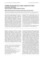
signs of arthritis. Celastrus-fed rats showed significantly
reduced disease severity compared to that of water-fed con-
trol rats (Figure 1a–c). The effect of Celastrus on clinical arthri-
tis was also validated by histological examination of arthritic
joints. The results (Table 1) show that synovial infiltration by
mononuclear cells and the damage to cartilage and bone were
significantly reduced in Celastrus-treated rats compared to
that in control water-fed rats. Thus, feeding Celastrus to LEW
rats significantly reduced the severity of subsequently induced
AA.
To define the mechanisms underlying the anti-arthritic activity
of Celastrus, we examined the changes in the immune
response to the disease-related antigen Bhsp65 as well as in
the production of a mediator of inflammatory arthritis (NO) in
Celastrus-treated LEW rats. The results of these investiga-
tions are described below.
Arthritis Research & Therapy Vol 9 No 4 Tong and Moudgil
Page 4 of 10
(page number not for citation purposes)
Celastrus feeding to LEW rats induces preferential
secretion of anti-inflammatory cytokines over pro-
inflammatory cytokines in response to Bhsp65
To test and compare the T cell proliferative and cytokine
response to the disease-related antigen Bhsp65 of Celastrus-
treated versus control (water-fed) LEW rats, the draining
LNCs of these arthritic rats were tested using the appropriate
assays on day 12 after Mtb immunization. The two groups of
rats had comparable (p > 0.05) levels of proliferative response
to Bhsp65/B177–191 (data not shown).
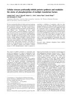
In regard to the cytokine response to Bhsp65, the levels of
IFN-γ (a pro-inflammatory cytokine) of the Celastrus-fed and
water-fed rats were comparable (Figure 2a), but the levels of
IL-10 (an anti-inflammatory cytokine; Figure 2b) were signifi-
cantly (p < 0.05) up-regulated in Celastrus-fed rats compared
to water-fed rats. The mean ratio of IFN-γ and IL-10 secreted
in recall response to Bhsp65 by Celastrus-fed rats (ratio 8.79)
was significantly lower than that of the water-fed rats (ratio
20.76), demonstrating that Celastrus preferentially facilitated
the secretion of IL-10 and, thereby, induced a relative skewing
(immune deviation) of the cytokine response towards a pre-
dominantly anti-inflammatory type.
Celastrus-fed LEW rats reveal enhanced antibody
response to Bhsp65
LEW rats were immunized s.c. with Mtb (1 mg/rat) after 4 days
of daily feeding of either Celastrus or water. Thereafter, these
rats continued to receive daily either Celastrus or water. The
sera collected from these rats at specific time points before
and after Mtb immunization were tested at different dilutions,
ranging from 1:50 to 1:8,100, by ELISA for total immunoglob-
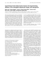
ulin against Bhsp65. The results (Figure 3) show that the lev-
els of anti-Bhsp65 antibodies increased gradually from day 0
through day 24. At both 1:100 and 1:200 serum dilutions, the
level of antibody response to Bhsp65 in Celastrus-treated rats
was significantly (p < 0.01) higher than that of water-fed rats
on day 18 and day 24. These anti-Bhsp65 antibodies were
composed mostly of IgG (data not shown). However, as
expected, the sera from both the test and control group of rats
had only minimal reactivity against KLH (control antigen), with
no significant difference between the two groups of rats (data
not shown). Thus, the increased antibody response to Bhsp65
was associated with the Celastrus-induced protection against
AA.
Reduced levels of NO in serum and LNC culture
supernatant of Celastrus-treated LEW rats
NO production is increased in patients with RA, and its pro-
duction correlates with the severity of arthritis [29,30,35].
Therefore, we reasoned that Celastrus might down-modulate
AA, in part by inhibiting the production of NO, and tested this
proposition in Celastrus-fed LEW rats. Our results show that
the levels of NO in the culture supernatant of LNCs of Mtb-
primed rats restimulated in vitro with Bhsp65 were
Figure 1
Feeding of Celastrus suppresses the induction of adjuvant arthritis (AA) in the Lewis (LEW) ratFeeding of Celastrus suppresses the induction of adjuvant arthritis (AA)
in the Lewis (LEW) rat. (a) LEW rats (n = 7 per group) were fed by gav-
age daily either Celastrus (triangles; 3 g/kg body weight, experimental
group) or water (circles; control group) starting on day 4 prior to M.
tuberculosis H37Ra (Mtb) immunization (1 mg/rat) and then continuing
throughout the observation period. Following Mtb injection, these rats
were scored regularly for signs of arthritis. The difference in the mean
arthritic scores of the Celastrus-fed and Water-fed rats during the
course of AA was significant (*p < 0.05 by Wilcoxon rank sum test). (b)
The results of an independent repeat experiment including two groups
of Celastrus-treated rats are shown in this section. The difference in
arthritic scores of Celastrus-fed versus Water-fed rats was significant
(*p < 0.05) for each of the groups tested (triangles; 1.5 g/kg, n = 4;
squares, 3 g/kg, n = 4). (c) The photograph shows the hind paw of a
representative LEW rat from the water-fed (left) and Celastrus-fed (mid-
dle) groups on day 16 after Mtb immunization. The hind paw of a naive
LEW rat (right) is also shown for comparison; each unit on the scale
equals 1 mm, with 10 units between numbered marks.
Available online />Page 5 of 10
(page number not for citation purposes)
significantly lower in Celastrus-fed rats than that in water-fed
rats on days 16 and 24 (Figure 4a). Similarly, the level of serum
NO in Celastrus-fed rats was significantly lower than that in
water-fed rats on days 16 and 24 (Figure 4b). However, the
NO levels in the sera of both these groups were much higher
than that in sera of naïve rats (Figure 4b). Taken together,
these results show that arthritic LEW rats produced NO in
response to Bhsp65, and that Celastrus feeding reduced the
levels of NO. This decrease in NO levels in turn correlates with
the reduced severity of arthritis in Celastrus-treated rats com-
pared to control rats.
Celastrus feeding suppresses the severity of ongoing AA
in the LEW rat and the level of this effect is comparable
to that of MTX
We have described above that feeding of Celastrus to naïve
LEW rats beginning prior to the induction of AA by Mtb injec-
tion can afford protection against AA (Figure 1). However,
from the clinical viewpoint of RA patients, it is critical that a
potentially beneficial anti-arthritic product displays not only a
preventive effect but also a therapeutic effect by suppressing
ongoing (established) arthritis. In this regard, we examined the
therapeutic potential of Celastrus in the AA model. Naïve LEW
rats were challenged with Mtb s.c. for the induction of AA.
Beginning at the onset of AA, and then continuing throughout
its course, one of the experimental groups of rats was fed
Celastrus (test group) and the control group received the vehi-
cle (water). Another group of experimental rats was fed an
established anti-arthritic compound, MTX (positive control). All
these rats were observed regularly for signs of arthritis. The
results (Figure 5a,b) show that both the Celastrus-fed and the
MTX-fed experimental groups had a significantly decreased
severity of AA compared to the Water-fed control rats, and
both the high (3 g/kg) and the low (1.5 g/kg) doses of Celas-
trus had comparable beneficial effects against AA (Figure 5b).
The severity of the disease in each of these two experimental
groups of rats (Celastrus-fed and MTX-fed) was significantly
reduced compared to control (water-fed) rats (Figure 5b).
Intriguingly, the level of the suppressive effect on arthritis of
Celastrus was comparable to that of MTX. Thus, Celastrus
showed both preventive as well as therapeutic anti-arthritic
activity in the AA model.
Discussion
Our results show that Celastrus aculeatus Merr. (Celastrus)
suppresses the induction of AA when fed to LEW rats prior to
Mtb challenge, as well as down-modulates the progression of
AA when administered to arthritic rats at the onset of the dis-
ease. The significant reduction in the severity of clinical AA fol-
lowing Celastrus feeding was further validated by limited
histological changes in the joints. Furthermore, the level of
suppression of ongoing AA by Celastrus was comparable to
that of MTX, a standard anti-arthritic agent used for the treat-
ment of arthritis. This attribute of Celastrus is an important one
because many regimens based on synthetic or natural com-
pounds can successfully prevent the induction of arthritis, but
they often fail to control the course of the ongoing disease. In
this regard, Celastrus is a promising anti-arthritic agent that
could be further explored as a therapeutic modality in control-
led pilot clinical trials on RA patients. As this is our first study
on the effect of Celastrus on AA, we have used the unfraction-
ated ethanol extract of the roots and stems to preserve as
much of the natural proportion of different constituents in the
mixture as possible. Accordingly, the dose of Celastrus fed to
rats is apparently high. However, in subsequent follow up
studies, we plan to use one or more of the purified compo-
nents of the crude extract. It has been reported by others that
various components of Celastrus possess anti-inflammatory
and anti-tumor properties, and these include a variety of ses-
quiterpene esters (for example, celastrol, celaphanol, celasdin,
orbiculin, esters with the β-dihydroagarofuran skeleton) and
flavonoids (for example, epiafzelechin) [9-15]. Some of the
Table 1
Quantification of histological changes in the hind paws of Celastrus-fed (experimental) versus water-fed (control) Lewis rats
Group (n) Cellular infiltrate
a
Synovial hyperplasia
a
Cartilage damage
a
Bone erosion
a
Water-fed
Day 12 (10) 2 (1–3) 3 (2–3) 2.5 (0–3) 2.5 (0–3)
Day 24 (10) 3 (2–3) 3 (3–3) 3 (1–3) 3 (0–3)
Celastrus-fed
Day 12 (10) 1 (0–2)* 0 (0–2)* 0 (0–2)* 0 (0–1)*
Day 24 (9) 1 (0–3)* 1 (0–2)* 1 (0–2)* 1 (0–1)*
The results shown are the median (interquartile range) scores of histological sections of the left or the right hind paw using the grading system
described in 'Materials and methods'.
a
Histological grading of each parameter was done as follows: 0 = absent; 1 = mild; 2 = moderate; 3 =
severe. *p < 0.01, comparing the results of the respective Celastrus-fed versus water-fed groups.
Arthritis Research & Therapy Vol 9 No 4 Tong and Moudgil
Page 6 of 10
(page number not for citation purposes)
reported pathways inhibited by these components are medi-
ated by nuclear factor kappa-B (NF-κB), inducible nitric oxide
synthase (iNOS), and cyclooxygenase (COX) [12-15,35].
We observed that the suppression of clinical arthritis in Celas-
trus-fed LEW rats was associated with significant changes in
the immune response to Bhsp65. Furthermore, both the cell-
mediated and the antibody responses to Bhsp65 were
affected. AA is driven by pro-inflammatory cytokines (IFN-γ and
tumor necrosis factor-α); in this context, Celastrus treatment
facilitated the secretion of the anti-inflammatory cytokine IL-10
over the pro-inflammatory cytokine IFN-γ, resulting in the over-
all skewing (immune deviation) of the cytokine response to an
anti-inflammatory type [26]. This relative deviation of the
cytokine response, caused either by decreased Th1-type
cytokines and/or by enhanced Th2-type cytokines leading to
the regression of an autoimmune disease, is reminiscent of
other compounds of synthetic (for example, peptides of
Figure 2
The cytokine response to mycobacterial hsp65 (Bhsp65) of lymph node cells (LNCs) of Celastrus-fed versus water-fed Lewis (LEW) ratsThe cytokine response to mycobacterial hsp65 (Bhsp65) of lymph
node cells (LNCs) of Celastrus-fed versus water-fed Lewis (LEW) rats.
Two groups of LEW rats (n = 6 to 9) were fed either Celastrus (3 g/kg)
or water as described in the legend to Figure 1. A sub-group of these
LEW rats was euthanized on day 12 after M. tuberculosis H37Ra
immunization, and the draining LNCs of these rats were cultured in a
96-well plate in the presence of the indicated recall antigens (HEL, hen
eggwhite lysozyme; B177, synthetic peptide 177–191 of Bhsp65). The
supernatant was collected after 72 h of cell culture and tested in ELISA
for (a) IFN-γ and (b) IL-10. The results are expressed as pg/ml (mean +
standard error of the mean). The difference in the level of IL-10 but not
of IFN-γ in response to Bhsp65 in Celastrus-fed versus water-fed rats is
statistically significant (*p < 0.05). The mean IFN-γ/IL-10 ratio in
response to Bhsp65 of Celastrus-treated (8.79) rats was significantly
reduced compared to that of the water-fed (20.76) rats.
Figure 3
Antibody response to mycobacterial hsp65 (Bhsp65) of Celastrus-fed Lewis (LEW) ratsAntibody response to mycobacterial hsp65 (Bhsp65) of Celastrus-fed
Lewis (LEW) rats. LEW rats (n = 4 to 6) were fed either Celastrus (3 g/
kg) or water as described in the legend to Figure 1. Blood samples
were collected from LEW rats immediately before (preimmune serum;
day 0) challenge with M. tuberculosis H37Ra (Mtb; 1 mg/rat) as well as
at different time points thereafter (days 10, 18 and 24). These sera
were tested separately at different dilutions (1:50 to 1:8,100) by ELISA
for total immunoglobulin against Bhsp65. The results are expressed as
optical density (O.D.) at 540 nm (mean + standard error of the mean).
At one representative concentration of sera (for example, 1:100 dilu-
tion), the level of antibody response to Bhsp65 in Celastrus-fed rats
was significantly higher than that of water-fed rats on days 18 and 24
(**p < 0.01 each).
Available online />Page 7 of 10
(page number not for citation purposes)
antigenic proteins or cytokines) [25,36,37] or natural origin
[38,39] that can successfully control disease in animal models
of arthritis.
Celastrus feeding to LEW rats immunized with Mtb led to
enhanced production of antibodies to Bhsp65 compared to
the control water-fed rats. Thus, a decrease in inflammatory
arthritis in LEW rats was associated with an increase in the
anti-Bhsp65 antibody response. This inverse association is
supported by previous work by others [25] and us [31] dem-
onstrating that anti-Bhsp65 antibodies produced during the
course of AA are disease-protective rather than being
pathogenic in nature. Unlike in other animal models of RA in
which antibodies are arthritogenic [40,41], in the AA model
certain subsets of anti-Bhsp65 antibodies generated either
during the course of AA [25,31] or following AA-protective tol-
Figure 4
Levels of nitric oxide (NO) in lymph node cell (LNC) culture supernatant and serum of Celastrus-treated Lewis (LEW) ratsLevels of nitric oxide (NO) in lymph node cell (LNC) culture supernatant
and serum of Celastrus-treated Lewis (LEW) rats. (a) LNC culture
supernatant and (b) sera were obtained from Celastrus-fed and Water-
fed rats as described in Materials and methods. The level of NO in
these samples was determined by a colorimetric assay. The results are
presented as μM (mean + standard error of the mean). The level of NO
secreted into the culture supernate following mycobacterial hsp65
(Bhsp65) restimulation of LNCs of Celastrus-fed rats was significantly
(**p < 0.005, *p < 0.05) lower than that of water-fed rats on days 16
and 24 following M. tuberculosis H37Ra (Mtb) immunization (a). The
levels of NO in sera of Celastrus-fed rats was significantly (**p < 0.01,
*p < 0.05) decreased at days 16 and 24 compared to those of water-
fed rats (b). However, the levels of NO in sera of both these Mtb-immu-
nized groups of rats were higher (++p < 0.01, +p < 0.05) compared to
those of naïve sera. In each section, some of the error bars are too
small to be detected.
Figure 5
Celastrus induces therapeutic down-modulation of adjuvant arthritis (AA) that is comparable to that when using methotrexate (MTX)Celastrus induces therapeutic down-modulation of adjuvant arthritis
(AA) that is comparable to that when using methotrexate (MTX). A
cohort of Lewis (LEW) rats was immunized subcutaneously with M.
tuberculosis H37Ra (Mtb; 1 mg/rat) at the base of the tail and then
split into different groups (n = 4 per group). Beginning day 9 thereafter,
coinciding with the onset of clinical signs of arthritis in the hind paws,
these rats were fed daily by gavage either Celastrus (experimental
group) or water (negative control group). (a) Experimental rats were fed
with 3 g/kg of Celastrus, or (b) with either 3 or 1.5 g/kg of Celastrus.
An additional group of experimental rats shown in (b) received MTX
(0.5 mg/kg; positive control). All these rats were observed and scored
regularly for the severity of AA. In both (a) and (b) the difference in the
mean arthritic score of each of the Celastrus-fed versus water-fed
group of rats was significant (*p < 0.05 by Wilcoxon rank sum test).
Similarly, in (b), a significant (*p < 0.05) difference in arthritic scores
was observed between MTX-fed and water-fed rats, whereas compara-
ble (p > 0.05) arthritic scores were observed for Celastrus-fed versus
MTX-fed rats.
Arthritis Research & Therapy Vol 9 No 4 Tong and Moudgil
Page 8 of 10
(page number not for citation purposes)
erization with Bhsp65 [42] contribute to disease regulation. It
has been proposed that the protective effect of antibodies in
AA is probably mediated by the induction of IL-10 production
from mononuclear cells [25]. In this regard, our finding of a
Celastrus-induced deviation of the cytokine response of
arthritic LEW rats towards IL-10 correlates very well with our
observation of enhanced anti-Bhsp65 antibody response in
Celastrus-treated rats, and the observed immune deviation
towards IL-10 might be attributable, in part, to the increased
antibody response to Bhsp65. We further suggest that the
anti-Bhsp65 antibodies might also contribute to the protection
against AA by modulating antigen processing and presenta-
tion [43] and, thereby, facilitating the induction of the immune
response to one or more of the regulatory T cell determinants
within Bhsp65 previously identified by others [23,25,36] and
us [24]. Thus, changes in both the cell-mediated and the anti-
body responses to Bhsp65 following Celastrus feeding might
cooperate to down-regulate the severity of AA in the LEW rat.
In addition to the disease-regulating changes in the immune
response to Bhsp65, the beneficial effect of Celastrus in AA
was also related to inhibition of the production of a well known
mediator of inflammation, namely NO [20,29,30,35]. We
observed antigen specificity in the production of NO; Bhsp65-
restimulated LNCs of Mtb-immunized water-fed (control) LEW
rats produced significantly higher levels of NO than those res-
timulated by the control antigen, HEL. Furthermore, Celastrus
treatment significantly reduced the levels of NO in both LNC
culture supernatant and sera of Mtb-immunized rats. Taken
together, these results document not only a direct association
between the levels of NO and the severity of AA, but also
provide insight into the in vivo anti-inflammatory activity of
Celastrus. These results of Celastrus-mediated suppression
of NO production in vivo are further corroborated by reports
by other investigators showing a similar effect of Celastrus in
vitro using macrophage cell lines (for example, RAW cells)
[12,15]. Furthermore, it has been reported that oral feeding of
B6 mice with the ethyl acetate extract of Tripterygium wilfordii
Hook F (TWHF) or its active component, triptolide, led to the
inhibition of both NO production and iNOS mRNA expression
by macrophages [44], and this decrease in NO production
was implicated in mediating the anti-inflammatory effects of
TWHF. One of the mechanisms by which Celastrus leads to
decreased NO production might involve NF-κB, which con-
trols the expression of genes encoding inducible enzymes,
such as iNOS and COX, which in turn generate some of the
critical mediators of the inflammatory response [14,15,35]. In
fact, some of the active components of Celastrus have been
shown to serve as inhibitors of the NF-κB pathway (for
example, celastrol and celaphanol A) [12,15] and the COX
pathway (for example, epiafzelechin) [13]. In addition, NF-κB
activity is inversely related to that of the heat-shock response
as the induction of heat-shock proteins is associated with a
decrease in NF-κB activity [14]. Celastrol can lead to the
induction of heat-shock protein gene expression by activation
of heat-shock factor-1 [14], and the enhanced response to self
hsp65 can, in turn, contribute to protection against AA
[23,32]. Thus, by regulating the activity of NF-κB, Celastrus
apparently influences multiple inter-connected pathways that
participate in the regulation of autoimmune arthritis.
Our results suggest that the ethanol extract of Celastrus as
well as its individual components should be explored further
for the treatment of RA through double-blind, placebo-control-
led preclinical and clinical trials in RA patients following the
strategy employed successfully by other investigators for
translational research on TWHF [8,45]. There is a compelling
need to fully examine multiple natural products such as TWHF
and Celastrus for their potential as anti-arthritic agents
because all RA patients may not respond equally well to any
single herbal medicine owing to differences in body constitu-
tion and genetics, and each natural plant product may have
unique compatibility with the standard mainstream medica-
tions when taken together. The availability of several different
natural plant products having anti-arthritic activity would
enlarge the scope of the use of CAM modalities for the treat-
ment of RA in conjunction with conventionally used drugs.
Conclusion
The ethanol extract of Celastrus aculeatus Merr. (Celastrus)
has potent anti-arthritic activity. Feeding Celastrus to LEW
rats offered protection against the subsequent induction as
well as progression of AA. The therapeutic effect of Celastrus
was comparable to that of MTX. Celastrus-induced protection
against AA involved significant modulation of both the cytokine
and antibody responses to the disease-related antigen
Bhsp65. In addition, Celastrus suppressed the production of
a known mediator of inflammation, NO. Celastrus should be
further tested in clinical trials on patients with RA to explore its
utility as a natural CAM product that might be beneficial either
alone or in combination with conventionally used drugs, with
the objective of complementing the beneficial anti-arthritic
effects and reducing the side effects of the latter group of
drugs.
Competing interests
The authors declare that they have no competing interests.
Authors' contributions
LT conducted all the experiments, recorded and analyzed the
raw data, prepared graphics, and participated in the interpre-
tation of data as well as writing of the manuscript. KDM
participated in the planning of experiments, data analysis,
interpretation of results and writing of the manuscript.
Acknowledgements
This work was supported by grants (AI059623 and AT001608) to KDM
from the National Institutes of Health (NIH), Bethesda, MD. LT was sup-
ported by an International Postdoctoral Fellowship Award (F05
AT002013) from the NCCAM, NIH. We thank Hong R Kim and Minjun
Yu for helping with experiments, Dr Xiao Changhong for advice and help
Available online />Page 9 of 10
(page number not for citation purposes)
in quantification of histological sections, and Dr Brian Berman (Center
for Integrative Medicine, UMB) for encouragement in pursuing research
in the area of CAM.
References
1. Kvien TK: Epidemiology and burden of illness of rheumatoid
arthritis. Pharmacoeconomics 2004, 22:1-12.
2. Barnes PM, Powell-Griner E, McFann K, Nahin RL: Complemen-
tary and alternative medicine use among adults: United
States, 2002. Adv Data 2004, 343:1-19.
3. Eisenberg DM, Davis RB, Ettner SL, Appel S, Wilkey S, Van
Rompay M, Kessler RC: Trends in alternative medicine use in
the United States, 1990–1997: results of a follow-up national
survey. JAMA 1998, 280:1569-1575.
4. Taibi D, Bourguignon C: The role of complementary and alter-
native therapies in managing rheumatoid arthritis. Fam Com-
munity Health 2003, 26:41-52.
5. Zhang GG, Lee W, Bausell B, Lao L, Handwerger B, Berman B:
Variability in the traditional Chinese medicine (TCM) diag-
noses and herbal prescriptions provided by three TCM practi-
tioners for 40 patients with rheumatoid arthritis. J Altern
Complement Med 2005, 11:415-421.
6. Kremers HM, Nicola P, Crowson CS, O'Fallon WM, Gabriel SE:
Therapeutic strategies in rheumatoid arthritis over a 40-year
period. J Rheumatol 2004, 31:2366-2373.
7. Olsen NJ, Stein CM: New drugs for rheumatoid arthritis. N Engl
J Med 2004, 350:2167-2179.
8. Cibere J, Deng Z, Lin Y, Ou R, He Y, Wang Z, Thorne A, Lehman
AJ, Tsang IK, Esdaile JM: A randomized double blind, placebo
controlled trial of topical Tripterygium wilfordii in rheumatoid
arthritis: reanalysis using logistic regression analysis. J
Rheumatol 2003, 30:465-467.
9. Spivey AC, Weston M, Woodhead S: Celastraceae sesquiterpe-
noids: biological activity and synthesis. Chem Soc Rev 2002,
31:43-59.
10. Guo YQ, Li X, Xu J, Li N, Meng DL, Wang JH: Sesquiterpene
esters from the fruits of Celastrus orbiculatus. Chem Pharm
Bull (Tokyo) 2004, 52:1134-1136.
11. Kim SE, Kim YH, Lee JJ, Kim YC: A new sesquiterpene ester
from Celastrus orbiculatus reversing multidrug resistance in
cancer cells. J Nat Prod 1998, 61:108-111.
12. Jin HZ, Hwang BY, Kim HS, Lee JH, Kim YH, Lee JJ: Antiinflam-
matory constituents of Celastrus orbiculatus inhibit the NF-
kappaB activation and NO production. J Nat Prod 2002,
65:89-91.
13. Min KR, Hwang BY, Lim HS, Kang BS, Oh GJ, Lee J, Kang SH, Lee
KS, Ro JS, Kim Y: (-)-Epiafzelechin: cyclooxygenase-1 inhibitor
and anti-inflammatory agent from aerial parts of Celastrus
orbiculatus. Planta Med 1999, 65:460-462.
14. Westerheide SD, Bosman JD, Mbadugha BN, Kawahara TL, Mat-
sumoto G, Kim S, Gu W, Devlin JP, Silverman RB, Morimoto RI:
Celastrols as inducers of the heat shock response and
cytoprotection. J Biol Chem 2004, 279:56053-56060.
15. Nam NH: Naturally occurring NF-kappaB inhibitors. Mini Rev
Med Chem 2006, 6:945-951.
16. Firestein GS: Immunologic mechanisms in the pathogenesis
of rheumatoid arthritis. J Clin Rheumatol 2005, 11:S39-44.
17. Antoniv TT, Ivashkiv LB: Dysregulation of interleukin-10-
dependent gene expression in rheumatoid arthritis synovial
macrophages. Arthritis Rheum 2006, 54:2711-2721.
18. Pearson CM: Development of arthritis, periarthritis and perios-
titis in rats given adjuvants. Proc Soc Exp Biol Med 1956,
91:95-101.
19. Taneja V, Taneja N, Behrens M, Griffiths MM, Luthra HS, David CS:
Requirement for CD28 may not be absolute for collagen-
induced arthritis: study with HLA-DQ8 transgenic mice. J
Immunol 2005, 174:1118-1125.
20. Brahn E, Banquerigo ML, Firestein GS, Boyle DL, Salzman AL,
Szabo C: Collagen induced arthritis: reversal by mercap-
toethylguanidine, a novel antiinflammatory agent with a com-
bined mechanism of action. J Rheumatol 1998, 25:1785-1793.
21. Mia MY, Durai M, Kim HR, Moudgil KD: Heat shock protein 65-
reactive T cells are involved in the pathogenesis of non-anti-
genic dimethyl dioctadecyl ammonium bromide-induced
arthritis. J Immunol 2005, 175:219-227.
22. van Eden W, Thole JE, van der Zee R, Noordzij A, van Embden JD,
Hensen EJ, Cohen IR: Cloning of the mycobacterial epitope rec-
ognized by T lymphocytes in adjuvant arthritis. Nature 1988,
331:171-173.
23. Quintana FJ, Carmi P, Mor F, Cohen IR: DNA fragments of the
human 60-kDa heat shock protein (HSP60) vaccinate against
adjuvant arthritis: identification of a regulatory HSP60 peptide.
J Immunol 2003, 171:3533-3541.
24. Moudgil KD, Chang TT, Eradat H, Chen AM, Gupta RS, Brahn E,
Sercarz EE: Diversification of T cell responses to carboxy-ter-
minal determinants within the 65-kD heat-shock protein is
involved in regulation of autoimmune arthritis. J Exp Med
1997, 185:1307-1316.
25. Ulmansky R, Cohen CJ, Szafer F, Moallem E, Fridlender ZG, Kashi
Y, Naparstek Y: Resistance to adjuvant arthritis is due to pro-
tective antibodies against heat shock protein surface epitopes
and the induction of IL-10 secretion. J Immunol 2002,
168:6463-6469.
26. Prakken BJ, Roord S, Ronaghy A, Wauben M, Albani S, van Eden
W: Heat shock protein 60 and adjuvant arthritis: a model for T
cell regulation in human arthritis. Springer Semin
Immunopathol 2003, 25:47-63.
27. Strober S, Holoshitz J: Mechanisms of immune injury in rheu-
matoid arthritis: evidence for the involvement of T cells and
heat-shock protein. Immunol Rev 1990, 118:233-255.
28. Celis L, Vandevyver C, Geusens P, Dequeker J, Raus J, Zhang J:
Clonal expansion of mycobacterial heat-shock protein-reac-
tive T lymphocytes in the synovial fluid and blood of rheuma-
toid arthritis patients. Arthritis Rheum 1997, 40:510-519.
29. Jang D, Murrell GA: Nitric oxide in arthritis. Free Radic Biol Med
1998, 24:1511-1519.
30. Bogdan C: Nitric oxide and the immune response. Nat Immunol
2001, 2:907-916.
31. Kim HR, Kim EY, Cerny J, Moudgil KD: Antibody responses to
mycobacterial and self heat shock protein 65 in autoimmune
arthritis: epitope specificity and implication in pathogenesis. J
Immunol 2006, 177:6634-6641.
32. Durai M, Gupta RS, Moudgil KD: The T cells specific for the car-
boxyl-terminal determinants of self (rat) heat-shock protein 65
escape tolerance induction and are involved in regulation of
autoimmune arthritis. J Immunol 2004, 172:2795-2802.
33. Hom JT, Estridge T, Cole H, Gliszczynski V, Bendele A: Effects of
various anti-T cell receptor antibodies on the development of
type II collagen-induced arthritis in mice. Immunol Invest 1993,
22:257-265.
34. Matthys P, Hatse S, Vermeire K, Wuyts A, Bridger G, Henson GW,
De Clercq E, Billiau A, Schols D: AMD3100, a potent and specific
antagonist of the stromal cell-derived factor-1 chemokine
receptor CXCR4, inhibits autoimmune joint inflammation in
IFN-gamma receptor-deficient mice. J Immunol 2001,
167:4686-4692.
35. Appleton I, Tomlinson A, Willoughby DA: Induction of cyclo-oxy-
genase and nitric oxide synthase in inflammation. Adv
Pharmacol 1996, 35:27-78.
36. Anderton SM, van der Zee R, Prakken B, Noordzij A, van Eden W:
Activation of T cells recognizing self 60-kD heat shock protein
can protect against experimental arthritis. J Exp Med 1995,
181:943-952.
37. Woods JM, Amin MA, Katschke KJ Jr, Volin MV, Ruth JH, Connors
MA, Woodruff DC, Kurata H, Arai K, Haines GK 3rd, et al.: Inter-
leukin-13 gene therapy reduces inflammation, vascularization,
and bony destruction in rat adjuvant-induced arthritis. Hum
Gene Ther 2002, 13:381-393.
38. Haqqi TM, Anthony DD, Gupta S, Ahmad N, Lee MS, Kumar GK,
Mukhtar H: Prevention of collagen-induced arthritis in mice by
a polyphenolic fraction from green tea. Proc Natl Acad Sci USA
1999, 96:4524-4529.
39. Chevrier MR, Ryan AE, Lee DY, Zhongze M, Wu-Yan Z, Via CS:
Boswellia carterii extract inhibits TH1 cytokines and promotes
TH2 cytokines in vitro. Clin Diagn Lab Immunol 2005,
12:575-580.
40. Wooley PH, Luthra HS, Krco CJ, Stuart JM, David CS: Type II col-
lagen-induced arthritis in mice. II. Passive transfer and sup-
pression by intravenous injection of anti-type II collagen
antibody or free native type II collagen. Arthritis Rheum 1984,
27:1010-1017.
Arthritis Research & Therapy Vol 9 No 4 Tong and Moudgil
Page 10 of 10
(page number not for citation purposes)
41. Kouskoff V, Korganow AS, Duchatelle V, Degott C, Benoist C,
Mathis D: Organ-specific disease provoked by systemic
autoimmunity. Cell 1996, 87:811-822.
42. Satpute SR, Soukhareva N, Scott DW, Moudgil KD: Mycobacte-
rial Hsp65-IgG-expressing tolerogenic B cells confer protec-
tion against adjuvant-induced arthritis in Lewis rats. Arthritis
Rheum 2007, 56:1490-1496.
43. Simitsek PD, Campbell DG, Lanzavecchia A, Fairweather N, Watts
C: Modulation of antigen processing by bound antibodies can
boost or suppress class II major histocompatibility complex
presentation of different T cell determinants. J Exp Med 1995,
181:1957-1963.
44. Wang B, Ma L, Tao X, Lipsky PE: Triptolide, an active component
of the Chinese herbal remedy Tripterygium wilfordii Hook F,
inhibits production of nitric oxide by decreasing inducible
nitric oxide synthase gene transcription. Arthritis Rheum 2004,
50:2995-2303.
45. Tao X, Younger J, Fan FZ, Wang B, Lipsky PE: Benefit of an
extract of Tripterygium wilfordii Hook F in patients with rheu-
matoid arthritis: a double-blind, placebo-controlled study.
Arthritis Rheum 2002, 46:1735-1743.