Báo cáo y học: "Heterogeneity of autoantibodies in 100 patients with autoimmune myositis: insights into clinical features and outcomes" ppt
Bạn đang xem bản rút gọn của tài liệu. Xem và tải ngay bản đầy đủ của tài liệu tại đây (328.68 KB, 13 trang )
Open Access
Available online />Page 1 of 13
(page number not for citation purposes)
Vol 9 No 4
Research article
Heterogeneity of autoantibodies in 100 patients with autoimmune
myositis: insights into clinical features and outcomes
Martial Koenig
1
, Marvin J Fritzler
2
, Ira N Targoff
3
, Yves Troyanov
1
and Jean-Luc Senécal
1
1
University of Montreal School of Medicine, and Laboratory for Research in Autoimmunity, Centre Hospitalier de l'Université de Montréal, M-4243,
1560 East Sherbrooke Street, Montreal, Quebec, Canada H2L 4M1
2
Faculty of Medicine HRB409, University of Calgary, 3330 Hospital Dr. NW, Calgary, Alberta, Canada T2N 4N1
3
Arthritis and Immunology, University of Oklahoma Health Sciences Center, 825 NE 13th Street Oklahoma City, OK 73104, and Oklahoma Medical
Research Foundation, Oklahoma City, Oklahoma, USA
Corresponding author: Jean-Luc Senécal,
Received: 22 May 2007 Revisions requested: 4 Jul 2007 Revisions received: 28 Jul 2007 Accepted: 9 Aug 2007 Published: 9 Aug 2007
Arthritis Research & Therapy 2007, 9:R78 (doi:10.1186/ar2276)
This article is online at: />© 2007 Koenig et al.; licensee BioMed Central Ltd.
This is an open access article distributed under the terms of the Creative Commons Attribution License ( />),
which permits unrestricted use, distribution, and reproduction in any medium, provided the original work is properly cited.
Abstract
The objective of this study was to determine the prevalence,
mutual associations, clinical manifestations, and diagnoses
associated with serum autoantibodies, as detected using
recently available immunoassays, in patients with autoimmune
myositis (AIM). Sera and clinical data were collected from 100
patients with AIM followed longitudinally. Sera were screened
cross-sectionally for 21 autoantibodies by multiplex addressable
laser bead immunoassay, line blot immunoassay,
immunoprecipitation of in vitro translated recombinant protein,
protein A assisted immunoprecipitation, and enzyme-linked
immunosorbent assay. Diagnoses were determined using the
Bohan and Peter classification as well as recently proposed
classifications. Relationships between autoantibodies and
clinical manifestations were analyzed by multiple logistic
regression. One or more autoantibodies encompassing 19
specificities were present in 80% of the patients. The most
common autoantibodies were anti-Ro52 (30% of patients), anti-
Ku (23%), anti-synthetases (22%), anti-U1RNP (15%), and anti-
fibrillarin (14%). In the presence of autoantibodies to Ku,
synthetases, U1RNP, fibrillarin, PM-Scl, or scleroderma
autoantigens, at least one more autoantibody was detected in
the majority of sera and at least two more autoantibodies in over
one-third of sera. The largest number of concurrent
autoantibodies was six autoantibodies. Overall, 44 distinct
combinations of autoantibodies were counted. Most
autoantibodies were unrestricted to any AIM diagnostic
category. Distinct clinical syndromes and therapeutic responses
were associated with anti-Jo-1, anti-fibrillarin, anti-U1RNP, anti-
Ro, anti-Ro52, and autoantibodies to scleroderma autoantigens.
We conclude that a significant proportion of AIM patients are
characterized by complex associations of autoantibodies.
Certain myositis autoantibodies are markers for distinct overlap
syndromes and predict therapeutic outcomes. The ultimate
clinical features, disease course, and response to therapy in a
given AIM patient may be linked to the particular set of
associated autoantibodies. These results provide a rationale for
patient profiling and its application to therapeutics, because it
cannot be assumed that the B-cell response is the same even in
the majority of patients in a given diagnostic category.
Introduction
Autoimmune myositis (AIM) is a syndrome characterized by
involvement of the cellular and humoral immune systems in
skeletal muscle pathology, immunogenetic modulation,
response to immunotherapies, and the presence of autoanti-
bodies in the serum of many patients [1,2]. Although AIM is
commonly classified using the original 1975 classification pro-
posed by Bohan and Peter [3,4], this approach has become
subject to increasing debate [5-7]. The Bohan and Peter clas-
sification has been criticized for over-diagnosing polymyositis
(PM) [8]; for loosely defining myositis in overlap (overlap
myositis [OM]) with another connective tissue disease (CTD)
AIM = autoimmune myositis; ALBIA = addressable laser bead immunoassay; CAM = cancer associated myositis; CENP = centromere protein; CTD
= connective tissue disease; DM = dermatomyositis; ELISA = enzyme-linked immunosorbent assay; IPP = immunoprecipitation; LIA = line immu-
noassay; MAA = myositis associated autoantibody; MSA = myositis specific autoantibody; OM = overlap myositis; PM = polymyositis; RNAPOLIII =
RNA polymerase III; SLE = systemic lupus erythematosus; SRP = signal recognition particle; SSc = systemic sclerosis; TNT = translation and tran-
scription; topo = topoisomerase I.
Arthritis Research & Therapy Vol 9 No 4 Koenig et al.
Page 2 of 13
(page number not for citation purposes)
[9]; for clinical, genetic, and immunologic heterogeneity in all
subsets [10]; and for being obsolete [11]. The discovery of
myositis specific antibodies (MSAs) and myositis-associated
antibodies (MAAs) led to a serologic approach complemen-
tary to the Bohan and Peter classification, because striking
associations of MSAs with clinical features, immunogenetics,
and survival were observed [10].
However, this approach has been limited by several con-
straints. First, until recently, sophisticated methods that are
costly, labor intensive, and not always routinely available were
required for identification of most MSAs, limiting their wide-
spread use. Second, because MSAs are relatively insensitive
markers for myositis [12], this serologic approach led to the
creation of a large and heterogeneous group of MSA-negative
patients, who were undefined with respect to diagnosis, prog-
nosis, and survival [13]. Third, the emphasis on MSAs has
resulted in a common perception among clinicians that AIM is
characterized by the presence of single autoantibody specifi-
cities, whereas associations between an MSA and MAAs are
not uncommon. However, the interrelationships between
these sets of autoantibodies and their clinical impact have yet
not been explored in depth. Taken together, these constraints
identify a need to develop more sensitive and less costly meth-
ods for detecting MSAs and MAAs, and for analyzing the inter-
relationships between these autoantibodies.
As a step toward resolving these issues, and with the objective
of improving AIM classifications, in this study we focus on AIM
autoantibodies by conducting an in-depth examination of their
prevalence, distribution and mutual associations, as well as
their corresponding diagnoses and clinical manifestations. We
took stock from our recently proposed novel approach to the
classification of AIM, which brings together strong clinical evi-
dence of myositis that is readily identifiable by clinicians and
the diagnostic value of MSA and MAA tests [14]. In the
present report, we examine the same cohort for an expanded
panel of 21 autoantibodies to major AIM autoantigens, using
recently available line immunoassay (LIA) and addressable
laser bead multiplex technologies. We also used multiple
logistic regression analysis to identify clinical manifestations
independently associated with these autoantibodies.
Patients and methods
Patients and data collection
We conducted a cross-sectional serum study of 100 adult
French Canadian patients with AIM, followed longitudinally,
who were seen up to 2001 at the Centre Hospitalier de l'Uni-
versité de Montréal, a tertiary care center. A list of AIM patients
was obtained from the medical records using discharge sum-
mary diagnostic codes corresponding to PM, dermatomyositis
(DM), myositis, mixed CTD, and overlap syndrome. The five
inclusion criteria were as follows. First, only French Canadian
patients were eligible. Second, the illness fulfilled Bohan and
Peter criteria for definite, probable, or possible PM or DM by
the end of follow up [3,4]. Patients with possible PM were
included because this diagnosis is not uncommon in clinical
practice and the prolonged follow up provided an opportunity
to examine its outcome. Third, patients had to be 18 years or
older at diagnosis of myositis (therefore juvenile DM, as
defined by Bohan and Peter, was excluded). Fourth, inclusion
body myositis, rare forms of AIM, and non-AIM causes of
myopathy (such as muscular dystrophies) were excluded. Also
excluded were patients diagnosed as having AIM in whom a
non-AIM myopathy was ultimately diagnosed upon follow up.
Finally, a frozen serum sample had to be available for immuno-
logic studies. At myositis diagnosis, according to the diagnos-
tic criteria proposed by Bohan and Peter [3,4], 36 definite, 45
probable, and 18 possible cases of myositis were seen, and a
single patient had a DM rash and a myopathic electromyo-
gram. At last follow up, there were 47 definite, 41 probable,
and 12 possible cases of myositis, and detailed features of
these patients have been described elsewhere [14].
Data on history, physical findings, and laboratory investiga-
tions were obtained by retrospective medical record review
using a standardized protocol. Written consent was obtained
from treating physicians to communicate with and examine the
patients for further data collection. The project was approved
by the Centre Hospitalier de l'Université de Montréal research
and ethics committees. The diagnoses of myositis were made
at Centre Hospitalier de l'Université de Montréal in 87
patients, and 13 additional patients were referred with an
established AIM diagnosis. A muscle biopsy and an electromy-
ogram were done in 87 and 88 patients, respectively. Among
the 13 patients in whom no muscle biopsy was taken, 12
(92%) patients had a DM rash (n = 8) and/or overlap CTD fea-
tures (n = 7). Five patients had possible myositis, three had
probable myositis, and five had definite myositis. Definitions for
overlap CTD features, target organ involvement, and clinical
characteristics are described in detail elsewhere [14] and are
summarized in Table 1. All living patients (n = 77) but one
were examined or contacted by us between June 1999 and
April 2001. The associations between autoantibodies and
clinical features were determined at myositis diagnosis,
whereas associations with myositis course and response to
therapy were based on cumulative data at last follow up. Defi-
nitions for monophasic myositis, response to prednisone
alone, and need for a second immunosuppressive drug were
as described previously [14].
AIM classifications
Patients were categorized at AIM diagnosis according to three
classifications, as shown in Table 1: the original Bohan and
Peter classification [3,4], a modified Bohan and Peter classifi-
cation developed by us [14], and a clinicoserologic classifica-
tion also developed by us [14]. The distribution of patients
using the modified Bohan and Peter classification was done
before results of AIM autoantibody testing were available.
Diagnosis of an associated CTD was made according to the
Available online />Page 3 of 13
(page number not for citation purposes)
American College of Rheumatology classification criteria for
systemic lupus erythematosus (SLE), rheumatoid arthritis, and
systemic sclerosis (SSc) [14]. Before AIM diagnosis, 16
patients fulfilled American College of Rheumatology criteria for
another CTD (seven SSc, six rheumatoid arthritis, and three
SLE patients), whereas at AIM diagnosis eight additional
patients fulfilled such criteria (mostly SSc).
Panel of 21 autoantibodies tested and screening
strategy
Serum samples were coded and frozen at -80°C. The timing of
serum samples relative to the diagnosis of myositis was as fol-
lows: nine sera were obtained at least 6 months before AIM
diagnosis, 45 sera were obtained at diagnosis, and 46 sera
were obtained at least 6 months after diagnosis, with 23 of
those more than 5 years after diagnosis.
The following MSAs and MAAs were studied. Anti-synthetas-
esencompassed Jo-1, OJ, EJ, KS, PL-7, and PL-12 aAb spe-
cificities [15-17]. SSc autoantibodies included autoantibodies
to centromere protein (CENP)-B, DNA topoisomerase I (topo;
Scl-70), Th/To (Th), and RNA polymerase III (RNAPOLIII) [18-
20]. Autoantibodies commonly associated with SSc in overlap
encompassed autoantibodies to PM-Scl, U1RNP, U2RNP,
fibrillarin, U5RNP, and Ku autoantigens [21-26]. Myositis
autoantibodies also included anti-signal recognition particle
(SRP) [27] and anti-nucleoporins [28,29]. Anti-Mi-2 (which
are DM specific when determined by immunodiffusion or
immunoprecipitation [IPP] and are not associated with overlap
manifestations) [30], as well as anti-Ro(SS-A) and anti-La (SS-
B; which are commonly associated with MSAs and MAAs),
were also tested for [14]. The prevalence of autoantibodies
was determined by systematic application of the methods that
follow to all sera.
Indirect immunofluorescence
Antibodies to centromeres and nucleoporins were detected
by indirect immunofluorescence on HEp-2 cells (Antibodies
Inc., Davis, CA, USA) [18,28].
Addressable laser bead immunoassay
Microspheres embedded with laser-reactive dyes (Luminex
Corporation, Austin, TX, USA), coupled with native Jo-1,
U1RNP, topo, La, and Sm antigens from calf thymus, or a
mixture of native Ro from calf thymus and recombinant Ro52
antigens, were part of a commercial kit (QuantaPlex™ SLE
Profile 8; INOVA Diagnostics Inc., San Diego, CA, USA).
Addressable laser bead immunoassay (ALBIA) allowed semi-
quantitative detection of autoantibodies to Jo-1, U1RNP, topo,
Ro, La, and Sm. The assay was performed at the Advanced
Diagnostics Laboratory of the University of Calgary. Briefly,
each test serum was diluted to 1:1,000, and 50 μl was added
to a well of a microtiter plate, mixed with the antigen-coated
beads that were preserved in the well, and incubated for 30
Table 1
Description of three classifications for autoimmune myositis
Classification Abbreviation Definition/description
Original Bohan and Peter classification PM Primary polymyositis
DM Primary dermatomyositis
CTM Myositis with another connective tissue disease
CAM Myositis associated with cancer
Modified Bohan and Peter classification PM Pure polymyositis
DM Pure dermatomyositis
OM Overlap myositis: with at least one clinical overlap feature
a
CAM Cancer-associated myositis: with clinical paraneoplasic features
b
Novel clinicoserologic classification PM Pure polymyositis
DM Pure dermatomyositis
OM Overlap myositis: with at least one clinical overlap feature and/or a myositis
autoantibody
c
CAM Cancer-associated myositis: with clinical paraneoplasic features and without a myositis
autoantibody or anti-Mi-2
a
Clinical overlap features: arthritis, Raynaud's phenomenon, sclerodactyly, scleroderma proximal to metacarpophalangeal joints, typical systemic
sclerosis-type calcinosis in the fingers, lower esophageal or small bowel hypomotility, lung involvement (carbon monoxide diffusing capacity <70%
of the normal predicted value, restrictive syndrome, and/or interstitial lung disease on chest radiogram or computed tomography scan), discoid
lupus, anti-native DNA antibodies plus hypocomplementemia, four or more of 11 American College of Rheumatology systemic lupus erythematosus
criteria, and antiphospholipid syndrome.
b
Clinical paraneoplasic features: cancer within 3 years of myositis diagnosis, plus absence of multiple
clinical overlap features, plus, if cancer was cured, myositis was cured as well.
c
Anti-synthethases (Jo-1, PL-7, PL-12, OJ, EJ, and KS), systemic
sclerosis autoantibodies (centromere protein [CENP]-B, DNA topoisomerase I, RNA polymerase III, and Th/To), autoantibodies commonly
associated with systemic sclerosis in overlap (U1RNP, U2RNP, U3RNP, U5RNP, PM-Scl, and Ku), anti-SRP (signal recognition particle), and anti-
nucleoporins. Autoantibodies to Mi-2, Ro, and La are not included. Modified from Troyanov and coworkers [14].
Arthritis Research & Therapy Vol 9 No 4 Koenig et al.
Page 4 of 13
(page number not for citation purposes)
min. Then, 50 μl of phycoerythrin-conjugated goat anti-human
immunoglobulin G (γ-chain specific; Jackson ImmunoRe-
search, Inc., West Grove, PA, USA) was added to each well
and incubated for an additional 30 min. The reactivity of the
antigen-coated beads was determined on a Luminex 100 dual-
laser flow cytometer. The antigens are each bound to distinct
fluorochrome-labeled microspheres, and this flow cytometer
can discriminate the color of each bead from the others as well
as measure the fluorescent intensity of the conjugate on each
bead [31]. The cut-off for a positive test result was based on
the reactivity of control samples. The control samples included
in the kit were titrated to provide high, medium, low, and neg-
ative values [32].
Line immunoassay
LIA was performed at the Advanced Diagnostics Laboratory
by Euroline-WB assay (Euroimmun AG, Luebeck, Germany).
Test strips are coated with sodium dodecyl sulphate extracted
and electrophoretically separated whole HeLa cell proteins
that are transferred to nitrocellulose strips that then allow
detection of autoantibodies against Mi-2, Ku-72, and Ku-86
autoantigens using a conventional immunoblot protocol. Each
strip also contains nitrocellulose chips on which recombinant
antigens (PM-Scl, PL-7, and PL-12) and native Jo-1 purified by
affinity chromatography were individually applied. The recom-
binant PM-Scl was a full length (100 kDa) PM-Scl derived from
a human cDNA expressed in baculovirus-infected insect cells.
The specificity of the reactivities was validated by using known
positive and negative controls. Using this LIA and sera from 70
patients with AIM, the following autoantibody frequencies
were observed: 6% anti-Mi-2, 14% anti-PM-Scl, 10% anti-Jo-
1, 6% anti-PL-7, 3% anti-PL-12, and 9% anti-Ku. These
autoantibodies were not observed in patients with SLE (n =
30; except for anti-PL-7 in none patient) or SSc (n = 20) or in
healthy blood donors (n = 50; data available online [33]). In
addition, anti-Mi-2 was not detected in sera from 100 normal
control individuals and 100 SLE patients at the Advanced
Diagnostics Laboratory of the University of Calgary [32]
(unpublished data).
Enzyme-linked immunosorbent assays
Anti-RNAPOLIII autoantibodies were detected by enzyme
linked immunosorbent assay (ELISA) using a recombinant
RNAPOLIII fragment containing the immunodominant epitope
(MBL Co., Nagoya, Japan) [20]. Positive controls were anti-
RNAPOLIII SSc sera provided by M Kuwana (Keio University
School of Medicine, Japan) [20]. The cut-off value was 11
units/ml, as recommended by the manufacturer. Anti-topo
autoantibodies were detected by ELISA using native full-
length topo extracted and purified from calf thymus (Immuno-
vision. Springdale, AR, USA) [34]. For sera positive for anti-Ro
by ALBIA, the specificity for anti-Ro52 and anti-Ro60 was fur-
ther determined by ELISA using recombinant human Ro52
expressed in Escherichia coli and native Ro60 from calf thy-
mus (INOVA Diagnostics Inc., San Diego, CA, USA). For sera
positive for anti-centromere autoantibodies by indirect immun-
ofluorescence, reactivity with CENP-B was confirmed by
ELISA using recombinant full-length CENP-B from baculovi-
rus-infected Sf9 cells (Diarect AG, Freiburg, Germany) [35].
Anti-fibrillarin assay
Sera were screened for anti-fibrillarin by ALBIA using purified
recombinant fibrillarin protein (Mikrogen GmbH, Neuried, Ger-
many) and test serum diluted to 1:1,000 [36]. Control nega-
tive and standard positive sera were included in each assay.
The presence of anti-fibrillarin was confirmed by translation
and transcription (TNT) of a full-length cDNA and IPP of the
radiolabeled recombinant protein [37,38]. This assay was ini-
tially validated in experimental autoimmunity and shown to
have greater than 90% specificity and 8% sensitivity for SSc
[39].
Protein A assisted IPP
IPP was performed by one of us (INT) for both nucleic acid and
protein analyses, along with double immunodiffusion as
described elsewhere [14-17]. Autoantibodies identified using
these tests include all of the described anti-synthetases, anti-
RNA polymerase III, anti-Th, anti-U2RNP, anti-U3RNP, anti-
U5RNP, and anti-SRP [14-17]. For IPP, nucleic acid analysis
used 3 to 5 mg of protein A-sepharose, 20 μl of patient serum,
and unlabeled HeLa cell extract (>10
6
cells). Immunoprecipi-
tates were analyzed by 7 to 8 mol/l urea and 10% polyacryla-
mide gel electrophoresis with silver stain development. Protein
analysis used 1 to 2 mg protein A-sepharose, 10 to 15 ml
serum, and 35S-methionine-labeled HeLa cell extract (>10
5
HeLa cells). Immunoprecipitates were analyzed by sodium
dodecyl sulphate polyacrylamide gel electrophoresis
(between 8% and 10%) [14-17].
Statistical analyses
Associations between categorical variables and comparisons
of the frequency of a given autoantibody between mutually
exclusive subsets of patients were based on two-tailed χ
2
tests. The frequency of OM versus other AIM diagnoses used
was analyzed using McNemar's test for comparing groups of
paired samples. To assess the relationships between a given
autoantibody and clinical manifestations, we employed multi-
ple logistic regression with forward selection of independent
variables. A separate multivariable logistic regression model
was obtained for each autoantibody, with a binary indicator of
its presence/absence as the dependent variable, and with clin-
ical characteristics as the candidate independent variables.
Adjustment for age and sex was also considered. To obtain
parsimonious multivariable models, and ensure the critical
ratio of at least five 'outcomes' (here presence of the respec-
tive autoantibody) per independent variable in the model, the
most significant variables were entered into the model one at
a time as long as the corresponding P value (from the two-
tailed Wald χ
2
test) did not exceed 0.05. Final results are
reported as adjusted odds ratios with 95% confidence inter-
Available online />Page 5 of 13
(page number not for citation purposes)
vals for those variables that were included in the final model. In
all analyses, two-tailed P < 0.05 was considered statistically
significant. Analyses were performed using SAS 9.1 statistical
software package (SAS Institute Inc, Cary, NC, USA).
Results
Frequency of autoantibodies to 21 autoantigens
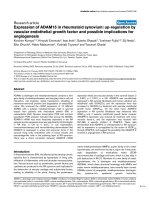
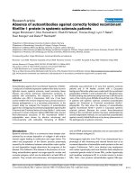
The frequency of each autoantibody in the cohort is shown in
Figure 1. Two features are noteworthy. First, the overall fre-
quency of autoantibodies, as detected using various methods,
was high, with 80% (80 patients) expressing one or more
autoantibody. In contrast, in our previous report using the
same serum samples [14] only 56% of the patients expressed
one or more autoantibodies. Second, the diversity of autoanti-
bodies was also high, as indicated by the detection of autoan-
tibodies to 19 of the 21 (90%) autoantigens tested. The most
common autoantibody, present in 31% (31 patients), was anti-
Ro (detected using ALBIA). Further analysis by ELISA for fine
specificity revealed that anti-Ro52 autoantibodies was most
common, occurring in 97% (30/31 patients) of anti-Ro posi-
tive sera, whereas anti-Ro60 autoantibodies were present in
35.5% (11/31 patients; Figure 1). Almost all sera with anti-
Ro60 (91%; 10/11 patients) displayed anti-Ro52 as well, con-
firming that among patients with AIM the most common anti-
Ro specificity is anti-Ro52 [40]. The next most common
autoantibodies were anti-Ku (23%; detected using LIA), anti-
Jo-1 (15%), anti-U1RNP (15%), and anti-fibrillarin and anti-La
(each 14%; Figure 1).
Of the 15 remaining potential autoantibody specificities, 13
specificities were present with frequencies ranging from 9%
(anti-PM-Scl) to 1% (for instance, anti-KS, anti-U2RNP, and
anti-U5RNP; Figure 1). Anti-Mi-2 and anti-nucleoporins were
detected in 6% and 3% of patients, respectively. Only anti-OJ
and anti-EJ (both anti-synthetases) were not detected.
Frequency of anti-synthetases and SSc autoantibodies
Anti-synthetases were present overall in 22% (22 patients;
Figure 1). Anti-Jo-1 and anti-PL-7 were the most common spe-
cificities, accounting for 68% (15/22 patients) and 23% (5/22
patients) of anti-synthetases, respectively, whereas anti-PL-12
and anti-KS were rare (one patient each). SSc autoantibodies
(anti-CENP, anti-topo, anti-Th, and anti-RNAPOLIII) were
uncommon (9%).
Multiple myositis autoantibodies frequently coexist
Table 2 shows that among the 80 patients with AIM autoanti-
bodies, multiple autoantibodies were found in 44 (55%)
patients. Thus, in the presence of the more common autoanti-
bodies (such as to Ku, synthetases, U1RNP, fibrillarin, PM-Scl,
or SSc autoantigens), at least one more autoantibody was
present in the majority (mean 78.5%, range 60% to 93%) of
Figure 1
Frequency of serum autoantibodies to 21 autoantigens in 100 French Canadian patients with autoimmune myositisFrequency of serum autoantibodies to 21 autoantigens in 100 French Canadian patients with autoimmune myositis. Autoantibodies were observed
to 19 (90%) of the specificities tested. Anti-OJ and anti-EJ (both anti-synthetases) were not detected. One or more autoantibodies were present in
80% of patients. Autoantibodies to synthetases (Jo-1, PL-7, PL-12, and KS) and systemic sclerosis autoantibodies were present overall in 22% and
9% of patients, respectively. The overall frequency is over 100% because 44% of patients had more than one autoantibody. Anti-Ro were deter-
mined by ALBIA whereas anti-Ro52 and anti-Ro60 fine specificities were identified by ELISA. See Materials and methods (in the text) for a descrip-
tion of immunoasssays. ALBIA, addressable laser bead immunoassay; CENP, centromere protein; ELISA, enzyme-linked immunosorbent assay;
RNAPOLIII, RNA polymerase III; SRP, signal recognition particle; TOPO, topoisomerase I.
Arthritis Research & Therapy Vol 9 No 4 Koenig et al.
Page 6 of 13
(page number not for citation purposes)
sera (Table 2). For example, of 22 patients with autoantibodies
to synthetases, 18 (82%) expressed one or more additional
autoantibodies. Furthermore, in the presence of the more com-
mon autoantibodies, at least two more autoantibodies were
present in 20% to 65% (mean 43.3%) of sera. For example,
10 out of 22 (46%) of patients with autoantibodies to syn-
thetases expressed two or more additional autoantibodies.
The largest number of concurrent autoantibodies was
observed in a single serum with six autoantibodies. Four addi-
tional patient sera displayed four specificities. Overall, not less
than 44 distinct combinations of autoantibodies were
counted. These data highlight that a major subset of AIM is
characterized by the simultaneous presence of two or more
autoantibodies rather than by single specificities.
Associations and exclusions between myositis
autoantibodies
As shown in Table 3, mutual exclusion was noted between
autoantibodies to synthetases, Mi-2, and SRP, as reported
previously [3].
Anti-Ro, anti-Ro52, and anti-Ro60
Anti-Ro autoantibodies were commonly associated with other
specificities (67%; 12/18 specificities), most commonly
autoantibodies to synthetases (45%; 14/31), notably Jo-1
(35%; 11/31), U1RNP (19%; 6/31), and Ku, fibrillarin and
PM-Scl (each 19%; 6/31). All anti-Ro positive patients with
anti-Ku, anti-fibrillarin, and anti-PM-Scl had anti-Ro52.
Anti-Ku
The most frequently associated autoantibodies were anti-fibril-
larin (35%), anti-Ro (26%), and anti-Jo-1 (17%).
Anti-fibrillarin
These were associated with 11 of the 18 (61%) other specif-
icities, most commonly anti-Ku (57%; 8/14).
Anti-synthetases
The frequency of anti-Ro was greater among patients with anti-
synthetases (64%; 14/22) than among all other patients
(22%; 17/78; odds ratio 6.8, 95% confidence interval 2.2 to
17.4; P = 0.0004). All patients with anti-synthetases and anti-
Ro exhibited anti-Ro52 autoantibodies as well (14/14), as
compared with 35% (5/14) for anti-Ro60. Anti-synthetases
were also associated with anti-Ku (32%; 7/22) and anti-fibril-
larin (18%; 4/22). Anti-Jo-1 autoantibodies were associated
with autoantibodies to Ro (73%; 11/15), Ku (27%), La (20%),
fibrillarin (13%), topo (13%), PM-Scl (7%), and CENP-B (7%;
Table 3).
Anti-Mi-2
Anti-Mi-2 autoantibodies detected by IPP only (anti-Mi-2-IPP;
3) were not associated with other autoantibodies, whereas
anti-Mi-2 autoantibodies detected by LIA only (anti-Mi-2-LIA;
3) were associated with other autoantibodies (Table 3).
Fine specificity of anti-Ku autoantibodies
All three anti-Ku positive sera by IPP reacted only with the Ku-
86 peptide in the LIA. Of the 20 anti-Ku negative sera by IPP,
nine (45%) reacted only with the Ku-86 peptide by LIA, five
(25%) reacted with the Ku-72 peptide only, and six (30%)
reacted with both peptides. This suggests that autoantigen
presentation in the LIA procedure revealed additional linear or
cryptic epitopes [41] or that stringent conditions used during
IPP may alter antigen binding.
Comparison of immunoassay sensitivities
For several autoantibodies, sensitivities concurred; for
instance, the 15 sera positive for anti-Jo-1 by IPP were positive
by LIA in all instances and by ALBIA in 14 (93.3%) cases. Sim-
ilarly, sera with anti-U1RNP detected by IPP were also positive
by ALBIA, and sera with anti-fibrillarin by ALBIA were also pos-
itive by TNT assay. Anti-PL-12 autoantibodies were detected
by both IPP and LIA. However, a discrepancy in sensitivity was
noted for anti-Ku, which was detected in 23 patients by LIA
but in only three (13%) of these patients by IPP. Finally, of the
six sera with anti-Mi-2, three were anti-Mi-2-IPP only and three
anti-Mi-2-LIA only.
Table 2
Frequency of multiple autoantibodies in 100 patients with autoimmune myositis
Additional
antibodies
a
Autoantibodies Patients (total)
Ku
(n = 23)
tRNA
(n = 22)
U1RNP
(n = 15)
Fibrillarin
(n = 14)
PM-Scl
(n = 9)
SSc
(n = 9)
NUP
(n = 3)
SRP
(n = 2)
Ro
(n = 31)
La
(n = 14)
Mi-2
(n = 6)
No antibody
(n = 20)
= 1 antibody
(n = 80)
None 7 (31) 4 (18) 6 (40) 1 (7) 2 (22) 1 (11) 2 (67) 2 (100) 4 (13) 3 (21) 3 (50) 0 36 (36)
b
1 more 7 (31) 8 (36) 6 (40) 4 (28) 4 (44) 3 (33) 1 (33) 0 12 (39) 4 (29) 2 (33) 0 26 (26)
2 more 5 (21) 6 (28) 2 (13) 6 (43) 1 (12) 3 (33) 0 0 10 (32) 4 (29) 0 0 12 (12)
≥3 more 4 (17) 4 (18) 1 (7) 3 (22) 2 (22) 2 (23) 0 0 5 (16) 3 (21) 1 (17) 0 6 (6)
Values are expressed as number (%). tRNA synthetases (tRNA) include Jo-1, PL-7, PL-12, and KS. Systemic sclerosis (SSc) autoantigens include topoisomerase I,
RNA polymerase III, centromere protein B, and Th.
a
Categories are mutually exclusive.
b
Includes an additional patient who had anti-U5RNP (not shown). NUP,
nucleoporins; RNP, ribonucleoprotein; SRP = signal recognition particle.
Available online />Page 7 of 13
(page number not for citation purposes)
Demographics, distribution of patients, and associated
autoantibodies according to the modified Bohan and
Peter classification at diagnosis
Most patients were female (female to male ratios: 13:1 for PM,
18:5 for DM, 41:19 for OM, and 3:0 for cancer-associated
myositis [CAM]). As shown in Table 4, autoantibodies to Ro,
Ku, fibrillarin, synthetases, U1RNP, La, PM-Scl, and nucleop-
orins were not restricted to a single diagnostic category. How-
ever, anti-Mi-2-IPP were restricted to patients with DM rashes
(two patients with DM, and one patients with CAM associated
with a DM rash), whereas anti-Mi-2-LIA (three patients) were
associated with OM. None of the patients with anti-Mi-2-LIA
developed DM rashes at follow up (mean duration 9.98 years,
range 9.2 to 10.4 years). Overall, OM accounted for the
majority (65% to 86%) of these various specificities, but only
anti-fibrillarin and anti-U1RNP were more common in OM than
in all other myositis patients (P = 0.007 and P = 0.024,
respectively; Table 4). In contrast, the frequency of anti-syn-
thetases (22 patients) was similar in PM (21.4%), DM
(17.4%), and OM (25%) patients (P = 0.46 for OM versus all
others). Anti-PM-Scl were present in both DM (three patients)
and OM (six patients; P = 0.73). Overall, except for anti-SRP
and anti-Mi-2, none of these autoantibodies segregated with a
unique AIM diagnostic category. Finally, the mean number
(and range) of autoantibodies in each diagnostic category was
as follows: 1.81 (0 to 6) for OM, 1.13 (0 to 3) for DM, 0.92 (0
to 3) for PM, and 0.33 (0 to 1) for CAM (P = 0.0055 by
Kruskall-Wallis test for difference of means, excluding CAM).
Thus, the greatest mean number of autoantibodies was
observed in OM. Interestingly, absence of autoantibodies was
associated with significantly decreased risk for OM (Table 4).
Impact of autoantibodies on myositis classification at
diagnosis
Because of the increased frequency of autoantibodies in the
present study in comparison with our previous study [14], we
used the novel AIM clinicoserologic classification to compare
the distribution of patients according to diagnosis in the cur-
rent versus the previous reports. As shown in Table 5, the fre-
quency of OM rose from 68% to 82%, not including anti-Ro
and anti-Mi-2, whereas the frequency of other diseases
decreased to 18% (versus the previous study: by McNemar
test, P < 0.001; versus the modified Bohan and Peter classifi-
cation, P < 0.001). This increase in the frequency of OM was
due to newly detected autoantibodies to Ku (eight), Ro (five),
PM-Scl (four), synthetases (three), U1RNP (two), and fibrillarin
(one). Of note is the overall decrease in PM frequency from
45% using the original Bohan and Peter classification to only
Table 3
Patterns of associations between single autoantibodies in 100 patients with autoimmune myositis
Ro Ku Jo-1 PL-7 PL-12 KS U1RNP Fibrillarin La PM-Scl Topo RNAPOLIII CENP-B Th Mi-2 NUP SRP U5RNP U2RNP
n 31 23 15 5 1 1 15 14 14 9 3 2 2 2 6 3 2 1 1
Methods of
detection
ALBIA LIA,
IPP
LIA,
IPP,
ALBIA
LIA,
IPP
LIA,
IPP
IPP IPP,
ALBIA
TNT,
ALBIA
ALBIA,
ELISA
LIA,
IPP
ALBIA,
ELISA
IPP,
ELISA
ELISA,
IIF
IPP LIA,
IPP
IPP IPP IPP IPP
Ro 4 61111 16 6 5 6 2 - 1 1 -
Ku 6 7 4 1- -2 8 4 2 - 1 - -1 -
Jo-1 11 4 2 - - 2 3 1 2 - 1 - - - -
PL-7 1 1 - 2 - 1 2 - - - - -
PL-12 1 - - - - 1 - - - - - - - - -
KS 1 - - - - - - - - - - - - -
U1RNP 6 2 - - - - 6 2 2 - - - - 1 1
Fibrillarin 6 8 2 1 1 - 2 1 2 1 - 1 1 -1 -
La 5 4 3 2 - - 2 2 3 11- - -11 1
PM-Scl 6 2 1 - - - - 1 1 2 1 - - -1 -
Topo 2 - 2 - - - - - 1 1 - - - -1 -
RNAPOLIII - 1 - - - - - 1 - - - - - -1 -
CENP-B 1 - 1 - - - - 1 - - - - - -
Th 1 - - - - - 1 - - - - - - 1 -
Mi-2 - 1 - - - - - 1 1 1 1 1 - - 3 -
NUP - - - - - - 1 - - - - 2 -
SRP - - - - - - - - - - - - 2
U5RNP - - - - - - - - - - - - - 1 -
U2RNP - - - - - -1 - 1 - - - - - - - -
Numbers in bold denote that a single autoantibody specificity was detected. -, absence of autoantibody; ALBIA, addressable laser bead immunoassay; CENP, centromere protein; ELISA, enzyme-linked
immunosorbent assay; IIF, indirect immunofluorescence; IPP, immunoprecipitation; LIA, line immunoassay; NUP, nucleoporins; RNAPOLIII, RNA polymerase III; SRP, signal recognition particle; TNT,
transcription and translation assay; Topo, topoisomerase I.
Arthritis Research & Therapy Vol 9 No 4 Koenig et al.
Page 8 of 13
(page number not for citation purposes)
7% using the clinicoserologic classification (Table 5). Taken
together, these data support previous observations that OM is
the most common AIM, and PM is the least common AIM [14].
Clinical features independently associated with AIM
autoantibodies
As shown in Table 6, interstitial lung disease, arthritis, fever,
and puffy hands (but neither mechanic's hands nor Raynaud's)
were associated with a higher frequency of anti-Jo-1. In con-
trast, both Raynaud's and lung involvement were associated
with anti-fibrillarin (Table 6). Raynaud's, arthritis, and sclero-
dactyly were strongly associated with anti-U1RNP. Tel-
angiectasias, sclerodactyly, and sclerodermatous skin
proximal to the MCP joints were associated with SSc autoan-
tibodies. A decreased risk for an associated American College
of Rheumatology criteria defined CTD was linked to anti-Jo-1,
whereas an increased risk was linked with anti-fibrillarin and
anti-U1RNP (Table 6).
Table 4
Demographics, associated autoantibodies, and distribution of 100 patients with autoimmune myositis according to the modified
Peter and Bohan classification at diagnosis
PM (n = 14) DM (n = 23) OM (n = 60) CAM (n = 3) P
a
Age at diagnosis (years; mean ± SD) 52.7 ± 16.8 45.4 ± 16.7 45.6 ± 13.5 56.3 ± 11 0.303
Mean follow-up period (years; mean ± SD) 8.15 ± 4.8 12.52 ± 9.2 8.05 ± 6.1 7.64 ± 2.7 0.013
Associated autoantibodies
Anti-Ro (n = 31) 4 (28.6) 5 (21.7) 22 (36.7) 0 0.185
Anti-Ku (n = 23) 3 (21.4) 5 (21.7) 15 (25) 0 0.633
Anti-synthetases (n = 22) 3 (21.4) 4 (17.4) 15 (25) 0 0.463
Anti-U1RNP (n = 15) 0 2 (8.7) 13(21.7) 0 0.024
b
Anti-fibrillarin (n = 14) 0 1 (4.3) 13 (21.7) 0 0.007
c
Anti-La (n = 14) 1 (7.1) 3 (13) 10 (16.7) 0 0.394
SSc autoantibodies (n = 9) 0 1 (4.3) 8 (13.3) 0 0.081
Anti-PM-Scl (n = 9) 0 3 (13) 6 (10) 0 0.737
Anti-Mi-2-IPP (n = 3) 0 2 (8.7) 0 1 (33)
d
-
Anti-Mi-2-LIA (n = 3) 0 0 3 (5) 0 -
Anti-NUP (n = 3) 1 (7.1) 0 2 (3.3) 0 1
Anti-SRP (n = 2) 0 0 2 (3.3) 0 0.515
None (n = 20) 5(35.7) 6 (26.1) 7(11.7) 2 (66) 0.02
e
Unless otherwise specified, values are expressed as number (%). Anti-Ro was detected using addressable laser bead immunoassay; for anti-Mi-2-
IPP and anti-Mi-2-LIA, autoantibody was present only by immunoprecipitation or line immunoassay.
a
For Associated autoantibodies: to avoid more
than 20% of values being <5, comparisons were made by Fisher's exact test between OM and all others.
b
Odds ratio 5.2, 95% confidence
interval 1.1 to 24.7.
c
Odds ratio 10.8, 95% confidence interval 1.35 to 86.
d
The patient also had a DM rash.
e
Odds ratio 0.27, 95% confidence
interval 0.1 to 0.77. CAM, cancer-associated myositis; DM, pure dermatomyositis; OM, overlap myositis; PM, pure polymyositis; SD, standard
deviation.
Table 5
Distribution of 100 patients at myositis diagnosis according to novel classifications for autoimmune myositis
Classifications PM DM OM CAM Total
Original Peter and Bohan classification: previous study
a
45 28 24 3 100
Modified Peter and Bohan classification: previous study 14 23 60 3 100
Novel clinicoserologic classification: previous study
b
10 20 68 2 100
Novel clinicoserologic classification: present study
b
7 8 82 3 100
Values are expressed as percentages. Frequency of OM versus non-OM by McNemar's test for comparing groups of paired samples: modified
versus original Bohan and Peter classifications, χ
2
= 34.03, P < 0.001; novel clinicoserologic classification (previous study) versus modified
classification, χ
2
= 6.12, P = 0.013; novel clinicoserologic classification (present study) versus modified classification: χ
2
= 20.04, P < 0.001; and
novel clinicoserologic classifications (present study versus previous study), χ
2
= 12.07, P < 0.001.
a
Data from Troyanov and coworkers [14].
b
Excluding anti-Ro and anti-Mi-2. CAM, cancer associated myositis; DM, pure dermatomyositis; OM, overlap myositis; PM, pure polymyositis.
Available online />Page 9 of 13
(page number not for citation purposes)
Association between therapeutic outcomes, myositis
course, and autoantibodies
Table 6 also shows that specific patterns of myositis respon-
siveness to standard therapy are independently associated
with certain autoantibodies. Thus, a decreased response to
prednisone alone and an increased need for a second-line
immunosuppressive drug were highly associated with anti-Jo-
1. In contrast, myositis responsive to prednisone alone and a
reduced need for a second-line drug were associated with
both anti-Ro and anti-Ro52 (Table 6). Finally, a monophasic
course of myositis was associated with anti-U1RNP.
Discussion
Several conclusions stem from the present study.
When using newly developed assays such as ALBIA and
LIA, the prevalence of autoantibodies in patients with
AIM is much higher than previously appreciated
Our report is the first to reassess the prevalence of autoanti-
bodies in the same serum samples obtained from the same
AIM cohort. Thus, 80% of patients expressed one or more
autoantibodies in the present report, whereas in the previous
report the frequency was only 56% [14]. The most common
autoantibody overall was anti-Ro52, which was present in
30% of patients, followed by anti-Ku (23%), and anti-Jo-1 and
anti-U1RNP (both 15%). Inter-cohort differences in the fre-
quency of autoantibodies have been reported. For example,
whereas the frequency of autoantibodies was only 53% in a
study of European myositis patients, it was 74% and 80% in
Italian and Polish cohorts, respectively [42-44]. However, to
our knowledge, a systematic study of intra-cohort differences
has not been reported.
Table 6
Independent associations of autoantibodies with specific sets of clinical features, therapeutic outcomes, and myositis course by
stepwise multiple logistic regression in 100 patients with autoimmune myositis
Autoantibodies Clinical features
a
OR 95% CI P
Anti-Jo-1
b
Interstitial lung disease 14.5 3.52 to 59.78 <0.001
Arthritis 11.6 2.92 to 45.65 <0.001
Fever 9.7 2.72 to 34.91 <0.001
Puffy hands 9.6 2.82 to 32.92 <0.001
Associated CTD 0.14 0.03 to 0.70 0.016
Response to prednisone alone 0.04 0.01 to 0.32 0.002
Need for second line drug 25.3 4.24 to 150 <0.001
Anti-fibrillarin Raynaud's phenomenon 10.9 2.83 to 42.60 <0.001
Any lung involvement 3.5 1.01 to 12.39 0.050
Associated CTD 4.1 1.14 to 14.50 0.031
Anti-U1RNP Raynaud's phenomenon 9.8 2.28 to 42.59 0.002
Arthritis 5.8 1.74 to 19.18 0.004
Sclerodactily 6.8 2.03 to 22.49 0.001
Associated CTD 6.8 1.76 to 26.55 0.006
Associated SSc 6.1 1.83 to 20.26 0.003
Monophasic myositis course 8.1 1.39 to 46.7 0.020
SSc autoantibodies Telangiectasias 19.3 2.05 to 181.51 0.010
Sclerodactyly 8.6 1.88 to 39.60 0.005
Scleroderma proximal to MCP 5.3 1.07 to 26.78 0.041
Associated SSc 5.8 1.29 to 25.78 0.022
Anti-Ro or anti-Ro52 Response to prednisone alone 6.2 1.23 to 31.64 0.027
Need for second line drug 0.12 0.03 to 0.54 0.006
Clinical associations were determined at myositis diagnosis, whereas associations with specific myositis courses and therapeutic responses were
determined at last follow up. Results were very similar when adjusted for age and sex. See Materials and methods (in the text) for definitions of
clinical findings.
a
Definitions were previously described by Troyanov and coworkers [14].
b
Anti-Jo-1 (or anti-synthetases overall) were also
associated with higher serum creatine kinase at myositis diagnosis (OR 5.3; P < 0.0001). CI, confidence interval; CTD, connective tissue disease;
DM, dermatomyositis; MCP, metacarpophalangeal joints; OR, odds ratio; SSc, systemic sclerosis.
Arthritis Research & Therapy Vol 9 No 4 Koenig et al.
Page 10 of 13
(page number not for citation purposes)
The higher frequency of autoantibodies is due to an
increased sensitivity of the detection methods employed
Certain autoantibodies were detected for the first time in 24
patients. The new specificities detected were Ku (12 patients),
fibrillarin (five), Ro (five), synthetases (three; all PL-7), PM-Scl
(three), and U1RNP (three). In our previous report, using a
highly specific IPP method, the frequency of anti-Ku was only
2% [14]. However, in the present study, we used a more sen-
sitive LIA method. Indeed, immunoblotting has been associ-
ated with a 16% to 33% frequency of anti-Ku in OM patients
[17,43,45]. Similarly, a 19% frequency of anti-Ku by immuno-
blotting was reported in PM and DM [41]. Ethnicity/genetic
background also influences the anti-Ku immune response,
because the highest frequency of anti-Ku has been reported in
Japanese and African American patients [17,46,47]. A combi-
nation of historical and geographic factors has resulted in rel-
ative genetic homogeneity in French Canadian patients, with
ensuing potential modulation of autoimmune responses
[18,48].
Anti-fibrillarin was not detected by IPP in our previous report,
although the IPP method used was not optimized for the
detection of this antibody [14]. In the present report we
screened for anti-fibrillarin by ALBIA using purified recom-
binant fibrillarin protein, yielding an autoantibody frequency of
14%. All positive anti-fibrillarin results were confirmed by
highly specific, protein-based TNT assay [37,38]. Anti-fibril-
larin autoantibodies are uncommon in conditions other than
SSc [23,24,46]. Interestingly, by multiple logistic regression
we found that the presence of an associated CTD, most com-
monly SSc, was linked to anti-fibrillarin. Furthermore, the asso-
ciation between anti-fibrillarin and myositis as well as lung
involvement has previously been reported in SSc patients
[24,49]. Finally, as mentioned for anti-Ku, immunogenetics
may have influenced anti-fibrillarin autoimmune response in
our French Canadian patients [18,48].
A major subset of AIM is characterized by complex
associations of autoantibodies and extremely marked
serologic heterogeneity
Our data expand on previous studies of autoantibodies in AIM
sera and show that the diversity of autoantibodies in AIM sera
is high and their mutual associations are complex [50]. In par-
ticular, anti-Jo-1 was almost always associated with autoanti-
bodies to one or more of seven autoantigens, notably anti-
Ro52. No less than 44 distinct combinations of autoantibodies
were identified, indicating remarkable heterogeneity of B-cell
responses in AIM. Such polyreactivity is common in other
CTDs such as SLE. However, polyreactivity has been
observed in only 0.8% of sera submitted to a clinical laboratory
for routine autoantibody analysis by ALBIA [51], suggesting
that polyreactivity is related to the primary CTD diagnosis.
Thus, clinicians should be aware that several autoantibodies
may be present in single AIM patient sera and that identifica-
tion of a common autoantibody specificity (for instance, anti-
Ro) does not preclude the presence of other autoantibodies
that may have useful diagnostic, prognostic, and even thera-
peutic significance. This issue is hindered by the fact that sev-
eral autoantibodies are not routinely detected in most clinical
diagnostic laboratories and, as currently constituted, are rela-
tively costly and labor intensive. Moreover, as ALBIA used
herein and other multiplexed autoantibody assays become
more widely available, it will be important for clinicians to
become aware that myositis subsets often are not single
autoantibody entities [32].
Taken together, these data suggest that a major subset of AIM
is characterized by complex associations of autoantibodies
rather than by single specificities. This has potential impor-
tance for future studies of the clinical associations and prog-
nostic value of myositis autoantibodies. It will be of interest to
dissect further whether there are differences within autoanti-
body defined groups based on whether they do or do not have
a particular set of associated autoantibodies. Furthermore,
these data provide a basis and a rationale for patient profiling
and its application to therapeutics, because it cannot be
assumed that the B-cell response is the same in all or even the
majority of patients in a given diagnostic category, as dis-
cussed below.
Myositis autoantibodies are not restricted to a unique
myositis diagnostic category
Except for the uncommon anti-SRP and anti-Mi-2 autoantibod-
ies, all autoantibodies occurred in at least two diagnostic cat-
egories, mainly OM, DM, and/or PM [8,42,43]. An intriguing
issue is the different diagnoses associated with anti-Mi-2
depending on the detection method. Anti-Mi-2 is strongly
associated with DM when it is detected by IPP of radiolabeled
cell extracts, and our data are in agreement because our
patients with anti-Mi-2-IPP had DM rashes [30]. However,
such diagnostic specificity was altered when ELISA with dif-
ferent fragments of recombinant Mi-2 was used for screening
a large AIM cohort [42]. Similarly, anti-Mi-2-LIA in our cohort
was observed only in OM patients. Anti-Mi-2 was recently
reported in PM patients with arthritis, Raynaud's phenomenon,
and interstitial lung disease, a cluster of manifestations similar
to OM according to the modified Bohan and Peter classifica-
tion [52]. Thus, the clinical significance of anti-Mi-2 appears to
depend on the detection method.
In a previous report [53], one of us (INT) focused on the char-
acterization of Mi-2 epitopes. It was shown that only 50% of
sera reactive with the 240 kDa major Mi-2 autoantigen immu-
noprecipitated from HeLa cells also reacted with Mi-2 from
HeLa cells on immunoblots. Failure to detect by immunoblot-
ting reactivity with this major 240 kDa Mi-2 protein could have
been due to exclusive reaction of these anti-Mi-2 sera with
conformational epitopes and not with the denatured proteins
used in immunoblots. Moreover, the uniform reactivity with the
240 kDa Mi-2 protein does not exclude additional reactivity of
Available online />Page 11 of 13
(page number not for citation purposes)
anti-Mi-2 sera with other Mi-2 epitopes. In the present study,
given that the LIA uses blotted proteins, it is possible that the
discrepant results between LIA and IPP are due to a similar
mechanism.
OM is the most common AIM and PM is least common
Using our proposed clinicoserologic classification to reassess
the diagnostic impact of autoantibody results obtained with
more sensitive immunoassays, an increase in the frequency of
OM in our cohort from 68% to 82% was noted, whereas PM
was least common. These drastic changes strengthen OM as
the most common AIM [8-11,14].
Certain myositis autoantibodies are markers for distinct
overlap syndromes and predict therapeutic outcomes
We identified statistically significant and independent associ-
ations between clinical manifestations and certain myositis
autoantibodies. Thus, interstitial lung disease, arthritis, fever,
puffy hands, higher serum creatine kinase, a decreased risk for
an associated CTD, a reduced myositis response to pred-
nisone alone, and an increased need for a second-line immu-
nosuppressive drug were strongly associated with anti-Jo-1. In
contrast, an increased risk for Raynaud's, lung involvement,
and an associated CTD were associated with anti-fibrillarin.
Raynaud's, arthritis, sclerodactyly, an associated CTD (partic-
ularly SSc), and a monophasic myositis course were strongly
linked with anti-U1RNP. Finally, myositis responsiveness to
prednisone alone and a decreased need for a second-line
drug were independently associated with anti-Ro and anti-
Ro52.
Taken altogether, these data are consistent with the view that
much of AIM is associated with overlap syndrome features
[5,11,13,14,43]. Moreover, certain myositis autoantibodies
such as anti-Jo-1, anti-fibrillarin, and anti-U1RNP are markers
for distinct overlap syndromes, irrespective of their current
classification as MSAs or MAAs. Given the independence of
the associations identified, the ultimate clinical features and
response to therapy in a given myositis patient may be linked
to the particular set of associated autoantibodies, and not
merely to the presence of a single autoantibody specificity.
Study limitations
Our study has a number of statistical limitations. It was proba-
bly underpowered to unveil clinical associations for the less
common autoantibodies or to identify a larger spectrum of
associations for the more prevalent autoantibodies (type II
error). Conversely, some significant associations observed by
multiple regression may be due to type I error. Finally, it is pos-
sible that some of the observed associations are the result of
ethnicity/geographic and immunogenetic features of our
French Canadian cohort. Therefore, our results will need to be
confirmed by independent and larger studies of AIM patients
from multiple ethnic and geographic areas.
Conclusion
The prevalence of autoantibodies in patients with AIM is much
higher than was previously appreciated. The higher frequency
of autoantibodies is due to increased sensitivity of the detec-
tion methods employed. A major subset of AIM is character-
ized by complex associations of autoantibodies and extremely
marked serologic heterogeneity. Myositis autoantibodies are
not restricted to a unique myositis diagnostic category. Cer-
tain myositis autoantibodies are markers for distinct overlap
syndromes and predict therapeutic outcomes. The ultimate
clinical features, disease course, and response to therapy in a
given AIM patient may be linked to the particular set of associ-
ated autoantibodies.
Competing interests
The authors declare that they have no competing interests.
Authors' contributions
JLS and MK participated in the design of the study. YT, JLS
and MK collected patient data. MK, JLS, MJF and IT performed
the experiments and data analysis. MK and JLS wrote the man-
uscript. JLS supervised the project. All authors read, cor-
rected, and approved the final manuscript.
Acknowledgements
This study was supported by research grants from Canadian Institutes
of Health Research (MT-14636 to JLS) and the Saskatchewan Chapter
of the Scleroderma Society (MJF), and a Fellowship grant from Scléro-
dermie Québec (MK). MJF holds the Arthritis Society Research Chair at
the University of Calgary, funds from which supported this study. The
work of INT was supported in part by Office of Research and Develop-
ment, Medical Research Service, US Department of Veterans Affairs.
We thank Michal Abrahamowicz, PhD, for assistance with statistical
interpretation, and Mark Fritzler, BSc, for assistance with ALBIA. We are
grateful to Mr Fernand Locas (deceased) and Mrs Gisèle Sarrazin-Locas
for generous donations in support of The Laboratory for Research in
Autoimmunity. The LIA and ALBIA kits were generous gifts from Mikro-
gen GmbH and INOVA Diagnostics, respectively.
References
1. Nagaraju K, Plotz PH, Miller FW: Inflammatory muscle disease:
etiology and pathogenesis. In Rheumatology Edited by: Hoch-
berg MC, Silman AJ, Smolen JS, Weinblatt ME, Weisman MH.
ELSEVIER, Toronto, Canada; 2003:1523-1535.
2. Targoff IN: Laboratory testing in the diagnosis and manage-
ment of idiopathic inflammatory myopathies. Rheum Dis Clin
North Am 2002, 28:859-890.
3. Bohan A, Peter JB: Polymyositis and dermatomyositis (first of
two parts). N Engl J Med 1975, 292:344-347.
4. Bohan A, Peter JB: Polymyositis and dermatomyositis (second
of two parts). N Engl J Med 1975, 292:403-407.
5. Amato AA, Griggs RC: Unicorns, dragons, polymyositis, and
other mythological beasts. Neurology 2003, 61:288-289.
6. Miller FW, Rider LG, Plotz PH, Isenberg DA, Oddis CV: Diagnos-
tic criteria for polymyositis and dermatomyositis. Lancet 2003,
362:1762-1763. author reply 1763.
7. Targoff IN, Miller FW, Medsger TA Jr, Oddis CV: Classification
criteria for the idiopathic inflammatory myopathies. Curr Opin
Rheumatol 1997, 9:527-535.
8. van der Meulen MF, Bronner IM, Hoogendijk JE, Burger H, van Ven-
rooij WJ, Voskuyl AE, Dinant HJ, Linssen WH, Wokke JH, de Visser
M: Polymyositis: an overdiagnosed entity. Neurology 2003,
61:316-321.
Arthritis Research & Therapy Vol 9 No 4 Koenig et al.
Page 12 of 13
(page number not for citation purposes)
9. Medsger TA Jr, Oddis CV: Classification and diagnostic criteria
for polymyositis and dermatomyositis. J Rheumatol 1995,
22:581-585.
10. Love LA, Leff RL, Fraser DD, Targoff IN, Dalakas M, Plotz PH, Miller
FW: A new approach to the classification of idiopathic inflam-
matory myopathy: myositis-specific autoantibodies define
useful homogeneous patient groups. Medicine (Baltimore)
1991, 70:360-374.
11. Kissel JT: Misunderstandings, misperceptions, and mistakes in
the management of the inflammatory myopathies. Semin
Neurol 2002, 22:41-51.
12. Oddis CV, Medsger TA Jr: Inflammatory muscle disease: clini-
cal features. In Rheumatology Edited by: Hochberg MC, Silman
AJ, Smolen JS, Weinblatt ME, Weisman MH. ELSEVIER, Toronto,
Canada; 2003:1537-1554.
13. Venables PJ: Polymyositis-associated overlap syndromes. Br J
Rheumatol 1996, 35:305-308.
14. Troyanov Y, Targoff I, Tremblay JL, Goulet JR, Raymond Y, Senécal
JL: Novel classification of idiopathic inflammatory myopathies
based on overlap clinical features and autoantibodies: analy-
sis of 100 French Canadian patients with myositis. Medicine
2005, 84:231-249.
15. Targoff IN: Autoantibodies to aminoacyl-transfer RNA syn-
thetases for isoleucine and glycine: two additional syn-
thetases are antigenic in myositis. J Immunol 1990,
144:1737-1743.
16. Targoff IN, Trieu EP, Miller FW: Reaction of anti-OJ autoantibod-
ies with components of the multi-enzyme complex of aminoa-
cyl-tRNA synthetases in addition to isoleucyl-tRNA synthetase.
J Clin Invest 1993, 91:2556-2564.
17. Arnett FC, Targoff IN, Minori T, Goldstein R, Warner NB, Reveille
JD: Interrelationship of major histocompatibility complex class
II alleles and autoantibodies in four ethnic groups with various
forms of myositis. Arthritis Rheum 1996, 39:1507-1518.
18. Scussel-Lonzetti L, Joyal F, Raynauld JP, Roussin A, Rich E, Goulet
JR, Raymond Y, Senécal JL: Predicting mortality in systemic
sclerosis: analysis of a cohort of 309 French Canadian patients
with emphasis on features at diagnosis as predictive factors
for survival. Medicine (Baltimore) 2002, 81:154-167.
19. Mitri GM, Lucas M, Fertig N, Steen VD, Medsger TA Jr: A compar-
ison between anti-Th/To- and anti-centromere antibody-posi-
tive systemic sclerosis patients with limited cutaneous
involvement.
Arthritis Rheum 2003, 48:203-209.
20. Kuwana M, Okano Y, Pandey JP, Silver RM, Fertig N, Medsger TA
Jr: Enzyme-linked immunosorbent assay for detection of anti-
RNA polymerase III antibody: analytical accuracy and clinical
associations in systemic sclerosis. Arthritis Rheum 2005,
52:2425-2432.
21. Marguerie C, Bunn CC, Copier J, Bernstein RM, Gilroy JM, Black
CM, So AK, Walport MJ: The clinical and immunogenetic fea-
tures of patients with autoantibodies to the nucleolar antigen
PM-Scl. Medicine (Baltimore) 1992, 71:327-336.
22. Burdt MA, Hoffman RW, Deutscher SL, Wang GS, Johnson JC,
Sharp GC: Long-term outcome in mixed connective tissue dis-
ease: longitudinal clinical and serologic findings. Arthritis
Rheum 1999, 42:899-909.
23. Okano Y, Steen VD, Medsger TA Jr: Autoantibody to U3 nucleo-
lar ribonucleoprotein (fibrillarin) in patients with systemic
sclerosis. Arthritis Rheum 1992, 35:95-100.
24. Tormey VJ, Bunn CC, Denton CP, Black CM: Anti-fibrillarin anti-
bodies in systemic sclerosis. Rheumatology 2001,
40:1157-1162.
25. Okano Y, Targoff IN, Oddis CV, Fujii T, Kuwana M, Tsuzaka K,
Hirakata M, Minori T, Craft J, Medsger TA Jr: Anti-U5 small
nuclear ribonucleoprotein (snRNP) antibodies: a rare anti-U
snRNP specificity. Clin Immunol Immunopathol 1996, 81:41-47.
26. Cooley HM, Melny BJ, Gleeson R, Greco T, Kay TW: Clinical and
serological associations of anti-Ku antibody. J Rheumatol
1999, 26:563-567.
27. Targoff IN, Johnson AE, Miller FW: Antibody to signal recognition
particle in polymyositis. Arthritis Rheum 1990, 33:1361-1370.
28. Dagenais A, Bibor-Hardy V, Senécal JL: A novel autoantibody
causing a peripheral fluorescent antinuclear antibody pattern
is specific for nuclear pore complexes. Arthritis Rheum 1988,
31:1322-1327.
29. Wilken N, Kossner U, Senécal JL, Scheer U, Dabauvalle MC:
Nup180, a novel nuclear pore complex protein localizing to the
cytoplasmic ring and associated fibrils.
J Cell Biol 1993,
123:1345-1354.
30. Targoff IN, Reichlin M: The association between Mi-2 antibodies
and dermatomyositis. Arthritis Rheum 1985, 28:796-803.
31. Vignali DA: Multiplexed particle-based flow cytometric assays.
J Immunol Methods 2000, 21:243-255.
32. Fritzler MJ: Advances and applications of multiplexed diagnos-
tic technologies in autoimmune diseases. Lupus 2006,
15:422-427.
33. Euroimmun [
]
34. Hénault J, Tremblay M, Clément I, Raymond Y, Senécal JL: Direct
binding of anti-DNA topoisomerase I autoantibodies to the cell
surface of fibroblasts in patients with systemic sclerosis.
Arthritis Rheum 2004, 50:3265-32274.
35. Vazquez-Abad D, Wallace S, Senécal JL, Joyal F, Roussin A, Earn-
shaw WC, Rothfield N: Anti-centromere autoantibodies. Evalu-
ation of an ELISA using recombinant fusion protein CENP-B as
antigen. Arthritis Rheum 1994, 37:248-252.
36. Eystathioy T, Chan EKL, Yang Z, Takeuchi K, Mahler M, Luft LM,
Zochodne DW, Fritzler MJ: Clinical and serological associations
of autoantibodies to a novel cytoplasmic autoantigen, GW182
and GW bodies. J Mol Med 2003, 81:811-818.
37. Arnett FC, Reveille JD, Goldstein R, Pollard KM, Leaird K, Smith
EA, Leroy EC, Fritzler MJ: Autoantibodies to fibrillarin in sys-
temic sclerosis (scleroderma). An immunogenetic, serologi-
cal, and clinical analysis. Arthritis Rheum 1996, 39:1151-1160.
38. Yang JM, Hildebrandt B, Luderschmidt C, Pollard KM: Human
scleroderma sera contain autoantibodies to protein compo-
nents specific to the U3 small nucleolar RNP complex. Arthritis
Rheum 2003, 48:210-217.
39. Takeuchi K, Turley SJ, Tan EM, Pollard KM: Analysis of the
autoantibody response to fibrillarin in human disease and
murine models of autoimmunity. J Immunol 1995,
154:961-971.
40. Rutjes SA, Vree Egberts WT, Jongen P, Van Den Hoogen F, Pruijn
GJ, Van Venrooij WJ: Anti-Ro52 antibodies frequently co-occur
with anti-Jo-1 antibodies in sera from patients with idiopathic
inflammatory myopathy. Clin Exp Immunol 1997, 109:403-405.
41. Yaneva M, Arnett FC: Antibodies against Ku protein in sera
from patients with autoimmune diseases. Clin Exp Immunol
1989, 76:366-372.
42. Brouwer R, Hengstman GJ, Vree Egberts W, Ehrfeld H, Bozic B,
Ghirardello A, Grondal G, Hietarinta M, Isenberg D, Kalden JR, et
al.: Autoantibody profiles in the sera of European patients with
myositis. Ann Rheum Dis 2001, 60:116-123.
43. Hausmanowa-Petrusewicz I, Kowalska-Oledzka E, Miller FW,
Jarzabeck-Chorzelzka M, Targoff IN, Blaszczyk-Kostanecka M, Jab-
lonska S: Clinical, serologic, and immunogenetic features in
Polish patients with idiopathic inflammatory myopathies.
Arthritis Rheum 1997, 40:1257-1266.
44. Ghirardello A, Zampieri S, Tarricone E, Iaccarino L, Bendo R, Briani
C, Rondinone R, Sarzi-Puttini P, Todesco S, Doria A: Clinical
implications of autoantibody screening in patients with
autoimmune myositis. Autoimmunity 2006, 39:217-221.
45. Ghirardello A, Zampieri S, Iaccarino L, Tarricone E, Tonello M,
Bendo R, Rondinone R, Cozzi F, Doria A: Myositis specific and
myositis associated autoantibodies in idiopathic inflammatory
myopathies: a serologic study of 46 patients. Reumatismo
2005, 57:22-28.
46. Hirakata M, Mimori T, Akizuki M, Craft J, Hardin JA, Homma M:
Autoantibodies to small nuclear and cytoplasmic ribonucleo-
proteins in Japanese patients with inflammatory muscle
disease. Arthritis Rheum 1992, 35:449-456.
47. Wang J, Satoh M, Kabir F, Shaw M, Domingo MA, Mansoor R, Beh-
ney KM, Dong X, Lahita RG, Richards HB, Reeves WH: Increased
prevalence of autoantibodies to ku antigen in African Ameri-
can versus white patients with systemic lupus erythematosus.
Arthritis Rheum 2001, 44:2367-2370.
48. Goldstein R, Sengar DP: Comparative studies of the major his-
tocompatibility complex in French Canadian and non-French
Canadian Caucasians with systemic lupus erythematosus.
Arthritis Rheum 1993, 36:1121-1127.
49. Steen V: Autoantibodies in systemic sclerosis. Semin Arthritis
Rheum 2005, 35:35-42.
50. Reichlin M, Arnett FC Jr: Multiplicity of antibodies in myositis
sera. Arthritis Rheum 1984, 27:1150-1156.
Available online />Page 13 of 13
(page number not for citation purposes)
51. Fritzler MJ, Behmanesh F, Fritzler ML: Analysis of human sera
that are polyreactive in an addressable laser bead
immunoassay. Clin Immunol 2006, 120:349-536.
52. Hengstman GJ, Vree Egberts WT, Seelig HP, Lundberg IE, Mout-
sopoulos HM, Doria A, Mosca M, Vencovsky J, van Venrooij WJ,
van Engelen BG: Clinical characteristics of patients with myosi-
tis and autoantibodies to different fragments of the Mi-2 beta
antigen. Ann Rheum Dis 2006, 65:242-245.
53. Ge Q, Nilasena DS, O'Brien CA, Frank MB, Targoff IN: Molecular
analysis of a major antigenic region of the 240-kD protein of
Mi-2 autoantigen. J Clin Invest 1995, 96:1730-1737.