Báo cáo y học: "Taurine chloramine differentially inhibits matrix metalloproteinase 1 and 13 synthesis in interleukin-1β stimulated fibroblast-like synoviocytes" doc
Bạn đang xem bản rút gọn của tài liệu. Xem và tải ngay bản đầy đủ của tài liệu tại đây (1.43 MB, 10 trang )
Open Access
Available online />Page 1 of 10
(page number not for citation purposes)
Vol 9 No 4
Research article
Taurine chloramine differentially inhibits matrix
metalloproteinase 1 and 13 synthesis in interleukin-1β stimulated
fibroblast-like synoviocytes
Kyoung Soo Kim
1
, Eun Kyung Park
1
, Seung Min Ju
1
, Hye-Sook Jung
1
, Jun Soo Bang
1
,
Chaekyun Kim
2
, Yeon-Ah Lee
3
, Seung-Jae Hong
3
, Sang-Hoon Lee
4
, Hyung-In Yang
4
and
Myung Chul Yoo
5
1
East-West Bone & Joint Research Center, East-West Neo Medical Center, Kyung Hee University, Sangil-dong, Gangdong-gu, Seoul, Republic of
Korea
2
Center for Advanced Medical Education by BK21 Project, Inha University School of Medicine, Incheon, Republic of Korea
3
Department of Internal Medicine, College of Medicine, Kyung Hee University, Hoegi-1-dong, Dongdaemun-gu, Seoul, Republic of Korea
4
Department of Internal Medicine, East-West Neo Medical Center, Kyung Hee University, Sangil-dong, Gangdong-gu, Seoul, Republic of Korea
5
Department of Orthopedic Surgery, East-West Neo Medical Center, Kyung Hee University, Sangil-dong, Gangdong-gu, Seoul, Republic of Korea
Corresponding author: Kyoung Soo Kim, Chul Yoo,
Received: 23 May 2007 Revisions requested: 19 Jun 2007 Revisions received: 23 Jul 2007 Accepted: 15 Aug 2007 Published: 1
5 Aug 2007
Arth
ritis Research & Therapy 2007, 9:R80 (doi:10.1186/ar2279)
This article is online at: />© 2007 Kim et al; licensee BioMed Central Ltd.
This is an open access article distributed under the terms of the Creative Commons Attribution License ( />),
which permits unrestricted use, distribution, and reproduction in any medium, provided the original work is properly cited.
Abstract
It has been suggested that taurine chloramine (TauCl) plays an
important role in the downregulation of proinflammatory
mediators. However, little is known about its effect on the
expression of matrix metalloproteinases (MMPs). In this study,
we investigated the effects of TauCl on synovial expression of
MMPs. The effects of TauCl on MMP expression in IL-1β
stimulated fibroblast-like synoviocytes (FLSs) were studied
using the following techniques. Real-time PCR and semi-
quantitative PCR were employed to analyze the mRNA
expression of MMPs. ELISA was used to determine protein
levels of MMPs. Western blot analyses were performed to
analyze the mitogen-activated protein kinase and inhibitor of
nuclear factor-κB (IκB) kinase signalling pathways. Finally,
electrophoretic mobility shift assay and immunohistochemistry
were used to assess localization of transcription factors. IL-1β
increased the transcriptional and translational levels of MMP-1
and MMP-13 in rheumatoid arthritis FLSs, whereas the levels of
MMP-2 and MMP-9 were unaffected. TauCl at a concentration
of 400 to 600 μmol/l greatly inhibited the transcriptional and
translational expression of MMP-13, but the expression of MMP-
1 was significantly inhibited at 800 μmol/l. At a concentration of
600 μmol/l, TauCl did not significantly inhibit phosphorylation of
mitogen-activated protein kinase or IκB degradation in IL-1β
stimulated rheumatoid arthritis FLSs. The degradation of IκB
was significantly inhibited at a TauCl concentration of 800 μmol/
l. The inhibitory effect of TauCl on IκB degradation was
confirmed by electrophoretic mobility shift assay and
immunochemical staining for localization of nuclear factor-κB.
TauCl differentially inhibits the expression of MMP-1 and MMP-
13, and inhibits expression of MMP-1 primarily through the
inhibition of IκB degradation, whereas it inhibits expression of
MMP-13 through signalling pathways other than the IκB
pathway.
Introduction
The characteristics of rheumatoid arthritis (RA) include
chronic proliferative synovitis, infiltration of inflammatory
immune cell types into the synovial fluid of joints, and cartilage
destruction. Proliferative fibroblast-like synoviocytes (FLSs)
play crucial roles in both joint damage and propagation of
inflammation because they produce many mediators of inflam-
mation and matrix metalloproteinases (MMPs), which
ELISA = enzyme-linked immunosorbent assay; EMSA = electrophoretic mobility shift assay; ERK = extracellular signal-regulated kinase; FLS = fibrob-
last-like synoviocyte; HOCl = hypochlorous acid; IκB = inhibitor of nuclear factor-κB; IL = interleukin; JNK = c-jun amino-terminal kinase; MAPK =
mitogen-activated protein kinase; MMP = matrix metalloproteinase; MTT = 3-(4,5-dimethylthiazol-2-yl)-2,5-diphenyltetrazolium bromide; NF-κB =
nuclear factor-κB; PBS = phosphate-buffered saline; PCR = polymerase chain reaction; PMSF = phenylmethylsuphonyl fluoride; RA = rheumatoid
arthritis; TauCl = taurine chloramines.
Arthritis Research & Therapy Vol 9 No 4 Kim et al.
Page 2 of 10
(page number not for citation purposes)
contribute to cartilage degradation in joints [1]. Immune cells
recruited into joint cavities by FLSs also contribute to progres-
sive destruction of cartilage in distal joints [2]. Among the
range of detrimental immune cells that are present in RA joints,
neutrophils have been a primary focus of research in RA
because of their number and function [3-7]. Once activated,
neutrophils secrete various mediators, including MMPs and, in
particular, the reactive oxygen intermediates nitric oxide and
hypochlorous acid (HOCl) [8,9]. Thus, neutrophils play an
important role in the pathogenesis of RA [9].
However, neutrophils also appear to possess homeostatic
mechanisms that can reduce the inflammatory response. Acti-
vated neutrophils contain substantial quantities of taurine,
which is one of the most abundant free intracellular amino
acids present in mammalian tissues and blood cells [10,11].
Taurine acts as a scavenger of HOCl, which is produced by
the myeloperoxidase/hydrogen peroxide/chloride system of
activated neutrophils and monocytes [12]. It reacts with HOCl
to form taurine chloramine (TauCl). Notably, TauCl has been
shown to play a major role in downregulating the expression of
inflammatory mediators such as chemokines, cytokines, cyclo-
oxygenase-2 and inducible nitric oxide synthase in various
types of cells [13-18]. Such inhibitory effects have also been
demonstrated in animal models of arthritis [19,20]. These
inhibitory effects may stem from the suppressive effects of
TauCl on expression of proinflammatory mediators (prostag-
landin E
2
, nitric oxide, and cytokines) and bone erosion related
enzymes, such as MMPs.
MMPs, which are primarily produced in fibroblast-like synovio-
cytes (FLSs) in RA, are proteases that participate in irrepara-
ble proteolytic degradation and in the remodelling of the
extracellular matrix. MMPs can be classified into five main
groups, according to their substrate specificities, primary
structures and cellular localizations [21]: collagenases (MMP-
1, MMP-8 and MMP-13), gelatinases (MMP-2 and MMP-9),
stromelysins (MMP-3 and MMP-10), matrilysins (MMP-7 and
MMP-26) and membrane-bound membrane-type MMPs
(MMP-14, MMP-15, MMP-16, MMP-17, MMP-24 and MMP-
25). The MMP-1 and MMP-13 collagenases play dominant
roles in RA and osteoarthritis because they are rate-limiting
components of the collagen degradation process [22,23]. In
particular, MMP-13 is a potent protease that is capable of
degrading a wide range of collagenous and noncollagenous
extracellular matrix macromolecules [24,25]. MMP-13 is
remarkably active against collagen type II, which is the pre-
dominant collagen in cartilage [26]. To date, investigations of
TauCl have focused on its inhibitory effects on the expression
of proinflammatory mediators. However, despite the important
roles played by MMPs in cartilage erosion, the effects of TauCl
on expression of MMPs are not well understood. In this report
we show that TauCl inhibits the increased expression of the
MMP-1 and MMP-13 genes in IL-1β stimulated RA FLSs.
Materials and methods
Primary culture of fibroblast-like synoviocytes
After obtaining informed consent, synovial tissues were col-
lected from RA patients who met the 1987 American College
of Rheumatology criteria for the diagnosis of RA and who were
undergoing therapeutic joint surgery. FLSs were isolated as
follows. Tissues were digested with gentle shaking in 20 ml
RPMI 1640 (Gibco-BRL, Grand Island, NY, USA) containing
1 mg/ml collagenase (Gibco-BRL) at 37°C for 90 min, filtered
through a 70 μm cell strainer and cultured in 75 cm
2
culture
flasks with Dulbecco's modified essential medium (Gibco-
BRL) supplemented with 20% (vol/vol) foetal bovine serum
(Gibco-BRL) and 1× antibiotic-antimycotic (Gibco-BRL). After
the cells had grown to confluence, they were detached with
0.25% trypsin (Gibco-BRL) and split at a 1:4 ratio. FLS pas-
sages three to six were used for all experiments. Visual exami-
nation of cell morphology under light microscopy and
fluorescence activated cell sorting analysis of cells stained
with anti-CD11b antibody (Santa Cruz Biotechnology, Santa
Cruz, CA, USA) confirmed that FLSs accounted for more than
95% of the cells.
Preparation of TauCl
TauCl was synthesized by mixing equimolar amounts of
sodium hypochlorite (Aldrich Chemical, Milwaukee, MI, USA)
and taurine (Sigma, St. Louis, MO, USA). TauCl formation was
verified by UV absorption (200 to 400 nM) [27]. Endotoxin-
free or low-endotoxin grade water and buffers were used.
Stock solutions of taurine and TauCl were kept at 4°C and
used within 3 days.
Semi-quantitative RT-PCR
TRIzol
®
reagent (Invitrogen, Carlsbad, CA, USA) was used to
extract total RNA from arthritic FLSs (2.5 × 10
5
cells/60-mm
dish/2 ml serum-free media) that had been starved in serum-
free media overnight and treated with IL-1β for 6 hours in the
presence or absence of TauCl. Complementary DNA was syn-
thesized from 1 μg total RNA in 20 μl reverse transcription
reaction mixture containing 5 mmol/l MgCl
2
, 1× RT buffer, 1
mmol/l dNTP, 1 U/μl RNase inhibitor, 0.25 U/μl AMV reverse
transcriptase, and 2.5 μmol/l random 9-mers. For semi-quanti-
tative PCR, aliquots of cDNA were amplified with the primers
in a 25 μl PCR mixture containing 1× PCR buffer, 0.625 units
of TaKaRa Ex Taq™ HS, and 0.2 μmol/l of specific upstream
primers, in accordance with the manufacturer's protocol
(TaKaRa Bio, Kyoto, Japan). The PCR conditions for the
MMPs were as follows: 30 to 33 cycles at 95°C for 45 s, 55
to 60°C for 45 s, and 72°C for 45 s. PCR products were sub-
jected to electrophoresis in 1.5% agarose gels containing
ethidium bromide, and the bands were visualized under UV
light. The primers were synthesized by Bioneer Co. Ltd (Seoul,
Republic of Korea), and their sequences are listed in Table 1.
Available online />Page 3 of 10
(page number not for citation purposes)
Real-time PCR
For real-time quantitative PCR analysis, the reaction was car-
ried out using the LightCycler PCR system (Roche Diagnos-
tics, Meylan, France), with the DNA-binding SYBR Green I dye
used to detect the PCR products. A serial dilution was used
to generate each standard curve. Each 20 μl reaction mixture
contained 1× LightCycler-DNA Master SYBR Green I, a spe-
cific primer, along with 2 μl cDNA. After 2 min denaturation at
95°C, the MMPs and β-actin underwent 40 reaction cycles at
95°C for 5 s, 55 to 60°C for 10 s (annealing) and 72°C for 13
s. Product specificity was determined by melting curve analy-
sis, as described in the LightCycler manual. The results are
expressed as ratios of MMP transcripts to β-actin transcripts,
with the quantity of transcripts in each sample expressed as a
copy number. The ratio of MMP/β-actin mRNA was assigned
a value of 100%, with the corresponding results calculated as
relative percentages.
Enzyme-linked immunosorbent assay
The levels of MMP-1 and MMP-13 secreted in the culture
media by IL-1β stimulated FLSs (5 × 10
5
cells/60-mm dish/2-
ml serum-free media) in the presence or absence of TauCl
were measured by ELISA (R&D Systems, Inc., Minneapolis,
MN, USA).
Western blot analysis
FLSs (5 × 10
5
cells) cultured in 60-mm dishes were serum
starved overnight and stimulated by IL-1β (10 ng/ml) for 10 or
30 min in the presence or absence of TauCl. The cells were
subsequently washed twice in phosphate-buffered saline
(PBS) and treated with 50 μl lysis buffer (20 mmol/l Tris-Cl
[pH 8.0], 150 mmol/l NaCl, 1 mmol/l EDTA, 1% Triton X-100,
20 μg/ml chymostatin, 2 mmol/l phenylmethylsuphonyl fluo-
ride [PMSF], 10 μmol/l leupeptin, and 1 mmol/l 4-[2-aminoe-
thyl]benzenesulfonyl fluoride). Cells were scraped using a
rubber policeman before addition of another 50 μl lysis buffer.
The cells were transferred to a microcentrifuge tube, incu-
bated on ice for 30 min with occasional agitation every 5 min
and centrifuged for 15 min at 12,000 rpm (16,090 g), and the
supernatant was then analyzed for protein concentration using
the Bio-Rad Protein Assay Kit (Bio-Rad, Hercules, CA, USA).
Thirty micrograms of cytoplasmic protein extract were then
boiled in 5× Laemmli sample buffer for 5 min. The samples
were separated by 12% SDS-PAGE and transferred to a
Hybond-ECL membrane (Amersham, Arlington Heights, IL,
USA). The membranes were blocked with 6% nonfat milk dis-
solved in TBST buffer (10 mmol/l Tris-Cl [pH 8.0], 150 mmol/
l NaCl, 0.05% Tween 20). The blots were probed with various
rabbit polyclonal antibodies for phosphorylated extracellular
signal regulated kinase-1/2 (phospho-ERK-1/2), phosphar-
ylated p38 (phospho-p38), phospharylated c-jun amino-termi-
nal kinase (phospho-JNK), and inhibitor of nuclear factor-κB
(IκB)α (Cell Signaling Technology, Beverly, MA, USA) diluted
1:1000 in Tris-buffered saline for 2 hours and incubated with
1:1000 dilutions of goat anti-rabbit IgG secondary antibody,
coupled with horseradish peroxidase. The blots were devel-
oped using the ECL method (Amersham). For re-probing, the
blots were incubated in the stripping buffer (100 mmol/l 2-
mercaptoethanol, 2% SDS, 62.5 mmol/l Tris-HCl [pH 6.7]) at
50°C for 30 min with occasional agitation.
Preparation of nuclear extracts
FLSs (2 × 10
6
cells) were seeded in 100-mm dishes and cul-
tured for 2 days. The cells were kept in serum-free medium
overnight and pretreated with TauCl 30 min before IL-1β (10
ng/ml) stimulation for 90 min. The cells were then washed with
cold PBS, and nuclear extracts were prepared by cell lysis
followed by nuclear lysis. In brief, cells were suspended in 400
μl of buffer A (10 mmol/l HEPES [pH 7.9], 1.5 mmol/l MgCl
2
,
10 mmol/l KCl, 0.5 mmol/l DTT, 1 μmol/l leupeptin and 0.2
mmol/l PMSF) and vortexed for 15 s. After incubation for 20
min at 4°C, the lysates were centrifuged at 10,000 g for 6 min.
Table 1
The sequence of PCR primers used in this experiment
Primer name Primer sequence Product size
MMP-1 sense 5'-CCT AGC TAC ACC TTC AGT GG-3' 338 bp
MMP-1 antisense 5'-GCC CAG TAC TTA TTC CCT TT-3'
MMP-13 sense 5'-TTG AGG ATA CAG GCA AGA CT-3' 311 bp
MMP-13 antisense 5'-TGG AAG TAT TAC CCC AAA TG-3'
MMP-2 sense 5'-ACT TCA GGC TCT TCT CCT TT-3' 288 bp
MMP-2 antisense 5'-TTC AGA CAA CCT GAG TCC TT-3'
MMP-9 sense 5'-TAC CCT ATG TAC CGC TTC AC-3' 345 bp
MMP-9 antisense 5'-GAA CAA ATA CAG CTG GTT CC-3'
β-actin sense 5'-TCA TGA GGT AGT CAG TCA GG-3' 305 bp
β-actin antisense 5'-CTT CTA CAA TGA GCT GCG TG-3'
bp, base pairs; MMP, matrix metalloproteinase.
Arthritis Research & Therapy Vol 9 No 4 Kim et al.
Page 4 of 10
(page number not for citation purposes)
The unclear pellet was re-suspended in buffer B (20 mmol/l
HEPES [pH 7.9], 25% glycerol, 420 mmol/l NaCl, 1.5 mmol/l
MgCl
2
, 0.2 mmol/l EDTA, 0.5 mmol/l DTT, 1 μmol/l leupeptin
and 0.2 mmol/l PMSF), incubated on ice for 40 min and cen-
trifuged at 10,000 g for 20 min. Protein concentrations were
determined using the Bradford method (Bio-Rad).
Electrophoretic mobility shift assay
The protein-DNA binding activity in nuclear factor-κB (NF-κB)
was determined using electrophoretic mobility shift assay
(EMSA). In brief, 10 μg nuclear protein was incubated with
0.25 μg of poly(dI-dC) (Amersham) and
32
P-labelled DNA
probe (5,000 counts per minute) in binding buffer (10 mmol/l
Tris-HCl [pH 7.5], 50 mmol/l NaCl, 1 mmol/l MgCl
2
, 0.5 mmol/
l EDTA, 5% glycerol and 0.5 mmol/l DTT) for 30 min at 30°C.
The protein-DNA complexes were then analyzed on 5% native
polyacrylamide gels. For the supershift experiment, antibodies
were included in the above reaction mixture and incubated at
4°C for 3 hours before the addition of the
32
P-labelled DNA
probe. The oligonucleotide sequences used to detect NF-κB
activity were as follows: 5'-AGT TGA GGG GAC TTT CCC
AGG-3' (sense) and 5'-GCC TGG GAA AGT CCC CTC AAC
T-3' (antisense).
Immunofluorescence staining
FLSs were cultured at 4 × 10
4
cells/well in four-well Lab-Tek
chamber slides (Falcon; Becton Dickinson Labware, Oxnard,
CA, USA) in order to visualize the translocation of NF-κB to
the nucleus under IL-1β stimulation. After serum starvation
overnight, the cells were stimulated with IL-1β at 10 ng/ml for
90 min, washed with cold PBS, and fixed with 4% paraformal-
dehyde in PBS for 20 min. Cells were permeabilized with PBS
and 0.5% Triton X-100 in PBS for 10 min, and were then incu-
bated for 30 min with the blocking buffer, 5% goat serum, in
order to prevent nonspecific binding. The cells were incubated
with 5 μg/ml rabbit polyclonal anti-NF-κB p65 antibody (Santa
Cruz Biotechnology) overnight, followed by incubation with 20
μg/ml cyan3-conjugated goat anti-rabbit antibody for 60 min
at room temperature. After washing, the cells were counter-
stained with 0.1 mg/ml DAPI for 30 min at room temperature.
The coverslips were fixed with mounting media (DakoCytoma-
tion, Carpinteria, CA, USA), and the slides were visualized
using confocal microscopy (Carl Zeiss, Oberkochen,
Germany).
In vitro cytotoxicity
TauCl cytotoxicity was assessed by a colorimetric assay using
3-(4,5-dimethylthiazol-2-yl)-2,5-diphenyltetrazolium bromide
(MTT). In brief, FLSs (2.5 × 10
5
cells/2 ml) were seeded into
six-well plates. After overnight incubation at 37°C, the medium
was replaced with serum-free medium and treated with TauCl
30 min before stimulation with IL-1β (10 ng/ml). Cells were
subsequently cultured for 24 hours, and MTT solution (5 mg/
ml) was added to each well at a final concentration of 0.5 mg/
ml. The plates were incubated for 4 hours at 37°C, and forma-
zan crystals were dissolved by the addition of 1 ml isopropanol
containing 0.04 M HCl. Finally, absorbance was measured at
595 nm.
Statistical analysis
All experiments were repeated three times, and the results are
expressed as the mean ± standard deviation. Statistical evalu-
ation was performed by means of a paired Student's t-test. Dif-
ferences were considered statistically significant at P < 0.05.
Results
TauCl differentially inhibits the expression of MMP-1
and MMP-13
The stimulation of arthritic FLSs using IL-1β greatly upregu-
lated the expressions of the MMP-1 and MMP-13 colla-
genases, as determined by ELISA (Figure 1a), real time PCR
(Figure 1b) and semi-quantitative RNA analysis (Figure 1c).
However, the expressions of the gelatinases MMP-2 and
MMP-9 remained unchanged (Figure 1). MMP-2 and MMP-9
remained unchanged at the mRNA level, even after 24 hours
of stimulation, which indicates that IL-1β did not stimulate
MMP-2 and MMP-9 (data not shown). Consistent with the
mRNA levels of MMP-1 and MMP-13, ELISA analyses of cul-
ture supernatants at 24 hours revealed that IL-1β upregulated
the expressions of MMP-1 and MMP-13 at the protein level by
about 30-fold and 15-fold, respectively (Figure 1a). The pro-
tein levels of the MMP-2 remained unchanged, whereas the
protein levels of the MMP-9 genes were below the ELISA
detection limit (data not shown).
To identify whether TauCl inhibits the expression of MMPs,
FLSs were treated with TauCl 30 min before 24 hours or 6
hours of IL-1β stimulation for protein analysis and RNA analy-
sis, respectively. Treatment with TauCl at concentrations of
400 and 600 μmol/l differentially inhibited the expressions of
MMP-1 and MMP-13. The expression of MMP-1 remained
unchanged at TauCl 400 and 600 μmol/l, whereas MMP-13
levels were reduced to about 50% or 20% of that observed in
the IL-1β treated group (with no TauCl treatment), respectively
(Figure 1a). However, at a TauCl concentration of 800 μmol/l,
the protein expressions of MMP-1 and MMP-13 diminished to
50% and 10%, respectively.
Consistent with the effects of TauCl on the protein levels of
MMP-1 and MMP-13, RNA analyses revealed that the levels of
MMP-13 were more sensitive to TauCl at a concentration of
600 μmol/l than were the levels of MMP-1. At a TauCl concen-
tration of 800 μmol/l, the transcriptional expressions of the
MMP-1 and MMP-13 genes diminished to 20% and 5%,
respectively (Figure 1b,c).
IL-1β stimulates the signalling pathways of both MAPK
and IκB kinase
IL-1β stimulates the signal transduction pathways of both
mitogen-activated protein kinase (MAPK) and IκB kinase in
Available online />Page 5 of 10
(page number not for citation purposes)
chondrocytes and astrocytes [28,29]. To identify the path-
ways that are involved in enhancing MMP-1 and MMP-13
expression under IL-1β stimulation, the levels of phospho-
ERK-1/2, phospho-JNK, phospho-p38MAPK and IκBα were
measured according to the duration of stimulation. IL-1β treat-
ment led to remarkable increases in the phosphorylation of
ERK-1/2 and p38MAPK by 10 min, and the increased levels
were maintained for 45 min. The level of phospho-JNK peaked
at 30 min. IκBα was completely degraded at 30 min but recov-
ered by 60 min, which indicates that IκBα was fully phospho-
rylated within 30 min of activation of IL-1β (Figure 2a).
TauCl primarily inhibits the IκBα pathway
We investigated the level of MAPK phosphorylation in an effort
to clarify the inhibitory mechanism of TauCl on MMPs. As
shown in Figure 2b, TauCl did not significantly inhibit the phos-
phorylation of ERK-1/2, p38, or JNK, even when the highest
test concentration of 800 μmol/l was used. At 800 μmol/l,
TauCl strongly blocked the IκB degradation that normally
occurs upon IL-1β stimulation, which suggests that TauCl pre-
vents NF-κB from migrating to the nucleus by inhibiting the
degradation of IκB. The effectiveness of TauCl as an inhibitor
of IκB was investigated by comparing it with MG132, which is
an NF-κB inhibitor that slows IκB degradation by deactivating
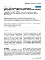
Figure 1
TauCl differentially inhibits the expression of MMPs in IL-β-stimulated RA FLSsTauCl differentially inhibits the expression of MMPs in IL-β-stimulated RA FLSs. The expressions of the collagenases (matrix metalloproteinase
[MMP]-1 and MMP-13) and the gelatinases (MMP-2 and MMP-9) were determined by (a) ELISA analysis, (b) real time PCR and (c) semi-quantita-
tive RNA analysis. Synovial cells (5 × 10
5
cells/60 mm dish/2 ml serum-free media) were treated with taurine chloramine (TauCl) 30 min before 24
hours of IL-1β (10 ng/ml) stimulation for MMP protein analysis by ELISA. Cells (2.5 × 10
5
cells/60 mm dish/2 ml serum-free media) were treated
with TauCl 30 min before 6 hours of stimulation with IL-1β (10 ng/ml) for RNA level analysis. IL-1β stimulated the expression of the MMP-1 and
MMP-13 genes, but it did not affect the expression of MMP-2 or MMP-9. TauCl differentially inhibited the expressions of MMP-1 and MMP-13.
Experiments were performed in duplicate with cells from three patients. Values are expressed as means ± standard deviation. *P < 0.01 versus con-
trol group (no IL-1β);
#
P < 0.05 and
##
P < 0.01 versus IL-1β treatment group without TauCl. FLS, fibroblast-like synoviocyte; PBS, phosphate-buff-
ered saline; RA, rheumatoid arthritis.
Arthritis Research & Therapy Vol 9 No 4 Kim et al.
Page 6 of 10
(page number not for citation purposes)
the proteasome [30]. TauCl inhibited IκB degradation as
potently as MG132, in this instance; however, the concentra-
tion of TauCl employed was greater than that of MG132 (Fig-
ure 3).
TauCl blocks NF-κB nuclear translocation through the
inhibition of IκB degradation
To further demonstrate that the effects of IκB degradation
extended to the transnuclear migration of NF-κB, levels of NF-
κB in the nucleus were assessed using EMSA (Figure 4) and
immunohistochemistry (Figure 5). As shown in Figure 4, at a
concentration of 800 μmol/l, TauCl completely blocked the
nuclear binding of NF-κB; however, at 600 αmol/l, TauCl did
not block binding activity. These results were confirmed by
confocal microscopy. After 90 min of IL-1β stimulation, the
majority of cytoplasmic NF-κB migrated into the nucleus, as
indicated by strong nuclear NF-κB staining following stimula-
tion and strong cytoplasmic staining before stimulation (Figure
5). Confirming previous findings, the migration of NF-κB into
the nucleus was not inhibited at TauCl concentrations of up to
600 μmol/l. However, at a concentration of 800 μmol/l, TauCl
blocked the transnuclear migration of NF-κB.
Figure 2
TauCl primarily inhibited the degradation of IκBTauCl primarily inhibited the degradation of IκB. (a) Synovial cells (5 × 10
5
cells/60 mm dish/2 ml serum-free media) were treated with IL-1β (10 ng/
ml). Shown are time courses of the signalling pathways activated during IL-1β stimulation. (b) Synovial cells (5 × 10
5
cells/60 mm dish/2 ml serum-
free media) were treated with taurine chloramine (TauCl) 30 min before 10 or 30 min of IL-1β (10 ng/ml) stimulation for Western blot analysis. A
TauCl concentration of 800 μmol/l significantly inhibited the inhibitor of nuclear factor-κB (IκB)/nuclear factor-κB (NF-κB) signalling pathway by
inhibiting the degradation of IκBα. The mitogen-activated protein kinase (MAPK) signalling pathway, including extracellular signal-regulated kinase
(ERK)-1/2, p38 and c-jun amino-terminal kinase (JNK), was unaffected. Three independent experiments were performed with cells from two patients.
p, phosphorylated.
Figure 3
TauCl inhibited IκBα degradation as potently as did a NF-κB inhibitor (MG132)TauCl inhibited IκBα degradation as potently as did a NF-κB inhibitor
(MG132). Synovial cells (5 × 10
5
cells/60 mm dish/2 ml serum-free
media) were treated with taurine chloramine (TauCl) or MG132 30 min
before IL-1β (10 ng/ml) stimulation for 30 min. At a concentration of
800 μmol/l, TauCl inhibited the degradation of inhibitor of nuclear fac-
tor-κB (IκB)α just as potently as did 1 μmol/l MG132. Three independ-
ent experiments were performed with cells from two patients. NF-κB,
nuclear factor-κB.
Figure 4
TauCl inhibited NF-κB binding activityTauCl inhibited NF-κB binding activity. Synovial cells (2 × 10
6
cells/
100-mm dish/5-ml serum-free media) were pretreated with taurine
chloramine (TauCl) or taurine (Tau) 30 min prior to IL-1β stimulation for
90 min. Nuclear extracts were prepared for electrophoretic mobility
shift assay (EMSA). IL-1β stimulation increased nuclear levels of
nuclear factor-κB (NF-κB). At a concentration of 800 μmol/l, TauCl
completely inhibited NF-κB binding. Antibodies against the p65 subunit
of NF-κB induced a gel shift in the NF-κB band. Three independent
experiments were performed with cells from two patients.
Available online />Page 7 of 10
(page number not for citation purposes)
Discussion
Because IL-1β is believed to play a major role in synovial
inflammation, RA FLSs stimulated with IL-1β in vitro have been
used to mimic the synovial proliferation that occurs in RA
patients suffering from inflammation [31]. IL-1β is also known
to stimulate many proinflammatory mediators in a variety of cell
types [32]. In addition, IL-1β is a potent inducer of
metalloproteinase production by FLSs; however, little investi-
gation has been conducted to determine its effects on the
gelatinases (MMP-2 and MMP-9) [33]. In the present study,
we found that IL-1β strongly stimulated the expression of col-
lagenases (MMP-1 and MMP-13). Gelatinase expression was
weakly activated by IL-1β stimulation. However, IL-1β is known
to induce high levels of gelatinase expression in other cell
types [34-36].
IL-1β activates different signalling pathways in different cell
types. Thus, we investigated signalling pathways in IL-1β stim-
ulated RA FLSs [37]. IL-1β stimulated the pathways of both
MAPK (ERK, p38 and JNK) and IκB kinase within 30 min, with
pathway activation subsiding to the basal levels of nonstimu-
lated cells by 60 min. The activation of these pathways led to
the activation of a number of transcriptional factors that
enhance the expression of various proinflammatory mediators.
Among these factors, NF-κB is a key regulator of inflammatory
gene transcription, and it is known to be activated in RA syno-
via and chondrocytes [38].
TauCl differentially inhibited the expression of MMPs in IL-1β
stimulated RA FLSs. The expression of MMP-13 was
significantly inhibited at concentrations of 400 to 600 μmol/l
TauCl, whereas the expression of MMP-1 was not significantly
inhibited at this concentration. To clarify the inhibitory mecha-
nism of TauCl on MMPs, the levels of both MAPK phosphor-
ylation and IκB degradation were investigated in IL-1β
stimulated RA FLSs. TauCl did not significantly inhibit the
phosphorylation of ERK-1/2, p38, or JNK, even at 800 μmol/l,
whereas IκB degradation was significantly inhibited at 800
μmol/l. These findings indicate that the inhibition of the IκB
signalling pathways by TauCl was primarily dependent on the
inhibition of IκB degradation. This finding is consistent with
previous reports showing that TauCl modifies the backbone of
IκB through amino acid oxidation of IκB, thus allowing IκB to
become resistant to degradation [39,40]. Confocal micro-
scopic examination of the NF-κB immunostaining results indi-
cated that a TauCl concentration of 800 μmol/l was required
to inhibit IκB degradation completely. Partial inhibition of IκB
degradation was seen at a TauCl concentration of 600 μ
mol/
l, as reflec
ted by NF-κB immunostaining in both the cytoplasm
and the nucleus. This may indicate that signalling pathways
other than the MAPK and IκB pathways are involved in the
stimulation of MMP-1 and MMP-13. In support of this idea,
protein kinase Cδ is known to play a key role in the stimulation
of MMP-13 via crosstalk with MAPKs in basic fibroblast
growth factor stimulated human adult articular chondrocytes
[41]. At concentrations lower than 800 μmol/l, TauCl may
inhibit or block minor pathways that are involved in the upreg-
Figure 5
TauCl inhibited the migration of NF-κB into the nucleusTauCl inhibited the migration of NF-κB into the nucleus. To visualize the translocation of nuclear factor-κB (NF-κB), synovial cells (4 × 10
4
cells/well
in four-well Lab-Tek chamber slides) were cultured. After serum starvation overnight, the cells were treated with taurine chloramine (TauCl) 30 min
before stimulation with IL-1β (10 ng/ml) for 90 min. IL-1β stimulation induced the migration of NF-κB from the cytoplasm into the nucleus (second
column), whereas NF-κB was found only in the cytoplasm of nonstimulated cells (first column). At a concentration of 800 μmol/l, TauCl completely
inhibited the migration of NF-κB into the nucleus (fifth column). All pictures were taken at a magnification of 200×. Three independent experiments
were performed in duplicate with cells from two patients.
Arthritis Research & Therapy Vol 9 No 4 Kim et al.
Page 8 of 10
(page number not for citation purposes)
ulation of MMP-1 and MMP-13. At a critical concentration
(600 to 800 μmol/l), IκBα degradation is completely inhibited,
thereby preventing the migration of NF-κB into the nucleus.
TauCl is less toxic than its precursor HOCl/OCl
-
, but cytotoxic
effects of TauCl at high concentrations have been reported. Its
toxicity appears to differ between cell types [42]. Kontny and
coworkers [43] reported that TauCl caused progressive
necrosis of RA FLSs at concentrations of 500 μmol/l or
greater. In our study, the RA FLSs used in the experiments
were not significantly affected by a TauCl concentration of
800 μmol/l for 24 hours, even though cytotoxicity was
detected in RA FLSs from some patients (Figure 6). TauCl tox-
icity appeared to vary between individual RA patients. In
addition, different cell passages might have contributed to the
variance in sensitivity to TauCl, because RA FLSs exhibit dif-
ferent characteristics according to passage [44,45]. Although
it remains uncertain whether the TauCl concentration used in
this experiment can be a physiologic concentration, TauCl may
remain at a high concentration in extracellular fluids because
the intracellular and extracellular concentrations of taurine in
mammalian tissues are 10 to 70 mmol/l and 20 to 100 μmol/
l, respectively [46].
The differential effects of TauCl on the expressions of MMP-1
and MMP-13 may also be related to other transcription factors
that are differentially involved in the activations of MMP-1 and
MMP-13. For example, Runxa2 was found to stimulate strongly
the transcriptional activation of MMP-13, but it had no effect
on MMP-1 expression in human chondrosarcoma cells [47]. In
addition, many transcriptional binding sites, such as activator
protein-1 and Ets/polymavirus enhancer 3 (OSE-2), have been
identified in the human MMP-13 proximal promoter [48-50].
An AG-rich element regulatory site was recently found in the
human MMP-13 proximal promoter [51]. This and other tran-
scription factors may contribute to the increased expression of
MMP-13 in IL-1β stimulated FLSs. The interaction of TauCl
with these as yet unidentified factors remains unknown.
Furthermore, these transcription factors may function at a
TauCl concentration that inhibits the degradation of IκB.
The degree of the inhibitory effect of TauCl was compared
with that of an NF-κB inhibitor, namely MG132. At a concen-
tration of 800 μmol/l, the inhibitory effect of TauCl on IκB deg-
radation was as potent as that of 1 μmol/l MG132. Because
MMP-13 exhibits the greatest activity toward the degradation
of type II collagen, a major component of the cartilage extracel-
lular matrix, the control of MMP-13 expression is crucial when
attempting to delay the degradation of cartilage [26]. At lower
concentrations of TauCl, inhibition of MMP-13 expression
would be a potentially effective strategy to control the destruc-
tion of joint cartilage in RA and osteoarthritis. Above all, TauCl
may be produced as a part of the homeostatic response to
infection and inflammation, thus playing a critical role in limiting
the duration and intensity of immune inflammation [52]. In sup-
Figure 6
Effect of TauCl on the viability of RA FLSsEffect of TauCl on the viability of RA FLSs. Rheumatoid arthritis (RA) fibroblast-like synoviocytes (FLSs) from two RA patients were treated with tau-
rine chloramine (TauCl) 30 min before the stimulation with IL-1β (10 ng/ml), and were incubated for 24 hours (as described in Materials and meth-
ods). Cell activity was then determined by 3-(4,5-dimethylthiazol-2-yl)-2,5-diphenyltetrazolium bromide (MTT) assay, and is expressed as the mean ±
standard deviation of three separate experiments. Three independent experiments were performed with cells from two patients. *P < 0.05 versus
untreated control.
Available online />Page 9 of 10
(page number not for citation purposes)
port of this hypothesis, synovial fluid neutrophils of RA patients
exhibit impaired generation of TauCl [53].
In summary, TauCl differentially inhibited the increased expres-
sion levels of MMP-1 and MMP-13 in IL-1β stimulated RA
FLSs. It inhibited the expression of MMP-1 primarily through
inhibition of IκB degradation, although it did not appear to
inhibit the expression of MMP-13 through inhibition of the IκB
signalling pathway.
Conclusion
Given that MMP-13, which is inhibited by TauCl, is remarkably
active against collagen type II, and that synovial fluid neu-
trophils of RA patients exhibit impaired generation of TauCl,
the involvement of TauCl in destruction of joint cartilage
should receive greater focus. This may yield insights into the
molecular mechanisms of joint destruction in RA.
Competing interests
The authors declare that they have no competing interests.
Authors' contributions
KSK participated in the data analysis and the design of the
study, and drafted the manuscript. EKP, SMJ, H-SJ and JSB
performed the experiments. CK supplied TauCl, performed
EMSA and helped to edit the manuscript. Y-AL, S-JH, S-HL
and H-IY provided clinical perspectives regarding the relation
of TauCl with RA. MCY provided the synovium from patients
and participated in the design of the study. All authors read
and approved the final manuscript.
Acknowledgements
This work was supported by a research grant from the Korean Ministry
of Health & Welfare (03-PJ9-PG6-SO01-002).
References
1. Mor A, Abramson SB, Pillinger MH: The fibroblast-like synovial
cell in rheumatoid arthritis: a key player in inflammation and
joint destruction. Clin Immunol 2005, 115:118-128.
2. Feldmann M, Brennan FM, Maini RN: Role of cytokines in rheu-
matoid arthritis. Annu Rev Immunol 1996, 14:397-440.
3. Bromley M, Woolley DE: Histopathology of the rheumatoid
lesion. Identification of cell types at sites of cartilage erosion.
Arthritis Rheum 1984, 27:857-863.
4. Harris ED Jr: Rheumatoid arthritis. Pathophysiology and impli-
cations for therapy. N Engl J Med 1990, 322:1277-1289.
5. Mohr W, Westerhellweg H, Wessinghage D: Polymorphonuclear
granulocytes in rheumatic tissue destruction. III. an electron
microscopic study of PMNs at the pannus-cartilage junction in
rheumatoid arthritis. Ann Rheum Dis 1981, 40:396-399.
6. Ottonello L, Cutolo M, Frumento G, Arduino N, Bertolotto M, Man-
cini M, Sottofattori E, Dallegri F: Synovial fluid from patients with
rheumatoid arthritis inhibits neutrophil apoptosis: role of ade-
nosine and proinflammatory cytokines. Rheumatology 2002,
41:1249-1260.
7. Wipke BT, Allen PM: Essential role of neutrophils in the initia-
tion and progression of a murine model of rheumatoid
arthritis. J Immunol 2001, 167:1601-1608.
8. Davies JM, Horwitz DA, Davies KJ: Potential roles of hypochlo-
rous acid and N-chloroamines in collagen breakdown by
phagocytic cells in synovitis. Free Radic Biol Med 1993,
15:637-643.
9. Edwards SW, Hallett MB: Seeing the wood for the trees: the
forgotten role of neutrophils in rheumatoid arthritis. Immunol
Today 1997, 18:320-324.
10. Learn DB, Fried VA, Thomas EL: Taurine and hypotaurine con-
tent of human leukocytes. J Leukoc Biol 1990, 48:174-182.
11. Vinton NE, Laidlaw SA, Ament ME, Kopple JD: Taurine concen-
trations in plasma and blood cells of patients undergoing
long-term parenteral nutrition. Am J Clin Nutr 1986,
44:398-404.
12. Thomas EL, Grisham MB, Melton DF, Jefferson MM: Evidenc
e for
a role of taurine in the in vitro oxidative toxicity of neutrophils
toward erythrocytes. J Biol Chem 1985, 260:3321-3329.
13. Kim C, Park E, Quinn MR, Schuller-Levis G: The production of
superoxide anion and nitric oxide by cultured murine leuko-
cytes and the accumulation of TNF-alpha in the conditioned
media is inhibited by taurine chloramine. Immunopharmacol-
ogy 1996, 34:89-95.
14. Kontny E, Rudnicka W, Kowalczewski J, Marcinkiewicz J, Maslinski
W: Selective inhibition of cyclooxygenase 2-generated pros-
taglandin E2 synthesis in rheumatoid arthritis synoviocytes by
taurine chloramine. Arthritis Rheum 2003, 48:1551-1555.
15. Marcinkiewicz J, Grabowska A, Bereta J, Bryniarski K, Nowak B:
Taurine chloramine down-regulates the generation of murine
neutrophil inflammatory mediators. Immunopharmacology
1998, 40:27-38.
16. Marcinkiewicz J, Grabowska A, Bereta J, Stelmaszynska T: Tau-
rine chloramine, a product of activated neutrophils, inhibits in
vitro the generation of nitric oxide and other macrophage
inflammatory mediators. J Leukoc Biol 1995, 58:667-674.
17. Park E, Schuller-Levis G, Quinn MR: Taurine chloramine inhibits
production of nitric oxide and TNF-alpha in activated RAW
264.7 cells by mechanisms that involve transcriptional and
translational events. J Immunol 1995, 154:4778-4784.
18. Schuller-Levis GB, Park E: Taurine and its chloramine: modula-
tors of immunity. Neurochem Res 2004, 29:117-126.
19. Kwasny-Krochin B, Bobek M, Kontny E, Gluszko P, Biedron R,
Chain BM, Maslinski W, Marcinkiewicz J: Effect of taurine chlo-
ramine, the product of activated neutrophils, on the develop-
ment of collagen-induced arthritis in DBA 1/J mice. Amino
Acids 2002, 23:419-426.
20. Wojtecka-Lukasik E, Gujski M, Roguska K, Maslinska D, Maslinski
S: Taurine chloramine modifies adjuvant arthritis in rats.
Inflamm Res 2005:S21-S22.
21. Murphy G, Knauper V, Atkinson S, Butler G, English W, Hutton M,
Stracke J, Clark I: Matrix metalloproteinases in arthritic disease.
Arthritis Res 2002:S39-S49.
22. Burrage PS, Mix KS, Brinckerhoff CE: Matrix metalloproteinases:
role in arthritis. Front Biosci 2006, 11:529-543.
23. Vincenti MP, Brinckerhoff CE: Transcriptional regulation of col-
lagenase (MMP-1, MMP-13) genes in arthritis: integration o
f
complex signaling pathways for the recruitment of gene-spe-
cific transcription factors. Arthritis Res 2002, 4:157-164.
24. Knauper V, Cowell S, Smith B, Lopez-Otin C, O'Shea M, Morris H,
Zardi L, Murphy G: The role of the C-terminal domain of human
collagenase-3 (MMP-13) in the activation of procollagenase-3,
substrate specificity, and tissue inhibitor of metalloproteinase
interaction. J Biol Chem 1997, 272:7608-7616.
25. Krane SM, Byrne MH, Lemaitre V, Henriet P, Jeffrey JJ, Witter JP,
Liu X, Wu H, Jaenisch R, Eeckhout Y: Different collagenase gene
products have different roles in degradation of type I collagen.
J Biol Chem 1996, 271:28509-28515.
26. Knauper V, Lopez-Otin C, Smith B, Knight G, Murphy G: Bio-
chemical characterization of human collagenase-3. J Biol
Chem 1996, 271:1544-1550.
27. Thomas EL, Grisham MB, Jefferson MM: Preparation and charac-
terization of chloramines. Methods Enzymol 1986,
132:569-585.
28. Rannou F, Francois M, Corvol MT, Berenbaum F: Cartilage break-
down in rheumatoid arthritis. Joint Bone Spine 2006, 73:29-36.
29. Zhao ML, Brosnan CF, Lee SC: 15-deoxy-delta (12,14)-PGJ2
inhibits astrocyte IL-1 signaling: inhibition of NF-kappaB and
MAP kinase pathways and suppression of cytokine and chem-
okine expression. J Neuroimmunol 2004, 153:132-142.
30. Snyder JG, Prewitt R, Campsen J, Britt LD: PDTC and Mg132,
inhibitors of NF-kappaB, block endotoxin induced vasodilation
of isolated rat skeletal muscle arterioles. Shock 2002,
17:304-307.
Arthritis Research & Therapy Vol 9 No 4 Kim et al.
Page 10 of 10
(page number not for citation purposes)
31. Kay J, Calabrese L: The role of interleukin-1 in the pathogene-
sis of rheumatoid arthritis. Rheumatology 2004:iii2-iii9.
32. Braddock M, Quinn A, Canvin J: Therapeutic potential of target-
ing IL-1 and IL-18 in inflammation. Expert Opin Biol Ther 2004,
4:847-860.
33. Dayer JM, de Rochemonteix B, Burrus B, Demczuk S, Dinarello
CA: Human recombinant interleukin 1 stimulates collagenase
and prostaglandin E2 production by human synovial cells. J
Clin Invest 1986, 77:645-648.
34. Eberhardt W, Huwiler A, Beck KF, Walpen S, Pfeilschifter J:
Amplification of IL-1 beta-induced matrix metalloproteinase-9
expression by superoxide in rat glomerular mesangial cells is
mediated by increased activities of NF-kappa B and activating
protein-1 and involves activation of the mitogen-activated pro-
tein kinase pathways. J Immunol 2000, 165:5788-5797.
35. Esteve PO, Chicoine E, Robledo O, Aoudjit F, Descoteaux A, Pot-
worowski EF, St-Pierre Y: Protein kinase C-zeta regulates tran-
scription of the matrix metalloproteinase-9 gene induced by
IL-1 and TNF-alpha in glioma cells via NF-kappa B. J Biol
Chem 2002, 277:35150-35155.
36. Yokoo T, Kitamura M: Dual regulation of IL-1 beta-mediated
matrix metalloproteinase-9 expression in mesangial cells by
NF-kappa B and AP-1. Am J Physiol 1996, 270:F123-F130.
37. Dinarello CA: The interleukin-1 family: 10 years of discovery.
FASEB J 1994, 8:1314-1325.
38. Miagkov AV, Kovalenko DV, Brown CE, Didsbury JR, Cogswell JP,
Stimpson SA, Baldwin AS, Makarov SS: NF-kappaB activation
provides the potential link between inflammation and hyper-
plasia in the arthritic joint. Proc Natl Acad Sci USA 1998,
95:13859-13864.
39. Barua M, Liu Y, Quinn MR: Taurine chloramine inhibits inducible
nitric oxide synthase and TNF-alpha gene expression in acti-
vated alveolar macrophages: decreased NF-kappaB activation
and IkappaB kinase activity. J Immunol 2001, 167:2275-2281.
40. Kanayama A, Inoue J, Sugita-Konishi Y, Shimizu M, Miyamoto Y:
Oxidation of Ikappa Balpha at methionine 45 is one cause of
taurine chloramine-induced inhibition of NF-kappa B
activation. J Biol Chem 2002, 277:24049-24056.
41. Im HJ, Muddasani P, Natarajan V, Schmid TM, Block JA, Davis F,
van Wijnen AJ, Loeser RF: Basic fibroblast growth factor stimu-
lates matrix metalloproteinase-13 via the molecular cross-talk
between the mitogen-activated protein kinases and protein
kinase Cdelta pathways in human adult articular
chondrocytes. J Bi
ol Chem 2007, 282:11110-11121.
42. Choi HS, Cha YN, Kim C: Taurine chloramine inhibits PMA-
stimulated superoxide production in human neutrophils per-
haps by inhibiting phosphorylation and translocation of
p47(phox). Int Immunopharmacol 2006, 6:1431-1440.
43. Kontny E, Rudnicka W, Chorazy-Massalska M, Marcinkiewicz J,
Maslinski W: Taurine chloramine inhibits proliferation of rheu-
matoid arthritis synoviocytes by triggering a p53-dependent
pathway. Inflamm Res 2006, 55:446-455.
44. Hirth A, Skapenko A, Kinne RW, Emmrich F, Schulze-Koops H,
Sack U: Cytokine mRNA and protein expression in primary-cul-
ture and repeated-passage synovial fibroblasts from patients
with rheumatoid arthritis. Arthritis Res 2002, 4:117-125.
45. Zimmermann T, Kunisch E, Pfeiffer R, Hirth A, Stahl HD, Sack U,
Laube A, Liesaus E, Roth A, Palombo-Kinne E, et al.: Isolation and
characterization of rheumatoid arthritis synovial fibroblasts
from primary culture: primary culture cells markedly differ
from fourth-passage cells. Arthritis Res 2001, 3:72-76.
46. Fukuda K, Hirai Y, Yoshida H, Nakajima T, Usui T: Free amino acid
content of lymphocytes nd granulocytes compared. Clin
Chem 1982, 28:1758-1761.
47. Pei Y, Harvey A, Yu XP, Chandrasekhar S, Thirunavukkarasu K:
Differential regulation of cytokine-induced MMP-1 and MMP-
13 expression by p38 kinase inhibitors in human chondrosar-
coma cells: potential role of Runx2 in mediating p38 effects.
Osteoarthritis Cartilage 2006, 14:749-758.
48. Jimenez MJ, Balbin M, Lopez JM, Alvarez J, Komori T, Lopez-Otin
C: Collagenase 3 is a target of Cbfa1, a transcription factor of
the runt gene family involved in bone formation. Mol Cell Biol
1999, 19:4431-4442.
49. Pendas AM, Balbin M, Llano E, Jimenez MG, Lopez-Otin C: Struc-
tural analysis and promoter characterization of the human col-
lagenase-3 gene (MMP13). Genomics 1997, 40:222-233.
50. Tardif G, Pelletier JP, Dupuis M, Hambor JE, Martel-Pelletier J:
Cloning, sequencing and characterization of the 5'-flanking
region of the human collagenase-3 gene. Biochem J 1997,
323:13-16.
51. Fan Z, Tardif G, Boileau C, Bidwell
JP, Geng C, Hum D, Watson
A, Pelletier JP, Lavigne M, Martel-Pelletier J: Identification in
human osteoarthritic chondrocytes of proteins binding to the
novel regulatory site AGRE in the human matrix metallopro-
tease 13 proximal promoter. Arthritis Rheum 2006,
54:2471-2480.
52. Antoniv TT, Ivashkiv LB: Dysregulation of interleukin-10-
dependent gene expression in rheumatoid arthritis synovial
macrophages. Arthritis Rheum 2006, 54:2711-2721.
53. Kontny E, Wojtecka LE, Rell-Bakalarska K, Dziewczopolski W,
Maslinski W, Maslinski S: Impaired generation of taurine chlo-
ramine by synovial fluid neutrophils of rheumatoid arthritis
patients. Amino Acids 2002, 23:415-418.