Báo cáo y học: "Critical role of the major histocompatibility complex and IL-10 in matrilin-1-induced relapsing polychondritis in mice" doc
Bạn đang xem bản rút gọn của tài liệu. Xem và tải ngay bản đầy đủ của tài liệu tại đây (939.3 KB, 8 trang )
Open Access
Available online />R484
Vol 6 No 5
Research article
Critical role of the major histocompatibility complex and IL-10 in
matrilin-1-induced relapsing polychondritis in mice
Ann-Sofie Hansson
1
, Åsa CM Johansson
2
and Rikard Holmdahl
2
1
Department of Clinical Immunology, Göteborg University, Göteborg, Sweden
2
Medical Inflammation Research, BMC, Lund University, Lund, Sweden
Corresponding author: Ann-Sofie Hansson,
Received: 21 Oct 2003 Revisions requested: 26 Nov 2003 Revisions received: 3 Jun 2004 Accepted: 30 Jun 2004 Published: 12 Aug 2004
Arthritis Res Ther 2004, 6:R484-R491 (DOI 10.1186/ar1218)
http://arthr itis-research.com/conte nt/6/5/R484
© 2004 Hansson et al.; licensee BioMed Central Ltd. This is an Open Access article: verbatim copying and redistribution of this article are permitted
in all media for any purpose, provided this notice is preserved along with the article's original URL.
Abstract
Relapsing polychondritis (RP) is an autoimmune disease that
affects extra-articular cartilage. Matrilin-1-induced relapsing
polychondritis (MIRP) is a model for RP and is useful for studies
of the pathogenic mechanisms in this disease. There are
indications that the major histocompatibility complex (MHC)
class II plays a major role in RP, since DR4
+
patients are more
commonly affected than controls. We have now addressed the
role of the MHC region, as well as the non-MHC contribution,
using congenic mouse strains. Of the MHC congenic strains,
B10.Q (H2
q
) was the most susceptible, the B10.P (H2
p
) and
B10.R (H2
r
) strains developed mild disease, while B10 strains
carrying the v, b, f, or u H2 haplotypes were resistant. A slight
variation of susceptibility of H2
q
strains (B10.Q> C3H.Q> DBA/
1) was observed and the (B10.Q × DBA/1)F
1
was the most
susceptible of all strains. Furthermore, macrophages and CD4
+
T cells were the most prominent cell types in inflammatory
infiltrates of the tracheal cartilage. Macrophages are the major
source of many cytokines, such as interleukin-10 (IL-10), which
is currently being tested as a therapeutic agent in several
autoimmune diseases. We therefore investigated B10.Q mice
devoid of IL-10 through gene deletion and found that they
developed a significantly more severe disease, with an earlier
onset, than their heterozygous littermates. In conclusion, MHC
genes, as well as non-MHC genes, are important for MIRP
induction, and IL-10 plays a major suppressive role in cartilage
inflammation of the respiratory tract.
Keywords: IL-10, matrilin-1, matrilin-1-induced relapsing polychondritis, major histocompatibility complex, relapsing polychondritis
Introduction
Autoimmune diseases that affect cartilage tissue are wide-
spread in the population. The most common one is rheuma-
toid arthritis (RA), in which joints are attacked by an erosive,
relapsing inflammation. In a related human disorder, relaps-
ing polychondritis (RP), mainly cartilage of the external
ears, nose, and respiratory tract is involved in the disease
process [1]. Joints are affected as a nonerosive, seronega-
tive arthritis [2] and 20% of patients with RP develop
nephritis, which is probably induced by the formation of
immune complexes [3].
Similar pathogenic mechanisms are thought to be involved
in RP and RA, partly because of the cartilage autoimmune
inflammation but also because both diseases have been
reported to be associated with the MHC allele HLA-DR4
[4-6]. Similarities, as well as differences, are also observed
in animal models that mimic these human diseases. Colla-
gen-induced arthritis (CIA), in which animals are immunized
with collagen type II (CII), is one of the most commonly
used and best-characterized models for RA [7,8]. In this
model, the H2
q
haplotype has been found to be the one
most strongly associated with CIA and the class II molecule
Aq has been reported to explain this association. Interest-
ingly, rheumatoid-associated class II molecules, such as
DR4 (DRB1*0401), when expressed in the mouse, mimic
the function of Aq. In one mouse strain, the human DQ6αβ
/8αβ transgenic mouse, immunization with CII induces
symptoms of arthritis as well as chondritis of the auricle that
mimic RP [9].
A mouse and rat model for RP, matrilin-1-induced relapsing
polychondritis (MIRP), was developed by our group to
investigate the pathogenic pathways in RP [10]. Matrilin-1
is a cartilage-specific protein expressed in upper-airway
cartilage [11], and consequently MIRP mimics the
CIA = collagen-induced arthritis; CII = collagen type II; COMP = cartilage oligomeric matrix protein; IL-10 = interleukin-10; MHC = major histocom-
patibility complex; MIRP = matrilin-1-induced relapsing polychondritis; RA = rheumatoid arthritis; RP = relapsing polychondritis.
Arthritis Research & Therapy Vol 6 No 5 Hansson et al.
R485
inflammatory attack of the nose and respiratory tract, phe-
nomena that are commonly seen in RP patients. There are
also morphological similarities, such as infiltrations of mac-
rophages and lymphocytes. In addition, a subgroup of
patients with RP produces an antibody response to matri-
lin-1, and serum antibodies from these patients inhibit the
binding of anti-matrilin-1-specific antibodies [12].
Surprisingly, when the MIRP and CIA models in rats are
compared, major genetic differences are found regarding
susceptibility to induction of disease symptoms. The DA rat
is recognized as highly susceptible in most arthritis models,
whereas it does not develop any sign of inflammation when
immunized with matrilin-1 [10,13,14]. In contrast, the
LEW.1F strain is a low responder to immunization with CII
[15] but is highly susceptible to MIRP. On the other hand,
the murine MIRP and CIA models are both dependent on B
cells for the induction of clinical symptoms [16,17]. In addi-
tion, the complement system plays a major role in the
pathogenesis of both diseases [16,18,19] and T cells are
required in order to induce disease [10,20].
No data have been reported on the role of cytokines in RP,
either in patients or in the corresponding animal models. In
the CIA model, several cytokines have been shown to play
major roles in the inflammatory process, anti-inflammatory
mediators as well as proinflammatory ones. The cytokine
interleukin-10 (IL-10) has been in focus for many years in
autoimmune arthritis and in other autoimmune diseases.
The human recombinant protein is currently being tested as
a therapeutic agent in several human inflammatory dis-
eases. Macrophages are the major source of IL-10 but this
cytokine is also produced by B cells, T helper 2 cells, and
monocytes [21-24]. IL-10 has an immunosuppressive
effect on several proinflammatory cytokines, such as TNF-
α and IL-1, both known as enhancers of the destructive
inflammation in RA. It is also known that IL-10 down-regu-
lates MHC class II on macrophages [25]. IL-10 was prima-
rily considered to only suppress the inflammatory response
in arthritis, but in recent years it has been shown to play a
more complex and pleiotropic role [26]. Our group recently
visualized this complexity. We showed that IL-10-deficient
mice immunized with CII develop a more severe disease
than their heterozygous littermates, while they are pro-
tected from antibody-transferred arthritis induced with CII-
specific monoclonal antibodies [27]. In addition, we
showed that IL-10 deficiency did not affect the proliferation
to CII or IFN-γ production in comparison with their hetero-
zygous littermates.
To further investigate the pathogenic pathways in RP, we
used the mouse MIRP model. We immunized several
strains of mice, including MHC congenic strains, to eluci-
date the role of MHC and non-MHC genes. We analyzed
parameters reflecting activity of the cellular as well as the
humoral immune response, such as influx of cells and anti-
body production. In addition, to investigate the role of
inflammatory mediators in MIRP, we immunized mice
devoid of IL-10 in order to determine whether this cytokine,
as in the CIA model, possesses significant effects on
autoimmune chondritis in the extra-articular cartilage.
Materials and methods
Mice
Mice were bred and kept at the animal department at Med-
ical Inflammation Research, Lund University. They were
used at age 8–13 weeks and kept in a climate-controlled
environment (temperature and humidity) with cycles of 12
hours light/dark and sound. IL-10-deficient mice were pro-
duced by a deletion in the IL-10 gene in a cross of C57BL/
6 × 129/Ola (originally provided by W Müller, Institute of
Genetics, Cologne, Germany). They were further back-
crossed into B10.Q (H2
q
) mice (originally from J Klein, Uni-
versity of Tübingen, Tübingen, Germany, as were the
B10.P mice [H2
p
]) background for nine generations and
intercrossed to provide homozygous littermates lacking IL-
10 [27]. Additional strains were kindly provided by collabo-
rators (C3H.Q [H2
q
], from DSchreffler, St Louis, MO, USA)
or purchased from Jackson Laboratories (Bar Harbor, ME,
USA). Here we refer to (B10.Q × DBA/1)F
1
mice as QD
mice. Approval for the animal experiments was obtained
from the ethical committee at Lund University.
Induction of disease
Mice were immunized at the base of the tail with 100 µg of
matrilin-1, purified as previously described [11], emulsified
in complete Freund's adjuvant (Difco, Detroit, MI, USA).
They were boosted at day 35 with 50 µg of matrilin-1 in
incomplete Freund's adjuvant (Difco). Control mice immu-
nized in the same way but with matrilin-1 omitted were used
in all experiments. Experimental mice were kept for 130
days. The severity of disease was scored using a modified
version of a scale previously developed for the rat model
[10]: 1, suspicion of respiratory distress; 2, discontinuous
inspiratory stridor; 3, continuous inspiratory stridor; 4, con-
tinuous inspiratory stridor and abnormal breathing pattern;
5, cyanosis. Mice developing severe respiratory distress,
indicated by score 5, were humanely killed at once.
Histology
Tissue samples were dissected in the acute phase at score
5 or at the end of the experiment at day 130. The tissue was
immediately either snap-frozen at -70°C or fixed in 4% para-
formaldehyde solution for 24 hours and further embedded
in paraffin. Joints were decalcified for 2–3 weeks in EDTA
solution. Sections 5–6 µm thick were stained with hema-
toxylin and erythrosine. Immunohistochemical staining was
performed in accordance with the standard protocol.
Briefly, sections were incubated for 2 hours at room tem-
perature with a primary antibody recognizing macrophages
Available online />R486
(defined as CD11b
+
cells), MHC II, CD4
+
cells, and CD8α
+
cells. A secondary biotinylated rabbit antirat Ig antibody
(DAKO A/S, Glostrup, Denmark) was incubated for
another 2 hours and binding was visualized with diami-
nobenzidine (Saveen Biotech, Malmö, Sweden). Immuno-
histochemical sections were scored by counting the mean
number of positive cells in two areas of the same size from
each section and were evaluated as follows: <5%, +; 5–
25%, ++; 25–50%, +++; and >50%, ++++.
Antibody detection
Sera were collected and stored at -20°C until assay. ELISA
was performed with sera diluted 1/10 and titrated in steps
of 10. Plates (Costar; Corning Life Sciences, Oneonta, NY,
USA) were coated with 1 µg/ml of matrilin-1, 10 µg/ml of
CII, or 10 µg/ml of cartilage oligomeric matrix protein
(COMP) in PBS + 0.02% sodium azide overnight at 4°C.
They were washed in washing buffer (0.1 M Tris/HCI+
0.05% Tween 20) and incubated for 2 hours at room tem-
perature in PBS buffer (PBS + 0.05% Tween 20 + 0.02%
sodium azide). Washing was repeated and the plates were
incubated for another 2 hours with conjugates detecting
sheep antimouse IgG Fcγ (Jackson ImmunoResearch Lab-
oratories, West Grove, PA, USA). The plates were devel-
oped with p-nitrophenol as the substrate and the amount of
antibody was estimated as absorbency at 405 nm by using
a Titertek Multiscan filter photometer. All plates detecting
the same antigen were analyzed at the same time point. A
positive control, consisting of a mixture of sera from DBA/
1 mice immunized with the protein in question, was used on
all plates assayed. An established ELISA protocol was
used for detection of anticollagen antibodies [28].
Statistical analysis
All assays were analyzed with the Mann–Whitney U test.
Unless indicated otherwise, P<0.05 was considered to
indicate significance.
Results
MHC genes and non-MHC genes influence susceptibility
to MIRP
To investigate the role of MHC in MIRP, we immunized sev-
eral strains of male mice carrying different MHC class II
molecules. The QD (H2
q
) strain [F
1
of a cross between
B10.Q (H2
q
) and DBA/1 (H2
q
)] was the most susceptible,
developing severe, relapsing respiratory distress and with a
significantly earlier onset of disease than any other strain
(Table 1; Fig. 1a,1b). The B10.Q strain was also a high
responder, as more than 50% of these mice were suscep-
tible to disease. A few mice of the C3H.Q (H2
q
) and DBA/
1 strains developed respiratory distress in the acute phase,
which in two mice had high scores. However, the symp-
toms in these strains did not proceed in relapses as they
did in the QD and B10.Q mice, and therefore resulted in a
lower mean score than for the other strains (Fig. 1c).
Table 1
Susceptibility of mouse strains to immunization with matrilin-1, as shown by their development of matrilin-1-induced relapsing
polychondritis (MIRP)
Mouse strain MHC Gender n Incidence of
disease (%)
Mean maximum
disease score
Day of onset of
symptoms
QD H2
q
m12924.8 ± 0.4
a
41 ± 1
b
B10.Q H2
q
m 16 56 2.6 ± 0.7 46 ± 4
B10.Q H2
q
f 8 25 2.0 ± 0 41 ± 0
C3H.Q H2
q
m 9 33 3.7 ± 1.2 46 ± 3
DBA/1 H2
q
m 12 50 3.2 ± 0.5 55 ± 8
c
Balb/c H2
d
m50
NOD H2
g7
m100
B10.P H2
p
m 5 40 3.0 ± 1.0 45 ± 0
B10.RIII H2
r
m 8 25 2.0 ± 0 47 ± 2
B10.V H2
v
m50
B10 H2
b
m50
B10.F H2
f
m50
B10.U H2
u
m10
Only affected mice were included in the statistical analysis.
a
QD mice developed higher mean maximum disease scores than mice from the
B10.Q, DBA/1, B10.P, and B10.RIII strains (P < 0.05).
b
QD mice developed disease symptoms earlier than all other strains (P < 0.05).
c
DBA/1
mice developed disease symptoms later than all other strains (P < 0.05). f, female; m, male; QD, (B10.Q × DBA/1)F
1
mice.
Arthritis Research & Therapy Vol 6 No 5 Hansson et al.
R487
Inflammation and erosion of the cartilage were observed in
sections from the nose, trachea, and larynx, and the degree
of pathologic changes was correlated with clinical scores.
The inflammatory infiltrates consisted of neutrophils, lym-
phocytes, and eosinophils. In addition, large numbers of
macrophages were detected in the acute as well as in the
chronic phase (Fig. 2). We did not detect any microscopic
sign of inflammation in nonresponding mice or in control
mice. In mice affected by respiratory distress, we observed
a drop in body weight, which confirmed the clinical scores.
Among mice of the QD strain, individuals that subsequently
developed cyanosis lost as much as 25% of their body
weight within a few days after the onset of respiratory
symptoms (Fig. 1). Major weight loss was observed in sev-
eral individual mice of other strains as well, but for strains
analyzed as a group, only the QD mice lost significantly
more body weight than the control group (data not shown).
In order to investigate the influence of gender, female mice
on the B10.Q background were immunized at the same
time as their male littermates. These females developed
disease symptoms less severe than those of the males,
with only mild respiratory distress for two or three days
being observed (Table 1). However, the group of female
mice produced levels of antibodies to matrilin-1 similar to
those in the males.
Antibodies to matrilin-1, CII, and COMP are produced
equally in susceptible and resistant strains
All strains that were immunized with matrilin-1 produced
antibodies to matrilin-1, and no difference in titers was
detected in comparisons of two defined groups of suscep-
tible and resistant strains (Fig. 3; Table 1). Balb/c (H2
d
)
mice produced the highest titers, while B10.P (H2
p
) were
low producers. However, when individual mice within each
strain were considered, a tendency was seen for mice pre-
senting severe respiratory distress, particularly those mice
with the highest clinical score, to mount the highest levels
of matrilin-1-specific antibodies. To investigate epitope
spreading, we analyzed antibody responses to collagen
type II (CII) and cartilage oligomeric matrix protein (COMP),
two additional cartilage proteins involved in the autoim-
mune process [29,30]. QD mice produced low titers of
antibodies to CII, and no CII-specific antibodies were
detected in the other strains. While all of the QD mice
responded to some degree to COMP, raised titers were
seen in only some mice from the other strains and without
any correlation with clinical score (data not shown).
Macrophages are important at the induction of MIRP
In order to define the infiltrating inflammatory cells in the
acute and chronic phases of murine MIRP, we stained tis-
sue sections dissected from cartilage of nasal, laryngeal,
and tracheal specimens. Tissue samples were collected in
the acute phase at the maximum of the clinical score
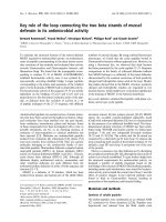
Figure 1
Disease course and weight in individual mice immunized with matrilin-1 to induce relapsing polychondritisDisease course and weight in individual mice immunized with matrilin-1
to induce relapsing polychondritis. (a, b) Two QD ([B10.Q × DBA/
1]F
1
) mice (QD 1 and QD 2) and (c) a C3H.Q mouse (CQ 1) were
scored for severity of disease on a scale from 0 to 5; see Materials and
methods. All control mice (n = 4) were scored at the same time, and
mean values of their weight are presented.
24
26
28
30
32
34
36
38
Weight (g)
25 50 75 100 125 150
mean weight controls
weight QD 1
0
1
2
3
4
5
Score
Days after immunization
score QD 1
24
26
28
30
32
34
36
38
Weight (g)
mean weight controls
weight QD 2
0
1
2
3
4
5
Score
25 50 75 100 125 150
Days after immunization
score QD 2
0
1
2
3
4
5
Score
25 50 75 100 125 150
Days after immunization
score CQ 1
24
26
28
30
32
34
36
38
Weight (g)
mean weight controls
weight CQ 1
(a)
(b)
(c)
Available online />R488
(around the day of onset) and at the end of the experiment
(on day 130). Two QD mice, two B10.Q mice, and two
controls were analyzed at each time point. Macrophages,
defined as CD11b
+
cells, comprised more than 50% of the
cells and were the most prominent cell type in the acute
phase, whereas fewer, less than 25%, were detected in the
chronic phase. In the chronic phase, there was a shift
towards higher levels of macrophages in nasal and laryn-
geal cartilage than in the trachea. The control mice had less
than 5% macrophages. T cells with a CD4
+
phenotype
comprised 5–25% of the cells in the acute phase and less
than 5% in the chronic phase. Low numbers of cells (fewer
than 5%) were positive for MHC class II or CD8, which
were found only in the acute phase of disease. No CD4
+
,
CD8
+
, or MHC-class-II-positive cells were detected in any
phase in the control mice.
IL-10 has a protective effect in MIRP
Our finding that macrophages are prominent cells in MIRP
led us to investigate the role of IL-10, an important product
of macrophages. Mice devoid of IL-10 and their hetero-
zygous littermates were immunized with matrilin-1 in
accordance with the standard protocol. Respiratory dis-
tress was observed in 9 of the 11 IL-10-deficient mice but
in only 4 of the 9 heterozygous littermates, indicating that
IL-10 acts in a suppressive fashion (Table 2). The mean
maximum score and the day of onset were significantly dif-
ferent in the homozygous group than in the heterozygous
one (Table 2). No difference was detected between the
two groups of mice in an analysis of the number of
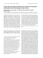
Figure 2
Tissue samples from a QD ([B10.Q × DBA/1]F
1
) mouse immunized with matrilin-1 to induce relapsing polychondritisTissue samples from a QD ([B10.Q × DBA/1]F
1
) mouse immunized with matrilin-1 to induce relapsing polychondritis. (a) Section from the tracheal
cartilage in the acute phase, showing inflammatory infiltrates and severe cartilage destruction. Cells detected in the infiltrates are macrophages, neu-
trophils, lymphocytes, and eosinophils. (b) Section from nasal septum, showing inflammatory infiltrates, fibrin deposition, and erosion of the cartilage.
Staining with hematoxylin and erythrosine. Original magnification ×200.
Figure 3
Titers of antibodies to matrilin-1 in mice immunized with matrilin-1Titers of antibodies to matrilin-1 in mice immunized with matrilin-1. Sera
analyzed at day 35, with values expressed as relative titers in compari-
son with a positive control used on all plates assayed. For detailed
information on the various strains and H2 haplotype, see Table 1.
0
0.5
1
1.5
2
2.5
Relative titer
(B10.QxDBA/1) F1
B10.Q
C3H.Q
DBA/1
Balb/c
NOD
B10.P
B10.RIII
B10.V
B10
B10.F
B10.U
Arthritis Research & Therapy Vol 6 No 5 Hansson et al.
R489
macrophages or of cells positive for MHC class II, CD4, or
CD8 in tests using immunohistochemical stainings of carti-
lage tissue from the nose, larynx, and trachea (two mice
from the acute phase and two from the chronic phase). As
was seen in the QD and B10.Q mice, more macrophages
were observed in the acute than in the chronic stage.
All the mice produced antibodies to matrilin-1 and there
was a tendency towards correlation between the titer of
anti-matrilin-1 antibodies and clinical symptoms, in both the
IL-10-deficient and the heterozygous mice. Surprisingly,
several of the IL-10 knockout mice, all of which were taken
off the experiment because of severe respiratory distress,
produced higher levels of CII-specific antibodies than were
detected in the QD mice (Fig. 4). Approximately half of the
mice in both groups produced antibodies to COMP com-
parable with the levels found in the other strains, as
described earlier (data not shown). No anticollagen or anti-
COMP antibodies were detected in nonimmunized mice.
Nor did we detect any inflammatory signs in joint sections
from any mouse.
Discussion
The pathogenic pathways in relapsing polychondritis are
largely unknown. In this paper we show that genes in the
MHC region as well as genes outside that region are impor-
tant for the induction of respiratory distress in murine MIRP.
Strains that carried the H2
q
haplotype were the most sus-
ceptible ones, and of these, the QD strain was the most
sensitive. We found that males were more severely affected
than females. All strains and both genders produced high
titers of antibodies to matrilin-1, with no significant correla-
tion to disease parameters at day 35. In addition, IL-10 was
an important immunomodulator in the pathogenesis of
MIRP.
The matrilin-1-induced symptoms appeared to be geneti-
cally controlled by the MHC region, as mice congenic at
the H2 region differed in susceptibility to disease. As in
mouse models for arthritis, mice carrying the H2
q
haplotype
were the most susceptible ones: all strains tested that had
this haplotype developed respiratory distress. However, the
influence of non-MHC genes in MIRP differs from that in
CIA, as the B10.Q mouse is relatively more resistant to
MIRP. These data further strengthen several publications
that indicate similarities in the MHC genetic control of RP
and RA, as both diseases are reported to be associated
with HLA-DR4 [4-6], whereas differences in non-MHC
genes contribute to the differing pathogeneses.
Surprisingly, we found no differences between strains in
the anti-matrilin-1 antibody titers at day 35. However, all
mice with clinical disease developed high levels of antibod-
ies to matrilin-1. We have recently shown that B-cell-defi-
cient mice are completely resistant to MIRP [16]. In
addition, in these experiments we induced inflammation
and erosion of the cartilage in the respiratory tract by inject-
ing matrilin-1-specific monoclonal antibodies into B-cell-
Table 2
Susceptibility to immunization with matrilin-1 in mice heterozygous or homozygous for an IL-10 gene deletion
Mice n Incidence (%) Mean maximum score
a
Day of onset of symptoms
IL-10
+/-
9 44 2.8 ± 0.8 68 ± 30
IL-10
-/-
11 82 3.5 ± 1.4* 41 ± 6*
All mice were bred on a C57BL/10 background carrying the H2
q
haplotype in the MHC class II region.
a
Score of severity of matrilin-1-induced
relapsing polychondritis, from a possible maximum of 5; see Materials and methods. *P < 0.05.
Figure 4
Anticollagen type II antibody response after immunization with matrilin-1 in (B10.Q × DBA/1)F
1
B10.Q mice devoid of IL-10 through gene dele-tion, and their heterozygous littermatesAnticollagen type II antibody response after immunization with matrilin-1
in (B10.Q × DBA/1)F
1
B10.Q mice devoid of IL-10 through gene dele-
tion, and their heterozygous littermates. Sera were analyzed for total
IgG levels at day 35 after immunization. For detailed information on the
experimental setup, see Materials and methods.
0
20
40
60
80
IgG (µg/ml)
(B10.QxDBA/1) F1
IL-10 –/–
IL-10 +/–
Available online />R490
deficient mice. This indicates that the matrilin-1-specific
humoral response plays an important role in the induction
phase of disease. The discrepancies between our earlier
results and the present findings of antibody titers could
possibly be explained by the fact that titers at day 35 do not
reflect the factors that are crucial for the initial triggering of
the matrilin-1-induced symptoms. There are likely to be
additional effector pathways of critical importance with
regard to maintenance of disease, as for example epitope
spreading. Unexpectedly, we found that some of the IL-10-
deficient mice with high clinical scores developed high
levels of anti-CII antibodies. We did not observe any clinical
signs of inflammation from the articular cartilage, which
indicated that these anti-CII specific antibodies were not
arthritogenic but rather were a result of the cartilage-
destructive inflammation in the trachea. However, the influ-
ence of IL-10 on immune reactivity to CII needs to be fur-
ther investigated.
Macrophages were the dominating cell type in the inflam-
matory infiltrates of laryngeal and nasal cartilage tissue sec-
tions. Macrophages produce large amounts of several
proinflammatory cytokines and are the major source of IL-
10, a pleiotropic cytokine with a significant effect on several
cell populations. Our finding that a lack of IL-10 increases
susceptibility to MIRP indicates that IL-10 acts in a sup-
pressive fashion in the MIRP model. This further highlights
the potential of IL-10 as a target for intervention in patients
with RP.
Conclusion
In conclusion, our results emphasize the contribution of
MHC as well as well as non-MHC genes in the autoimmune
chondritis model MIRP. We further show that macro-
phages and CD4
+
T cells as well as IL-10 play major roles
in the pathogenesis of cartilage inflammation of the respira-
tory tract. Additional investigations of the genetic control as
well as the pathogenic pathways, particularly regarding
inflammatory cytokines, are needed to elucidate the com-
plexity of the autoimmune inflammation in cartilage tissue.
Finally, we found major similarities between our MIRP
model and the commonly used models for RA, indicating
that pathogenesis and, as a consequence, therapeutic
strategies similar to those for RA should be considered for
RP.
Competing interests
None declared.
Acknowledgement
We would like to thank Prof Dick Heinegård at the section for Connec-
tive Tissue Biology at Lund University for contributing with the matrilin-1
production.
References
1. McAdam LP, O'Hanlan MA, Bluestone R, Pearson CM: Relapsing
polychondritis: prospective study of 23 patients and a review
of the literature. Medicine (Baltimore) 1976, 55:193-215.
2. O'Hanlan M, McAdam LP, Bluestone R, Pearson CM: The
arthropathy of relapsing polychrondritis. Arthritis Rheum 1976,
19:191-194.
3. Chang-Miller A, Okamura M, Torres VE, Michet CJ, Wagoner RD,
Donadio JV Jr, Offord KP, Holley KE: Renal involvement in
relapsing polychondritis. Medicine (Baltimore) 1987,
66:202-217.
4. Stastny P: Association of the B-cell alloantigen DRw4 with
rheumatoid arthritis. N Engl J Med 1978, 298:869-871.
5. Lang B, Rothenfusser A, Lanchbury JS, Rauh G, Breedveld FC,
Urlacher A, Albert ED, Peter HH, Melchers I: Susceptibility to
relapsing polychondritis is associated with HLA-DR4. Arthritis
Rheum 1993, 36:660-664.
6. Zeuner M, Straub RH, Rauh G, Albert ED, Scholmerich J, Lang B:
Relapsing polychondritis: clinical and immunogenetic analysis
of 62 patients. J Rheumatol 1997, 24:96-101.
7. Wooley PH, Luthra HS, Stuart JM, David CS: Type II collagen-
induced arthritis in mice. I. Major histocompatibility complex (I
region) linkage and antibody correlates. J Exp Med 1981,
154:688-700.
8. Cremer MA, Pitcock JA, Stuart JM, Kang AH, Townes AS: Auricu-
lar chondritis in rats. An experimental model of relapsing poly-
chondritis induced with type II collagen. J Exp Med 1981,
154:535-540.
9. Bradley DS, Das P, Griffiths MM, Luthra HS, David CS: HLA-DQ6/
8 double transgenic mice develop auricular chondritis follow-
ing type II collagen immunization: a model for human relaps-
ing polychondritis. J Immunol 1998, 161:5046-5053.
10. Hansson AS, Heinegard D, Holmdahl R: A new animal model for
relapsing polychondritis, induced by cartilage matrix protein
(matrilin-1). J Clin Invest 1999, 104:589-598.
11. Paulsson M, Heinegard D: Purification and structural character-
ization of a cartilage matrix protein. Biochem J 1981,
197:367-375.
12. Hansson AS, Heinegard D, Piette JC, Burkhardt H, Holmdahl R:
The occurrence of autoantibodies to matrilin 1 reflects a tis-
sue-specific response to cartilage of the respiratory tract in
patients with relapsing polychondritis. Arthritis Rheum 2001,
44:2402-2412.
13. Griffiths MM, DeWitt CW: Immunogenetic control of experi-
mental collagen-induced arthritis in rats. II. ECIA susceptibility
and immune response to type II collagen (CALF) are linked to
RT1. J Immunogenet 1981, 8:463-470.
14. Vingsbo C, Sahlstrand P, Brun JG, Jonsson R, Saxne T, Holmdahl
R: Pristane-induced arthritis in rats: a new model for rheuma-
toid arthritis with a chronic disease course influenced by both
major histocompatibility complex and non-major histocom-
patibility complex genes. Am J Pathol 1996, 149:1675-1683.
15. Hansson A-S, Lu S, Holmdahl R: Extraarticular cartilage affected
in collagen-induced, but not in pristane-induced, arthritis
models. Clin Exp Immunol 2002, 127:37-42.
16. Hansson AS, Johannesson M, Svensson L, Nandakumar KS, Hein-
egard D, Holmdahl R: Relapsing polychondritis, induced in mice
with matrilin 1, is an antibody-and complement-dependent
disease. Am J Pathol 2004, 164:959-966.
17. Svensson L, Jirholt J, Holmdahl R, Jansson L: B cell-deficient
mice do not develop type II collagen-induced arthritis (CIA).
Clin Exp Immunol 1998, 111:521-526.
18. Wang Y, Kristan J, Hao L, Lenkoski CS, Shen Y, Matis LA: A role
for complement in antibody-mediated inflammation: C5-defi-
cient DBA/1 mice are resistant to collagen-induced arthritis. J
Immunol 2000, 164:4340-4347.
19. Hietala MA, Jonsson IM, Tarkowski A, Kleinau S, Pekna M: Com-
plement deficiency ameliorates collagen-induced arthritis in
mice. J Immunol 2002, 169:454-459.
20. Goldschmidt TJ, Holmdahl R: Anti-T cell receptor antibody treat-
ment of rats with established autologous collagen-induced
arthritis: suppression of arthritis without reduction of anti-type
II collagen autoantibody levels. Eur J Immunol 1991,
21:1327-1330.
21. de Waal Malefyt R, Abrams J, Bennett B, Figdor CG, de Vries JE:
Interleukin 10 (IL-10) inhibits cytokine synthesis by human
Arthritis Research & Therapy Vol 6 No 5 Hansson et al.
R491
monocytes: an autoregulatory role of IL-10 produced by
monocytes. J Exp Med 1991, 174:1209-1220.
22. Fiorentino DF, Bond MW, Mosmann TR: Two types of mouse T
helper cell. IV. Th2 clones secrete a factor that inhibits
cytokine production by Th1 clones. J Exp Med 1989,
170:2081-2095.
23. Pistoia V: Production of cytokines by human B cells in health
and disease. Immunol Today 1997, 18:343-350.
24. Spits H, de Waal Malefyt R: Functional characterization of
human IL-10. Int Arch Allergy Immunol 1992, 99:8-15.
25. Fiorentino DF, Zlotnik A, Mosmann TR, Howard M, O'Garra A: IL-
10 inhibits cytokine production by activated macrophages. J
Immunol 1991, 147:3815-3822.
26. Moore KW, O'Garra A, de Waal Malefyt R, Vieira P, Mosmann TR:
Interleukin-10. Annu Rev Immunol 1993, 11:165-190.
27. Johansson AC, Hansson AS, Nandakumar KS, Backlund J, Holm-
dahl R: IL-10-deficient B10.Q mice develop more severe colla-
gen-induced arthritis, but are protected from arthritis induced
with anti-type II collagen antibodies. J Immunol 2001,
167:3505-3512.
28. Holmdahl R, Klareskog L, Andersson M, Hansen C: High antibody
response to autologous type II collagen is restricted to H-2q.
Immunogenetics 1986, 24:84-89.
29. Courtenay JS, Dallman MJ, Dayan AD, Martin A, Mosedale B:
Immunisation against heterologous type II collagen induces
arthritis in mice. Nature 1980, 283:666-668.
30. Carlsen S, Hansson AS, Olsson H, Heinegard D, Holmdahl R:
Cartilage oligomeric matrix protein (COMP)-induced arthritis
in rats. Clin Exp Immunol 1998, 114:477-484.