Báo cáo khoa học: " Distinct effects of rectum delineation methods in 3D-confromal vs. IMRT treatment planning of prostate cancer" pdf
Bạn đang xem bản rút gọn của tài liệu. Xem và tải ngay bản đầy đủ của tài liệu tại đây (1.12 MB, 11 trang )
Radiation Oncology
BioMed Central
Open Access
Research
Distinct effects of rectum delineation methods in 3D-confromal vs.
IMRT treatment planning of prostate cancer
Matthias Guckenberger*, Jürgen Meyer, Kurt Baier, Dirk Vordermark and
Michael Flentje
Address: Department of Radiation Oncology, University of Wuerzburg, Josef-Schneider-Str. 11, 97080 Wuerzburg, Germany
Email: Matthias Guckenberger* - ; Jürgen Meyer - ;
Kurt Baier - ; Dirk Vordermark - ;
Michael Flentje -
* Corresponding author
Published: 06 September 2006
Radiation Oncology 2006, 1:34
doi:10.1186/1748-717X-1-34
Received: 27 July 2006
Accepted: 06 September 2006
This article is available from: />© 2006 Guckenberger et al; licensee BioMed Central Ltd.
This is an Open Access article distributed under the terms of the Creative Commons Attribution License ( />which permits unrestricted use, distribution, and reproduction in any medium, provided the original work is properly cited.
Abstract
Background: The dose distribution to the rectum, delineated as solid organ, rectal wall and rectal
surface, in 3D conformal (3D-CRT) and intensity-modulated radiotherapy treatment (IMRT)
planning for localized prostate cancer was evaluated.
Materials and methods: In a retrospective planning study 3-field, 4-field and IMRT treatment
plans were analyzed for ten patients with localized prostate cancer. The dose to the rectum was
evaluated based on dose-volume histograms of 1) the entire rectal volume (DVH) 2) manually
delineated rectal wall (DWH) 3) rectal wall with 3 mm wall thickness (DWH3) 4) and the rectal
surface (DSH). The influence of the rectal filling and of the seminal vesicles' anatomy on these dose
parameters was investigated. A literature review of the dose-volume relationship for late rectal
toxicity was conducted.
Results: In 3D-CRT (3-field and 4-field) the dose parameters differed most in the mid-dose region:
the DWH showed significantly lower doses to the rectum (8.7% ± 4.2%) compared to the DWH3
and the DSH. In IMRT the differences between dose parameters were larger in comparison with
3D-CRT. Differences were statistically significant between DVH and all other dose parameters and
between DWH and DSH. Mean doses were increased by 23.6% ± 8.7% in the DSH compared to
the DVH in the mid-dose region. Furthermore, both the rectal filling and the anatomy of the
seminal vesicles influenced the relationship between the dose parameters: a significant correlation
of the difference between DVH and DWH and the rectal volume was seen in IMRT treatment.
Discussion: The method of delineating the rectum significantly influenced the dose representation
in the dose-volume histogram. This effect was pronounced in IMRT treatment planning compared
to 3D-CRT. For integration of dose-volume parameters from the literature into clinical practice
these results have to be considered.
Page 1 of 11
(page number not for citation purposes)
Radiation Oncology 2006, 1:34
Background
Dose escalation has been effective in radiotherapy treatment of localized prostate cancer. Especially intermediate
risk patients benefit from doses higher than 70Gy,
whether low and high risk patients do so is controversial
[1].
Late rectal toxicity, in particular late rectal bleeding,
turned out to be the limiting factor in dose escalation [2].
The Patterns of Care Study stated that the incidence of
severe rectal and bladder complications almost doubled
when dose levels were increased beyond 70Gy with conventional treatment [3]. Three dimensional conformal
radiotherapy (3D-CRT) in comparison to conventional
radiotherapy resulted in lower rates of late rectal toxicity
[4] and allowed the safe administration of doses up to
80Gy. Intensity-modulated radiotherapy (IMRT) has been
indicated to be beneficial in comparison with 3D-CRT
and made further dose escalation to 86.4Gy possible [5].
The improvements from conventional RT to 3D-CRT and
from 3D-CRT to IMRT are due to more conformal dose
distributions with the high dose region confined to the
target volume and sparing of organs-at-risk [6,7]. The correlation between the volume of the rectum within the
high dose region and the risk for late rectal toxicity suggested a dose volume effect [8].
Dose-volume histograms (DVH) are widely used to evaluate treatment plans and to estimate the risk for toxicity.
For solid organs like most tumors, liver or parotid gland
the DVH is based on the volume encompassed by the
outer contour of the organ. For "hollow" organs like the
rectum or bladder, the use of the DVH is controversial as
this implicates that rectum and bladder are solid organs.
From a radiobiological point of view the rectal wall without its filling defines the critical structure. The content of
the hollow organ is irrelevant in terms of risk of complication. Therefore dose-wall histogram (DWH) and dose-surface histogram (DSH) have been suggested to describe the
dose to hollow organs in a more appropriate way.
Whereas DVH and DWH calculate dose distributions to
3D volumes (entire rectal volume and rectal wall respectively) DSH calculates dose distributions to 2D surfaces,
e.g. the outer contour of the rectal wall.
This study compared and analyzed the dose distribution
of the rectal DVH, DWH and DSH in 3D-CRT and IMRT
treatment planning for prostate cancer. A literature review
of the association of these dose parameters with late rectal
toxicity was conducted.
Materials and methods
This retrospective planning study included ten consecutive patients treated for localized prostate cancer at the
/>
Department of Radiation Oncology of the University of
Wuerzburg, Germany, between August 2003 and November 2003.
A spiral planning computed tomography (CT) scan was
acquired in the supine position. Slice thickness was 5 mm.
Patients were advised to have an empty bowel and a full
bladder. A full bladder was advised to keep larger parts of
the bladder outside the treatment fields. Simultaneously,
a distended rectum has been demonstrated to be not
reproducible during the total time of treatment [9].
Patients with a distended rectum in the planning CT
received a second CT study in the first or second week of
treatment. If the rectal filling was significantly smaller, a
new treatment plan based on the second planning CT was
generated.
Oncentra™ Treatment Planning (OTP) Version 1.3
(Nucletron, Veenendaal, Netherlands), now Masterplan™,
was utilized for treatment planning.
The clinical target volume (CTV) encompassed the prostate gland and seminal vesicles to simulate treatment
plans with high risk of vesicle involvement. This target
volume concept was used because IMRT is particularly
beneficial for concave targets wrapped around organs-atrisk (OAR) [10]. The planning target volume 1 (PTV 1)
was generated with a 3D margin of 5 mm around the GTV.
PTV 1 was not allowed to overlap with the rectum. PTV 2
was generated by defining a 3D margin of 10 mm around
the CTV but only 7 mm in posterior direction.
The bladder (as a solid organ) and both femoral heads
were defined as OARs. The rectum was contoured in four
different ways: 1) rectal wall based on manual delineation
of the inner and outer contour of the rectal wall 2) rectal
wall based on manual delineation of the outer contour of
the rectal wall and automatic calculation the inner contour using a 3 mm margin [11]3) entire rectal volume
including the rectal wall and the rectal lumen 4) rectal surface as the outer contour of the rectal wall. For all four
approaches the rectum was confined to 1 cm above to 1
cm below PTV 2 in superior-inferior direction. Therefore,
the delineated OAR rectum was different from the anatomical anal canal and rectum as the most superior and
inferior parts were not included into the OAR. Anal canal
and rectum were not delineated as different OARs to make
the analysis and presentation of results more straight-forward [12].
Treatment was planned for a Siemens PRIMUS™ linear
accelerator with 6 MV and 18 MV photon energy and a
multi-leaf collimator with 1 cm leaf width. The isocenter
was placed in the geometrical center of PTV 2. Two 3DCRT treatment plans were generated for each patient with
Page 2 of 11
(page number not for citation purposes)
Radiation Oncology 2006, 1:34
/>
a prescription dose of 70Gy to PTV 2 according to ICRU
50. Three-field plans with gantry angles of 0° (6 MV),
100° (18 MV) and 260° (18 MV) and four-field plans
with gantry angles of 0° (6 MV), 90° (18 MV), 180° (18
MV) and 270° (18 MV) were generated.
utilized. Differences were considered significant for p <
0.05.
Results
The three-field treatment plans compared with the fourfield plans resulted in significantly decreased doses to the
rectum in the low dose region D70 and D90. The relationship between rectal DWH3, DWH, DVH and DSH was not
different between the three-field and the four-field plans.
Therefore, only results of the 3-field plans are reported in
the following and referred to as 3D-CRT in comparison to
results from the IMRT treatment plans.
A third treatment plan with step-and-shoot IMRT was generated for each patient using optimization objectives
listed in Table 1. A simultaneous-integrated boost (SIB)
[10] concept with a prescription dose of 66Gy to PTV 2
and a prescription dose of 73Gy to PTV 1 in 33 fractions
was applied. Seven beams with 6 MV photon energy were
used; the isocentre was placed in the centre of the PTV2.
Five intensity levels were allowed for the optimization
with a minimum segment size of 2 cm2 and a maximum
of 10 segments per beam.
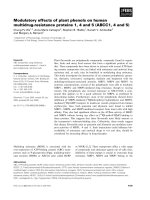
The relationship between DWH3, DWH, DVH and DSH in
3D-CRT treatment planning is shown in Figure 1a. In the
high-dose region D5 to D20 an almost identical dose distribution to the rectum was shown by all four approaches.
In the mid-dose region D30 to D50 the doses displayed in
the DWH were significantly lower compared to doses in
the DSH and the DWH3: mean difference of 8.7% ± 4.2%
(mean ± SD). In the low-dose region of D70 and D90 the
DVH showed significantly higher dose of 6.8% ± 2.2%.
After plan generation the dose distribution was calculated
for targets and OARs of each treatment plan. For the rectum the dose distribution to the manually delineated rectal wall (DWH), to the semi-automatic delineated rectal
wall with 3 mm wall thickness (DWH3) and to the solid
rectum including the lumen (DVH) were calculated. The
dose distribution to the outer surface of the rectal wall
(DSH) was calculated using the CERR software developed
at Washington University in St. Louis [13]. Dx (Gy)
denotes the minimal dose (Gy) delivered to x volume percent (x area percent for the DSH) of the evaluated volumeof-interest (VOI).
Correlation between corresponding dose parameters was
investigated by the nonparametric Spearman's rank test. A
highly significant linear correlation between pairs of
DWH3, DWH, DVH and DSH parameters was shown. Best
correlation was seen between DSH and DWH3 (R2 =
0.996), worst correlation between DVH and DWH (R2 =
0.939). The slope of linear fit lines ranged between 0.997
(DSH and DWH3) and 1.03 (DVH vs. DWH3).
Dose parameters were compared using student's t-test for
matched pairs. The Spearman's rank correlation was utilized to test the correlation between pairs of values. For
statistical analysis Statistica 6.0 (Statsoft, Tulsa, USA) was
Comparing 3D-CRT with IMRT treatment plans, more
pronounced differences between dose parameters were
seen for the latter (Fig 1b). In IMRT the differences were
Table 1: IMRT optimization objectives for OTP planning system
Organs-at-risk
Full Volume Dose (Gy)
Bladder
Right femoral head
Left femoral head
Rectum
Max. Dose (Gy)
Over Dose Volume (%)
Limit Dose (Gy)
23
27
27
23
50
41
41
50
23
9
9
21
75
50
50
73
Target volumes
Min. Dose (Gy)
PTV 1
PTV 2
Prescription Dose (Gy)
Under Dose (%)
Limit Dose (Gy)
68
61
73
66
5
5
81
81
The nomenclature of the OTP TPS was used for description of dose volume objectives: For organs-at-risk: the minimum dose should be lower
than "full volume dose"; "maximum dose" and "over dose volume" defines one DVH objective; "limit dose" is the maximum dose;
For targets: "Under dose (%)" is the volume (%) that is allowed receiving less than the prescription dose; "limit dose" is the maximum dose;
Page 3 of 11
(page number not for citation purposes)
Radiation Oncology 2006, 1:34
/>
investigated. Patients were equally divided into two subgroups according to the rectal volume.
100
DVH
90
DWH
80
DSH
DWH3
Volume (%)
70
60
50
40
30
20
10
0
0
10
20
30
40
50
60
70
80
Dose (Gy)
100
90
DVH
DWH
80
DSH
Volume (%)
70
DWH3
60
50
40
30
20
10
0
0
10
20
30
40
50
60
70
80
Dose (Gy)
Figure 1 and in Fig 1b) IMRT treatment planning all n
1a)patients) based on DWH3, DWH, DVH and DSH in Fig =
10 3D-CRT
Dose-volume histogram of the rectum (averaged over
Dose-volume histogram of the rectum (averaged over all n =
10 patients) based on DWH3, DWH, DVH and DSH in Fig
1a) 3D-CRT and in Fig 1b) IMRT treatment planning.
statistically significant between DVH and all other dose
parameters, between DWH and DSH but not between
DWH and DWH3 and between DSH and DWH3. In the
high-, mid- and low-dose region the DSH showed significantly higher doses to the rectum compared to the DVH.
Doses in the DSH were increased by 23.6% ± 8.7% compared to the DVH in the mid-dose region; differences were
smaller in the high-dose region (9.2% ± 6.6%) and in the
low-dose region (6.2% ± 3.9%). The DWH showed
decreased doses compared with the DWH3 in all dose
regions.
In Fig. 2 the corresponding results of DWH3, DWH, DVH
and DSH were plotted and linear fit lines were calculated.
In general correlation between dose parameters was worse
in IMRT plans compared to 3D-CRT plans. Best correlation was seen between DSH and DWH3 (R2 = 0.994) and
worst correlation between DSH and DVH (R2 = 0.930);
the slope of linear fit lines ranged between 1.01 (DWH vs.
DWH3) and 1.11 (DVH vs. DSH).
The influence of the rectal volume, the degree of rectal filling, on the relationship between the dose parameters was
In 3D-CRT treatment planning, no significant difference
was seen between DWH3, DVH and DSH for patients with
small rectal volumes (n = 5). For patients with a distended
rectum (n = 5) DSH and DWH showed identical results
but DVH showed significantly lower dose to the rectum in
the mid-dose region by 6.3% ± 7.2%. In the IMRT treatment plans the influence of the rectal volume on the relationship between dose parameters was different. The
order of the dose distribution to the rectum was not different between the sub-groups: DSH > DWH3 > DWH >
DVH. However, differences between dose parameters
were larger in the sub-group with a distended rectum. In
the mid-dose region the difference between DVH and
DWH was 7.5% ± 3.9% and 19.1% ± 4.9% in the subgroup with small rectal volumes and with a distended rectum, respectively. A statistical significant correlation (r =
0.81) between the rectal volume and the difference
between DVH and DWH was observed (Fig 3).
Furthermore, the influence of the anatomy of the seminal
vesicles on the relationship between the dose parameters
was tested. Two sub-groups were generated with five
patients each. The criterion was how far the seminal vesicles were wrapped around the rectum.
In the 3D-CRT plans a significant difference between
DSH/DWH3 vs. DVH was seen for patients with the seminal vesicles confined to the anterior rectal wall. With the
seminal vesicles wrapped around the rectum no difference
between DSH, DWH3 and DVH was found. Contrary, in
the IMRT treatment plans the anatomy of the seminal vesicles influenced the relationship between the dose parameters only marginally.
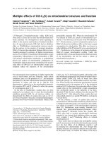
Dose distribution to the rectum was compared between
IMRT and 3D-CRT treatment. Depending on the way of
contouring the rectum the benefit of IMRT in sparing the
rectum was different (Fig. 4). Comparing IMRT and 3DCRT the IMRT technique resulted in 23% ± 15% decreased
doses to the rectal DVH in the mid dose region. Based on
DWH3 the benefit of the IMRT technique was 11% ± 11%
and based on DSH the benefit was reduced to 7% ± 10%.
Discussion
Reducing rectal toxicity represents a major challenge in
radiotherapy treatment planning for prostate cancer.
Treatment with escalated doses was shown to result in
improved rates of local control [14,15] but simultaneously higher doses to the rectum were found to be correlated with increased rates of late rectal toxicity. Reliable
tools in treatment planning for estimating the risk of toxicity are therefore essential. The dose-volume histogram is
Page 4 of 11
(page number not for citation purposes)
Radiation Oncology 2006, 1:34
/>
Figure 2
Correlation between dose parameters in IMRT treatment planning
Correlation between dose parameters in IMRT treatment planning. S (slope of linear fit line).
Page 5 of 11
(page number not for citation purposes)
Radiation Oncology 2006, 1:34
/>
100
4F
90
3F
80
IMRT
Volume (%)
70
60
50
40
30
20
10
0
0
10
20
30
40
50
60
70
80
Dose (Gy)
100
4F
90
3F
80
IMRT
Figure 3
tate cancer
rectal DVH of rectal volume and relative difference between
Correlation and DWH in IMRT treatment planning of prosCorrelation of rectal volume and relative difference between
rectal DVH and DWH in IMRT treatment planning of prostate cancer.
Volume (%)
70
60
50
40
30
20
10
0
0
10
20
30
40
50
60
70
80
Dose (Gy)
100
4F
90
3F
80
IMRT
70
Volume (%)
a common tool to express the dose that is delivered to targets and OARs. Though dose-volume histograms do not
provide spatial information, i.e. the location of the highand low-dose regions ("hot" and "cold" spots) inside the
volume of interest, multiple studies have shown correlation between dose-volume-histogram parameters and rectal toxicity. In table 2 a literature review about these
studies is given.
60
50
40
30
20
However, the transfer of the results from table 2 into clinical practice is complicated by the different way of contouring the rectum, different toxicity endpoints and
different classifications of rectal toxicity in the literature.
Within this retrospective planning study it was demonstrated that the method of contouring the rectum significantly influenced the "dose to the rectum" represented in
the dose-volume histogram. In general, delineation of the
rectal volume as a solid organ underestimated the exposure of the rectum compared to delineation of the rectal
surface or the rectal wall. The differences were larger in
IMRT treatment planning compared to 3D-CRT. For one
single patient the dose to the rectum in the mid-dose
region was 35% higher in the DSH compared to the DVH.
The rectum was delineated from 1 cm superior to 1 cm
inferior the PTV. The delineated OAR rectum constituted
a fairly constant fraction of the anatomical anus/rectum
averaged over all patients (73% ± 4%). Portions of the rectum outside the beam, receiving very low doses, were
therefore excluded from analysis. Differences between
10
0
0
10
20
30
40
50
60
70
80
Dose (Gy)
DWH3 4
plans with 4b) and dose based (4F)
Comparison of 3-field (3F), 4-field4c) DVH (Fig 4a), the
Figure (Figthe rectalthe DSH (Figon theand IMRT treatment
Comparison of 3-field (3F), 4-field (4F) and IMRT treatment
plans with the rectal dose based on the DVH (Fig 4a), the
DWH3 (Fig 4b) and the DSH (Fig 4c).
dose parameters would have been smaller if the complete
anatomical anus and rectum would have been contoured.
It was also demonstrated that there was no constant relationship between dose parameters DWH3, DWH, DVH
and DSH for all patients. Both the rectal volume, the
degree of the rectal filling, and the anatomy of the seminal
vesicles were shown to be relevant. The pattern how these
anatomic characteristics influenced the relationship
between DWH3, DWH, DVH and DSH was different in
Page 6 of 11
(page number not for citation purposes)
Patients Follow up
Persription
Doses
Treatment
technique
Classification
of toxicity
Endpoint
Events
Dosimetric
parameter
Rectum delineation
Results
41
Minimum 4
years
50.4Gy
25.2CGE
4 field Perineal
proton boost
RTOG
≥ Grad I
rectal
bleeding
14
DWH ant.
RW
From superior limit of
anus to 2 cm superior to
prostate
Cut-off:
Continuously between 60Gy to 70%
and 75Gy to 30%
Boersma 1998 [30]
130
Median 24
months
70 – 76Gy
3 field 3D-CRT
SOMA/LENT
and RTOG/
EORTC
≥ Grad III
rectal
bleeding
2
DWH
15 mm caudal to the
apex of the prostate to
boarder to sigmoid
Cut-off:
≥ 65Gy to >40%
≥ 70Gy to >30%
≥ 75Gy to >5%
(no correlation for grade I/II rectal
bleeding)
Storey 2000 [31]
189
Minimum 2
years
70Gy 78Gy
4 field box 4
field box, 6 field
3D-CRT boost
Modified
RTOG
≥ Grad II
late rectal
toxicity
28
DVH
Rectum included within
11 cm of initial APPA
field
For patients treated to 78Gy:
Cut-off:
≥ 70Gy to >25%
Jackson 2001 [32]
451
Minimum
30 months
70.2Gy
75.6Gy
6 field
arrangement
3D-CRT
RTOG
≥ Grad III
late rectal
bleeding
49
DWH
below sigmoid flexure to
above anal verge
Correlation with:
# area under the average percent
volume DWH
# Exposure to ~62% and to ~102% of
prescription dose
Fenwick 2001 [33]
79
Minimum 2
years
60 – 64Gy
3 field
• 3D-CRT
• Conventional
RTOG
Grade I –
III rectal
bleeding
?
DSH
up to level of
rectosigmoid junction
Correlation with:
% of RS exposed to > 57Gy
Wachter 2001 [34]
Radiation Oncology 2006, 1:34
Hartford 1996 [29]
109
Median 30
months
66Gy
4 field 3D-CRT
EORTC/
RTOG
Grade II
rectal
bleeding
15
DVH
From lower to upper
boarder of 4 field
Cut-off:
≥ 60Gy to >57%
Kupelian 2002 [35]
128
Median 24
months
78Gy 70Gy
4 field (42Gy) 6
field boost
(36Gy): 3D-CRT
IMRT (SD
2.5Gy)
RTOG
Grade I –
III rectal
bleeding
9
DVH
From 1 cm above to 1
cm below the target
Cut-off:
Absolute rectal volume:
≥ 78Gy to >15 cm3
Huang 2002 [36]
163
Median 62
months
74 – 78Gy
4 field
conventional
(46Gy) 6 field
boost 3D-CRT
Modified
RTOG
≥ Grad II
late rectal
toxicity
38
DVH
11 cm in length starting
at 2 cm below the
inferiormost aspect of
the ischial tuberosities
Cut-off:
V60 below 40%
V70 below 25%
V75.6 below 15%
V78 below 5%
Page 7 of 11
Author
(page number not for citation purposes)
/>
Table 2: Literature review of dose-volume relationship for late rectal bleeding in radiotherapy of prostate cancer
Median 2
years
70 – 78Gy
3 to 4 field 3DCRT
Modified
RTOG
Grade II –
III rectal
bleeding
23
DVH
Above anal verge to
sigmoid
Cut-off:
V50 below 60–65%
V60 below 50–55%
V70 below 25–30%
Greco 2003 [38]
135
Median 28
months
76Gy
6 field 3D-CRT
RTOG
≥ Grad II
late rectal
toxicity
24
DVH
from just below the
sigmoid flexure to just
above the anal verge
Cut-off:
V40 below 60%
V50 below 50%
V60 below 25%
V72 below 15%
V76 below 5%
Akimoto 2004 [39]
52
Median 31
months
69Gy SD
3Gy
unblocked 4 field
technique to the
prostate
RTOG
≥ Grad II
late rectal
toxicity
13
DVH
above anal verge to
point at which it turns
into the sigmoid colon
Cut-off (equivalent 83Gy prescription
dose):
V30 (V24.9) to ≥ 60%
V50 (V41.5) to ≥ 40%
V80 (V66.4) to ≥ 40%
V90 (V74.7) to ≥ 15%
266
Minimum 2
years
66Gy
Conventional (n
= 125) 3 field
3D-CRT (n =
123)
RTOG
≥ Grad I
late rectal
toxicity
57%
47%
DVH
(separately
for proximal,
middle and
distal part of
rectum)
length of intestinal
structures was limited to
cranial and caudal field
borders
Correlation with:
Distal rectal volume exposed to ≥
90% tumor dose
Lee 2005 [41]
212
Median 86
months 35
months
66 70 – 74Gy
Conventional
3D-CRT
Modified
RTOG/Lent
and RTOG
≥ Grad II
late rectal
toxicity
34
DVH
?
Cut-offs:
≥ 60Gy to >51.5%
≥ 70Gy to >41.5%
Vargas 2005 [11]
331
Median 19
months
70.2Gy to
79.2Gy
Adaptive 3DCRT
CTC 2.0
≥ Grad II
late rectal
toxicity
43
DVH, DWH
from the anal verge or
ischial tuberosities
(whichever was higher)
to the sacroiliac joints or
rectosigmoid junction
(whichever was lower)
Association with:
DWH: V50, V60, V66.6, V70, V72
DVH V60–V72
Peeters 2006 [42]
614
Median: 44
months
68Gy vs
78Gy
3D-CRT
Adapted
RTOG/
EORTC
≥ Grad II
rectal
bleeding
31
DWH
anorectal, rectal, and
anal wall dose volume
histogram
Correlation with:
anorectal V55–V65
Page 8 of 11
245
Koper 2004 [40]
Radiation Oncology 2006, 1:34
Fiorino 2003 [37]
(page number not for citation purposes)
/>
Table 2: Literature review of dose-volume relationship for late rectal bleeding in radiotherapy of prostate cancer (Continued)
Radiation Oncology 2006, 1:34
IMRT and 3D-CRT treatment planning. Because of significant differences between dose parameters and because
dose volume histograms do not provide spatial information the importance of reviewing the dose distribution in
every single CT slice and not only relying on dose parameters has to be stressed.
Others studies compared rectal DVH, DWH and DSH in
treatment planning of the prostate [16-20]. Using a cylindrical model for the rectum Fiorino et al. described substantial differences between DVH and DWH for a "full"
rectum but only small differences for an "empty" rectum.
For patients with a distended rectum the DSH was close to
the DWH. Boehmer et al. [20] showed that the length of
delineating the rectum in superior-inferior direction significantly influenced the dose to the rectum and therefore
should be standardized. However, all these studies are
based on 3D-CRT. In this work it has been clearly demonstrated that a one-to-one transfer of the results from 3DCRT to IMRT treatment planning is not possible.
Another interesting result of this study was the finding
that the dose to the manually delineated rectal wall
(DWH) was different from the dose to the semi-automatically generated rectal wall with 3 mm wall thickness
(DWH3). The choice of the 3-mm wall thickness is supported by the study of Rasmussen, in which the rectal wall
thickness measured by ultrasound was found to have a
median of 2.6 mm [21]. Tucker et al. reported only small
differences of the DWH for rectal wall thicknesses ranging
between 2 mm and 5 mm [19]. As the patients in this
study were treated in a supine position the intra-rectal
feces moved to the posterior rectal wall due to gravity.
With CT density values of the rectal wall often very similar
to the density of the filling a precise delineation of the
inner contour of the rectal wall was difficult for some
patients resulting in asymmetric rectal wall thicknesses
between anterior (within high-dose region) and posterior
(within mid- to low-dose region) rectal wall. It is likely
that this explains the differences between DWH3 and
DWH and because of this difficulty and uncertainty we do
not advocate delineating the inner contour of the rectum
manually. Though automatic generation of the DWH3
reduced uncertainties compared to DWH, the thickness of
the rectal wall is dependent on the rectal distension and
consequently not constant. Meijer et al. described a more
sophisticated method of automatic DWH generation [18]:
based on the delineated outer rectal contour the inner
contour was generated automatically taking the rectal distension into account.
Delineation of the outer contour of the rectum was found
to be associated with small intra- and inter-observer variability [22,23]. Consequently, in analysis of DVH and
DSH uncertainties are expected to be lower compared to
/>
DWH analysis. Furthermore, generation of the DSH and
the DVH are known to be sensitive to parameters such as
voxel dimensions and dose calculation grid size [16].
These facts could partially be responsible for differences
between dose parameters.
Recently, de Crevoisier et al. showed an increased risk of
local failure and simultaneously a lower incidence of late
rectal bleeding for patients with a distended rectum on the
planning CT study [9]. Treatment planning based on a
planning CT with distended rectum introduced a systematic error with the prostate and the anterior rectal wall
moving posterior out of the high-dose-region during the
treatment. Repetition of the planning CT study in case of
a distended rectum was suggested to avoid this error.
Additionally, good agreement between DVH and DWH
was shown in case of an empty rectum making transfer of
constraints form the literature to treatment planning
more reliable.
The fact that one single planning CT study is only a snapshot of the patients' anatomy has to be considered for the
interpretation of dose-volume histograms. Image-guided
treatment techniques are thought to correct differences
between treatment planning and the current anatomy at
the time of treatment [24-27]. Recently, technologies
introduced 3D volume imaging into the treatment room
with sufficient soft-tissue contrast for visualization of the
prostate and OARs [28]. Such image-guided treatment
protocol are expected to allow a substantial reduction of
safety margins and consequence in a further escalation of
the treatment dose [25].
Comparison of 3D-CRT and IMRT in terms of sparing the
rectum was not aim of this study. A simultaneous integrated boost concept was applied for the IMRT plans
whereas a homogenous dose distribution without field
size reduction was planned for the 3D-CRT plans. It was
interesting to note that the "benefit" of IMRT in comparison to 3D-CRT was strongly dependent on the way of contouring the rectum. Doses to the rectum were reduced in
the IMRT plan by 23%, 11% and 7% with the calculation
based on the rectal DVH, DWH3 and the DSH.
Conclusion
This study demonstrated that the method of delineating
the rectum significantly influenced the dose representation in external beam radiotherapy of localized prostate
cancer. Differences between the dose parameters, based
on delineation of the rectal wall, rectal volume and rectal
surface, were larger in IMRT treatment planning compared
with 3D-CRT. It was shown that the patient's anatomy,
both the rectal filling and the anatomy of the seminal vesicles, influenced the relationship between the four evaluated parameters. For integration of dose-volume
Page 9 of 11
(page number not for citation purposes)
Radiation Oncology 2006, 1:34
parameters from the literature into treatment planning
these results have to be considered: a one-to-one transfer
of the results from 3D-CRT to IMRT treatment planning
may be associated with substantial errors.
/>
9.
10.
Competing interests
The author(s) declare that they have no competing interests.
11.
Authors' contributions
All authors read and approved the final manuscript.
12.
MG designed the analysis, generated the treatment plans,
performed the analysis and drafted the manuscript.
13.
JM was involved in the statistical analysis and revised the
manuscript.
KB participated in the study design and revised the manuscript.
14.
15.
DV participated in the study design and revised the manuscript.
16.
MF participated in the study design and revised the manuscript.
17.
18.
References
1.
2.
3.
4.
5.
6.
7.
8.
Kupelian P, Kuban D, Thames H, Levy L, Horwitz E, Martinez A,
Michalski J, Pisansky T, Sandler H, Shipley W, Zelefsky M, Zietman A:
Improved biochemical relapse-free survival with increased
external radiation doses in patients with localized prostate
cancer: The combined experience of nine institutions in
patients treated in 1994 and 1995. Int J Radiat Oncol Biol Phys
2005, 61(2):415-419.
Schultheiss TE, Lee WR, Hunt MA, Hanlon AL, Peter RS, Hanks GE:
Late GI and GU complications in the treatment of prostate
cancer. Int J Radiat Oncol Biol Phys 1997, 37(1):3-11.
Leibel SA, Hanks GE, Kramer S: Patterns of care outcome studies: results of the national practice in adenocarcinoma of the
prostate. Int J Radiat Oncol Biol Phys 1984, 10(3):401-409.
Dearnaley DP, Khoo VS, Norman AR, Meyer L, Nahum A, Tait D,
Yarnold J, Horwich A: Comparison of radiation side-effects of
conformal and conventional radiotherapy in prostate cancer: a randomised trial. Lancet 1999, 353(9149):267-272.
Zelefsky MJ, Fuks Z, Hunt M, Yamada Y, Marion C, Ling CC, Amols
H, Venkatraman ES, Leibel SA: High-dose intensity modulated
radiation therapy for prostate cancer: early toxicity and biochemical outcome in 772 patients. Int J Radiat Oncol Biol Phys
2002, 53(5):1111-1116.
Oh CE, Antes K, Darby M, Song S, Starkschall G: Comparison of 2D
conventional, 3D conformal, and intensity-modulated treatment planning techniques for patients with prostate cancer
with regard to target-dose homogeneity and dose to critical,
uninvolved structures. Med Dosim 1999, 24(4):255-263.
Zelefsky MJ, Fuks Z, Happersett L, Lee HJ, Ling CC, Burman CM,
Hunt M, Wolfe T, Venkatraman ES, Jackson A, Skwarchuk M, Leibel
SA: Clinical experience with intensity modulated radiation
therapy (IMRT) in prostate cancer. Radiother Oncol 2000,
55(3):241-249.
Lee WR, Hanks GE, Hanlon AL, Schultheiss TE, Hunt MA: Lateral
rectal shielding reduces late rectal morbidity following high
dose three-dimensional conformal radiation therapy for clinically localized prostate cancer: further evidence for a significant dose effect. Int J Radiat Oncol Biol Phys 1996, 35(2):251-257.
19.
20.
21.
22.
23.
24.
25.
26.
de Crevoisier R, Tucker SL, Dong L, Mohan R, Cheung R, Cox JD,
Kuban DA: Increased risk of biochemical and local failure in
patients with distended rectum on the planning CT for prostate cancer radiotherapy. Int J Radiat Oncol Biol Phys 2005,
62(4):965-973.
Bos LJ, Damen EM, de Boer RW, Mijnheer BJ, McShan DL, Fraass BA,
Kessler ML, Lebesque JV: Reduction of rectal dose by integration of the boost in the large-field treatment plan for prostate irradiation. Int J Radiat Oncol Biol Phys 2002, 52(1):254-265.
Vargas C, Yan D, Kestin LL, Krauss D, Lockman DM, Brabbins DS,
Martinez AA: Phase II dose escalation study of image-guided
adaptive radiotherapy for prostate cancer: use of dose-volume constraints to achieve rectal isotoxicity. Int J Radiat Oncol
Biol Phys 2005, 63(1):141-149.
Guckenberger M, Pohl F, Baier K, Meyer J, Vordermark D, Flentje M:
Adverse effect of a distended rectum in intensity-modulated
radiotherapy (IMRT) treatment planning of prostate cancer.
Radiother Oncol 2006.
Deasy JO, Blanco AI, Clark VH: CERR: a computational environment for radiotherapy research. Med Phys 2003, 30(5):979-985.
Pollack A, Zagars GK, Starkschall G, Antolak JA, Lee JJ, Huang E, von
Eschenbach AC, Kuban DA, Rosen I: Prostate cancer radiation
dose response: results of the M. D. Anderson phase III randomized trial. Int J Radiat Oncol Biol Phys 2002, 53(5):1097-1105.
Zietman AL, DeSilvio ML, Slater JD, Rossi CJJ, Miller DW, Adams JA,
Shipley WU: Comparison of conventional-dose vs high-dose
conformal radiation therapy in clinically localized adenocarcinoma of the prostate: a randomized controlled trial. Jama
2005, 294(10):1233-1239.
Fiorino C, Gianolini S, Nahum AE: A cylindrical model of the rectum: comparing dose-volume, dose-surface and dose-wall
histograms in the radiotherapy of prostate cancer. Phys Med
Biol 2003, 48(16):2603-2616.
Li S, Boyer A, Lu Y, Chen GT: Analysis of the dose-surface histogram and dose-wall histogram for the rectum and bladder.
Med Phys 1997, 24(7):1107-1116.
Meijer GJ, van den Brink M, Hoogeman MS, Meinders J, Lebesque JV:
Dose-wall histograms and normalized dose-surface histograms for the rectum: a new method to analyze the dose distribution over the rectum in conformal radiotherapy. Int J
Radiat Oncol Biol Phys 1999, 45(4):1073-1080.
Tucker SL, Dong L, Cheung R, Johnson J, Mohan R, Huang EH, Liu HH,
Thames HD, Kuban D: Comparison of rectal dose-wall histogram versus dose-volume histogram for modeling the incidence of late rectal bleeding after radiotherapy. Int J Radiat
Oncol Biol Phys 2004, 60(5):1589-1601.
Boehmer D, Kuczer D, Badakhshi H, Stiefel S, Kuschke W, Wernecke
KD, Budach V: Influence of organ at risk definition on rectal
dose-volume histograms in patients with prostate cancer
undergoing external-beam radiotherapy. Strahlenther Onkol
2006, 182(5):277-282.
Rasmussen SN, Riis P: Rectal wall thickness measured by ultrasound in chronic inflammatory diseases of the colon. Scand J
Gastroenterol 1985, 20(1):109-114.
Fiorino C, Vavassori V, Sanguineti G, Bianchi C, Cattaneo GM, Piazzolla A, Cozzarini C: Rectum contouring variability in patients
treated for prostate cancer: impact on rectum dose-volume
histograms and normal tissue complication probability. Radiother Oncol 2002, 63(3):249-255.
Foppiano F, Fiorino C, Frezza G, Greco C, Valdagni R: The impact
of contouring uncertainty on rectal 3D dose-volume data:
results of a dummy run in a multicenter trial (AIROPROS0102). Int J Radiat Oncol Biol Phys 2003, 57(2):573-579.
Litzenberg DW, Balter JM, Hadley SW, Sandler HM, Willoughby TR,
Kupelian PA, Levine L: Influence of intrafraction motion on
margins for prostate radiotherapy. Int J Radiat Oncol Biol Phys
2006, 65(2):548-553.
Wu Q, Ivaldi G, Liang J, Lockman D, Yan D, Martinez A: Geometric
and dosimetric evaluations of an online image-guidance
strategy for 3D-CRT of prostate cancer. Int J Radiat Oncol Biol
Phys 2006, 64(5):1596-1609.
Bos LJ, van der Geer J, van Herk M, Mijnheer BJ, Lebesque JV, Damen
EM: The sensitivity of dose distributions for organ motion
and set-up uncertainties in prostate IMRT. Radiother Oncol
2005, 76(1):18-26.
Page 10 of 11
(page number not for citation purposes)
Radiation Oncology 2006, 1:34
27.
28.
29.
30.
31.
32.
33.
34.
35.
36.
37.
38.
39.
40.
41.
42.
Guckenberger M, Meyer J, Vordermark D, Baier K, Wilbert J, Flentje
M: Magnitude and clinical relevance of translational and rotational patient setup errors: A cone-beam CT study. Int J
Radiat Oncol Biol Phys 2006, 65(3):934-942.
Smitsmans MH, de Bois J, Sonke JJ, Betgen A, Zijp LJ, Jaffray DA, Lebesque JV, van Herk M: Automatic prostate localization on conebeam CT scans for high precision image-guided radiotherapy. Int J Radiat Oncol Biol Phys 2005, 63(4):975-984.
Hartford AC, Niemierko A, Adams JA, Urie MM, Shipley WU: Conformal irradiation of the prostate: estimating long-term rectal bleeding risk using dose-volume histograms. Int J Radiat
Oncol Biol Phys 1996, 36(3):721-730.
Boersma LJ, van den Brink M, Bruce AM, Shouman T, Gras L, te Velde
A, Lebesque JV: Estimation of the incidence of late bladder and
rectum complications after high-dose (70-78 GY) conformal
radiotherapy for prostate cancer, using dose-volume histograms. Int J Radiat Oncol Biol Phys 1998, 41(1):83-92.
Storey MR, Pollack A, Zagars G, Smith L, Antolak J, Rosen I: Complications from radiotherapy dose escalation in prostate cancer: preliminary results of a randomized trial. Int J Radiat Oncol
Biol Phys 2000, 48(3):635-642.
Jackson A, Skwarchuk MW, Zelefsky MJ, Cowen DM, Venkatraman
ES, Levegrun S, Burman CM, Kutcher GJ, Fuks Z, Liebel SA, Ling CC:
Late rectal bleeding after conformal radiotherapy of prostate cancer. II. Volume effects and dose-volume histograms.
Int J Radiat Oncol Biol Phys 2001, 49(3):685-698.
Fenwick JD, Khoo VS, Nahum AE, Sanchez-Nieto B, Dearnaley DP:
Correlations between dose-surface histograms and the incidence of long-term rectal bleeding following conformal or
conventional radiotherapy treatment of prostate cancer. Int
J Radiat Oncol Biol Phys 2001, 49(2):473-480.
Wachter S, Gerstner N, Goldner G, Potzi R, Wambersie A, Potter R:
Rectal sequelae after conformal radiotherapy of prostate
cancer: dose-volume histograms as predictive factors. Radiother Oncol 2001, 59(1):65-70.
Kupelian PA, Reddy CA, Carlson TP, Willoughby TR: Dose/volume
relationship of late rectal bleeding after external beam radiotherapy for localized prostate cancer: absolute or relative
rectal volume? Cancer J 2002, 8(1):62-66.
Huang EH, Pollack A, Levy L, Starkschall G, Dong L, Rosen I, Kuban
DA: Late rectal toxicity: dose-volume effects of conformal
radiotherapy for prostate cancer. Int J Radiat Oncol Biol Phys
2002, 54(5):1314-1321.
Fiorino C, Sanguineti G, Cozzarini C, Fellin G, Foppiano F, Menegotti
L, Piazzolla A, Vavassori V, Valdagni R: Rectal dose-volume constraints in high-dose radiotherapy of localized prostate cancer. Int J Radiat Oncol Biol Phys 2003, 57(4):953-962.
Greco C, Mazzetta C, Cattani F, Tosi G, Castiglioni S, Fodor A, Orecchia R: Finding dose-volume constraints to reduce late rectal
toxicity following 3D-conformal radiotherapy (3D-CRT) of
prostate cancer. Radiother Oncol 2003, 69(2):215-222.
Akimoto T, Muramatsu H, Takahashi M, Saito J, Kitamoto Y, Harashima K, Miyazawa Y, Yamada M, Ito K, Kurokawa K, Yamanaka H,
Nakano T, Mitsuhashi N, Niibe H: Rectal bleeding after hypofractionated radiotherapy for prostate cancer: correlation
between clinical and dosimetric parameters and the incidence of grade 2 or worse rectal bleeding. Int J Radiat Oncol Biol
Phys 2004, 60(4):1033-1039.
Koper PC, Heemsbergen WD, Hoogeman MS, Jansen PP, Hart GA,
Wijnmaalen AJ, van Os M, Boersma LJ, Lebesque JV, Levendag P:
Impact of volume and location of irradiated rectum wall on
rectal blood loss after radiotherapy of prostate cancer. Int J
Radiat Oncol Biol Phys 2004, 58(4):1072-1082.
Lee CM, Lee RJ, Handrahan DL, Sause WT: Comparison of late
rectal toxicity from conventional versus three-dimensional
conformal radiotherapy for prostate cancer: analysis of clinical and dosimetric factors. Urology 2005, 65(1):114-119.
Peeters ST, Lebesque JV, Heemsbergen WD, van Putten WL, Slot A,
Dielwart MF, Koper PC: Localized volume effects for late rectal
and anal toxicity after radiotherapy for prostate cancer. Int J
Radiat Oncol Biol Phys 2006, 64(4):1151-1161.
/>
Publish with Bio Med Central and every
scientist can read your work free of charge
"BioMed Central will be the most significant development for
disseminating the results of biomedical researc h in our lifetime."
Sir Paul Nurse, Cancer Research UK
Your research papers will be:
available free of charge to the entire biomedical community
peer reviewed and published immediately upon acceptance
cited in PubMed and archived on PubMed Central
yours — you keep the copyright
BioMedcentral
Submit your manuscript here:
/>
Page 11 of 11
(page number not for citation purposes)