Báo cáo y học: "NF-κB inhibitor dehydroxymethylepoxyquinomicin suppresses osteoclastogenesis and expression of NFATc1 in mouse arthritis without affecting expression of RANKL, osteoprotegerin or macrophage colony-stimulating factor" docx
Bạn đang xem bản rút gọn của tài liệu. Xem và tải ngay bản đầy đủ của tài liệu tại đây (493.78 KB, 10 trang )
Open Access
Available online />Page 1 of 10
(page number not for citation purposes)
Vol 9 No 5
Research article
NF-κB inhibitor dehydroxymethylepoxyquinomicin suppresses
osteoclastogenesis and expression of NFATc1 in mouse arthritis
without affecting expression of RANKL, osteoprotegerin or
macrophage colony-stimulating factor
Tetsuo Kubota
1
, Machiko Hoshino
1
, Kazuhiro Aoki
2
, Keiichi Ohya
2
, Yukiko Komano
3
,
Toshihiro Nanki
3
, Nobuyuki Miyasaka
3
and Kazuo Umezawa
4
1
Department of Microbiology and Immunology, Tokyo Medical and Dental University Graduate School of Health Sciences, Tokyo, Japan
2
Department of Hard Tissue Engineering, Tokyo Medical and Dental University Graduate School, Tokyo, Japan
3
Department of Medicine and Rheumatology, Tokyo Medical and Dental University Graduate School, Tokyo, Japan
4
Department of Applied Chemistry, Keio University, Kanagawa, Japan
Corresponding author: Tetsuo Kubota,
Received: 13 Jun 2007 Revisions requested: 9 Aug 2007 Revisions received: 25 Aug 2007 Accepted: 25 Sep 2007 Published: 25 Sep 2007
Arthritis Research & Therapy 2007, 9:R97 (doi:10.1186/ar2298)
This article is online at: />© 2007 Kubota et al., licensee BioMed Central Ltd.
This is an open access article distributed under the terms of the Creative Commons Attribution License ( />),
which permits unrestricted use, distribution, and reproduction in any medium, provided the original work is properly cited.
Abstract
Inhibition of NF-κB is known to be effective in reducing both
inflammation and bone destruction in animal models of arthritis.
Our previous study demonstrated that a small cell-permeable
NF-κB inhibitor, dehydroxymethylepoxyquinomicin (DHMEQ),
suppresses expression of proinflammatory cytokines and
ameliorates mouse arthritis. It remained unclear, however,
whether DHMEQ directly affects osteoclast precursor cells to
suppress their differentiation to mature osteoclasts in vivo. The
effect of DHMEQ on human osteoclastogenesis also remained
elusive. In the present study, we therefore examined the effect of
DHMEQ on osteoclastogenesis using a mouse collagen-
induced arthritis model, and using culture systems of fibroblast-
like synovial cells obtained from patients with rheumatoid
arthritis, and of osteoclast precursor cells from peripheral blood
of healthy volunteers. DHMEQ significantly suppressed
formation of osteoclasts in arthritic joints, and also suppressed
expression of NFATc1 along the inner surfaces of bone lacunae
and the eroded bone surface, while serum levels of soluble
receptor activator of NF-κB ligand (RANKL), osteoprotegerin
and macrophage colony-stimulating factor were not affected by
the treatment. DHMEQ also did not suppress spontaneous
expression of RANKL nor of macrophage colony-stimulating
factor in culture of fibroblast-like synovial cells obtained from
patients with rheumatoid arthritis. These results suggest that
DHMEQ suppresses osteoclastogenesis in vivo, through
downregulation of NFATc1 expression, without significantly
affecting expression of upstream molecules of the RANKL/
receptor activator of NF-κB/osteoprotegerin cascade, at least in
our experimental condition. Furthermore, in the presence of
RANKL and macrophage colony-stimulating factor,
differentiation and activation of human osteoclasts were also
suppressed by DHMEQ, suggesting the possibility of future
application of NF-κB inhibitors to rheumatoid arthritis therapy.
Introduction
Prevention of bone destruction in affected joints is one of the
most important goals in the treatment of rheumatoid arthritis
(RA), and many clinical trials of newly developed biologic
agents include assessment of radiographic changes before
and after treatment. For example, a significant effect of anti-
TNF therapy in halting the progression of joint structural dam-
age in active RA has been reported [1-3]. There are still some
patients with persistently active disease, however, despite the
use of currently available agents; further development of small,
DHMEQ = dehydroxymethylepoxyquinomicin; DMEM = Dulbecco's modified Eagle's medium; ELISA = enzyme-linked immunosorbent assay; FCS =
fetal calf serum; FLS = fibroblast-like synovial cells; IL = interleukin; M-CSF = macrophage colony stimulating factor; MMP = matrix metalloprotease;
NFAT = nuclear factor of activated T cells; NF = nuclear factor; OPG = osteoprotegerin; PBS = phosphate-buffered saline; RA = rheumatoid arthritis;
RANK = receptor activator of NF-κB; RANKL = receptor activator of NF-κB ligand; sRANKL = soluble receptor activator of NF-κB ligand; TNF =
tumor necrosis factor; TRAP = tartrate-resistant acid phosphatase
Arthritis Research & Therapy Vol 9 No 5 Kubota et al.
Page 2 of 10
(page number not for citation purposes)
cell-permeable agents that specifically interrupt the critical
intracellular pathways involved in bone destruction could
prove beneficial.
Recent studies have revealed the prominent contribution of
osteoclasts to bone resorption that may be dissociated from
inflammation in RA pathophysiology. For example, human TNF
transgenic mice were protected from bone destruction
despite severe arthritis when they were crossed with c-fos-
deficient mice lacking osteoclasts [4]. In early RA patients
treated with methotrexate and infliximab, radiographic pro-
gression was slowed even in cases with elevated time-aver-
aged levels of C-reactive protein or erythrocyte sedimentation
rate or elevated time-averaged swollen joint counts [3]. Oste-
oclasts are multinucleated cells formed by fusion of mononu-
clear progenitors of the monocyte/macrophage lineage. The
osteoclasts develop a specialized cytoskeleton that permits
them to establish an isolated microenvironment between
themselves and the underlying bone, within which matrix deg-
radation occurs by a process involving proton transport to
acidify the extracellular microenvironment [5]. Acidification of
this compartment leads to the activation of tartrate-resistant
acid phosphatase (TRAP) and cathepsin K, which are the
enzymes responsible for degradation of bone mineral and col-
lagen matrices [6].
NF-κB is a transcription factor implicated in diverse receptor-
mediated signaling pathways including differentiation and acti-
vation of osteoclasts [7,8]. Several lines of in vitro and in vivo
studies have demonstrated that inhibition of NF-κB results in
suppression of osteoclastogenesis [9-12]. As regards mecha-
nisms underlying the involvement of NF-κB in osteoclastogen-
esis, Takatsuna and colleagues [12] demonstrated that
expression of NFATc1, a key transcriptional factor of osteo-
clastogenesis induced by macrophage colony-stimulating fac-
tor (M-CSF) and receptor activator of NF-κB ligand (RANKL)
in a culture of murine precursor cells [13], was inhibited by the
NF-κB inhibitor dehydroxymethylepoxyquinomicin (DHMEQ).
DHMEQ is a unique NF-κB inhibitor designed in our laboratory
based on the structure of the antibiotic epoxyquinomicin C,
which acts at the level of nuclear translocation of NF-κB [14].
An in vivo anti-inflammatory effect of DHMEQ has already
been demonstrated in various models, including collagen-
induced mouse arthritis [15-17]. Since inflammation and bone
resorption could be considerably dissociated as mentioned
above, and many factors besides RANKL and M-CSF are
thought to affect osteoclastogenesis [18], the effect of
DHMEQ on in vivo osteoclastogenesis needed further investi-
gation. In the present study, therefore, we looked into the
effect of DHMEQ focusing on in vivo osteoclastogenesis in
collagen-induced arthritis. In addition, we tested the effect of
this compound on human osteoclast differentiation in vitro, to
explore the possibility of future development of novel RA
therapy.
Materials and methods
Inhibitor of NF-κB
The (-)-enantiomer of DHMEQ, which is simply represented as
DHMEQ in this manuscript, is a more potent inhibitor of NF-κB
than its (+)-enantiomer, and was synthesized as described
previously [19].
Induction of collagen-induced arthritis
Animal experiments were approved by the Institutional Animal
Care and Use Committee of Tokyo Medical and Dental Univer-
sity. Male 8-week-old DBA/1J mice were purchased from Ori-
ental Yeast (Tokyo, Japan). Bovine collagen type II (Collagen
Research Center, Tokyo, Japan) was dissolved in 50 mM ace-
tic acid at 4 mg/ml and was emulsified in an equal volume of
Freund's complete adjuvant (Difco Laboratories, Detroit, MI,
USA). Mice were immunized with 100 μl emulsion intrader-
mally at the base of the tail. After 21 days (day 0), the same
amount of the antigen emulsified in the same adjuvant was
intradermally injected at the base of the tail as a booster immu-
nization. From day 0 to day 10, 100 μg DHMEQ (5 mg/kg
body weight) dissolved in 50 μl dimethyl sulfoxide was
injected subcutaneously every day to mice in the experimental
group. Mice in the control group received 50 μl dimethyl sul-
foxide similarly injected.
The thickness of each hind paw was measured on day -4 and
on day 10 using a pair of digital slide calipers. Radiographs of
both ankle joints were obtained on day 10 and were scored on
a scale of 1–3 (1 = no change, 2 = mild osteoporosis without
bone erosion, 3 = severe osteoporosis with or without bone
erosion) by three investigators who were blinded to the
assignment of mouse groups.
Histochemical staining of osteoclasts
Mice were sacrificed on day 10, 3 hours after the last injection,
and their hind paws were excised for experiments. After the
skin was scarified with a surgical blade, the left hind paws
were preserved in 10% buffered formalin for 3 hours, and the
skin was totally removed. The paws were then decalcified in
10% ethylenediamine tetraacetic acid, 5% polyvinylpyro-
lidone, 100 mM Tris (pH 7.4) for 4 weeks, were dehydrated in
graded ethanol, were permeated serially by methyl benzoate
and benzene, and were embedded into paraffin in a vacuum
oven. Longitudinally sectioned paraffin blocks were fixed in cit-
rate-acetone (2:3 mixture of 380 mM citrate and acetone) for
30 seconds, and were stained with 0.5 mg/ml naphthol AS-BI
phosphoric acid and 0.3 mg/ml fast red violet LB salt (Sigma-
Aldrich, St Louis, MO, USA) in 27 mM sodium tartrate and 100
mM sodium acetate (pH 5.2) for 1 hour at 37°C. Nuclei were
stained with hematoxylin. The mean number of TRAP-positive
giant cells with four or more nuclei in the individual ankle joints
of arthritic mice was counted under a microscope by two
investigators in a manner blinded to the assignment of mouse
groups.
Available online />Page 3 of 10
(page number not for citation purposes)
Immunohistological staining of NFATc1 and NF-κB
The right hind paws were frozen in liquid nitrogen, and bone-
containing sections were prepared using Cryofilm (Finetec,
Tokyo, Japan) [20]. Phycoerythrin-labeled anti-NFATc1 mono-
clonal antibody 7A6 was purchased from Santa Cruz Biotech-
nology (Santa Cruz, CA, USA). Monoclonal antibody to the
nuclear localization signal in the p65 subunit of NF-κB was
purchased from Chemicon (Temecula, CA, USA), was labeled
with FITC (Wako, Tokyo, Japan), and was dialyzed against 100
mM Tris, 200 mM NaCl (pH 7.4). After fixing with acetone for
10 min, permiabilization with PBS containing 0.5% Triton X-
100 for 10 minutes, and blocking with PBS containing 10%
FCS for 1 hour, the sections were incubated with a mixture of
the two antibodies for 1 hour, and observed under a confocal
microscope (Olympus, Tokyo, Japan).
Measurement of soluble RANKL, osteoprotegerin and
M-CSF
Serum samples were obtained at euthanasia and the concen-
trations of soluble receptor activator of NF-κB ligand
(sRANKL), osteoprotegerin(OPG) and M-CSF were meas-
ured by ELISA. The ELISA kits for sRANKL and OPG were
purchased from Biomedica (Vienna, Austria), and the ELISA
for M-CSF was from R&D Systems (Minneapolis, MN, USA).
Estimation of RANKL and M-CSF expressed by
fibroblast-like synovial cells obtained from patients with
RA
Synovial tissues were obtained at the time of total knee joint
replacement from six patients with RA; these patients were
female, aged (mean ± standard deviation) 67.3 ± 9.1 years,
and their serum C-reactive protein levels were 3.4 ± 2.4 mg/
dl. Of the six RA patients, five women took prednisolone, four
women took methotrexate, two women took bucillamine, and
one woman took leflunomide. Signed consent forms were
obtained prior to the operation, and the experimental protocol
was approved in advance by the Ethics Committee of Tokyo
Medical and Dental University. RA was diagnosed according
to the criteria of the American College of Rheumatology [21].
RA fibroblast-like synovial cells (FLS) were prepared from the
synovial tissues as described previously [22], and were cul-
tured in DMEM with heat-inactivated 10% FCS (Sigma-
Aldrich). After incubation with or without DHMEQ for 24 h,
RA-FLS were collected and lysed with RIPA lysis buffer
(Upstate, Lake Placid, NY, USA). After debris was eliminated
by centrifugation, 5 μg proteins in the supernatant were sepa-
rated by 10% SDS-PAGE under reducing conditions, and
were transferred to a polyvinylidene difluoride membrane. The
membranes were blocked with 4% Block Ace (Snow Brand
Milk Products, Sapporo, Japan) in PBS containing 0.1%
Tween 20 overnight, were incubated with anti-RANKL mono-
clonal antibody 70513 (R&D Systems) or with anti-β-actin
monoclonal antibody AC-15 (Sigma-Aldrich) in PBS contain-
ing 0.1% Tween 20 with 0.4% Block Ace for 1 hour, and were
then incubated with peroxidase-conjugated rabbit anti-mouse
IgG (Dako Cytomation, Carpinteria, CA, USA) for 1 hour. The
immunoblots were detected by enhanced chemiluminescence
(Amersham Pharmacia Biotech, Piscataway, NJ, USA). To
analyze M-CSF production by RA-FLS, cells (2 × 10
4
/well)
were cultured in 96-well plates with or without DHMEQ for 24
hours. The culture supernatants were collected and the con-
centration of M-CSF was measured using an ELISA kit (Bio-
Source, Camarillo, CA, USA).
Estimation of human osteoclastogenesis and production
of matrix metalloprotease-9
Peripheral blood mononuclear cells from healthy donors were
collected by Ficoll-Conray gradient centrifugation, and mono-
cytes were positively selected using MACS microbeads
(Miltenyi Biotec, Auburn, CA, USA). The monocytes (5 × 10
4
/
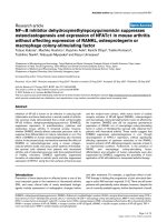
Figure 1
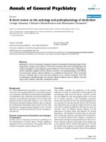
Effect of dehydroxymethylepoxyquinomicin on inflammation and bone destruction in collagen-induced mouse arthritisEffect of dehydroxymethylepoxyquinomicin on inflammation and bone
destruction in collagen-induced mouse arthritis. (a) Increase (%) of the
sum of the thickness of the right and left hind paws in each mouse dur-
ing day -4 and day 10. Horizontal bars represent the mean. DHMEQ,
dehydroxymethylepoxyquinomicin. (b) Radiographic scores of the ankle
joints were determined as described in Materials and methods, and
were normalized to the normal mice. Values are expressed as the mean
± standard deviation, and represent data obtained by three independ-
ent investigators. Data were compared by Student's t test.
Arthritis Research & Therapy Vol 9 No 5 Kubota et al.
Page 4 of 10
(page number not for citation purposes)
well) were incubated in 96-well plates in αMEM with heat-
inactivated 10% FCS (Sigma-Aldrich), 25 ng/ml M-CSF
(Peprotech, Rocky Hill, NJ, USA) and 40 ng/ml RANKL
(Peprotech). The indicated concentration of DHMEQ was
added throughout the culture period. On day 3 the medium
was replaced with fresh medium. After incubation for a further
4 days, the number of TRAP-positive multinucleated cells with
three or more nuclei was counted under a microscope. To ana-
lyze the expression of matrix metalloprotease-9 (MMP-9), oste-
oclasts were differentiated as above without DHMEQ; then
DHMEQ was added after medium replacement at day 7, and
the culture supernatant was collected at day 8. The concentra-
tion of MMP-9 in the supernatant was measured using an
ELISA kit (GE Healthcare Bio-Sciences, Tokyo, Japan).
Results
Suppression of in vivo osteoclastogenesis by DHMEQ
We first confirmed the effect of DHMEQ on collagen-induced
arthritis by comparing the paw thickness and radiographic
changes in the mice treated with DHMEQ (n = 12) and vehicle
alone (n = 13), as well as in nonimmunized age-matched nor-
mal mice (n = 6), 10 days after booster immunization. As
shown in Figure 1, treatment with DHMEQ ameliorated both
inflammation and bone destruction.
To examine the effect of DHMEQ on in vivo differentiation of
osteoclasts, the ankle joints of the mice were excised and
processed for histochemical staining. The specimens from
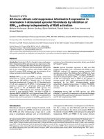
Figure 2
Effect of dehydroxymethylepoxyquinomicin on differentiation of osteoclasts in ankle joints of mice with collagen-induced arthritisEffect of dehydroxymethylepoxyquinomicin on differentiation of osteoclasts in ankle joints of mice with collagen-induced arthritis. After taking radio-
graphs (a-c), the ankle joints were histochemically examined for tartrate-resistant acid phosphatase-positive cells (d-i). (a), (d) and (g) Typical joint of
an arthritic mouse treated with vehicle alone. (b), (e) and (h) Typical joint of an arthritic mouse treated with dehydroxymethylepoxyquinomicin. (c), (f)
and (i) Joint of an age-matched normal mouse. Arrow, multinucleated giant osteoclasts.
Available online />Page 5 of 10
(page number not for citation purposes)
arthritic mice treated with vehicle alone showed marked syno-
vitis accompanying invasion of pannus into the marrow space
(Figure 2d). Numerous TRAP-positive cells were attached on
the eroded bone surface and the inner surfaces of bone lacu-
nae, and some of them were multinucleated (Figure 2g). Radi-
ographically, the ankles of these mice showed remarkable
periarticular osteoporosis and bone erosion (Figure 2a). In
contrast, the joints of arthritic mice treated with DHMEQ
showed milder synovial inflammation. Osteoclasts were mainly
observed on the inner surfaces of the bone marrow, and their
number and size were less than those in vehicle-treated mice
(Figure 2e, h). Radiographs showed mild periarticular oste-
oporosis (Figure 2b). In ankle joints of normal control mice, vir-
tually no TRAP-positive cells were observed (Figure 2f, i).
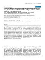
For quantitative evaluation, the number of TRAP-positive giant
cells with four or more nuclei in each ankle joint was counted.
As shown in Figure 3, the mice treated with DHMEQ exhibited
significantly fewer osteoclasts than those given vehicle alone,
indicating the suppressive effect of DHMEQ on in vivo osteo-
clastogenesis.
Effect of DHMEQ on production of sRANKL,
osteoprotegerin and M-CSF
Many in vitro studies adopt a culture system in which mono-
cyte/macrophage precursor cells are stimulated with sRANKL
and M-CSF for induction of osteoclasts. In the in vivo bone
metabolism, the naturally occurring decoy receptor OPG also
plays a key role by preventing the binding of RANKL to its
receptor, receptor activator of NF-κB (RANK). The ratio of cir-
culating OPG to sRANKL in early RA patients has been dem-
onstrated to predict later joint destruction [23]. We therefore
tested whether administration of DHMEQ changed expression
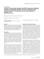
of these soluble factors. As shown in Figure 4a, serum levels
of OPG in arthritic mice treated with DHMEQ and with vehicle
alone were both significantly higher than those of normal mice.
sRANKL in both arthritic groups also tended to be higher than
nonarthritic normal mice, although this was not statistically sig-
nificant (Figure 4b). No differences in OPG, sRANKL or the
sRANKL/OPG ratio were observed, however, between the
DHMEQ-treated and vehicle-treated groups (Figure 4a–c). In
addition, no significant difference was observed in the serum
levels of M-CSF among three groups (Figure 4d).
To further examine the effect of DHMEQ on expression of
RANKL and M-CSF, we carried out in vitro experiments using
human RA-FLS. As shown in the results of western blotting,
RA-FLS spontaneously expressed RANKL without the addi-
tion of proinflammatory cytokines, and incubation with
DHMEQ did not change the level of RANKL expression (Fig-
ure 5a). Similarly, the result of ELISA revealed that RA-FLS
secreted M-CSF without any stimulation. Incubation with
DHMEQ did not suppress the levels of M-CSF, but rather
enhanced it slightly at 3 μg/ml (Figure 5b). Stimulation with
TNFα did not further increase the production of RANKL or M-
CSF by RA-FLS (data not shown). These results suggest that
production of RANKL and M-CSF by proliferating RA-FLS are
not particularly dependent on NF-κB, and the suppressive
effect of DHMEQ on osteoclastogenesis resulted from the
downregulation of proosteoclastogenic factors other than
RANKL, RANK or OPG.
Suppression of NFATc1 expression by DHMEQ in
arthritic joints
In the presence of RANKL and M-CSF, DHMEQ inhibits differ-
entiation of osteoclasts in cultures of mouse bone-marrow-
derived monocyte/macrophage precursor cells by downregu-
lation of NFATc1 [12]. We therefore examined the expression
of NFATc1 as well as NF-κB in the joints of arthritic mice by
immunofluorescent staining. Using monoclonal antibody that
recognizes only an activated form of the p65 subunit of NF-κB,
distinct staining was observed along the inner surface of bone
lacunae (Figure 6a) and in eroded regions of arthritic bone
from mice treated with vehicle alone, but was not observed in
those mice treated with DHMEQ (Figure 6d). Staining of
NFATc1 was also obvious on the inner surfaces of bone
lacunae (Figure 6b) and in the eroded regions of vehicle-
treated mice, but not from DHMEQ-treated mice (Figure 6e).
Normal control mice exhibited no staining of NF-κB (Figure
6g) nor of NFATc1 (Figure 6h). These results suggest that
inhibition of NF-κB activation by DHMEQ leads to suppression
of NF-κB-dependent expression of NFATc1 by osteoclasts in
arthritic joints.
Figure 3
Quantitative estimation of the suppressive effect of dehydroxymethyle-poxyquinomicin on in vivo osteoclastogenesisQuantitative estimation of the suppressive effect of dehydroxymethyle-
poxyquinomicin on in vivo osteoclastogenesis. The mean number of tar-
trate-resistant acid phosphatase-positive giant cells with four or more
nuclei in the individual ankle joints of arthritic mice treated with vehicle
alone (n = 13), of mice treated with dehydroxymethylepoxyquinomicin
(DHMEQ) (n = 12), and of normal mice (n = 6) were counted under a
microscope by two investigators in a blinded manner to the assignment
of mouse groups. The results shown are the mean ± standard error of
the mean of four independent counts, and were compared by Student's
t test.
Arthritis Research & Therapy Vol 9 No 5 Kubota et al.
Page 6 of 10
(page number not for citation purposes)
Suppression of human osteoclastogenesis and MMP-9
expression by DHMEQ
Human peripheral blood monocytes cultured with M-CSF and
RANKL differentiate into osteoclasts [24]. To test whether the
suppressive effect of DHMEQ on osteoclastogenesis can be
applied to human cells, monocytes from peripheral blood of
healthy volunteers were cultured with DHMEQ together with
M-CSF and RANKL. The result showed that the number of
TRAP-positive multinucleated cells was decreased by incuba-
tion with DHMEQ in a dose-dependent manner (Figure 7a).
MMP-9 is one of the enzymes released by osteoclasts, and the
enzyme plays a role in degradation of the extracellular matrix.
Its expression is reported to be modulated by NFATc1 [25],
and to be upregulated in serum of patients with active RA [26].
To examine the effect of DHMEQ on MMP-9 production by
human osteoclasts, DHMEQ was added to the culture after
formation of mature osteoclasts and secreted MMP-9 was
measured. The results showed that concentration of MMP-9 in
the culture supernatant was partially but significantly
decreased by DHMEQ (Figure 7b). These results indicate that
DHMEQ suppresses osteoclast differentiation from human
peripheral blood monocytes as well as the activity of mature
osteoclasts.
Discussion
In the present study, we investigated the effect of DHMEQ on
in vivo osteoclastogenesis using a mouse arthritis model, and
showed that DHMEQ significantly suppresses differentiation
of osteoclasts in arthritic joints. Serum levels of sRANKL, OPG
and M-CSF, and the sRANKL/OPG ratio, were not affected by
this treatment regimen with DHMEQ, whereas expression of
NFATc1 in the joints was suppressed in DHMEQ-treated
mice. In accordance with these observations, spontaneous
expression of RANKL and M-CSF in cultures of RA-FLS were
not suppressed by DHMEQ in concentrations at which it has
Figure 4
Effect of dehydroxymethylepoxyquinomicin on serum factors involved in osteoclastogenesisEffect of dehydroxymethylepoxyquinomicin on serum factors involved in osteoclastogenesis. Effect of dehydroxymethylepoxyquinomicin (DHMEQ)
on serum levels of (a) osteoprotegerin (OPG), (b) soluble receptor activator of NF-κB ligand (sRANKL), (c) sRANKL/OPG ratio and (d) macro-
phage colony-stimulating factor. Serum levels of these cytokines in individual arthritic mice 3 hours after the last treatment with vehicle alone (n =
13) or with DHMEQ (n = 12), and in age-matched normal mice (n = 4–6), were determined by ELISA. Horizontal lines represent the median. Data
were analyzed by the Mann-Whitney test. P < 0.05 was considered significant; ns, not significant.
Available online />Page 7 of 10
(page number not for citation purposes)
been demonstrated to suppress expression of proinflamma-
tory cytokines [16]. These results indicate that in a RANKL/
RANK/OPG signaling cascade, expression of NFATc1, a key
downstream regulator of this cascade, is more susceptible
than that of upstream molecules to treatment with DHMEQ.
Expression of RANKL and OPG is coordinated to regulate
bone resorption positively and negatively by controlling the
activation state of RANK on osteoclasts. The crucial role of the
RANKL/RANK/OPG signaling pathways in regulating bone
metabolism is underscored by findings that genetic mutations
that activate RANK and that inhibit the RANKL binding proper-
ties of OPG are associated with familial expansile osteolysis
[27] and with juvenile Paget's disease [28], respectively. In
addition, a cyclic peptide with sequence homology to a pre-
dicted ligand contact surface on RANK has been reported to
inhibit RANKL-induced signaling and osteoclastogenesis [29].
Proinflammatory cytokines, such as TNFα and IL-1, are
thought to modulate this system primarily by stimulating M-
CSF production (thereby increasing the pool of preosteoclas-
tic cells) and by directly increasing RANKL expression [30].
Aberrant expression of RANKL especially on RA-FLS stimu-
lated by TNFα or IL-1 is therefore supposed to be the main
contributor to bone destruction in active RA [31].
We previously demonstrated that 10 μg/ml DHMEQ sup-
presses expression of IL-1β and IL-6, as well as CC
chemokines CCL2 and CCL5, in culture of TNFα-stimulated
RA-FLS [16]. We therefore speculated that expression of M-
CSF, RANKL, OPG, or the sRANKL/OPG ratio may be modu-
lated in DHMEQ-treated mice. Serum levels of these factors,
however, were not significantly affected by treatment with
DHMEQ. Even in vitro, the expression of RANKL and M-CSF
by RA-FLS was not enhanced by TNFα, and was not sup-
pressed by 10 μg/ml DHMEQ. Taken together, FLS of RA
patients – and presumably of mice – are suggested, once acti-
vated, to express RANKL and M-CSF rather constitutively, and
they are resistant to treatment with DHMEQ. The ineffective-
ness of DHMEQ on RANKL suppression may possibly be
ascribed to insensitivity of the transcription mechanism of the
RANKL gene to DHMEQ. Regulation of the rate of gene
expression is a complex process involving several transcription
factors and gene activator/repressor proteins. For example, it
has been recently reported that NF-κB collaborates with other
transcription factors (early growth response-2 and early
growth response-3) in expression of the RANKL gene [32].
Even in molecules whose expression is demonstrated to be
NF-κB dependent in a certain assay condition, therefore, the
molecules' dependency on NF-κB or sensitivity to DHMEQ
treatment varies among the molecules under other conditions.
The second possible reason may involve the stability of
RANKL once expressed on the surface of FLS. We detected
RANKL by western blotting in the lysates of RA-FLS that had
been cultured for a few weeks without addition of proinflam-
matory cytokines (Figure 5); this is consistent with the obser-
vation of other investigators [33].
Downstream of the RANKL/RANK/OPG system, a significant
part of the genetic regulation of osteoclastogenesis is per-
formed by NF-κB. The critical role of this transcription factor is
underscored by the report of Franzoso and colleagues that
mice lacking the p50 and p52 subunits of NF-κB develop
osteopetrosis [7]. A few years later, the same group reported
that expression of p50 and p52 is not required for formation of
RANK-expressing osteoclast progenitors but is essential for
RANK-expressing osteoclast precursors to differentiate into
osteoclasts in response to RANKL and other osteoclastogenic
Figure 5
Effect of dehydroxymethylepoxyquinomicin on human fibroblast-like synovial cellsEffect of dehydroxymethylepoxyquinomicin on human fibroblast-like
synovial cells. Effect of dehydroxymethylepoxyquinomicin (DHMEQ) on
expression of receptor activator of NF-κB ligand (RANKL) and of mac-
rophage colony-stimulating factor (M-CSF) by fibroblast-like synovial
cells obtained from patients with rheumatoid arthritis (RA-FLS). The
RA-FLS were incubated with DHMEQ, with vehicle (dimethyl sulfoxide
(DMSO)), or with PBS for 24 hours. (a) Cell lysates were analyzed by
western blotting with anti-RANKL or with anti-β-actin monoclonal anti-
body. Representative data of similar results obtained using cell lines
from two patients with RA are shown. (b) Concentration of M-CSF in
the culture supernatant measured by ELISA; results expressed as rela-
tive values compared with PBS. Data are the mean ± standard error of
the mean of independent experiments carried out in triplicate using cell
lines obtained from six patients with RA, and were compared by Stu-
dent's t test. *P < 0.05.
Arthritis Research & Therapy Vol 9 No 5 Kubota et al.
Page 8 of 10
(page number not for citation purposes)
cytokines [8]. In a rat overiectomized model of estrogen defi-
ciency, administration of NF-κB decoy oligodeoxynucleotides
attenuated the increase of TRAP activity, accompanied by a
significant increase in calcium concentration in the tibia and
femur [9]. A cell-permeable peptide inhibitor of the IκB kinase
complex reduced the number of osteoclasts in the joints of col-
lagen-induced arthritic mice [10].
How NF-κB is involved in osteoclastogenesis, however, had
not been elucidated until Takatsuna and colleagues demon-
strated that DHMEQ suppresses osteoclastogenesis by
downregulation of NFATc1 in a culture system of mouse bone
marrow-derived monocyte/macrophage precursor cells stimu-
lated with RANKL and M-CSF [12]. The essential role of
NFATc1 in osteoclastogenesis was also demonstrated in a
recent in vivo study using osteoclast-deficient Fos
-/-
mice [34].
In the present study, we found that expression of NFATc1
along the inner surfaces of bone lacunae and eroded bone sur-
face in arthritic joints is suppressed by DHMEQ, suggesting
that in vivo expression of NFATc1 is significantly regulated by
NF-κB in agreement with the in vitro studies. RANKL induces
NFATc1 expression via three intracellular signaling pathways;
an NF-κB pathway, a mitogen-activated protein kinase path-
way, and a c-Fos pathway. RANKL also evokes Ca
2+
oscilla-
tion, which leads to calcineurin-mediated activation of NFATc1
[13]. DHMEQ does not inhibit activation of mitogen-activated
protein kinases or inhibit Ca
2+
oscillation [12]; the present
study therefore also indicates that the NF-κB pathway has pri-
ority over other pathways to induce NFATc1 expression.
Conclusion
In vivo administration of the NF-κB inhibitor DHMEQ sup-
pressed differentiation of osteoclasts in collagen-induced
mouse arthritis. In addition, DHMEQ exhibited suppressive
effects on in vitro differentiation and activation of human oste-
oclasts, suggesting the possible clinical application of this
compound.
Figure 6
Effect of dehydroxymethylepoxyquinomicin on NF-κB activation and NFATc1 expression in joints of collagen-induced arthritisEffect of dehydroxymethylepoxyquinomicin on NF-κB activation and NFATc1 expression in joints of collagen-induced arthritis. Fresh frozen sections
of each ankle joint were double-stained: (a, d, g) with FITC-labeled antibody to an activated form of the p65 subunit of NF-κB, and (b, e, h) with phy-
coerythrin-labeled antibody to NFATc1. (c, f, i) Transmission microscopy images of the same slides to show the articular structure. First row, a typi-
cal joint of an arthritic mouse treated with vehicle alone (a-c). Second row, a typical joint of an arthritic mouse treated with
dehydroxymethylepoxyquinomicin (d-f). Third row, a joint of an age-matched normal mouse (g-i). White arrow (a), staining by anti-NF-κB p65 anti-
body; blue arrow (b), staining by anti-NFATc1 antibody of the cells along the inner surfaces of bone lacunae.
Available online />Page 9 of 10
(page number not for citation purposes)
Competing interests
The authors declare that they have no competing interests.
Authors' contributions
MH, KA and KO carried out in vivo experiments using a mouse
model. YK carried out in vitro experiments using human cells.
KU synthesized a critical chemical. TN and NM participated in
the design of the study and helped to draft the manuscript. TK
conceived of the study, and participated in its design and
drafted the manuscript. All authors read and approved the final
manuscript.
Acknowledgements
The authors thank Fumiko Inoue and Dr Soichiro Ito (Tokyo Medical and
Dental University) for their expert technical assistance.
References
1. Lipsky PE, van der Heijde DM, St Clair EW, Furst DE, Breedveld
FC, Kalden JR, Smolen JS, Weisman M, Emery P, Feldmann M, et
al.: Infliximab and methotrexate in the treatment of rheumatoid
arthritis. Anti-tumor necrosis factor trial in rheumatoid arthritis
with concomitant therapy study group. N Engl J Med 2000,
343:1594-1602.
2. van der Heijde D, Klareskog L, Rodriguez-Valverde V, Codreanu C,
Bolosiu H, Melo-Gomes J, Tornero-Molina J, Wajdula J, Pedersen
R, Fatenejad S: Comparison of etanercept and methotrexate,
alone and combined, in the treatment of rheumatoid arthritis.
Two-year clinical and radiographic results from the TEMPO
study, a double-blind, randomized trial. Arthritis Rheum 2006,
54:1063-1074.
3. Smolen JS, van der Heijde DMFM, St Clair EW, Emery P, Bathon
JM, Keystone E, Maini RN, Kalden JR, Schiff M, Baker D, et al.: Pre-
dictors of joint damage in patients with early rheumatoid
arthritis treated with high-dose methotrexate with or without
concomitant infliximab. Results from the ASPIRE trial. Arthritis
Rheum 2006, 54:702-710.
4. Redlich K, Hayer S, Ricci R, David J-P, Tohidast-Akrad M, Kollias
G, Steiner G, Smolen JS, Wagner EF, Schett G: Osteoclasts are
essential for TNF-α-mediated joint destruction. J Clin Invest
2002, 110:1419-1427.
5. Teitelbaum SL: Bone resorption by osteoclasts. Science 2000,
289:1504-1508.
6. Boyle WJ, Simonet WS, Lacey DL: Osteoclast differentiation
and activation. Nature 2003, 423:337-342.
7. Franzoso G, Carlson L, Xing L, Poljak L, Shores EW, Brown KD,
Leonardi A, Tran T, Boyce BF, Siebenlist U: Requirement for NF-
κB in osteoclast and B-cell development. Genes Dev 1997,
11:3482-3496.
8. Xing L, Bushnell TP, Carlson L, Tai Z, Tondravi M, Siebenlist U,
Young F, Boyce BF: NF-κB p50 and p52 expression is not
required for RANK-expressing osteoclast progenitor forma-
tion but is essential for RANK- and cytokine-mediated
osteoclastogenesis. J Bone Miner Res 2002, 17:1200-1210.
9. Shimizu H, Nakagami H, Tsukamoto I, Morita S, Kunugiza Y, Tomita
T, Yoshikawa H, Kaneda Y, Ogihara T, Morishita R: NF-κB decoy
oligodeoxynucleotides ameliorates osteoporosis through
inhibition of activation and differentiation of osteoclasts. Gene
Ther 2006, 13:933-941.
Figure 7
Effect of dehydroxymethylepoxyquinomicin on human osteoclastogenesis and production of matrix metalloprotease-9 by human osteoclastsEffect of dehydroxymethylepoxyquinomicin on human osteoclastogenesis and production of matrix metalloprotease-9 by human osteoclasts. (a)
Peripheral blood monocytes were incubated in 96-well plates with macrophage colony-stimulating factor (M-CSF), receptor activator of NF-κB lig-
and (RANKL), and the indicated concentrations of dehydroxymethylepoxyquinomicin (DHMEQ). At day 7, the total number of tartrate-resistant acid
phosphatase (TRAP)-positive multinucleated cells (MNC) with three or more nuclei/well was counted. Representative data of three independent
experiments are shown. *P < 0.01, DHMEQ versus dimethyl sulfoxide (DMSO). (b) Peripheral blood monocytes were incubated in 96-well plates
with M-CSF and RANKL without DHMEQ. At day 7, the medium was replaced with fresh medium and the indicated concentrations of DHMEQ were
added. The culture supernatant was collected at day 8, and the matrix metalloprotease-9 (MMP-9) concentration was measured by ELISA. Repre-
sentative data of two independent experiments are shown. Data represent the mean ± standard error of the mean of triplicate wells, and were com-
pared by Student's t test. *P < 0.05, DHMEQ versus DMSO.
Arthritis Research & Therapy Vol 9 No 5 Kubota et al.
Page 10 of 10
(page number not for citation purposes)
10. Jimi E, Aoki K, Saito H, D'Acquisto F, May MJ, Nakamura I, Sudo T,
Kojima T, Okamoto F, Fukushima H, et al.: Selective inhibition of
NF-κB blocks osteoclastogenesis and prevents inflammatory
bone destruction in vivo. Nat Med 2004, 10:617-624.
11. Abu-Amer Y, Dowdy SF, Ross FP, Clohisy JC, Teitelbaum SL: TAT
fusion proteins containing tyrosine 42-deleted IκBα arrest
osteoclastogenesis. J Biol Chem 2001, 276:30499-30503.
12. Takatsuna H, Asagiri M, Kubota T, Oka K, Osada T, Sugiyama C,
Saito H, Aoki K, Ohya K, Takayanagi H, Umezawa K: Inhibition of
RANKL-induced osteoclastogenesis by (-)-DHMEQ, a novel
NF-κB inhibitor, through downregulation of NFATc1. J Bone
Miner Res 2005, 20:653-662.
13. Takayanagi H, Kim S, Koga T, Nishina H, Isshiki M, Yoshida H,
Saiura A, Isobe M, Yokochi T, Inoue J, et al.: Induction and activa-
tion of the transcription factor NFATc1 (NFAT2) integrate
RANKL signaling in terminal differentiation of osteoclasts. Dev
Cell 2002, 3:889-901.
14. Ariga A, Namekawa J, Matsumoto N, Inoue J, Umezawa K: Inhibi-
tion of tumor necrosis factor-α-induced nuclear translocation
and activation of NF-κB by dehydroxymethylepoxyquinomicin.
J Biol Chem 2002, 277:24625-24630.
15. Miyajima A, Kosaka T, Seta K, Asano T, Umezawa K, Hayakawa M:
Novel nuclear factor κB activation inhibitor prevents inflamma-
tory injury in unilateral ureteral obstruction. J Urol 2003,
169:1559-1563.
16. Wakamatsu K, Nanki T, Miyasaka N, Umezawa K, Kubota T: Effect
of a small molecule inhibitor of nuclear factor-κB nuclear
translocation in a murine model of arthritis and cultured
human synovial cells. Arthritis Res Ther 2005, 7:R1348-R1359.
17. Chiba T, Kondo Y, Shinozaki S, Kaneko E, Ishigami A, Maruyama
N, Umezawa K, Shimokado K: A selective NF
κB inhibitor,
DHMEQ, reduced atherosclerosis in apoE-deficient mice. J
Atheroscler Thromb 2006, 13:308-313.
18. Lee S-K, Lorenzo J: Cytokines regulating osteoclast formation
and function. Curr Opin Rheumatol 2006, 18:411-418.
19. Suzuki Y, Sugiyama C, Ohno O, Umezawa K: Preparation and
biological activities of optically active dehydroxymethylepoxy-
quinomicin, a novel NF-κB inhibitor. Tetrahedron 2004,
60:7061-7066.
20. Tadokoro M, Hattori K, Takakura Y, Ohgushi H: Rapid preparation
of fresh frozen tissue-engineered bone sections for histologi-
cal, histomorphological and histochemical analyses. Biomed
Mater Eng 2006, 16:405-413.
21. Arnett FC, Edworthy SM, Bloch DA, Mcshane DJ, Fries JF, Cooper
NS, Healey LA, Kaplan SR, Liang MH, Luthra HS, et al.: The Amer-
ican Rheumatism Association 1987 revised criteria for the
classification of rheumatoid arthritis. Arthritis Rheum 1988,
31:315-324.
22. Nanki T, Nagasaka K, Hayashida K, Saita Y, Miyasaka N: Chemok-
ines regulate IL-6 and IL-8 production by fibroblast-like syn-
oviocytes from patients with rheumatoid arthritis. J Immunol
2001, 167:5381-5385.
23. Geusens PP, Landewé RBM, Garnero P, Chen D, Dunstan CR,
Lems WF, Stinissen P, van der Heijde DMFM, van der Linden S,
Boers M: The ratio of circulating osteoprotegerin to RANKL in
early rheumatoid arthritis predicts later joint destruction.
Arthritis Rheum 2006, 54:1772-1777.
24. Komano Y, Nanki T, Hayashida K, Taniguchi K, Miyasaka N: Iden-
tification of a human peripheral blood monocyte subset that
differentiates into osteoclasts. Arthritis Res Ther 2006, 8:R152.
25. Sundaram K, Nishimura R, Senn J, Youssef RF, London SD, Reddy
SV: RANK ligand signaling modulates the matrix metallopro-
teinase-9 gene expression during osteoclast differentiation.
Exp Cell Res 2007, 313:168-178.
26. Fiedorczyk M, Klimiuk PA, Sierakowski S, Gindzienska-Sieskiewicz
E, Chwiecko J: Serum matrix metalloproteinases and tissue
inhibitors of metalloproteinases in patients with early rheuma-
toid arthritis. J Rheumatol 2006, 33:1523-1529.
27. Hughes AE, Ralston SH, Marken J, Bell C, MacPherson H, Wallace
RGH, van Hul W, Whyte MP, Nakatsuka K, Hovy L, Anderson DM:
Mutations in TNFRSF11A, affecting the signal peptide of RANK,
cause familial expansile osteolysis. Nat Genet 2000, 24:45-48.
28. Whyte MP, Obrecht SE, Finnegan PM, Jones JL, Podgornik MN,
McAlister WH, Mumm S: Osteoprotegerin deficiency and juve-
nile Paget's disease. N Engl J Med 2002, 347:175-184.
29. Aoki K, Saito H, Itzstein C, Ishiguro M, Shibata T, Blanque R, Mian
AH, Takahashi M, Suzuki Y, Yoshimatsu M, et al.: A TNF receptor
loop peptide mimic blocks RANK ligand-induced signaling,
bone resorption, and bone loss. J Clin Invest 2006,
116:1525-1534.
30. Khosla S: The OPG/RANKL/RANK system. Endocrinology
2001, 142:5050-5055.
31. Takayanagi H: Mechanistic insight into osteoclast differentia-
tion in osteoimmunology. J Mol Med 2005, 83:170-179.
32. Fionda C, Nappi F, Piccoli M, Frati L, Santoni A, Cippitelli M: 15-
Deoxy-Δ
12,14
-prostaglandin J
2
negatively regulates rankl gene
expression in activated T lymphocytes: role of NF-κB and early
growth response transcription factors. J Immunol 2007,
178:4039-4050.
33. Lee CK, Lee EY, Chung SM, Mun SH, Yoo B, Moon HB: Effects
of disease-modifying antirheumatic drugs and antiinflamma-
tory cytokines on human osteoclastogenesis through interac-
tion with receptor activator of nuclear factor κB,
osteoprotegerin, and receptor activator of nuclear factor κB
ligand. Arthritis Rheum 2004, 50:3831-3843.
34. Asagiri M, Sato K, Usami T, Ochi S, Nishina H, Yoshida H, Morita
I, Wagner EF, Mak TW, Serfling E, Takayanagi H: Autoamplifica-
tion of NFATc1 expression determines its essential role in
bone homeostasis. J Exp Med 2005, 202:1261-1269.