Báo cáo khoa học: " Radiosensitization by 2-benzoyl-3-phenyl-6,7-dichloroquinoxaline 1,4-dioxide under oxia and hypoxia in human colon cancer cells" pdf
Bạn đang xem bản rút gọn của tài liệu. Xem và tải ngay bản đầy đủ của tài liệu tại đây (455.07 KB, 13 trang )
BioMed Central
Page 1 of 13
(page number not for citation purposes)
Radiation Oncology
Open Access
Research
Radiosensitization by 2-benzoyl-3-phenyl-6,7-dichloroquinoxaline
1,4-dioxide under oxia and hypoxia in human colon cancer cells
Wafica Itani
1
, Fady Geara
2
, Joelle Haykal
1
, Makhluf Haddadin
3
and
Hala Gali-Muhtasib*
1
Address:
1
Department of Biology, American University of Beirut, Beirut, Lebanon,
2
Department of Radiation Oncology, American University of
Beirut, Beirut, Lebanon and
3
Department of Chemistry, American University of Beirut, Beirut, Lebanon
Email: Wafica Itani - ; Fady Geara - ; Joelle Haykal - ;
Makhluf Haddadin - ; Hala Gali-Muhtasib* -
* Corresponding author
Abstract
Background: The sensitizing effects of 2-benzoyl-3-phenyl-6,7-dichloroquinoxaline 1,4-dioxide
(DCQ) and ionizing radiation (IR) were determined in four colon cancer cells and in FHs74Int
normal intestinal cells.
Methods: Cell cycle modulation, TUNEL assay, clonogenic survival and DNA damage were
examined under oxia or hypoxia. Effects on apoptotic molecules and on p-Akt and Cox-2 protein
expression were investigated.
Results: The four cell lines responded differently to DCQ+IR; HT-29 cells were most resistant.
Combination treatment caused significant increases in preG
1
(apoptosis) in HCT-116, while G
2
/M
arrest occurred in DLD-1. DCQ potentiated IR effects more so under hypoxia than oxia. Pre-
exposure of DLD-1 to hypoxia induced 30% apoptosis, and G
2
/M arrest in oxia. The survival rate
was 50% lower in DCQ+IR than DCQ alone and this rate further decreased under hypoxia.
FHs74Int normal intestinal cells were more resistant to DCQ+IR than cancer cells.Greater ssDNA
damage occurred in DLD-1 exposed to DCQ+IR under hypoxia than oxia. In oxia, p-Akt protein
expression increased upon IR exposure and drug pre-treatment inhibited this increase. In contrast,
in hypoxia, exposure to IR reduced p-Akt protein and DCQ restored its expression to the
untreated control. Apoptosis induced in hypoxic DLD-1 cells was independent of p53-p21
modulation but was associated with an increase in Bax/Bcl-2 ratio and the inhibition of the Cox-2
protein.
Conclusion: DCQ is a hypoxic cell radiosensitizer in DLD-1 human colon cancer cells.
Background
Oxygen is known to help in stabilizing the radiation-
induced DNA damage [1]. The lack of oxygen in solid
malignant tumors results in their resistance to radiation
therapy [1,2]. Attempts to overcome this resistance
include the use of "oxygen-mimetic" radiosensitizers [3];
compounds which offer an attractive alternative for
increasing the therapeutic window [4].
Published: 03 January 2007
Radiation Oncology 2007, 2:1 doi:10.1186/1748-717X-2-1
Received: 15 September 2006
Accepted: 03 January 2007
This article is available from: />© 2007 Itani et al; licensee BioMed Central Ltd.
This is an Open Access article distributed under the terms of the Creative Commons Attribution License ( />),
which permits unrestricted use, distribution, and reproduction in any medium, provided the original work is properly cited.
Radiation Oncology 2007, 2:1 />Page 2 of 13
(page number not for citation purposes)
Quinoxaline 1,4-dioxides (QdNOs) share the di-N-oxide
moiety with the clinically used drug Tirapazamine. These
hypoxia-selective compounds are known to be redox-acti-
vated DNA-cleaving agents [5]. DNA cleavage by QdNOs
requires enzymatic one-electron reduction of the com-
pound to an activated, oxygen-sensitive intermediate [6].
This one-electron reduction is more likely to occur in the
reducing conditions of hypoxic cells, targeting the toxicity
of these compounds to hypoxic cells. Recent studies have
shown that the nature of the substituent on the benzo-
ring of the QDNO influences its potency [7]. Mild elec-
tron withdrawing groups in the 6(7) position increase the
potency of these compounds under hypoxic conditions
[7].
We have shown that the compound, 2-benzoyl-3-phenyl-
6,7-dichloroquinoxaline 1,4-dioxide (DCQ), is a hypoxic
cytotoxin [8]. Treatment of human colon cancer T-84 cells
with DCQ reduced the expression levels of HIF-1α mRNA
and protein [8]. The decrease in HIF-1α mRNA and pro-
tein expression by DCQ was later documented in EMT6
mouse mammary adenocarcinoma and Lewis lung carci-
noma cells [9]. DCQ was also shown to reduce the expres-
sion levels of vascular endothelial growth factor (VEGF)
and to inhibit hypoxia-induced angiogenesis [9]. Subse-
quent experiments performed by our group established
that DCQ is an effective radiosensitizer both in vitro and
in vivo [9]. When DCQ was combined with radiation,
doses of 2.5–5 μM resulted in a dramatic decrease in clo-
nogenic survival of EMT6 cells. The mechanism of radi-
osenitization by DCQ in EMT6 cells was found to involve
the induction of G
2
/M arrest and apoptosis (unpublished
results). Radiosensitization effects were also seen in vivo
when LLC tumors were injected into C57BL/J6 mice and
the effects of DCQ+IR on tumor volume were observed
over 20 days [9].
This study aims, for the first time, to determine DCQ radi-
osensitizing activities in several human colorectal cancer
cell lines and to investigate its cell cycle modulatory effects
under both oxic and hypoxic conditions. Drug sensitiza-
tion was examined in the FHs74Int normal human intes-
tinal cell line to determine the sensitivity of normal cells
to DCQ. In addition, the DNA damaging potential of
DCQ and its effects on the protein expression levels of the
oncogene Akt and on key molecules of apoptosis was
investigated.
Methods
Cell culture
FHs74Int normal human intestinal cells were cultured in
Hybri-Care medium supplemented with 30 ng/ml epider-
mal growth factor. Human colon cancer cell lines (DLD-
1, HT-29, HCT-116, and SW-480) were grown in RPMI
1640 containing L-Glutamine and 25 mm HEPES. All
media were supplemented with 10% heat-inactivated FBS
and 1% Penicillin-Streptomycin (50 μg/ml). Cells were
cultured in a humidified incubator (95% air 5% CO
2
) at
37°C (Forma Scientific Inc. Ohio, USA).
Drug preparation
DCQ was synthesized from 5,6-dichlorobenzofurazan
oxide and dibenzoylmethane according to the Beirut
Reaction [10]. A fresh stock of 10 mg of DCQ was dis-
solved in 1 ml of filtered DMSO. Before treatment, DCQ
was diluted 1 in 10 using media containing 10% FBS and
1% Penicillin-Streptomycin (50 μg/ml).
Radiation experiments
Cells cultured in 25 cm
2
T-flasks were treated either with
DCQ (0–10 μM), irradiation (0–6 Gy) or combinations.
Irradiation was administered by a JL Shepherd, 143-68
Cesium-137 Laboratory Irradiator with an output activity
of 1683 Ci. Immediately after irradiation, cells were
replenished with fresh media containing no drugs and left
in the incubator for 24 hours for studies on cell cycle reg-
ulation and DNA damage (COMET) as described below.
Hypoxia treatment
DLD-1 or FHs 74Int cells cultured in 25 cm
2
flasks were
treated at 50% confluency with DCQ for 4 hours, after
which they were placed in a tightly sealed chamber (37°C,
1% O
2
) for 1 hour. The desired oxygen level was opti-
mized by injecting N
2
gas into the chamber, and the levels
were measured every 15 minutes using an Ohmeda
Oxymeter (Datex-Ohmeda, Louisville, CO). Immediately
after hypoxia the flasks were sealed and the cells were irra-
diated. Later, cells were replenished with fresh media con-
taining no drugs and incubated for another 24 hours.
Clonogenic survival
Oxic or hypoxic DLD-1 cells cultured in 25 cm
2
T-flasks
were treated with DCQ (0–100 μM, 1 hour), after which
they were irradiated (2 Gy). FHs74Int cells were treated
under oxic conditions with DCQ (0–10 μM) for 1 hour
prior to irradiation (2 Gy). Immediately after irradiation,
both cell lines were re-plated at known dilutions with
fresh media for 10 days. After 10 days of incubation, col-
onies were stained with crystal violet and counted. The
number of colonies containing more than 50 cells was
counted and the percentage of survival rates at each dose
was calculated according to the formula: (colony no. in
treatment/colony no. in control) × 100.
Cell cycle analysis using flow cytometry
Following treatment, cells were harvested, fixed in ice cold
70% ethanol and stored at -20°C. On the day of DNA
staining, cells were incubated for 75 minutes in 200 μg/ml
RNase A at 37°C, and stained with 50 μg/ml propidium
iodide. Cell cycle analysis was performed using a FACScan
Radiation Oncology 2007, 2:1 />Page 3 of 13
(page number not for citation purposes)
flow cytometry (Becton Dickinson, Research Triangle,
NC) and the percentage of cells in preG
1
, G
1
, S, and G
2
/M
phases was determined using the Cell Quest program.
Apoptosis TUNEL assay
Fragmented DNA was detected by Terminal deoxy-trans-
ferase (TdT)-mediated dUTP nick-end labeling (TUNEL
assay) (Roche Diagnostics, Mannheim, Germany) to
assess the induction of apoptosis. Following treatment,
cells were harvested and the pellet was suspended in 100
μl freshly prepared PBS in 4% formaldehyde, incubated at
room temperature for 30 minutes, and centrifuged at 300
g/2000 rpm for 10 minutes. The pellet was washed once
with 200 μl PBS. Followed by suspension in 100 μl of a
solution containing 1× PBS, 0.1% sodium citrate, and
0.1% Triton X-100 for 2 minutes on ice. Cells were then
washed twice with 1× PBS. The pellet was resuspended in
50 μl tunnel reaction mixture (45 μl labeling solution and
5 μl enzyme solution), incubated for 1 h at 37°C in a
humidified atmosphere in the dark, then washed twice
with 1× PBS and suspended in 1× PBS for reading by flow
cytometry. Cells suspended in 50 μl labeling solution
served as the negative control. The samples were exam-
ined by FACScan flow cytometer to determine the percent-
age of apoptotic cells in treated samples as compared to
the control samples.
Single Cell Gel Electrophoresis (SCGE)/comet assay
DNA damage, including single strand breaks (SSB) and
alkali labile sites (ALS), was measured using the alkaline
SCGE assay in DLD-1 cells treated with DCQ (5 μM, 1
hour) IR (2 Gy) or combinations under oxia or hypoxia.
Immediately after IR, cells were scraped and collected in
RPMI medium. Comet assay was performed as described
previously [11]. For electrophoresis, an electric current of
25 volts and 300 mA was applied for 30 minutes, after
which the slides were placed in a neutralizing buffer for 5
minutes. This neutralizing procedure was repeated two
more times. Finally, 50 μl of YOYO stain (0.25 μM YOYO,
2.5% DMSO and 0.5% sucrose) (Molecular Probes –
Eugene, Oregon, USA) was added to each slide and ana-
lyzed immediately using a fluorescence microscope
(AXIOVERT 200, ZEISS Flourescence and optical micro-
scope with ZEISS AXIOCAM HRC and KS 300 V3 image
analysis software). Images of 100 randomly selected non-
overlapping cells (magnification 100×) were analyzed for
each sample with the help of Tri-Tek CometScore™ soft-
ware, a fully automatic image analysis system. The follow-
ing parameters were used to assess DNA damage: total
fluorescence of the comet, fluorescence of the tail, per-
centage of DNA in the tail region and tail moment
(%DNA in tail multiplied by tail length). The comet data
values were expressed as mean ± S.D. Statistical compari-
sons were made by t-test and the P-values < 0.05 or P <
0.01 were considered significant.
Protein expression by Western Blotting
DLD-1 cells cultured in 75 cm
2
T-flasks were treated with
DCQ (5 μM, 1 hour), IR (2 Gy) or combinations under
oxic or hypoxic conditions. Cellular proteins were
extracted by SDS-lysis buffer (50 mM Tris-HCL, pH 7.5,
150 mM NaCl, 1% Nonidet P40, 0.5% Sodium deoxycho-
late, 4% protease inhibitors and 1% phosphatase inhibi-
tors). Protein extracts were centrifuged for 10 minutes at
14,000 rpm. Proteins were quantified using the DC Bio-
Rad Protein Assay kit with BSA as a standard. Whole cell
lysates (40–60 μg) were loaded on 12% SDS-polyacryla-
mide gels and then transferred onto PVDF membranes
(Amersham Pharmacia Biotech, Amersham, England).
The membranes were incubated with the primary anti-
bodies: p21 (F-5), p53 (FL-393), p-p53, Bcl-2 (N-19),
Cox-2 (all from Santa Cruz, CA), Bax (Biosource, Califor-
nia, USA), pS473 Akt (44-622G) (Chemicon Interna-
tional, California, USA). The GAPDH antibody
(Biogenesis, Poole, UK) was used as a loading control. The
membrane was then washed 3 times for 10 minutes each
in wash buffer (TBS containing 0.05%–0.1% Tween 20)
and probed with the appropriate secondary antibody
(IgG-HRP, antirabbit IgG-HRP, or antigoat IgG-HRP from
Santa Cruz) for 1 hour at room temperature. After wash,
the membrane was exposed to X-ray film (Hyperfilm ECL,
Lebanon) using a chemiluminescent substrate (Amer-
sham Pharmacia Biotech, Amersham, England). The
bands were quantified using LabWorks 4.0 software.
Results
Cell cycle modulation in four human colon cancer cell lines
under oxia
To study cell cycle modulation by DCQ+IR, cells were
incubated with DCQ (5 or 10 μM) for either 1 hour (DLD-
1 and HCT116) or 4 hours (SW-480 and HT-29), and then
irradiated (2 Gy). The times, 1 or 4 hours, were chosen
based on differences in the sensitivity of the four cell lines
to the drug. While SW-480 and HT-29 survived after 4
hour exposure to DCQ, DLD-1 and HCT-116 died when
drug treatment was extended for more than 1 hour (data
not shown). Twenty four hours after treatment, cells were
harvested for flow cytometry analysis and the percentage
of cells in preG
1
and G
2
/M phases were plotted as these
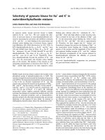
phases were the most modulated. The response of the four
cell lines to DCQ+IR was different; HT-29 cells were the
most resistant followed by SW-480 (Figure 1A and 1B).
HCT116 and DLD-1 were sensitive to DCQ+IR, but
responded differently. Treatment with 10 μM DCQ+IR
caused 11 fold increases in the preG
1
portion in HCT-116
(Figure 1C), however, in DLD-1 cells 2-fold increases in
the percentage of G
2
/M cells was observed (Figure 1D).
Radiation Oncology 2007, 2:1 />Page 4 of 13
(page number not for citation purposes)
Cell cycle modulation in HCT116 and SW-480 cells under
hypoxia
Since DCQ is a hypoxic cytotoxin [9], we then investigated
whether it could potentiate IR effects more so under
hypoxia than oxia. The hypoxia toxicity of DCQ was first
studied in the two cell lines, HCT116 and SW-480. Cells
were incubated in DCQ (5 μM, 1 or 4 hours) under oxic
or hypoxic conditions, after which they were irradiated,
then replenished with media containing no DCQ, and
harvested 24 hours later for cell cycle analysis (Figures 2
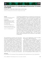
and 3). In both cell lines, hypoxia treatment alone caused
G
2
/M arrest (1.5–2.0 fold increase). Exposure of HCT-116
cells to oxic or hypoxic conditions prior to IR resulted in
no difference in their sensitivity to the drug (% of cells in
preG
1
phase was 36% in oxia and 23% in hypoxia) (Figure
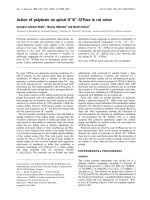
2). However, SW-480 showed a significant increase in
preG
1
cells when combination treatment was done under
hypoxia (Figure 3). Considering that HCT116 and SW-
480 were sensitive to hypoxia, no further studies were
done with these cell lines.
Cell cycle modulation and clonogenic survival in DLD-1
cells under hypoxia
To investigate the hypoxic cytotoxicity of DCQ in DLD-1,
we compared its efficacy in cells incubated in oxia or
hypoxia prior to irradiation. DLD-1 cells were treated with
Effect of DCQ, IR and their combinations on cell cycle regulation in four different human colon cancer cell lines (SW-480, HT-29, HCT116 and DLD-1)Figure 1
Effect of DCQ, IR and their combinations on cell cycle regulation in four different human colon cancer cell lines (SW-480, HT-
29, HCT116 and DLD-1). Cells were treated with DCQ (0, 5, 10 μM), IR (2 Gy) or combinations. Immediately after radiation
or drug treatment, cells were replenished with fresh medium containing no drug and incubated for another 24 hours. Control
cells were treated with DMSO (0.1%). Cell cycle changes were assessed using Propidium Iodide stain with flow cytometry as
described in "Materials and Methods". The percentage of cells in preG
1
and G
2
/M phases were plotted as a function of DCQ
dose. Results are representative of at least two independent experiments each performed in duplicates.
A
Control IR DCQ5μM IR+DCQ5μM DCQ10μM IR+DCQ10μM
Treatment
SW-480
Fold increase (relative to control)
preG
1
G
2
/M
12
10
8
6
4
2
0
C
HCT116
Treatment
preG
1
G
2
/M
12
10
8
6
4
2
0
Control IR DCQ5μM IR+DCQ5μM DCQ10μM IR+DCQ10μM
Fold increase (relative to control)
Control IR DCQ5μM IR+DCQ5μM DCQ10μM IR+DCQ10μM
Treatment
B
HT-29
preG
1
G
2
/M
12
10
8
6
4
2
0
Fold increase (relative to control)
Control IR DCQ5μM IR+DCQ5μM DCQ10μM IR+DCQ10μM
D
preG
1
G
2
/M
DLD-1
Treatment
12
10
8
6
4
2
0
Fold increase (relative to control)
Radiation Oncology 2007, 2:1 />Page 5 of 13
(page number not for citation purposes)
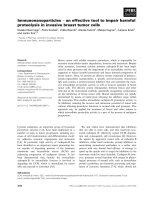
DCQ (5 μM) + IR and harvested after 24 hours for cell
cycle analysis (Figure 4). Treatment under oxia resulted in
the accumulation of 63% of the cells in G
2
/M phase and
4% in preG
1
. More pronounced effects were observed in
hypoxia, as 33% of apoptotic cells accumulated in preG
1
(Figure 4). Therefore treatment of DLD-1 cells with
DCQ+IR caused G
2
/M arrest in oxia and preG1 arrest in
hypoxia.
Using TUNEL assay, the level of apoptosis in cells treated
with DCQ+IR under oxic and hypoxic conditions was
found to be 3.9% and 30% respectively (Figure 5) con-
firming that the increases in preG1 observed by flow
cytometry are due to apoptosis.
To confirm the hypoxic effects of DCQ, DLD-1 cells were
treated with DCQ (1–100 μM) in oxia or hypoxia, irradi-
ated (2 Gy) and then re-plated at known dilutions. Ten
days after re-plating, the surviving colonies were counted.
The survival curves for DCQ+IR and DCQ alone show a
more pronounced decrease in cell survival under hypoxia
than oxia (Figure 6). Exposing DLD-1 cells to IR alone did
not reduce the absolute survival rate of cells under
hypoxia as compared to oxia (Figure 6C). When DLD-1
cells were exposed to DCQ alone (10 μM), the surviving
fraction determined with respect to the untreated cells was
0.49 (SD ± 0.04) in oxia and 0.20 (SD ± 0.02) in hypoxia
(Figure 6A). However, when DCQ (10 μM) was combined
with IR, the surviving fraction determined with respect to
the irradiated cells dropped to 0.29 (SD ± 0.03) in oxia
and 0.04 (SD ± 0.01) in hypoxia (Figure 6B).
The hypoxia cytotoxicity ratio (HCR), i.e. the concentra-
tion of drug required under oxia relative to hypoxia to
Effect of DCQ, IR and their combinations on cell cycle regulation in HCT116 cells exposed to oxic or hypoxic conditionsFigure 2
Effect of DCQ, IR and their combinations on cell cycle regulation in HCT116 cells exposed to oxic or hypoxic conditions. Cells
were treated with 5 μM DCQ or DMSO (0.1%) and exposed to hypoxia or incubated in oxia for 1 hour, then irradiated (2
Gy). Immediately after radiation or drug treatment, cells were replenished with fresh medium containing no drug and incubated
for another 24 hours. Cell cycle changes were assessed using Propidium iodide stain with flow cytometry as described in
"Materials and Methods". Bar graphs are a summary of at least three independent experiments each performed in duplicates.
preG1: 2.7 ± 0.6
Go/G1: 44.8 ± 1.9
S: 10.9 ± 1.7
G
2
/M: 41.7 ± 2.5
preG1: 2.4 ± 0.5
Go/G1: 39.5 ± 3.8
S: 13.7 ± 1.2
G
2
/M: 44.8 ± 2.1
preG1: 22.9 ± 2.4
Go/G1: 29.5 ± 2.1
S: 13.4 ± 0.6
G
2
/M: 34.2 ± 2.5
preG1: 5.1 ± 1.6
Go/G1: 37.1 ± 1.1
S: 7.5 ± 0.8
G
2
/M: 49.2 ± 1.5
200 400 600 200 400 600
200 400 600
200 400 600
FL2-A FL2-A FL2-AFL2-A
Hypoxia Control IR 2Gy DCQ 10μM IR +DCQ
120
0
120
0
120
0
120
0
Counts
Counts
Counts
Counts
preG1: 12.5 ± 1.6
Go/G1: 26.7 ± 1.2
S: 11.6 ± 1.0
G
2
/M: 46.9 ± 1.3
Counts
200 400 600
120
0
200 400 600
preG1: 3.3 ± 0.1
Go/G1: 46.8 ± 3.9
S: 24.1 ± 2.5
G
2
/M: 23.1 ± 1.2
Counts
FL2-A
preG1: 36.1 ± 1.5
Go/G1: 28.3 ± 1.2
S: 15.3 ± 1.3
G
2
/M: 19.0 ± 0.6
120
0
Counts
200 400 600
FL2-A
preG1: 5.8 ± 1.6
Go/G1: 48.7 ± 2.9
S: 10.61 ± 1.7
G
2
/M: 34.3 ± 1.6
200 400 600
FL2-A
Oxia Control IR 2Gy DCQ 10μM IR +DCQ
120
0
Counts
FL2-A
120
0
Counts
14
12
10
8
6
4
2
0
Control IR2Gy DCQ10μM IR+DCQ
Oxia
Hypoxia
Fold increase
(relative to control)
preG
1
Control IR2Gy DCQ10μM IR+DCQ
Oxia
Hypoxia
Fold increase
(relative to control)’
G
2
/M
4
3
2
1
0
HCT116
Radiation Oncology 2007, 2:1 />Page 6 of 13
(page number not for citation purposes)
produce 90% cell death, was 4 fold higher when DCQ was
combined with IR (HCR = 12) as compared to DCQ alone
(HCR = 3). This provided additional evidence that the
drug is a potent radio-sensitizer in hypoxic cells.
DCQ radiosensitization in the FHs74Int normal intestinal
cell line
After establishing effects of DCQ and IR in cancer cells, we
compared DCQ efficacy in normal cells. For this purpose,
FHs74Int normal human intestinal cells were pre-treated
with DCQ (1.25–10 μM, 1 hour), irradiated, and then re-
plated at known dilutions and the surviving colonies were
determined 10 days later. At 5 μM DCQ, the survival rate
was 0.68 (SD ± 0.02), and this rate was reduced to 0.46
(SD ± 0.01) when DCQ (5 μM) was combined with IR
(Figure 6D). A comparison of the extent of decrease in cell
survival in DCQ+IR in normal FHs74Int v.s. DLD-1 cancer
cells confirms the greater radio-sensitizing effects of this
drug in cancer cells.
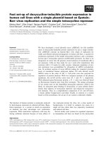
DNA damage by DCQ in irradiated DLD-1 cells under oxia
and hypoxia
To determine if DCQ is a DNA-targeting agent, the extent
of DNA damage was measured by the alkaline COMET
assay in oxic or hypoxic DLD-1 cells exposed to DCQ (5
μM, 1 hour), IR or combinations. The COMET assay meas-
ures single strand DNA breaks by the increase in the elec-
trophoretic mobility of denatured genomic DNA in an
agarose gel. Figure 7A shows an example of different
grades of DNA fragmentation. In the first image, the DNA
of a largely non-fragmented cell is depicted. The next 2
images represent cells with increasingly fragmented DNA;
thus giving the comet its tail. The last image shows a cell
with highly fragmented DNA. Treatment with DCQ+IR
Effect of DCQ, IR and their combinations on cell cycle regulation in SW-480 cells exposed to oxic or hypoxic conditionsFigure 3
Effect of DCQ, IR and their combinations on cell cycle regulation in SW-480 cells exposed to oxic or hypoxic conditions. Cells
were treated with 5 μM DCQ or DMSO (0.1%) and exposed to hypoxia or incubated in oxia for 4 hours, then irradiated (2
Gy). Immediately after radiation or drug treatment, cells were replenished with fresh medium containing no drug and incubated
for another 24 hours. Cell cycle changes were assessed using Propidium iodide stain with flow cytometry as described in
"Materials and Methods". Bar graphs are a summary of at least three independent experiments each performed in duplicates.
200 400 600
preG1: 0.7 ± 0.1
Go/G1: 51.8 ± 2.6
S: 20.0 ± 1.2
G
2
/M: 27.1 ± 1.7
preG1: 4.4 ± 0.3
Go/G1: 38.2 ± 1.8
S: 22.7 ± 1.9
G
2
/M: 30.9 ± 1.4
200 400 600
FL2-A
FL2-A
120
0
Counts
preG1: 4.45 ± 1.1
Go/G1: 38.3 ± 2.9
S: 22.4 ± 2.8
G
2
/M: 33.6 ± 1.7
preG1: 6.4 ± 0.8
Go/G1: 36.8 ± 1.5
S: 14.0 ± 1.0
G
2
/M: 42.1 ± 1.3
200 400 600
200 400 600
FL2-A
FL2-A
120
0
120
0
Counts
Counts
Oxia Control IR 2Gy DCQ 5μM IR +DCQ
120
0
Counts
Hypoxia Control
IR 2Gy DCQ 5μM IR +DCQ
200 400 600
200 400 600 200 400 600 200 400 600
120
0
120
0
120
0
120
0
Counts
Counts
Counts
Counts
preG1: 0.4 ± 0.2
Go/G1: 39.4 ± 2.7
S: 22.5 ± 2.2
G
2
/M: 38.2 ± 2.7
preG1: 10.6 ± 1.9
Go/G1: 37.8 ± 1.3
S: 13.7 ± 1.8
G
2
/M: 38.4 ± 1.9
preG1: 20.7 ± 1.3
Go/G1: 31.4 ± 2.8
S: 17.9 ± 1.6
G
2
/M: 26.6 ± 1.0
preG1: 5.0 ± 1.2
Go/G1: 35.3 ± 1.4
S: 16.8 ± 1.3
G
2
/M: 42.0 ± 2.3
FL2-A FL2-A
FL2-A
FL2-A
60
50
40
30
20
10
0
Control IR2Gy DCQ5μM IR+DCQ
Oxia
Hypoxia
Fold increase
(relative to control)
preG
1
Oxia
Hypoxia
Control IR2Gy DCQ5μM IR+DCQ
Fold increase
(relative to control)
G
2
/M
SW-480
Radiation Oncology 2007, 2:1 />Page 7 of 13
(page number not for citation purposes)
resulted in a statistically significant increase (p < 0.01) in
DNA damage in hypoxia compared to oxia. The mean per-
centage of DNA damage was 95 (SD ± 5.65) in cells
exposed to DCQ+ IR under hypoxia as compared to only
60.5 (SD ± 2.12) under oxia (Figure 7B).
Digital images were further analyzed using Comet Score
software that allows quantitative measurements of vari-
ous comet assay end-points, in particular, the mean aver-
age of comet length, tail length, and percentage of DNA in
the tail (Figure 7C). In addition, tail moment was calcu-
lated as the product of the percentage of DNA in the
comet tail multiplied by the total comet length. Such end-
points are the most accepted parameters for assessing
DNA damage. It is important to note that 1 hour exposure
of the cells to hypoxia did not induce a major change in
any of the measured comet assay end-points.
Several end-point measures indicated that DCQ is a more
potent DNA damaging agent in irradiated hypoxic cells:
1) significant (p < 0.05) increase in mean tail moment in
hypoxia compared to oxia (24.69 in oxia v.s. 72.3 in
hypoxia); 2) greater relative amount of damage, quanti-
fied by measuring the distance that DNA moves in the gel
or the length of the comet tail; 3) greater amount of DNA
present in the tail in hypoxic cells (11 fold increase in tail
DNA in hypoxia v.s. 7-fold increase in oxia) (Figure 7C).
DCQ effects on radiation-induced p53, p-p53 and p21
expression
To investigate the effects of DCQ on key apoptotic mole-
cules, DLD-1 cells were treated with DCQ, IR or combina-
tions under oxic or hypoxic conditions and the expression
levels of p53, p-p53 and p21 proteins were determined
(Figure 8). The phosphorylation of p53 normally stabi-
Combination effects of DCQ and IR in DLD-1 cells under oxic and hypoxic conditionsFigure 4
Combination effects of DCQ and IR in DLD-1 cells under oxic and hypoxic conditions. Cells were treated with 5 μM DCQ or
DMSO (0.1%) and exposed to hypoxia or incubated in oxia for 1 hour, then irradiated (2 Gy). Immediately after radiation or
drug treatment, cells were replenished with fresh medium containing no drug and incubated for another 24 hours. Cell cycle
changes were assessed using Propidium iodide stain with flow cytometry as described in "Materials and Methods". Bar graphs
are a summary of at least three independent experiments each performed in duplicates.
Hypoxia
Oxia
preG1: 2.2 ± 0.6
Go/G1: 42.9± 1.8
S: 24.3 ± 2.3
G
2
/M: 27.0 ± 1.9
preG1: 2.4 ± 0.7
Go/G1: 42.6 ± 2.9
S: 15.9 ± 1.2
G
2
/M: 39.5 ± 3.9
preG1: 4.2 ± 1.0
Go/G1: 20.2 ± 1.9
S: 11.2 ± 1.5
G2/M: 63.8 ± 3.9
Go/G1
G2/M
S
preGo
preG1: 0.8 ± 0.1
Go/G1: 39.9 ± 2.5
S: 23.9 ± 1.9
G
2
/M: 33.5 ± 2.9
200 400 600
120
0
200 400 600
Counts
FL2-A
200 400 600
FL2-A
Control IR 2Gy DCQ 5μM IR +DCQ
120
0
Counts
120
0
120
0
Counts
Counts
200 400 600
FL2-AFL2-A
preG1: 1.3 ± 0.8
Go/G1: 49.5 ± 2.8
S: 17.6 ± 1.8
G
2
/M: 29.8 ± 2.9
preG1: 10.4 ± 1.8
Go/G1: 23.7 ± 2.9
S: 9.5 ± 1.4
G
2
/M: 54.3 ± 3.9
preG1: 32.3 ± 2.9
Go/G1: 18.8 ± 1.4
S: 13.3 ± 1.6
G
2
/M: 34.9 ± 2.3
preG1: 3.3 ± 0.2
Go/G1: 37.8 ± 2.9
S: 24.0 ± 2.6
G
2
/M: 35.5 ± 1.9
120
0
Counts
200 400 600
200 400 600
200 400 600
200 400 600
FL2-A FL2-A
FL2-A
FL2-A
120
0
Counts
120
0
Counts
120
0
Counts
Control IR 2Gy DCQ 5μM IR +DCQ
DLD-1
Control IR2Gy DCQ5μM IR+DCQ
Oxia
Hypoxia
Fold increase
(relative to control)
preG
1
Oxia
Hypoxia
Control IR2Gy DCQ5μM IR+DCQ
Fold increase
(relative to control)
G
2
/M
Radiation Oncology 2007, 2:1 />Page 8 of 13
(page number not for citation purposes)
lizes the protein [12,13] which in turn activates and stabi-
lizes p21 leading to cell cycle arrest [14,15]. In hypoxia,
the IR-induced p53 protein expression levels were reduced
by 0.3 fold in cells exposed to DCQ prior to IR (Figure
8A). A much greater increase in the expression levels of p-
p53 protein was evident in cells exposed to DCQ+IR
under oxia (8 fold) than hypoxia (1.3 fold) (Figure 8A).
This increase was associated with an increase in p21 pro-
tein expression levels under oxia (3.7 fold) and hypoxia
(1.5 fold) (Figure 8A). This finding aligns with the fact
that the induction of p21 under hypoxia may be inde-
pendent of p53 status.
DCQ effects on radiation-induced Bax/Bcl-2 expression
We then investigated whether DCQ radiosensitization is
associated with changes in the levels of the anti-apoptotic
Bcl-2 and pro-apoptotic Bax proteins. Up regulation of
Bax and down regulation of Bcl-2 favor the pro-apoptotic
over the anti-apoptotic response in the cell leading to the
release of cytochrome c and promoting cell death. Treat-
ment with DCQ+IR in oxic cells did not induce changes in
the Bax/Bcl-2 ratio (Figure 8B). However, DCQ+IR in
hypoxic cells increased Bax/Bcl-2 expression by 2.3 fold.
DCQ effects on radiation-induced p-Akt expression
Since the Akt survival oncogene is known to be involved
in the transition to G
2
/M [16], its inhibition may lead to
cell cycle arrest at G
2
/M phase. In oxic cells, p-Akt protein
expression levels increased upon exposure to IR; pretreat-
ment with DCQ inhibited this increase in p-Akt protein
(Figure 8C). In contrast, in hypoxic cells, exposure to IR
reduced p-Akt protein expression levels and DCQ restored
those levels to the untreated control (Figure 8C). It
appears that the inhibition of p-Akt by DCQ under oxia
results in enhanced susceptibility of DLD-1 cells to IR,
thus leading to cell cycle arrest at G
2
/M.
DCQ effects on radiation-induced Cox-2 expression
Cox-2 is an anti-apoptotic protein the expression of which
is reduced at high Bax/Bcl-2 protein expression levels [17].
Therefore, we examined whether DCQ radiosensitization
is associated with changes in the Cox-2 protein (Figure 8).
Recent studies show that Cox-2 inhibition can restore p53
levels in response to hypoxia and thereby render the cells
more sensitive to therapeutic agents [18]. DLD-1 cells
exposed to hypoxia had 1.7 fold higher levels of Cox-2
protein than those exposed to oxia (Figure 8C). Pre-treat-
ment with DCQ was found to inhibit the IR-induced lev-
TUNEL assay showing that the combination of DCQ and IR induces apoptosis in DLD-1 cells under oxic and hypoxic condi-tionsFigure 5
TUNEL assay showing that the combination of DCQ and IR induces apoptosis in DLD-1 cells under oxic and hypoxic condi-
tions. Cells were treated with 5 μM DCQ or DMSO (0.1%) and exposed to hypoxia or incubated in oxia for 1 hour, then irra-
diated (2 Gy). Immediately after radiation or drug treatment, cells were replenished with fresh media containing no drugs and
left in the incubator for 24 hours. The extent of DNA fragmentation was determined by TUNEL assay and measured by flow
cytometry. The percentage of apoptotic cells was determined using CellQuest. Results are representative of at least two inde-
pendent experiments.
Oxia
Hypoxia
Control IR2Gy DCQ5μM IR+DCQ
40
30
20
10
0
%Apoptotic Cells
Treatment
DLD-1
Radiation Oncology 2007, 2:1 />Page 9 of 13
(page number not for citation purposes)
els of Cox-2 protein by 0.2 fold in oxic cells and by 9.8
fold in hypoxic cells. It is interesting to note that the sig-
nificant inhibition of Cox-2 protein by DCQ in hypoxic
and irradiated cells is associated with increased p-p53 pro-
tein levels and Bax/Bcl-2 ratio (Figure 8C). Such protein
modulation may be responsible for the greater DCQ radi-
osensitization in hypoxic cells.
Discussion
The use of non-toxic drugs that are activated in hypoxic
regions of tumors are known to enhance the killing effects
of radiation therapy and to be the most effective treatment
modality so far [19]. Here, we demonstrate that DCQ is a
DNA-damaging radiosensitizer with greater efficacy
towards hypoxic tumor cells. This is the first report of
DCQ sensitization when combined with IR against
human colon cancer cells.
All four human colon cancer cell lines were sensitive to
DCQ+IR, but to a different extent. Although HT-29 cell
line was resistant, the three other cell lines (HCT116, SW-
480, DLD-1) showed relative sensitivity towards the com-
bination of DCQ and radiation. The efficacy of the drug
was enhanced when the cells were exposed to hypoxia
prior to irradiation. The combination of drug and radia-
tion treatment under hypoxia resulted in apoptosis, while
such treatment induced G
2
/M arrest in oxic cells. This
indicates that DCQ enhances IR effects to a different
Survival curves of DLD-1 cancer cells and FHs74Int normal cells exposed to DCQ alone or DCQ and irradiationFigure 6
Survival curves of DLD-1 cancer cells and FHs74Int normal cells exposed to DCQ alone or DCQ and irradiation. A. DLD-1
cells were exposed to 1 hour oxia or hypoxia in the presence of DCQ and the surviving fraction was determined as a percent-
age with respect to the untreated cells. B. DLD-1 cells were exposed to 1 hour oxia or hypoxia in the presence of DCQ and
then irradiated (2 Gy) and the surviving fraction was determined as a percentage with respect to the irradiated cells. C. Abso-
lute survival rates of DLD-1 cells exposed to DCQ, IR or their combinations under oxic and hypoxic conditions. D. FHs74Int
cells were exposed to 1 hour oxia in the presence of DCQ and then irradiated. After irradiation, cells were re-plated and the
colonies were stained with crystal violet and counted 10 days later. Each data point was calculated as percent of untreated cells
of two independent experiments each performed in duplicates.
DCQ DCQ + IR 2Gy
IR 2Gy
1μM10μM100μM1μM10μM100μM
Oxia
0.61 0.43 0.29 0.04 0.42 0.18 0.031
Hypoxia
0.52 0.32 0.12 0.0026 0.27 0.022 0.001
Oxia
Hypoxia
DCQ (μM)
Surviving Fraction
(% Control)
100
10
1
0.1
0 1 10 100
DLD-1
A
Oxia
Hypoxia
DCQ (μM) + IR (200 cGy)
Surviving Fraction
(% Control)
100
10
1
0.1
0 1 10 100
DLD-1
B
C
DCQ
+ IR (200cGy)
DCQ (μM)
Surviving Fraction
(% Control)
100
10
1
0.1
0 1.25 2.5 5 10
FHs74Int
D
Radiation Oncology 2007, 2:1 />Page 10 of 13
(page number not for citation purposes)
extent according to the cell type, and G
2
/M arrest and
apoptosis are involved in the mechanism of radiosensiti-
zation by the drug. Interestingly, normal cells were less
sensitive to DCQ sensitization than cancer cells.
Using the alkaline Comet assay, DCQ was found to be a
redox-activated DNA-damaging agent when combined
with radiation, with selective toxicity against hypoxic
cells. Recent evidence indicates that the hypoxia selective
cytotoxic activity of quinoxaline 1,4-dioxides involves
enzymatic reduction of the compound to a crucial oxy-
gen-sensitive radical intermediate capable of cleaving the
DNA [7]. Many QdNOs are known as "chemical nucle-
ases" that efficiently "nick" the DNA [20]. Most promi-
nent among these compounds is 3-amino-1,2,4-
benzotriazine1,4-dioxide (tirapazamine TPZ), a heterocy-
clic di-N-oxide that is selectively toxic to hypoxic tumor
cells. TPZ is also involved in transferring oxygen atoms
from its N-oxide functional groups to these radicals, con-
verting them to base-labile strand cleavage sites [7].
A significant increase in DNA single strand breaks, meas-
ured as alkaline tail moment, was observed in DLD-1 cells
exposed to DCQ and IR under hypoxic conditions. How-
ever, DCQ and IR under oxic conditions predominantly
induced relatively non-cytotoxic single-strand breaks.
DNA single strand breaks or alkali labile sites are by far the
largest number of lesions in DNA in general. Therefore,
the decrease in cell survival and induction of apoptosis in
DLD-1 cells was likely due to the additive effects of DNA
damage produced by DCQ and IR upon hypoxia. On the
basis of structural correlation between TPZ and the qui-
Induction of DNA damage in DLD-1 cells after treatment with DCQ, IR or combinations under oxic and hypoxic conditionsFigure 7
Induction of DNA damage in DLD-1 cells after treatment with DCQ, IR or combinations under oxic and hypoxic conditions.
Cells were treated with 5 μM DCQ for 1 hour, 2 Gy IR or combinations. Immediately after treatment, DNA damage was
assessed using alkaline single cell microgel electrophoresis (Comet) assay as mentioned in the "Materials and methods" section.
A. The figure shows different grades of DNA fragmentation in DLD-1 cells. Magnification: 100×. B. An average of 100 cells per
slide were counted and analyzed, and the mean of damaged cells is represented as the percentage of control untreated cells. C.
Quantitative measurements of various comet assay end-points as analyzed using Comet Score software.
Control IR 200cGy DCQ 5μM DCQ+IR
Oxia
Hypoxia
120
100
80
60
40
20
0
% Damaged Cells
A
B
No Damage Intermediate Damage Maximum Damage
1 2 3 4
Comet Length (µm) Tail Length (µm) %DNA in Tail Tail moment
Control
40.95 ± 6.72 3.08 ± 2.90 5.95 ± 1.65 0.18 ± 0.017
Oxia IR 200 cGy
50.96 ± 4.51 20.39 ± 2.95 26.96 ± 1.79 5.50 ± 1.67
DCQ 5µM
46.40 ± 9.56 16.96 ± 2.48 28.82 ± 1.15 4.89 ± 1.06
DCQ + IR
93.06 ±1.40 38.09 ± 0.38 64.83 ± 1.50 24.69 ± 1.87
Control
45.95 ± 1.13 6.08 ± 1.03 7.94 ± 1.56 0.48 ± 0.09
Hypoxia
IR 200 cGy
80.90 ± 3.43 39.39 ± 1.38 36.52 ± 2.71 14.56 ± 1.14
DCQ 5µM
76.40 ± 2.96 28.98 ± 2.08 33.28 ± 1.97 9.79 ± 1.45
DCQ + IR
124.65 ± 8.36 91.82 ± 5.28 78.74 ± 3.63 72.30 ± 3.16
C
Radiation Oncology 2007, 2:1 />Page 11 of 13
(page number not for citation purposes)
noxaline 1,4-dioxide DCQ, the latter compound can be
considered as a more potent DNA radical oxidant by oxi-
dizing such DNA radicals to cytotoxic DNA strand break
[3].
Studies have reported the influence of the cellular p53 sta-
tus on radiosensitivity, due to the function of this tumor
suppressor gene in the cellular response to DNA damage
[21]. Activation of p53 following genotoxic damage is
achieved by the induction of p53 levels and by the phos-
phorylation of the p53 protein, in particular, at serine 15
and 20 [22]. Here we show that irradiating hypoxic DLD-
1 cells reduced the protein expression levels of p-p53,
while DCQ in combination with IR caused no changes in
p53 or p-p53 protein. This suggests that the enhanced
response of hypoxic DLD-1 cells to the combination treat-
ments is probably due to the radiation-induced reduction
of p53 as a result of increased DNA instability at various
loci [23]. However, p-p53 protein levels were increased in
DLD-1 cells treated with DCQ and IR under oxic condi-
tions, indicating that p53 may be involved in the mecha-
nism by which DCQ and IR induce cell cycle arrest at G
2
/
M phase; the most radiosensitive phase of the cell cycle.
The mechanism by which p53 induces cell-cycle arrest is
highly dependent upon the transcriptional induction of
p21, which inhibits cyclin dependent kinase activity that
is necessary for G
2
/M transitions [24]. Our findings show
that p53-p21 signaling pathways may be involved in DCQ
radiosensitization under oxia but not under hypoxia in
DLD-1 cells. This indicates that hypoxia enhances DCQ's
potent activity as radiosensitizer through a different
mechanistic pathway than what is observed under oxia.
It appears that the induction of cell death in hypoxic DLD-
1 cells after combination treatments involves the induc-
tion of Bax/Bcl-2 expression levels. Among the variety of
proteins that control the apoptotic program are the mem-
bers of the Bcl-2 family that act as inhibitors (Bcl-2, Bcl-Xl
and Bcl-W), and those that act as promoters of apoptosis
Effects of DCQ and IR on the expression levels of p53, p-p53, p21 (A), Bax/Bcl2 (B), p-Akt and Cox-2 (C) proteinsFigure 8
Effects of DCQ and IR on the expression levels of p53, p-p53, p21 (A), Bax/Bcl2 (B), p-Akt and Cox-2 (C) proteins. DLD-1
cells were treated under oxic and hypoxic conditions with 5 μM DCQ, 2 Gy IR or combinations. After 24 hours, 40 μg cell
lysates were subjected to SDS-PAGE. Fold induction of protein levels was based on densitometry measurments. Protein levels
in treated cells were defined as percentage of control. All plots were re-probed with GAPDH to ensure equal protein loading.
A
B
C
C
o
n
t
r
o
l
D
C
Q
5
μ
M
I
R
2
0
0
c
G
y
D
C
Q
+
I
R
C
o
n
t
r
o
l
D
C
Q
5
μ
M
I
R
2
0
0
c
G
y
D
C
Q
+
I
R
Oxia Hypoxia
p53
p-p53
p21
GAPDH
1.0 4.3 6.9 8.0 1.0 1.7 0.3 1.2
1.0 0.9 1.1 0.9 1.0 0.8 0.8 0.8
1.0 2.0 1.2 3.7 1.0 0.4 0.8 1.5
Bax
Bcl-2
Bax/Bcl-2
GAPDH
C
o
n
t
r
o
l
D
C
Q
5
μ
M
I
R
2
0
0
c
G
y
D
C
Q
+
I
R
C
o
n
t
r
o
l
D
C
Q
5
μ
M
I
R
2
0
0
c
G
y
D
C
Q
+
I
R
Oxia Hypoxia
1.0 0.8 0.8 1.0 1.0 1.4 1.9 2.3
Cox-2
p-Akt
GAPDH
C
o
n
t
r
o
l
D
C
Q
5
μ
M
I
R
2
0
0
c
G
y
D
C
Q
+
I
R
C
o
n
t
r
o
l
D
C
Q
5
μ
M
I
R
2
0
0
c
G
y
D
C
Q
+
I
R
Oxia Hypoxia
1.0 1.4 1.8 1.6 1.0 0.7 1.3 1.9
1.0 1.5 1.9 1.4 1.0 1.4 0.6 1.0
Radiation Oncology 2007, 2:1 />Page 12 of 13
(page number not for citation purposes)
(Bax, Bad, Bak and Bcl-Xs) [25]. We showed that hypoxia
enhances the expression of Bcl-2 protein and reduces Bax
protein expression levels, thereby inhibiting apoptosis.
DNA damage could trigger apoptosis via a p53-mediated
pathway that includes the upregulation of the pro-apop-
totic protein Bax [26]. In our study, treatment with DCQ
plus radiation under hypoxic conditions in DLD-1 cells
down regulated the protein expression levels of Bax. This
is further confirmation that p53 may not be involved in
the induction of apoptosis by DCQ in hypoxic DLD-1
cells. Alternatively, apoptosis triggering via Bax/Bcl-2
induction might arise via another pathway.
In addition, DCQ radiosensitization effects were found to
be associated with changes in the Cox-2 signaling mole-
cule. The anti-apoptotic Cox-2 is an enzyme that converts
arachidonic acid to prostaglandins, and is inducible by
various stimuli including interleukin-1, hypoxia, radia-
tion, epidermal growth factor, transforming growth fac-
tor-β, tumor necrosis factor-α, and several oncogenes
[16]. Recent evidence suggests that Cox-2 inhibition may
arrest cells in G
2
/M phase through p53 inactivation. How-
ever, in the present study, Cox-2 does not appear to be
involved in G
2
/M phase arrest of DLD-1 cells when com-
bination treatments were done under oxia, as p-p53 pro-
tein expression levels were induced. Since the modulation
of protein expression levels was studied in DLD-1 cells,
these results may not be extrapolated to other colon can-
cer cell lines that showed different features with regard to
hypoxic radiosensitization.
Our present data show that pretreatment with DCQ under
hypoxic conditions induces cell death in DLD-1 cells
probably through the reduction of Cox-2 protein. One
mechanism for the pro-apoptotic activity of Cox-2 has
been the down-regulation of Bcl-2. Although the precise
link between Cox-2 and Bcl-2 has not been elucidated, it
is interesting to speculate on the potential role of DCQ in
enhancing the sensitivity of hypoxic DLD-1 cells to radia-
tion upon the inhibition of Cox-2 and Bcl-2. More
recently, celecoxib, a potent and selective Cox-2 inhibitor,
was shown to induce apoptosis in human prostate cancer
cells by blocking Akt activation, independent of Bcl-2 sig-
naling [27]. Our results correlate with that of celecoxib,
since hypoxic treatment with DCQ inhibited the phos-
phorylation of the Akt prosurvival gene upon IR exposure.
Evidence suggests that the Akt/PKB pathway promotes
growth factor-mediated cell survival and inhibits apopto-
sis via modifying the anti-apoptotic and pro-apoptotic
activities of members of the Bcl-2 gene family [16]. Cox-2
may represent a downstream mediator of the Akt/PKB
pathways.
Conclusion
In summary, the data presented here indicate that DCQ
could be used as a model radiosensitizer to understand
the crosstalk between signaling molecules involved in
radiation enhancement. This hypoxic cell radiosensitizer
is a potentially useful drug that enhances the response of
DLD-1 human colon cancer cells to IR. The radiosensitiz-
ing efficacy of DCQ is related to the oxygenation status of
the cell and the type of tumor cell. In addition, DCQ
seems to generate lethal single stranded DNA breaks upon
IR exposure. DCQ radiosensitization effects in DLD-1
cells occur mostly through the enhanced induction of G
2
/
M arrest under oxia and apoptosis induction under
hypoxia. Apoptosis by DCQ in DLD-1 cells is associated
with the inhibition of Cox-2 protein levels and the
increase in Bax/Bcl-2 ratio.
Competing interests
The author(s) declare that they have no competing inter-
ests.
Authors' contributions
WI participated in the design of the study, contributed to
data acquisition and analysis and in drafting the paper. FG
was involved in revising the manuscript critically for
important intellectual content. JH participated in per-
forming the Comet assay. MH provided the compound
and critically reviewed the manuscript. HM conceived of
the study, and participated in its design and coordination
and drafted the manuscript. All authors read and
approved the final manuscript.
Acknowledgements
This study was supported by the University Research Board of the Ameri-
can University of Beirut and the Lebanese National Council for Scientific
Research. We thank members of the Central Research Science Laboratory
for helping with flow cytometry.
References
1. Hockel M, Vaupel P: Tumor Hypoxia: Definitions and Current
Clinical, Biologic, and Molecular Aspects. J Natl Cancer Inst
2001, 93:266-276.
2. Vaupel P: Tumor Microenvironmental Physiology and its
Implications for Radiation Oncology. Semin Radiat Oncol 2004,
14:198-206.
3. Weinmann M, Welz S, Bamberg M: Hypoxic radiosensitizers and
hypoxic cytotoxins in radiation oncology. Curr Med Chem: Anti-
Cancer Agents 2003, 3:364-374.
4. Phillips RM, Jaffar M, Maitland DJ: Pharmacological and biological
evaluation of a series of substituted 1,4-naphthoquinone
bioreductive drugs. Biochem Pharmacol 2004, 68:2107-2116.
5. Brown JM: Exploiting the hypoxic cancer cell: mechanisms
and therapeutic strategies. Mol Med Today 2000, 6:157-162.
6. Ganley B, Chowdhury G, Bhansali J, Daniels JS, Gates KS: Redox-
Activated, Hypoxia-Selective DNA Cleavage by Quinoxaline
1,4-di-N-Oxide. Bioorganic & Medicinal Chemistry 2001,
9:2395-2401.
7. Ortega MA, Morancho MJ, Martinez-Crespo FJ: New quinoxaline
carbonitrite 1,4-di-N-oxide derivatives as hypoic cytotoxic
agents. Eur J Med Chem 2000, 35:21-30.
Publish with BioMed Central and every
scientist can read your work free of charge
"BioMed Central will be the most significant development for
disseminating the results of biomedical research in our lifetime."
Sir Paul Nurse, Cancer Research UK
Your research papers will be:
available free of charge to the entire biomedical community
peer reviewed and published immediately upon acceptance
cited in PubMed and archived on PubMed Central
yours — you keep the copyright
Submit your manuscript here:
/>BioMedcentral
Radiation Oncology 2007, 2:1 />Page 13 of 13
(page number not for citation purposes)
8. Diab-Assef M, Haddadin M, Yared P, Assaad C, Gali-Muhtasib H: Qui-
noxaline 1,4-Dioxides: Hypoxia-Selective Therapeutic
Agents. Molecular Carcinogenesis 2002, 33:198-205.
9. Gali-Muhtasib H, Sidani M, Geara F, Assaf-Diab M, Al-Hmaira J, Hadd-
adin M, Zaatari G: Quinoxaline 1,4-dioxides are novel angio-
genesis inhibitors that potentiate antitumor effects of
ionizing radiation. Int J Onco 2004, 24:1121-1131.
10. Haddadin M, Issidorides C: The Beirut Reaction. Heterocycles
1993, 35:1503-1525.
11. Dhawan A, Bajpayee M, Pandy AK, Parmar D: Protocol For The
Single Gel Electrophoreisis/Comet Assay For Rapid Genoto-
xicity Assessment. ITRC: The SCGE/Comet Protocol 2005.
12. Kubbutat M, Jones S, Vousden K: Regulation of p53 stability by
Mdm2. Nature 1997, 387:299-303.
13. El-Deiry W: Regulation of p53 downstream genes. Cancer Biol
1998, 8:345-357.
14. Brugarolas J, Moberg K, Boyd S: Inhibition of cyclin-dependent
kinase 2 by p21 is necessary for retinoblastoma protein-
mediated G1 arrest after gamma-irradiation. Proc Natl Acad
Sci USA 1999, 961:1002-1007.
15. Fukuchi K, Watanabe H, Tomoyasu S: Phosphotidyl 3-kinase
inhibitors, Wortmannin or LY294002 inhibited accumula-
tion of p21 protein after γ-irradiation by stabilization of the
protein. Biochim Biophys Acta 1496:207-220.
16. Davis TW, Hunter N, Trifan OC, Milas L, Masferrer JL: COX-2
Inhibitors as Radiosensitizing Agents for Cancer Therapy.
Am J Clin Oncol (CCT) 2003, 26:S58-S61.
17. Ueta E, Yoneda K, Kimura : Mn-SOD antisense upregulates in
vivo apoptosis of squamous cell carcinoma cells by antican-
cer drugs and γ-rays regulating expression of the BcL-2 fam-
ily proteins, COX-2 and p21. Int J Cancer 2001, 94:545-550.
18. Liu X, Kirschenbaum A, Yu K, Yao S, Levine A: Cyclooxygenase-2
Suppresses Hypoxia-induced Apoptosis via a Combination of
Direct and Indirect Inhibition of p53 Activity in a Human
Prostate Cancer Cell Line. J Biol Chem 2005, 280:3817-3823.
19. Kaanders J, Bussink J, van der Kogel A: Clinical Studies of Hypoxia
Modification in Radiotherapy. Semin Radiat Oncol 2004,
14:233-240.
20. Staszewska A, Stefanowicz P, Szewczuk Z: Direct solid-phase syn-
thesis of quinoxaline-containing peptides. Tetrahedron Lett
2005, 46:5525-5528.
21. Matsui Y, Tsuchida Y, Keng PC: Effects of p53 mutations on cel-
lular sensitivity to ionizing radiation. Am J Clin Oncol 2001,
24:486-90.
22. Chandel NS, Vander HM, Thompson CB, Schumacker PT: Redox
regulation of p53 during hypoxia. Oncogene 2000, 19:3840-8.
23. Samuni A, Kasid U, Chuang E: Effects of Hypoxia on Radiation-
Responsive Stress-Activated Protein Kinase, p53, and Cas-
pase 3 Signals in TK6 Human Lymphoblastoid Cells. Cancer
Res 2005, 65:579-86.
24. El-Deiry W: The role of p53 in chemosensitivity and radiosen-
sitivity. Oncogene 2003, 22:7486-7495.
25. Cuisnier O, Serduc R, Lavieille JP, Longuet M, Reyt E, Riva C:
Chronic hypoxia protects against γ-irradiation-induced
apoptosis by inducing bcl-2 up-regulation and inhibiting
mitochondrial translocation and conformational change of
bax protein. Int J Onco 2003, 23:1033-1041.
26. Prise KM, Schettino G, Folkard M, Held KD: New insights on cell
death from radiation exposure. Lancet Oncol 2005, 6:520-28.
27. Choy H, Milas L: Enhancing Radiotherapy With Cyclooxygen-
ase-2 Enzyme Inhibitors: A Rational Advance? J Natl Cancer
Inst 2003, 95:1440-52.