Báo cáo khoa học: " Influence of different treatment techniques on radiation dose to the LAD coronary artery" pdf
Bạn đang xem bản rút gọn của tài liệu. Xem và tải ngay bản đầy đủ của tài liệu tại đây (578.26 KB, 7 trang )
BioMed Central
Page 1 of 7
(page number not for citation purposes)
Radiation Oncology
Open Access
Research
Influence of different treatment techniques on radiation dose to the
LAD coronary artery
Carsten Nieder*
1
, Sabine Schill
2
, Peter Kneschaurek
2
and Michael Molls
2
Address:
1
Radiation Oncology Unit, Nordlandssykehuset HF, 8092 Bodø, Norway and
2
Department of Radiation Oncology, Klinikum rechts der
Isar der Technischen Universität München, Ismaninger Str. 22, 81675 Munich, Germany
Email: Carsten Nieder* - ; Sabine Schill - ; Peter Kneschaurek -
muenchen.de; Michael Molls -
* Corresponding author
Abstract
Background: The purpose of this proof-of-principle study was to test the ability of an intensity-
modulated radiotherapy (IMRT) technique to reduce the radiation dose to the heart plus the left
ventricle and a coronary artery. Radiation-induced heart disease might be a serious complication
in long-term cancer survivors.
Methods: Planning CT scans from 6 female patients were available. They were part of a previous
study of mediastinal IMRT for target volumes used in lymphoma treatment that included 8 patients
and represent all cases where the left anterior descending coronary artery (LAD) could be
contoured. We compared 6 MV AP/PA opposed fields to a 3D conformal 4-field technique and an
optimised 7-field step-and-shoot IMRT technique and evaluated DVH's for several structures. The
planning system was BrainSCAN 5.21 (BrainLAB, Heimstetten, Germany).
Results: IMRT maintained target volume coverage but resulted in better dose reduction to the
heart, left ventricle and LAD than the other techniques. Selective dose reduction could be
accomplished, although not to the degree initially attempted. The median LAD dose was
approximately 50% lower with IMRT. In 5 out of 6 patients, IMRT was the best technique with
regard to heart sparing.
Conclusion: IMRT techniques are able to reduce the radiation dose to the heart. In addition to
dose reduction to whole heart, individualised dose distributions can be created, which spare, e.g.,
one ventricle plus one of the coronary arteries. Certain patients with well-defined vessel pathology
might profit from an approach of general heart sparing with further selective dose reduction,
accounting for the individual aspects of pre-existing damage.
Background
Intensity-modulated radiation therapy (IMRT) can be
used to reduce the dose to critical organs such as the heart
in mediastinal radiotherapy [1-6]. This might impact on
long-term side effects especially in highly curable diseases,
e.g., in patients with Hodgkin's and non-Hodgkin's lym-
phoma [7-10]. The current study in female patients with
target volumes typical for lymphoma treatment examines
the ability of a previously developed IMRT technique [5]
to spare not only the heart as a complete organ and its
chambers, for example the left ventricle, but also another
well-defined region within the heart, such as, a coronary
Published: 5 June 2007
Radiation Oncology 2007, 2:20 doi:10.1186/1748-717X-2-20
Received: 28 March 2007
Accepted: 5 June 2007
This article is available from: />© 2007 Nieder et al; licensee BioMed Central Ltd.
This is an Open Access article distributed under the terms of the Creative Commons Attribution License ( />),
which permits unrestricted use, distribution, and reproduction in any medium, provided the original work is properly cited.
Radiation Oncology 2007, 2:20 />Page 2 of 7
(page number not for citation purposes)
artery. As recently suggested, arteries appear to be particu-
larly vulnerable to the effects of ionising radiation [11].
Radiation of the endothelium might cause early func-
tional alterations such as pro-inflammatory responses and
other changes, which are slowly progressive and might
interact fatally with atherosclerotic lesions. An IMRT plan
optimisation that takes the localisation of a critical vessel
into account in individuals with known, localised coro-
nary artery stenosis might allow for selective dose reduc-
tion. The present study evaluates the feasibility of an
already heart-sparing IMRT technique to create such an
individualised dose distribution.
Materials and methods
We used the original data set of 8 female patients that
formed the basis for development of our heart-sparing
IMRT technique to identify the most suitable coronary
artery for this study, i.e. the artery that could be reliably
contoured in as many patients as possible. Females were
chosen because of the challenge to obtain low breast
doses in addition to heart sparing. Our attempt to reliably
identify one of the coronary arteries was successful in 6 of
the 8 patients. The left anterior descending artery (LAD)
could be contoured with the help of a radiologist and was
therefore further explored for the purpose of this study.
These 6 patients were older than the others and some of
them had slight vessel calcifications, which facilitated
delination.
As already described [5], the planning computed tomog-
raphy (CT) scans were performed in standard supine posi-
tion during free breathing. The CT scanner was a Siemens
Somatom Plus4. The scans were performed with 8 mm
slice-thickness, scanned without gap. No contrast media
were administered. Three different clinical target volume
(CTV) scenarios were studied. The first one included the
paraclavicular and upper mediastinal lymph nodes
(median size 749 ccm, range 566–860 ccm). Expanded
CTV's also including a. the lower mediastinum (median
size 1008 ccm, range 774–1337 ccm) and b. the lower
mediastinum and both hilar regions (median size 1142
ccm, range 936–1664 ccm) were examined too. Figures 1
and 2 provide examples of target volumes and organs at
risk. We contoured left and right lung, esophagus, spinal
cord, breasts, heart, left ventricle and LAD.
As in the original 8 patients, the coplanar single-isocenter
7-field step-and-shoot IMRT technique developed by our
group (gantry angles of 0, 51, 102, 153, 204, 255, and
306°) was compared with AP/PA opposed fields and a
coplanar single-isocenter 4-field technique (beam angles
0, 180, 90, and 270°). A Siemens Mevatron KD-2 linear
accelerator with a 58 leaf multi-leaf collimator and leaf
width of 1 cm (6 MV photons) was used. The 7 IMRT
fields consisted of 13–18 sub-segments each (median 15).
The gantry angles remained unchanged for all 6 cases, i.e.
no individual optimization was attempted. A dose of 2 Gy
per fraction was chosen for a total dose of 30 Gy, reflecting
current concepts in many types of lymphoma. These doses
were prescribed to the isocenter. The PTV was to be sur-
rounded by the 95% isodose line. In IMRT, 100% of the
PTV was to receive 95% of the prescribed dose. The con-
straints for organs at risk in IMRT were chosen as follows:
absolute maximum dose to the left ventricle 50% of the
prescription dose (i.e. 1 Gy), dose to 25% of the volume
25% (0.5 Gy), dose to 50% of the volume 25% (0.5 Gy),
dose to 75% of the volume 20% (0.4 Gy). The heart
should receive an absolute maximum dose of 75% and
not more than 20% to 70% of the volume, 40% to 40% of
the volume, and 60% to 20% of the volume. The planning
system used for all techniques and scenarios was BrainS-
CAN 5.21 (BrainLAB, Heimstetten, Germany), which uses
a pencil beam algorithm and heterogeneity corrections.
BrainSCAN offers the option of adjusting the priority of
each organ at risk relative to the others by specifying organ
at risk guardian values. We assigned equally high priority
to the heart, left ventricle and LAD (guardian 100%),
because patients with coronary artery disease will often
have myocardial damage in addition. The calculation grid
size was 4 mm.
We used the Kruskal-Wallis-test for global statistic evalua-
tion of differences in PTV and organ at risk DVH's, fol-
lowed by post hoc analysis with the Mann-Whitney test
(all performed with the SPSS software). A p-value < 0.05
was considered statistically significant.
Results
The two patients that could not be included in the present
LAD study were relatively young and had a. the smallest
PTV and heart from the original group of 8 patients and b.
intermediate values for these two parameters, respectively.
No other special anatomic features distinguished the non-
eligible two cases from the eligible 6 cases.
Dose to the heart and left ventricle
The results of DVH analysis remained essentially
unchanged in the study group of 6 patients compared to
the original group. The small target volume excluded most
of the heart. Therefore, no difference between the 3 tech-
niques could be observed [5]. For both other target vol-
umes, the maximum doses were comparable.
Nevertheless, IMRT resulted in better dose reduction to
the heart and left ventricle than both other techniques.
Better heart sparing was achieved when looking at the
median dose, the volume receiving 30 Gy, i.e. 100% of the
prescribed dose, and all dose levels down to the 15% isod-
ose. The heart volume receiving 10% or less of the pre-
scribed dose was similar for all techniques. Compared to
IMRT with dose constraints only to heart and left ventri-
Radiation Oncology 2007, 2:20 />Page 3 of 7
(page number not for citation purposes)
cle, addition of LAD sparing had no consistent impact on
heart DVH's, while high-dose areas in the left ventricle
tended to be slightly reduced in 5 out of 6 patients. As dis-
cussed in the next paragraph, the main problem of adding
the LAD as organ at risk was to maintain target volume
coverage.
Dose to the LAD coronary artery
The median contoured volume was 1.94 ccm (range 1.28–
2.86). As shown in Table 1, the AP/PA and the 4-field
technique gave very similar results for most parameters,
with some advantages for 4 fields in the intermediate and
large target volume scenarios. Even with IMRT, high-dose
areas could not be avoided completely, because the dis-
tance between PTV and LAD was too small (Figure 2).
However, IMRT resulted in the lowest median LAD dose
in all 3 scenarios and in the smallest volume of LAD
receiving 100% of the prescribed dose. The median LAD
dose was reduced by at least 44% with even larger reduc-
tions in the volume of LAD receiving 100% of the pre-
scribed dose. The advantage of IMRT disappeared in most
cases below or around the 25% isodose level. We stepwise
tested several strong LAD dose constraints, still aiming at
the desired PTV coverage. However, unacceptable PTV
underdosage required the assignment of looser con-
straints. The strongest ones that were acceptable and
finally used were a maximum dose of 60%, 20% dose to
75% of the LAD volume, 40% dose to 50% of the volume,
and 50% dose to 20% of the volume. In all patients, even
these relatively generous constraints were not exactly met.
The typical failure consisted of higher maximum doses
than 60%. Table 2 summarizes the results and optimal
technique for each given patient and Figure 3 displays a
typical dose-volume histogram.
Dose to the other organs at risk and PTV
Compared to the results in the original group of 8
patients, the dose distributions in the other contoured
organs at risk remained essentially unchanged. The same
holds true for the finding of similar PTV coverage with all
three techniques. Interestingly, the PTV was more sensi-
tive than the organs at risk to introduction of strong LAD
dose constraints.
Discussion
The present extension of our systematic IMRT treatment
planning study was performed in a very challenging
patient population, i.e. females with different sizes of par-
aclavicular and mediastinal target volumes and presumed
cardiac disease, necessitating selective sparing of vulnera-
ble structures within the heart. The IMRT technique was
previously developed and optimized with regard to beam
angles and dose constraints by our group [5]. We found
during this process, that 7 equally-spaced beams resulted
in satisfactory PTV coverage, dose homogeneity, and
adherence to the normal tissue constraints. Other groups
have also shown that 7-field IMRT can be a useful tech-
nique in this region of the body, e.g. in esophageal cancer
[1,2].
The aim of general heart sparing was best achieved with
IMRT. Yet, high doses to some parts of the heart will still
occur if the distance between the heart and PTV is mar-
ginal or even absent. It is certainly important whether
such high-dose areas are located in regions with better or
compromised perfusion. Therefore we decided to perform
this proof-of-principle-study aiming at selective protec-
tion of a well-defined small substructure. With the availa-
ble CT equipment, the LAD was the most suitable
structure, which could be contoured in 6 patients. It
should be noted that cardiac CT imaging protocols that
use, e.g., a smaller slice thickness, would enable us to
depict longer segments of the coronary tree and smaller
branches [12]. In addition, the vessel contours could be
delinated more sharply and a higher contrast-to-noise
ratio could be obtained. Therefore, our LAD contours
might underestimate the true extent of the vessel. Irrespec-
Treatment planning computed tomography scan with con-toured left anterior descending coronary artery (and part of the left circumflex artery) in green color, left ventricle in orange and heart in purpleFigure 1
Treatment planning computed tomography scan with con-
toured left anterior descending coronary artery (and part of
the left circumflex artery) in green color, left ventricle in
orange and heart in purple.
Radiation Oncology 2007, 2:20 />Page 4 of 7
(page number not for citation purposes)
tive of scanner parameters, the small branches will even-
tually disappear within the myocardium of the left
ventricle, which is feeded by these branches and which
was also considered as organ at risk. In our study, IMRT
resulted in the lowest median LAD dose in all 3 scenarios
and in the smallest volume of LAD receiving 100% of the
prescribed dose and eventually provided the most suitable
plan in 5 out of 6 patients. In IMRT with dose constraints
to the heart and left ventricle only, maximum doses of
113% in the LAD occurred [5]. In the present study, the
maximum was reduced to 106%. But more importantly,
the LAD volume receiving doses ≥100% was smaller and
a pronounced sparing from intermediate doses could be
obtained.
To our knowledge, no firm human data allow us to
answer the question of which doses are most damaging to
the coronary arteries, i.e. the "a lot to a little or a little to a
lot" question. Intuitively, and supported by the data dis-
cussed by Schultz-Hector and Trott [11], one would like to
reduce the whole area under the DVH as much as possi-
ble, but in addition obtain pronounced reductions in the
high-dose regions, because it can not be assumed that a
coronary artery reacts like a parallel organ. Whether the
significant improvements in dose distribution by IMRT
translate into clinical benefits, requires prospective confir-
mation. In addition, the individual magnitude of benefit
from selective dose-reduction might depend on the extent
of pre-existing damage. Optimal planning of such individ-
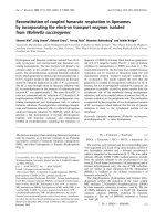
Treatment planning computed tomography scan with contoured organs at risk (incl. left anterior descending coronary artery in red color, on the small images in green color), clinical target volume (both intermediate and large scenario in the same patient) and isodose distributions for the intermediate scenario with ap-pa (upper left), 4-field (lower left), and 7-field IMRT technique (right) in the same patientFigure 2
Treatment planning computed tomography scan with contoured organs at risk (incl. left anterior descending coronary artery in
red color, on the small images in green color), clinical target volume (both intermediate and large scenario in the same patient)
and isodose distributions for the intermediate scenario with ap-pa (upper left), 4-field (lower left), and 7-field IMRT technique
(right) in the same patient.
Intermediate and large target volume
LAD coronary artery
Radiation Oncology 2007, 2:20 />Page 5 of 7
(page number not for citation purposes)
ualised dose distributions beyond a proof-of-principle
study will require more information than that provided
by standard-CT. Angiography, cardio-CT and/or magnetic
resonance imaging will likely have to add data on individ-
ual patient anatomy and pre-existing damage. Another
important question is how reliably such individual dose
distributions can be transferred into daily routine, where
breathing, swallowing, cardiac motion and set-up errors
need to be taken into account. Therefore, assessment of
the influence of motion artefacts and the need for defini-
tion of a safety margin around the LAD is necessary. It
might be possible to use virtual volumes to protect small
structures at risk, as described by Girinsky et al. [4] who
found this strategy superior to dose constraints assigned
to individual organs. However, a detailed discussion of
gating, high-precision and 4-D radiotherapy methodol-
ogy [13,14], is beyond the scope of this article.
As described earlier, there is a price to pay for optimized
heart sparing with IMRT compared to AP/PA: both a
higher mean lung dose (of lesser concern with a total dose
of only 30 Gy) and higher exposure of breast parenchyma
to low radiation doses [5]. However, both the mean lung
dose, V20 and V30 is not higher with IMRT than with 3-D
4 fields, which is important for patients with non-lym-
phoma mediastinal malignancy, where AP/PA techniques
clearly are inappropiate because the total doses required
are much higher than 30 Gy. The IMRT disadvantages
described in our patients were also found in the plan com-
parisons by Girinsky et al. [4]. It appears therefore neces-
sary to select very carefully the patients where heart
sparing is of utmost importance and to weigh the benefits
against the disadvantages. Further refinement is hoped to
result from continued optimization of target volume con-
cepts, e.g., based on positron emission tomography and
early response evaluation during chemotherapy [15,16],
as toxicity risks will decrease with further reduction of the
Table 2: Results for each of the 6 patients
Patient Nr. Magnitude of IMRT advantage in LAD sparing for both
intermediate and large target volume scenarios
Would a decision for IMRT have been the preferred option also with
regard to heart and left ventricle sparing?
1 IMRT outperformed the other techniques to just below
the 25% isodose
Yes, IMRT was optimal
2 IMRT outperformed the other techniques to just below
the 25% isodose
Yes, IMRT was optimal
3 IMRT outperformed the other techniques to just below
the 25% isodose
Yes, IMRT was optimal
4 IMRT outperformed the other techniques down to the
25% isodose
Yes, IMRT was optimal
5 IMRT outperformed the other techniques down to the
50% isodose
IMRT and 4-field were very similar regarding total heart, but IMRT was
slightly better regarding median and mean left ventricle dose (maximum
doses were similar, as were ≤25% isodose levels)
6 IMRT and 4-field very similar, both outperformed AP/PA
down to the 50% isodose
No, 4-field was best (lowest median heart dose and volume receiving 2 Gy,
no disadvantage regarding maximum dose and the various low- dose
parameters)
LAD: left anterior descending coronary artery
Table 1: Median doses to the left anterior descending coronary artery (LAD) in [Gy] for 3 differently sized target volumes
Maximum dose (range)* Median dose (range) Median volume receiving 100% Median volume receiving 25%*
Small, AP-PA 28.5 Gy (27.3–29.4) 23.4 Gy (18.3–27.0) 0% (0-0) 69% (65–80)
Small, 4-field 29.7 Gy (28.5–30.0) 21.3 Gy (13.2–26.1) 0% (0-0) 72% (56–80)
Small, IMRT 28.5 Gy (21.3–29.7) 11.1 Gy (8.7–14.1) 0% (0-0) 70% (53–75)
Intermediate, AP-PA 30.6 Gy (30.0–31.2) 30.0 Gy (26.4–30.0) 50% (1–82) 98% (74–100)
Intermediate, 4-field 30.6 Gy (30.0–30.9) 29.0 Gy (19.2–30.3) 23% (0–93) 100% (89–100)
Intermediate, IMRT 26.9 Gy (23.7–30.0) 15.9 Gy (10.8–29.4) 0.25% (0–26) 92% (78–100)
Large, AP-PA 31.5 Gy (29.7–31.8) 30.5 Gy (30.2–30.6) 90% (60–98) 100% (100-100)
Large, 4-field 31.2 Gy (30.6–31.8) 28.5 Gy (21.9–29.4) 23% (18–25) 100% (100-100)
Large, IMRT 29.1 Gy (27.3–31.2) 15.9 Gy (15.0–21.9) 3% (0–9) 88% (82–95)
* Statistical testing was not performed for these parameters.
p < 0.01 for comparison of the median LAD dose with ap-pa vs. IMRT and 4-field vs. IMRT.
p < 0.01 for comparison of the median volume receiving 100% with ap-pa vs. both 4-field and IMRT (for both intermediate and large PTV). The
differences between 4-field and IMRT are also statistically significant.
Small volume: upper mediastinal plus paraclavicular nodal areas (566–860 ccm), intermediate volume: lower mediastinal nodes in addition (774–
1337 ccm), large volume: hilar nodes in addition (936–1664 ccm)
Radiation Oncology 2007, 2:20 />Page 6 of 7
(page number not for citation purposes)
target volumes [17]. Proton treatment of thoracic target
volumes appears to reduce the dose to normal tissues sig-
nificantly, compared with photon therapy, either 3D-con-
formal or IMRT [18]. However, no planning studies of
selective dose reduction to certain substructures of the
heart have yet been performed and the issue of precise
delivery of such plans to patients is not less complicated
in proton radiotherapy.
Conclusion
The 7-field IMRT technique provided better heart sparing
than traditional approaches in the majority of patients. In
addition to dose reduction to the whole organ, individu-
alised dose distributions can be created, which spare, e.g.,
one ventricle plus one of the coronary arteries. Certain
patients with well-defined vessel pathology might profit
from an approach of general heart sparing with further
selective dose reduction, accounting for the individual
aspects of pre-existing damage.
Competing interests
The author(s) declare that they have no competing inter-
ests.
Authors' contributions
CN and MM participated in the conception and design of
the study and the target volume definition. SS and PK cre-
ated the treatment plans and performed data acquisition.
CN and SS performed data analysis and interpretation
and drafted the manuscript. All authors read and
approved the final manuscript.
References
1. Chandra A, Guerrero TM, Liu HH, Tucker SL, Liao Z, Wang X, Bonnen
MD, Garg AK, Stevens CW, Chang JY, Jeter MD, Mohan R, Cox JD,
Komaki R: Feasibility of using intensity-modulated radiother-
apy to improve lung sparing in treatment planning for distal
esophageal cancer. Radiother Oncol 2005, 77:247-253.
2. Woudstra E, Heijmen BJ, Storchi PR: Automated selection of beam
orientations and segmented intensity-modulated radiother-
apy (IMRT) for treatment of oesophagus tumors. Radiother
Oncol 2005, 77:254-261.
3. Goodman KA, Toner S, Hunt M, Wu EJ, Yahalom J: Intensity-modu-
lated radiotherapy for lymphoma involving the mediastinum.
Int J Radiat Oncol Biol Phys 2005, 62:198-206.
4. Girinsky T, Pichenot C, Beaudre A, Ghalibafian M, Lefkopoulos D: Is
intensity-modulated radiotherapy better than conventional
radiation treatment and three-dimensional conformal radio-
therapy for mediastinal masses in patients with Hodgkin's dis-
ease, and is there a role for beam orientation optimization
and dose constraints assigned to virtual volumes? Int J Radiat
Oncol Biol Phys 2006, 64:218-226.
5. Nieder C, Schill S, Kneschaurek P, Molls M: Comparison of three
different mediastinal radiotherapy techniques in female
patients: impact on heart sparing and dose to the breasts.
Radiother Oncol 2007, 82:301-307.
6. Dobler B, Lorenz F, Wertz H, Polednik M, Wolff D, Steil V, Lohr F,
Wenz F: Intensity-modulated radiation therapy (IMRT) with
different combinations of treatment-planning systems and
linacs: issues and how to detect them. Strahlenther Onkol 2006,
182:481-488.
7. Prosnitz RG, Chen YH, Marks LB: Cardiac toxicity following tho-
racic radiation. Semin Oncol 2005, 32:S71-80.
8. Eich HT, Haverkamp U, Engert A, Kocher M, Skripnitchenko R, Brillant
C, Sehlen S, Duhmke E, Diehl V, Muller RP: Biophysical analysis of
the acute toxicity of radiotherapy in Hodgkin's lymphoma – a
comparison between extended field and involved field radio-
therapy based on the data of the German Hodgkin Study
Group. Int J Radiat Oncol Biol Phys 2005, 63:860-865.
9. Hull MC, Morris CG, Pepine CJ, Mendenhall NP: Valvular dysfunc-
tion and carotid, subclavian, and coronary artery disease in
survivors of Hodgkin lymphoma treated with radiation ther-
apy. JAMA 2003, 290:2831-2837.
10. Heidenreich PA, Schnittger I, Strauss HW, Vagelos RH, Lee BK, Maris-
cal CS, Tate DJ, Horning SJ, Hoppe RT, Hancock SL: Screening for
coronary artery disease after mediastinal irradiation for
Hodgkin's disease. J Clin Oncol 2007, 25:43-49.
11. Schultz-Hector S, Trott KR: Radiation-induced cardiovascular dis-
eases: is the epidemiologic evidence compatible with the radi-
obiologic data? Int J Radiat Oncol Biol Phys 2007, 67:10-18.
12. Becker CR, Ohnesorge BM, Schoepf UJ, Reiser MF: Current devel-
opment of cardiac imaging with multidetector-row CT. Eur J
Radiol 2000, 36:97-103.
13. Schwarz M, van der Geer J, van Herk M, Lebesque JV, Mijnheer BJ,
Damen EM: Impact of geometrical uncertainties on 3D CRT
and IMRT dose distributions for lung cancer treatment. Int J
Radiat Oncol Biol Phys 2006, 65:1260-1269.
14. Weiss E, Wijesooriya K, Dill SV, Keall PJ: Tumor and normal tissue
motion in the thorax during respiration: Analysis of volumet-
ric and positional variations using 4D CT. Int J Radiat Oncol Biol
Phys 2007, 67:296-307.
15. Grosu AL, Piert M, Weber WA, Jeremic B, Picchio M, Schratzenstaller
U, Zimmermann FB, Schwaiger M, Molls M: Positron emission tom-
ography for radiation treatment planning. Strahlenther Onkol
2005, 181:483-499.
16. Yahalom J: Transformation in the use of radiation therapy of
Hodgkin lymphoma: new concepts and indications lead to
modern field design and are assisted by PET imaging and
intensity modulated radiation therapy (IMRT). Eur J Haematol
Suppl 2005, 66:90-97.
17. Koh ES, Tran TH, Heydarian M, Sachs RK, Tsang RW, Brenner DJ, Pin-
tilie M, Xu T, Chung J, Paul N, Hodgson DC: A comparison of man-
tle versus involved-field radiotherapy for Hodgkins
lymphoma: reduction in normal tissue dose and second can-
cer risk. Radiat Oncol 2007, 2:13.
18. Chang JY, Zhang X, Wang X, Kang Y, Riley B, Bilton S, Mohan R,
Komaki R, Cox JD: Significant reduction of normal tissue dose
by proton radiotherapy compared with three-dimensional
Dose-volume histogram for the left anterior descending cor-onary artery with the 7-field IMRT technique in the interme-diate target volume scenarioFigure 3
Dose-volume histogram for the left anterior descending cor-
onary artery with the 7-field IMRT technique in the interme-
diate target volume scenario.
Publish with BioMed Central and every
scientist can read your work free of charge
"BioMed Central will be the most significant development for
disseminating the results of biomedical research in our lifetime."
Sir Paul Nurse, Cancer Research UK
Your research papers will be:
available free of charge to the entire biomedical community
peer reviewed and published immediately upon acceptance
cited in PubMed and archived on PubMed Central
yours — you keep the copyright
Submit your manuscript here:
/>BioMedcentral
Radiation Oncology 2007, 2:20 />Page 7 of 7
(page number not for citation purposes)
conformal or intensity-modulated radiation therapy in Stage
I or Stage III non-small-cell lung cancer. Int J Radiat Oncol Biol Phys
2006, 65:1087-1096.