Báo cáo khoa học: " Hypofractionated intensity modulated irradiation for localized prostate cancer, results from a phase I/II feasibility study" ppsx
Bạn đang xem bản rút gọn của tài liệu. Xem và tải ngay bản đầy đủ của tài liệu tại đây (308.13 KB, 10 trang )
BioMed Central
Page 1 of 10
(page number not for citation purposes)
Radiation Oncology
Open Access
Research
Hypofractionated intensity modulated irradiation for localized
prostate cancer, results from a phase I/II feasibility study
Sara Junius
1
, Karin Haustermans*
1
, Barbara Bussels
2
, Raymond Oyen
3
,
Bianca Vanstraelen
4
, Tom Depuydt
4
, Jan Verstraete
4
, Steven Joniau
5
and
Hendrik Van Poppel
5
Address:
1
Radiation Oncology, University Hospital Gasthuisberg, Herestraat 49, 3000 Leuven, Belgium,
2
Radiation Oncology, H. Hartziekenhuis,
Wilgenstraat 2, 8800 Roeselare, Belgium,
3
Radiology, University Hospital Gasthuisberg, Herestraat 49, 3000 Leuven, Belgium,
4
Physics, University
Hospital Gasthuisberg, Herestraat 49, 3000 Leuven, Belgium and
5
Urology, University Hospital Gasthuisberg, Herestraat 49, 3000 Leuven,
Belgium
Email: Sara Junius - ; Karin Haustermans* - ;
Barbara Bussels - ; Raymond Oyen - ;
Bianca Vanstraelen - ; Tom Depuydt - ;
Jan Verstraete - ; Steven Joniau - ; Hendrik Van
Poppel -
* Corresponding author
Abstract
Background: To assess acute (primary endpoint) and late toxicity, quality of life (QOL), biochemical or clinical failure
(secondary endpoints) of a hypofractionated IMRT schedule for prostate cancer (PC).
Methods: 38 men with localized PC received 66 Gy (2.64 Gy) to prostate,2 Gy to seminal vesicles (50 Gy total) using
IMRT.
Acute toxicity was evaluated weekly during radiotherapy (RT), at 1–3 months afterwards using RTOG acute scoring
system. Late side effects were scored at 6, 9, 12, 16, 20, 24 and 36 months after RT using RTOG/EORTC criteria.
Quality of life was assessed by EORTC-C30 questionnaire and PR25 prostate module. Biochemical failure was defined
using ASTRO consensus and nadir+2 definition, clinical failure as local, regional or distant relapse.
Results: None experienced grade III-IV toxicity. 10% had no acute genito-urinary (GU) toxicity, 63% grade I; 26% grade
II. Maximum acute gastrointestinal (GI) scores 0, I, II were 37%, 47% and 16%. Maximal acute toxicity was reached weeks
4–5 and resolved within 4 weeks after RT in 82%.
Grade II rectal bleeding needing coagulation had a peak incidence of 18% at 16 months after RT but is 0% at 24–36
months. One developed a urethral stricture at 2 years (grade II late GU toxicity) successfully dilated until now. QOL
urinary symptom scores reached a peak incidence 1 month after RT but normalized 6 months later. Bowel symptom
scores before, at 1–6 months showed similar values but rose slowly 2–3 years after RT. Nadir of sexual symptom scores
was reached 1–6 months after RT but improved 2–3 years later as well as physical, cognitive and role functional scales.
Emotional, social functional scales were lowest before RT when diagnosis was given but improved later. Two years after
RT global health status normalized.
Published: 8 August 2007
Radiation Oncology 2007, 2:29 doi:10.1186/1748-717X-2-29
Received: 14 May 2007
Accepted: 8 August 2007
This article is available from: />© 2007 Junius et al; licensee BioMed Central Ltd.
This is an Open Access article distributed under the terms of the Creative Commons Attribution License ( />),
which permits unrestricted use, distribution, and reproduction in any medium, provided the original work is properly cited.
Radiation Oncology 2007, 2:29 />Page 2 of 10
(page number not for citation purposes)
Conclusion: This hypofractionated IMRT schedule for PC using 25 fractions of 2.64 Gy did not result in severe acute
side effects. Until now late urethral, rectal toxicities seemed acceptable as well as failure rates. Detailed analysis of QOL
questionnaires resulted in the same conclusion.
Background
Radiotherapy (RT) is one of the established primary
modalities for treating prostate cancer. About 30% of all
prostate cancer patients, who are treated with curative
intent, receive RT [1] and a substantial proportion of these
patients will be cured. The most common RT technique
for treating prostate cancer is external beam radiotherapy,
often delivered conformally to spare as much normal tis-
sue as possible. A great deal of effort has been put into
improving radiotherapeutic regimens for prostate cancer
through brachytherapy and intensity-modulated radio-
therapy (IMRT). Less attention has, however, been paid to
fraction size.
Brenner and Hall [2] suggested in 1999 an α/β ratio for
prostate cancer of 1.5, much lower than the typical value
of 10 Gy for many other tumours and even lower than the
late-responding tissues (3–4 Gy). This conclusion was
based on a modelling comparison of the doses of 65–80
Gy used for external beams and the higher doses used for
permanent 125-I seed implants which resulted in similar
freedom from biochemical failure rates.
Recent analysis of clinical data (Fowler et al. [3]; Brenner
and Martinez [4]; Bentzen et al. [5]) showed remarkable
agreement with the conclusions of Brenner and Hall's
1999 paper. These estimates are consistent with the very
slow proliferation characteristics of prostate tumours in
comparison with other malignancies. Most prostate
tumours have an extremely low proportion of cycling cells
with an average potential doubling time (Tpot) before
treatment of 40 days ranging from 15 to more than 60
days, compared with about 5 days for many other types of
tumour [6-8].
A recent publication done by Williams et al. [9] supports
the concept of a low α/β ratio but their data are more con-
sistent with a value in the range of 2 to 5 Gy.
The disparity between the α/β value of 3–4 Gy for late
complications and < 2 Gy for prostate tumours raises the
prospect that we might be able to widen the therapeutic
window by treating prostate cancer with hypofractionated
radiation [10,11]. A similar rationale (but in the opposite
direction) has worked out well in hyperfractionation for
head and neck tumors [12]. In addition to possible radio-
biological gains there are other benefits to a hypofraction-
ation scheme. The shorter overall treatment time increases
convenience for the patients and decreases cost. At
present, the main concern is uncertainty about normal tis-
sue toxicity of such hypofractionated protocols. So far the
results and the toxicity are acceptable, but there is still a
lack of long-term follow-up data.
In 12/2002 we started a phase I/II hypofractionation pro-
tocol in prostate cancer. The primary endpoint was assess-
ment of the feasibility of a hypofractionation schedule to
deliver a total dose of 66 Gy in 25 fractions of 2.64 Gy in
five weeks for patients with localized prostate cancer using
IMRT. Here we present our results for a group of 38 men
treated between 12/2002 and 05/2006.
Patients and methods
Patients characteristics
From 12/2002 until 6/2005, 38 men with biopsy proven
prostate adenocarcinoma and a clinically localized stage
(cT1–T4 N0M0, using the UICC 2002 TNM classification)
were recruited in this single institution study. Ethical com-
mittee of UZ Gasthuisberg Leuven approved the protocol
and all patients provided written informed consent. WHO
performance status ranged from 0–1. Mean age was 71
years (range: 54–79 years). Median pre-treatment PSA was
9.2 μg/l (range: 2.77–45.6 μg/l). Gleason scores ranged
from 5 to 10. Table 1 shows the disease characteristics.
According to the d'Amico prognostic factors 18% were
low risk, 50% intermediate risk and 32% high risk
patients.
31/38 patients received hormonal treatment (HT) with
LHRH agonist +/- antiandrogen therapy varying from 6
months to a total of 4 years and in all cases concurrently
with radiotherapy. Exclusion criteria were previous irradi-
ation in the pelvic area, previous surgery for prostate can-
cer, nodal or distant metastasis proven by a CT pelvis or
bone scan, presence of any psychological, familial, geo-
graphical or sociological condition potentially hampering
compliance with study protocol and follow-up schedule.
End points
Primary endpoint of the study was the occurrence of any
grade II or more acute GU or GI toxicity during and within
three months after RT, scored by using the RTOG scoring
system. Secondary endpoints were late GU or GI toxicity
scored by RTOG/EORTC scoring system; QOL with the
EORTC 30 questionnaire and PR25 prostate module; bio-
chemical free survival as defined by the 1997 American
Society of Therapeutic Radiation and Oncology (ASTRO)
Radiation Oncology 2007, 2:29 />Page 3 of 10
(page number not for citation purposes)
consensus definition [13,14] and nadir + 2 definition
[15,16] or clinical failure defined as local, regional or dis-
tant relapse.
Dose and technique
All 38 patients were treated by the same hypofractionated
schedule to a total dose of 66 Gy in 25 fractions in five
weeks of 2.64 Gy to the prostate with 50 Gy in 25 fractions
of 2 Gy to the seminal vesicles using IMRT. For late effects,
characterized by an α/β of 3 Gy, this is an isoeffective
schedule compared to our current schedule of 74 Gy in 37
fractions of 2 Gy. For the prostate tumor the chosen dose
is equivalent to 78 Gy in 39 fractions of 2 Gy for an α/β of
1.5 Gy.
All patients were simulated in supine position with feet
fixation. Skin marks representing the isocenter were
placed at both sides of the hips, epigastric and at the level
of the pubis. Lateral and anterior simulation X-rays were
taken in order to document the position of the isocenter.
Patients were instructed to empty their bladder before
simulation and drink a steady amount of 250 cc water
before scanning. A rectal enema was used to empty the
rectum as much as possible. A CT-scan in treatment posi-
tion with IV contrast with 3 mm slices taken from the anal
verge to the level of the acetabulum was performed, fol-
lowed by an MRI the same day. CT and MRI images were
fused. Prostate, seminal vesicles and organs at risk (OAR's:
bladder, rectum and anterior rectal wall) were outlined on
the MRI. Rectum and anterior rectal wall were outlined
from the anal verge to the rectosigmoid junction and the
whole bladder was included.
The CTV1 included the prostate; CTV2 was used for the
seminal vesicles. PTV1 was defined as CTV1 + 1 cm, PTV2
as CTV2 + 0.5 cm. The PTV1s were planned to receive a
D99% of 59.4 Gy, D95% of 62.7 Gy, D50% of 66 Gy. The
PTV2s were planned to receive a D99% of 45 Gy, D95%
of 47.5 Gy, D50% of 50 Gy.
The OAR's planning limits were based on prior studies
(17). Less than or equal to 25%, 50% and 70% of the rec-
tum volume could receive respectively 70 Gy (2 Gy/fx), 45
Gy (2.64 Gy/fx), 38 Gy (2.64 Gy/fx) with a maximum tol-
erated dose of 76 Gy (2 Gy/fx). For the rectum the DVH's
were recalculated to the equivalent dose in 2 Gy per frac-
tion using the LQ model assuming α/β = 3 Gy and only
for the dose above 50 Gy (25 fractions). Below 50 Gy, the
original DVH was used as we preferred to overestimate
rectal doses instead of underestimating them. Maximum
dose to the anterior rectal wall was set at 66.5 Gy with a
maximal dose never exceeding 13.3 Gy/week. Fifty per-
cent of the bladder volume could receive up to 70 Gy (2
Gy/fx).
IMRT with inverse treatment planning on the Eclipse
planning system (Varian) was performed using a five field
18 MV photon beam set-up. Pre-treatment verification of
the dose distribution was done with an IMRT phantom
and an amorphous silicon imager. During treatment the
patient was advised to have a full bladder and to empty
his rectum before treatment. The patient was localized
daily using the BAT transabdominal ultrasound system (n
= 14) or portal imaging of bony structures (n = 24).
Toxicity
Acute side effects were scored weekly during RT, weekly
afterwards until acute effects were resolved, at 1 and 3
months after RT using the RTOG scoring system. Late
effects were scored at 6, 9, 12, 16, 20, 24, 36 months using
the RTOG/EORTC late morbidity scoring system.
Table 1: disease parameters (iPSA: initial pretreatment PSA; HT: hormonal treatment).
Parameters Number (%)
iPSA < 10 18 (47%)
10–20 16 (42%)
> 20 4 (11%)
Stage T1c 6 (16%)
T2a 10 (26%)
T2c 10 (26%)
T3a 10 (26%)
T4 2 (6%)
Gleason score < or = 5 2 (6%)
6–7 25 (66%)
8–10 11 (28%)
HT No 7 (18%)
Yes 31 (82%)
Radiation Oncology 2007, 2:29 />Page 4 of 10
(page number not for citation purposes)
Quality of Life (QOL)
QOL was scored at baseline; 1 and 6 months; 1, 2 and 3
years after RT using EORTC-C30 questionnaire and PR25
prostate module.
Failure rates
Evaluation of tumour response was performed by digital
rectal examination and PSA levels 3-monthly the first year
after RT, 4-monthly the second and third year and from
then on every 6 months until the fifth year after RT when
it was on yearly basis. On suspicion of tumour recurrence
or progression a CT scan of the pelvis, ultrasonography of
the prostate and a bone scan were performed. Prostate
biopsies were not systematically performed. We defined
failure as biochemical or clinical failure. Biochemical fail-
ure was defined according to (ASTRO) consensus guide-
lines [13,14] as three consecutive rises in PSA level after
the nadir. The nadir + 2 definition was also used as recent
publications [15,16] pointed out that this definition
appears to be optimal and may be selected as the new
RTOG-ASTRO definition. Clinical failure included local,
regional or nodal relapse and distant metastasis.
Statistics
The Fleming one stage testing procedure was used [18].
The hypotheses were: (1–P0) is the highest probability of
toxicity which, if true, implies that the irradiation sched-
ule does not warrant further investigation, in this trial P0
has been taken as 50% incidence of grade II or more; (1–
P1) is the lowest probability of toxicity which, if true,
implies that the irradiation schedule does warrant further
clinical investigation; in this trial P1 has been taken as
70%; α is the accepted probability of recommending for
further trials the regimen if the toxicity is equal to or
higher than 30%, in this trial α has been taken as 0.1; β is
the accepted probability of rejecting from further trials the
regimen if the stated toxicity is equal or less than 50%; in
this trial β has been taken as 0.1. Under these hypotheses
a total sample size of 38 patients was calculated.
Results
Compliance with the study protocol was excellent. All 38
patients were scored according to protocol and filled in
QOL questionnaires.
Median follow-up was 20 months (range 6–36) after com-
pleting RT.
Dosimetric parameters
Table 2 shows that the mean delivered doses for the PTV1
D99%, D95%, D50% and PTV2 D99%, D95%, D50%
were higher than the constraints and confirmed the RT
schedule. Mean delivered doses for the OAR were lower
than set up constraints.
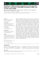
Acute GU symptoms (Figure 1)
Four patients (10%) had no acute GU toxicity while 63%
(n = 24) experienced a maximum of Grade I and 26% (n
= 10) Grade II during RT. None developed a grade III/IV
acute GU toxicity. Acute GU toxicity reached its maximum
in weeks 4 and 5 and resolved within 4 weeks after RT in
82% (n = 31) of the patients. At three months after RT, 5
patients (13%) had Grade I GU toxicity.
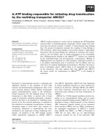
Acute GI symptoms (Figure 2)
Maximum acute GI grades of 0, I and II were respectively
37% (n = 14), 47% (n = 18) and 16% (n = 6). Detailed
scoring of rectal mucus or blood loss resulted probably in
the rather high incidence of Grade II toxicity. No Grade
III/IV toxicity was found. At 3 months after RT, 6 men
(16%) had Grade I toxicity.
Late GU symptoms
At 6 months after RT only one (3%) patient had Grade I
GU toxicity. At one year (n = 26), at 16 (n = 16), and 20
months (n = 14) after RT, none of the patients experienced
GU toxicity. At two years (n = 10) one patient was diag-
nosed with a stricture of the urethra scored as Grade II late
GU toxicity. After single dilatation dysuria disappeared. At
36 months (n = 6) no late GU toxicity was found.
Late GI symptoms (Table 3)
At 6 months after RT, 6/38 (16%) had Grade I toxicity due
to slight rectal discharge or mildly increased bowel move-
ments. 1/38 (3%) experienced a Grade II toxicity due to
intermittent rectal bleeding with rectoscopy proven tel-
angiectasia, needing coagulation. At one year after RT 5/
26 men (19%) had Grade I toxicity because of persisting
slight rectal discharge, 1/26 (4%) were scored as Grade II
as described above. At 16 months after RT 5/16 (31%) had
Grade I toxicity; three of them because of persisting slight
rectal discharge, the other two because of mild rectal
bleeding. One had telangiectasia where no therapy was
performed, for the other one no cause for the rectal bleed-
ing was found. 3/16 patients (18%) complained at that
time of intermittent bleeding. Telangiectasia were docu-
mented by rectoscopy and coagulation was performed
with an excellent result in one patient. The other patient
received a second coagulation at 20 months after RTwith
a good result until now (Grade II toxicity). No Grade III
toxicity was seen with a median follow up of 20 months.
At 24 and 36 months there were still respectively 3/10 and
2/6 patients with Grade I toxicity due to persistent slight
rectal bleeding not needing coagulation, but no Grade II
or III toxicity was found.
QOL
Following scoring procedures [19,20] we calculated the
mean values of urinary, bowel and sexual symptom scales;
functional scales (physical, role, emotional, cognitive and
Radiation Oncology 2007, 2:29 />Page 5 of 10
(page number not for citation purposes)
social functioning) and global health status as summa-
rized in Table 4. Urinary symptom scores reached a peak
incidence one month after RT but normalized 5 months
later. They stabilized at the mean value of 4 at one, two
and three years after RT which was lower than the starting
value of 9.6. Bowel symptom scores before, at one and six
months after RT showed a similar value of 2.5, but rose
slowly to 3.1 at one year, 5 at two and 5.8 at three years
after RT. A nadir of sexual symptom scores was reached
from one to six months after RT but this improved to a
value of 40 at two and three years after RT (compared with
a value of 44 before radiation). Physical, cognitive and
role functional scales showed the same pattern of a lower
value at one month after RT, an increase to a maximum at
one year and a slowly decrease at two and three years after
RT. Emotional and social functional scales showed the
lowest score before RT when diagnosis was given and
improved gradually over the following months and years.
Table 2: mean (range) delivered doses to PTV1 (prostate), PTV2 (seminal vesicles), OAR's (rectum, anterior rectal wall, bladder)
Volume Constraints Mean delivered dose
PTV1 99% 59.4 Gy 62.2 Gy (60.7–63)
95% 62.7 Gy 63.7 Gy (62.7–64.6)
50% 66 Gy 66 Gy (64–66.6)
PTV2 99% 45 Gy 47.6 Gy (45–49.8)
95% 47.5 Gy 49.6 Gy (47–52.3)
50% 50 Gy 56.8 Gy (52–61)
Rectum 25% 70 Gy (2 Gy) 55.3 Gy (30–65.4)
50% 45 Gy (2.64 Gy) 35.7 Gy (17–43)
70% 38 Gy (2.64 Gy) 22.7 Gy (6.6–34.4)
Max Dose 76 Gy (2 Gy) 74.5 Gy (66–74.9)
Anterior rectal wall 66.5 Gy (2.64 Gy) 66.1 Gy (65–66.7)
Bladder 50% 70 Gy (2 Gy) 35.1 Gy (5.5–58.3)
acute GU toxicity in all 38 patientsFigure 1
acute GU toxicity in all 38 patients.
Acute GU toxicity (N=38)
0
5
10
15
20
25
30
35
40
we
ek
1
we
ek
2
w
e
ek
3
w
e
ek
4
w
e
ek
5
w
e
ek
6
w
e
ek
7
1
m
o
nt
h
3 months
Time
Number of patients
G 0
G I
G II
G III
G IV
Radiation Oncology 2007, 2:29 />Page 6 of 10
(page number not for citation purposes)
Two years after RT global health status reached about the
same value as before therapy. The lowest value was found
at one month after RT.
Biochemical or clinical failure
At time of assessment, biochemical failure defined as
three consecutive rises after the nadir according to ASTRO
consensus definition occurred in 3/38 patients, one at 12
and two at 16 months posttherapy. In these three cases a
6 months course of HT was given concurrently with radi-
otherapy. However if nadir + 2 definition was used, no
biochemical failure is reported until now. Clinical failure
in terms of nodal relapse was seen in one patient one year
after RT for which salvage HT was started. Unfortunately
the disease became hormone refractory one year later and
due to alcohol induced severe liver disorder the patient
could not receive chemotherapy which resulted in death a
few months later. One patient developed lung metastasis
three months after RT, due to a secondary colorectal ade-
nocarcinoma. PSA-levels of this patient are still below
detection level. The latter also occurred in another patient
who unfortunately died due to a metastasized lung carci-
noma diagnosed 4 months after RT for prostate cancer.
Discussion
The main objective of this study was to assess the feasibil-
ity in terms of acute genito-urinary and gastro-intestinal
(primary endpoint) and late (secondary endpoint) toxic-
ity of delivering a hypofractionated schedule of 25 frac-
tions of 2.64 Gy to a total dose of 66 Gy in five weeks to
acute GI toxicity in all 38 patientsFigure 2
acute GI toxicity in all 38 patients.
Acute GI toxicity (N=38)
0
5
10
15
20
25
30
35
40
w
e
e
k
1
w
e
e
k
2
w
e
e
k
3
w
e
e
k
4
w
e
e
k
5
w
e
e
k
6
w
e
e
k
7
1
mo
n
t
h
3m
o
n
t
h
s
Time
Number of patients
G 0
G I
G II
G III
G IV
Table 3: late GI toxicity
6 months 9 months 12 months 16 months 20 months 24 months 36 months
N° patients 38 36 26 16 14 10 6
Grade 03129208 9 7 4
Grade I*6555432
Grade II*1213100
Grade III0000000
Grade IV0000000
*grade I: mucosal discharge, mild rectal bleeding not needing coagulation
*grade II: rectal bleeding with telangiectasia on rectoscopy and coagulation
Radiation Oncology 2007, 2:29 />Page 7 of 10
(page number not for citation purposes)
patients with localized prostate cancer using IMRT. The
hypofractionated schedule is iso-effective (at α/β = 3) for
late effects with a schedule of 74 Gy (2 Gy). According to
Fowler et al. [21] this hypofractionation regimen with an
overall treatment time of 5 weeks and fraction number of
25 is estimated to be very unlikely to result in significantly
increased late effects.
Previous late toxicity reports of radiation in prostate can-
cer at the Leuven University Hospital were given by
Vanuytsel and Van Poppel [22] for a schedule of 2 Gy frac-
tions to 60 Gy. They found no grade III of higher late side
effects in a subset of patients prospectively randomized in
EORTC trial 22911 evaluating the role of postoperative
radiotherapy in pT3 patients. If the present study proved
to be feasible, a multicenter phase III trial could be started
comparing conventional fractionation of 74 Gy in 2 Gy
fraction with hypofractionation giving 66 Gy in 25 frac-
tions of 2.64 Gy in patients with localized prostate cancer.
Assuming a value of 3% per Gy for the slope of the tumor
control probability curve, this strategy could lead to an
increase in bNED from 70% at 5 years to a bNED of 82%
at 5 years.
Acute toxicity
Acute effects observed in this hypofractionated regimen
were comparable to those reported by others [23-26]. A
26% grade II acute GU toxicity and 16% grade II acute GI
toxicity was found with a peak incidence weeks 4 and 5 of
the regimen. No grade III/IV acute GU and GI toxicity was
found. This is comparable with the findings of Peeters et
al. [27] in their 68–78 Gy (2 Gy) trial. Pollack et al. [28]
reported in their randomized trial somewhat higher fig-
ures of grade II (40%) and grade III (8%) acute GU toxic-
ity in the H-IMRT arm (70.2 Gy in 26 fractions of 2.7 Gy)
although PTV margins were slightly smaller. Inclusion of
lymph nodes in high-risk patients, the use of a modified
RTOG scale and mean biological doses to the prostate
exceeding 80 Gy were held responsible for these findings.
Acute GI toxicity figures were similar to those reported by
others. They found no statistical differences in acute GU
and GI toxicity between the C-IMRT (76 Gy in 38 fractions
of 2 Gy) and above mentioned H-IMRT arm. Slightly
higher figures of acute GI and GU toxicity were also
recently reported by Soete et al. [29] in a phase II multi-
institutional study were 36 prostate cancer patients
received a total dose of 56 Gy in 16 fractions over 4 weeks
to the prostate. Lukka et al. [30] compared 66 Gy in 33
fractions of 2 Gy to 52.5 Gy in 20 fractions over 28 days
and found higher acute urinary (5.1 vs 9.2%) and rectal
(2.8 vs 4.3%) in the hypofractionated arm. Kupelian et al.
[31-33] compared acute toxicity of a three-dimensional
conformal radiotherapy scheme of 78 Gy in 39 fractions
of 2 Gy for prostate cancer patients with a later IMRT
scheme of 70 Gy in 28 fractions of 2.5 Gy. Comparable
rates of acute GU (20% conformal vs 21% IMRT) and GI
(19% conformal vs 14% IMRT) were found. Up until now
reports on the degree of acute toxicity of prostate cancer
patients treated with a hypofractionated radiotherapy reg-
imen are not consistent, probably due to different organ
at risk constraints, different radiotherapy techniques (con-
formal vs IMRT) and PTV margins used. Another question
that needs to be answered is the influence of hormonal
treatment (HT) on acute toxicity in men with prostate can-
cer treated with radiotherapy. Peeters et al. [34] concluded
that neo-adjuvant HT appeared to be an independent
prognostic factor for acute toxicity, resulting in less acute
GI, but more acute GU toxicity. The first could be
explained by the shrinking of the prostate and seminal
vesicles with subsequent smaller RT fields and less expo-
sure of the rectal wall [35,36]; but for the latter no obvious
explanation was suggested. An additive effect of androgen
suppression and external irradiation on local control by
induction of apoptosis is reported by several authors
[37,38]. This phenomenon could have an increasing effect
Table 4: mean values of urinary, bowel, sexual symptom scales; functional scales; global health status
before 1 month 6 months 1 year 2 years 3 years
N° patients 38 38 38 26 10 6
Symptom scales
urinary 9.6 15.9 8.3 4.6 4.5 4.3
bowel 2.5 2.7 2.6 3.1 5 5.8
sexual44 17.117.219 40 40
Functional scales
physical89.986.987.990.889.484.4
role 90.4 85.1 92.5 90.4 88.4 83.3
emotional85.989.994.395.694.297.2
cognitive 87.3 86.4 88.5 89.5 81.7 77.2
social 93.5 93.6 96.5 94.9 98.4 100
Global health
status
82.7 60.3 78.1 79.1 81.6 75
Radiation Oncology 2007, 2:29 />Page 8 of 10
(page number not for citation purposes)
on normal tissue toxicity and explain the higher acute GU
toxicity rates in the hormonal therapy (HT) arm of the
Peeters study. Also in our hypofractionated regimen acute
toxicity figures could be influenced by this phenomenon
as 31 of the 38 patients received HT concurrently with RT.
Late toxicity
No late GU toxicity was found at 6, 9, 12, 20 months after
RT. At two years one patient was diagnosed with a stricture
of the urethra scored as a Grade II late GU toxicity. After
dilatation his symptoms of dysuria disappeared. Late GI
toxicity and especially rectal bleeding seems more impor-
tant. Yeoh et al [39] reported in their randomized trial a
sustained increase in GI toxicity at two years after RT com-
pared with baseline in both arms (conventional 64 Gy in
32 fractions versus hypofractionation 55 Gy in 22 frac-
tions). In the hypofractionated arm they found a slightly
greater percentage of patients experiencing mild rectal
bleeding at two years, but this difference was not statisti-
cally significant. In this study grade II rectal bleeding that
needs coagulation has reached a peak incidence of 18% at
16 months after radiotherapy and is now 0% at 24 and 36
months. We believe that intensive detailed scoring for rec-
tal bleeding followed by rectoscopy and immediate coag-
ulation if telangiectasia was present, contributed to these
figures. The majority of these patients had significant car-
diac morbidity and the large use of anticoagulants could
also be responsible for earlier recognition of rectal blood
loss.
The influence of HT on late radiotherapy toxicity has been
examined in a retrospective study by Jani et al [40]. They
observed similar late GU and GI toxicity rates in 455
patients who did (n = 197) and did not (n = 248) receive
HT. These findings are not consistent with other investiga-
tions that demonstrated a greater rate of late GI toxicity
and especially late rectal bleeding with the use of HT. San-
guineti et al. [41] reported in a multivariate analysis 2-year
estimates of grade II-IV late rectal toxicity of 30.3% in
patients receiving HT versus 14% in patients without HT.
As all the present patients with grade II rectal bleeding
received concurrently HT in our study, we believe that
these late rectal bleeding figures could be strongly influ-
enced by the HT addition.
QOL
Urinary symptom scores reached a peak incidence 1
month after RT, but normalized 5 months later. At one,
two and three years after RT a stabilisation was noticed
and the mean value of 4 was lower than the starting value
of 9.6, probably due to prostate shrinkage and tumour
control. Bowel symptoms scores before, at one and six
months after RT showed the same value of 2.5 but slowly
rose to 3.1 at one year, 5 at two and 5.8 at three years after
RT which can be explained by detailed reporting of rectal
bleeding. One to six months after RT the lowest value of
sexual symptoms scores was reached probably due to the
concurrent use of HT with radiotherapy. Two and three
years later a value of 40 (compared with the value of 44
before therapy) was found. A possible explanation for this
phenomenon could be the short duration of HT in most
of the patients, but also the use of 5 fosfodiësterase inhib-
itors especially in the younger ones. Physical, cognitive
and role functional scales showed the same pattern of a
lower value at one month after RT, an increase to a maxi-
mum value at one year after RT and a slow decrease at two
and three years probably due to aging of the patient pop-
ulation. Emotional and social functional scales showed
the lowest score before RT when diagnosis was given and
improved gradually in the months and years after RT. Two
years after RT global health status reached about the same
value as before. The lowest value was also in this case
reached at one month after RT.
Failure
In one patient biochemical failure according to ASTRO
consensus definition was noticed one year after RT and in
two others at 16 months. A major issue is the use of a
short course (6 months) of HT concurrently with RT in
these cases. After cessation of HT, a transient increase in
PSA may occur as a result of recovery of prostate tissue
from testosterone suppression. This may lead to false-pos-
itive results with ASTRO definition and a recalculation
with nadir + 2 definition was performed. With this defini-
tion no biochemical failure was seen until now. Clinical
failure in terms of nodal relapse was seen in one patient
one year after therapy for which salvage HT was started
but resulted in death one year later.
Conclusion
In conclusion, this phase I/II study shows acceptable acute
GU and GI toxicity rates resulting from a hypofractionated
regimen of 66 Gy in 25 fractions of 2.64 Gy for localized
prostate cancer. Late urethral toxicity and rectal bleeding
rates may be influenced by the addition of hormonal ther-
apy but seem acceptable, although we are aware that
longer follow up is needed to see if these figures can be
maintained. Detailed analysis of different QOL scales
resulted in the same conclusion. The future of all this is
likely to include fewer and larger fractions in the radiation
treatment of prostate cancer, keeping overall treatment
time not too short like three or four fractions a week. The
important thing, until more and more tumour results
come through and we can see what α/β for tumours really
is, is to keep normal tissue reactions under control.
Acknowledgements
We thank Jack Fowler for careful reading of the manuscript and helpful dis-
cussion.
Radiation Oncology 2007, 2:29 />Page 9 of 10
(page number not for citation purposes)
References
1. Jones GW, Mettlin C, Murphy GP, Guinan P, Herr HW, Hussey DH,
Chmiel JS, Fremgen AM, Clive RE, Zuber-Ocwieja KE: Patterns of
care for carcinoma of the prostate gland. Results of a
national survey of 1984 and 1990. J Am Coll Surg 1995,
180:545-554.
2. Brenner DJ, Hall EJ: Fractionation and protraction for radio-
therapy of prostate carcinoma. Int J Radiat Oncol Biol Phys 1999,
43(5):1095-1101.
3. Fowler JP, Chappell R, Ritter M: Is alpha/beta for prostate
tumors really low? Int J Radiat Oncol Biol Phys 2001,
50(4):1021-1031.
4. Brenner DJ, Martinez AA, Edmundson GK, Mitchell C, Thames HD,
Armour EP: Direct evidence that prostate tumors show high
sensivity to fractionation (low alpha/beta) comparable with
late responding normal tissue. Int J Radiat Oncol Biol Phys 2002,
52(1):6-13.
5. Bentzen SM, Ritter MA: The alpha/beta ratio for prostate can-
cer: What is it, really? Radiother Oncol 2005, 76:1-3.
6. Haustermans KM, Hofland I, Van Poppel H, Oyen R, Van de Voorde
W, Begg AC, Fowler JF: Cell kinetics measurements in prostatic
cancer. Int J Radiat Oncol Biol Phys 1997, 37:1067-1070.
7. Haustermans K, Fowler JF: A comment on proliferation rates in
human prostate cancer (letter). Int J Radiat Oncol Biol Phys 2000,
48(1):297-297.
8. Scrivner DL, Meyer DS, Rujanavech N, Fathman A, Scully T: Cell
kinetics by bromodeoxyuridine labelling and deoxyribonu-
cleic acid ploidy in prostatic carcinoma needle biopsies. J Urol
1991, 146:1034-1039.
9. Williams S, Taylor J: Use of individual fraction size data from
3756 patients to directly determine the alpha/beta ratio of
prostate cancer. Int J Radiat Oncol Biol Phys 2007, 68(1):24-33.
10. Van der Kogel AJ, Jarrett KA, Paciotti MA, Raju MR: Radiation tol-
erance of the rat rectum fractionated X-Rays and pimesons.
Radiother Oncol 1988, 12:225-232.
11. Brenner DJ: Fractionation and late rectal toxicity.
Int J Radiat
Oncol Biol Phys 2004, 60:1013-1015.
12. Stuschke M, Thames HDl: Fractionation sensitivities and dose-
control relations of head and neck carcinomas: analysis of
the randomized hyperfractionation trials. Radiother Oncol
1999, 51:113-121.
13. American Society for Therapeutic Radiology and Oncology Consen-
sus Panel: Consensus Statement: Guidelines for PSA following
radiation therapy. Int J Radiat Oncol Biol Phys 1997, 37:1035-1041.
14. American Society for Therapeutic Radiology and Oncology Consen-
sus Panel: PSA Relapse Definitions. In Radiology and Oncology New
Orleans, LA, American Society for Therapeutics; 2002.
15. Chueng R, Tucker SL, Lee AL, Dong L, Kamat A, Pisters L, Kuban DA:
Assessing the impact of an alternative biochemical failure
definition on radiation dose response for high-risk prostate
cancer treated with external beam radiotherapy. Int J Radiat
Oncol Biol Phys 61(1):14-9. 2005; jan 1
16. Williams SG, Duchesne GM, Gogna NK, Millar JL, Pickles T, Pratt GR,
Turner S: An international multicenter study evaluating the
impact of an alternative biochemical failure definition on the
judgment of prostate cancer risk. Int J Radiat Oncol Biol Phys
65(2):351-7. 2006 June 1
17. Storey MR, Pollack A, Zagers G, Smith L, Antolak J, Rosen I: Com-
plications from radiotherapy dose escalation in prostate can-
cer: preliminary results of a randomized trial. Int J Radiat Oncol
Biol Phys 2000, 48:635-642.
18. Fleming TR: One-sampling multiple testing procedure for
phase II clinical trials. Biometrics 1982, 38:143-151.
19. EORTC Quality of life group QLQ-C30 reference values. CD rom version 1.0
1998.
20. Van der Gucht S, Van Audenhove C: De beoordeling van de kwaliteit van
leven van patienten met gelokaliseerde prostaatkanker. Evaluatie van een
meetinstrument. Thesis Faculteit Psychologie en Pedagogische Wetenschap-
pen 2004.
21. Fowler JF, Ritter MA, Chappell RJ, Brenner DJ: What hypofraction-
ated protocols should be tested for prostate cancer? Int J
Radiat Oncol Biol Phys 2003, 56(4):
1093-1104.
22. Vanuytsel L, Van Poppel H: Adjuvant radiotherapy after radical
prostatectomy. In Radiotherapy of prostate cancer Edited by: Greco
C, Zelefsky M. London, Harwood, GmbH; 1999:377-384.
23. Lloyd-Davies RW, Collins CD, Swan AV: Carcinoma of prostate
treated by radical external beam radiotherapy using hypof-
ractionation: Twenty-two years' experience (1962–1984).
Urology 1990, 36:107-111.
24. Logue JP, Cowan RA, Hendry JH: Hypofractionation for prostate
cancer. Int J Radiat Oncol Biol Phys 2001, 49:1522-1523.
25. Duncan W, Warde P, Catton CN, Murro AJ, Lakier R, Gadalla T,
Gospodarowicz MK: Carcinoma of the prostate: Results of rad-
ical radiotherapy (1970–1985). Int J Radiat Oncol Biol Phys 1993,
26:203-210.
26. Logue JP: Hypofractionation for prostate cancer: letter to the
editor. Int J Radiat Oncol Biol Phys 2001, 49(5):1522-1522.
27. Peeters ST, Heemsbergen WD, Van Putten WL, Slot A, Tabak H,
Mens JW, Lebesque JV, Koper PC: Acute and late complications
after radiotherapy for prostate cancer: Results of a multi-
center randomized trial comparing 68 to 78 Gy. Int J Radiat
Oncol Biol Phys 2005, 61:1019-1034.
28. Pollack A, Hanlon AL, Horwitz EM, Feigenberg SJ, Konski AA, Mousas
B, Greenberg RE, Uzzo RG, Ma CM, McNeeley SW, Buyyounouski
MK, Price RA Jr: Dosimetry and preliminary acute toxicity in
the first 100 men treated for prostate cancer on a rand-
omized hypofractionation dose escalation trial. Int J Radiat
Oncol Biol Phys 2006, 64(2):518-526.
29. Soete G, Arcangeli S, De Meerleer G, Landoni V, Fonteyne V, Arcan-
geli G, De Neve W, Storme G: Phase II study of a four-week
hypofractionated external beam radiotherapy regimen for
prostate cancer: report on acute toxicity. Radiat Oncol 2006,
80(1):78-81. Epub 2006 Jul 7
30. Lukka H, Hayter CH, Julian JA, Warde P, Morris WZ, Gospodarowicz
M, Levine M, Sathya J, Choo R, prichard H, Brundage M, Kwan W:
Randomized trial comparing two fractionation schedules for
patients with localized prostate cancer. Journal of Clinical Oncol-
ogy 2005, 23:6132-6138.
31. Kupelian PA, Reddy CA, Carlson TP, Altsman KA, Willoughby TR:
Preliminary observations on biochemical relapse-free sur-
vival rates after short-course intensity modulated radiother-
apy (70 Gy at 2.5 Gy/fraction) for localized prostate cancer.
Int J Radiat Oncol Biol Phys 2002, 53:904-912.
32. Kupelian PA, Reddy CA, Klein EA, Willoughby TR: Short-course
intensity modulated radiotherapy (70 Gy at 2.5 Gy/fraction)
for localized prostate cancer: preliminary results on late tox-
icity and quality of life. Int J Radiat Oncol Biol Phys 2001,
51:988-993.
33. Kupelian PA, Thakkar VV, Khuntia D, Reddy CA, Klein EA,
Mahadevan A: Hypofractionated intensity-modulated radio-
therapy (70 Gy at 2.5 Gy per fraction) for localized prostate
cancer: Long term outcomes. Int J Radiat Oncol Biol Phys 2005,
63(5):1463-1468.
34. Peeters ST, Hoogeman MS, Heemsbergen WD, Slot A, Tabak H,
Koper PC, Lebesque JV: Volume and hormonal effects for acute
side effects of rectum and bladder during conformal radio-
therapy for prostate cancer. Int J Radiat Oncol Biol Phys 2005,
63(4):1142-1152.
35. Zelefsky MJ, Harrison A: Neoadjuvant androgen ablation prior
to radiotherapy for prostate cancer: Reducing the potential
morbidity of therapy. Urology 1997, 49:38-45.
36. Tsai HK, Manola J, Abner A, Talcott JA, D'Amico AV, Beard C:
Patient-reported acute gastro-intestinal toxicity in men
receiving 3-dimensional conformal radiation therapy for
prostate cancer with or without neoadjuvant androgen sup-
pression therapy. In Urologic Oncology Volume 23. Seminars and
Original investigations; 2005:230-237.
37. Joon DL, Hasegawa M, Sikes C, Khoo VS, Terry NH, Zagars GK, Meis-
trich ML, Pollack A: Supraadditive apoptotic respone of R3327-
G rat prostate tumors to androgen ablation and radiation.
Int J Radiat Oncol Biol Phys 1997, 38:1071-1077.
38. Zietman AL, Prince EA, Nakfoor BM, Park JJ: Androgen depriva-
tion and radiation therapy; sequencing studies using the
Shionogi in vivo tumor system. Int J Radiat Oncol Biol Phys 1997,
38:1067-1070.
39. Yeoh EE, Fraser RJ, Borg MF, McGowan RE, Botten RJ, Di Matteo AC,
Roos DE, Penniment MG, Borg MF: Evidence for efficacy without
increased toxicity of hypofractionated radiotherapy for pros-
tate carcinoma: early results of a Phase III randomized trial.
Int J Radiat Oncol Biol Phys 2003, 55(4):943-955.
Publish with BioMed Central and every
scientist can read your work free of charge
"BioMed Central will be the most significant development for
disseminating the results of biomedical researc h in our lifetime."
Sir Paul Nurse, Cancer Research UK
Your research papers will be:
available free of charge to the entire biomedical community
peer reviewed and published immediately upon acceptance
cited in PubMed and archived on PubMed Central
yours — you keep the copyright
Submit your manuscript here:
/>BioMedcentral
Radiation Oncology 2007, 2:29 />Page 10 of 10
(page number not for citation purposes)
40. Jani AB, Gratzle J: Late radiotherapy toxicity after prostate
cancer treatment: influence of hormonal therapy. Urology
2005, 66(3):566-70.
41. Sanguineti G, Agostinelli S, Foppiano F, Franzone P, Garelli S, Marce-
naro M, Orsatti M, Vitale V: Adjuvant androgen deprivation
impacts late rectal toxicity after conformal radiotherapy for
prostate carcinoma. Br J Cancer 2002, 86:1843-1847.