Báo cáo khoa học: " Dosimetric comparison between coplanar and non coplanar field radiotherapy for ethmoid sinus cancer" pps
Bạn đang xem bản rút gọn của tài liệu. Xem và tải ngay bản đầy đủ của tài liệu tại đây (385.84 KB, 9 trang )
BioMed Central
Page 1 of 9
(page number not for citation purposes)
Radiation Oncology
Open Access
Research
Dosimetric comparison between coplanar and non coplanar field
radiotherapy for ethmoid sinus cancer
Antoine Serre
1
, Katia Idri
2
, Pascal Fenoglietto
2
, Norbert Ailleres
2
,
Lore Santoro
2
, Claire Lemanski
1
, Renaud Garrel
3
, Marc Makeieff
3
, Ali Allaw
1
,
Jean-Bernard Dubois
1
and David Azria*
1
Address:
1
Department of Radiation Oncology, Val d'Aurelle Cancer Institute, Montpellier, France,
2
Radiophysics Unit, Val d'Aurelle Cancer
Institute, Montpellier, France and
3
Department of Head and Neck Surgery, University Hospital Gui De Chauliac, Montpellier, France
Email: Antoine Serre - ; Katia Idri - ;
Pascal Fenoglietto - ; Norbert Ailleres - ;
Lore Santoro - ; Claire Lemanski - ; Renaud Garrel - r-garrel@chu-
montpellier.fr; Marc Makeieff - ; Ali Allaw - ; Jean-Bernard Dubois - jean-
; David Azria* -
* Corresponding author
Abstract
Background: To compare non coplanar field (NCF) with coplanar field (CF) -intensity-modulated
radiotherapy (IMRT) planning for ethmoid cancer.
Methods: Seven patients treated with NCF IMRT for ethmoid cancer were studied. A CF IMRT
optimization was prepared with the same constraints as for the NCF treatment. The maximum
point doses (D max) obtained for the different optic pathway structures (OPS) should differ no
more than 3% from those achieved with the NCF IMRT plan. The distribution of the dose in the
target volume and in the critical structures was compared between the two techniques, as well as
the Conformity (CI) and the Homogeneity Indexes (HI) in the target volume.
Results: We noted no difference between the two techniques in the OPS for the D1, D2, and
D5%, in the inner ear and controlateral lens for the average Dmax, in the temporo-mandibular
joints for the average mean dose, in the cord and brainstem for the average D1%. The dose-volume
histograms were slightly better with the NCF treatment plan for the planning target volume (PTV)
with a marginally better HI but no impact on CI. We found a great improvement in the PTV
coverage with the CF treatment plan for two patients with T4 tumors.
Conclusion: IMRT is one of the treatment options for ethmoid cancer. The PTV coverage is
optimal without compromising the protection of the OPS. The impact of non coplanar versus
coplanar set up is very slight.
Background
Ethmoid sinus cancers are rare malignant tumors of the
paranasal sinuses. They are often diagnosed at a late stage
and are often, at that point, locally advanced. Despite the
lack of randomised studies [1-3] we have a multidiscipli-
nary approach with initial surgery and adjuvant radiother-
Published: 18 September 2007
Radiation Oncology 2007, 2:35 doi:10.1186/1748-717X-2-35
Received: 27 July 2007
Accepted: 18 September 2007
This article is available from: />© 2007 Serre et al; licensee BioMed Central Ltd.
This is an Open Access article distributed under the terms of the Creative Commons Attribution License ( />),
which permits unrestricted use, distribution, and reproduction in any medium, provided the original work is properly cited.
Radiation Oncology 2007, 2:35 />Page 2 of 9
(page number not for citation purposes)
apy. Planning the radiation treatment is a challenge for
the radiophysician due to the proximity of critical and
radiosensitive structures. The outcome is suboptimal with
locoregional failure and treatment morbidity [4-9]. The
implementation of intensity modulated radiotherapy
(IMRT) for this pathology offers a better bilateral sparing
of the optic pathways and probably increases the thera-
peutic ratio. Nevertheless, the coverage of the target vol-
ume depends on the dose delivered to optic structure [10].
In daily therapeutic practice, the physician has to make a
decision based on the probabilities for locoregional con-
trol and the risks of loosing binocular vision. The possible
contribution of non coplanar field (NCF) as opposed to
coplanar field (CF) IMRT is not well known. In our hospi-
tal, 7 patients were treated for ethmoid sinus carcinoma
by IMRT with a non coplanar technique. The dose deliv-
ered to the optic structures depended on initial staging of
the pathology and was at the discretion of the physician.
For all these patients, we came up with a coplanar treat-
ment plan, with the same maximum doses to optic struc-
tures as those obtained with NCF. The aim of this paper
was to compare the dose distribution in the target vol-
umes and in other various critical structures in coplanar
and non coplanar field IMRT.
Methods
Patients
Between July 2004 and April 2005, 7 consecutive patients
(3 males and 4 females) with node-negative ethmoid
sinus tumors (based on the CT scan) were treated in the
radiotherapy department of the CRLC Val D'Aurelle –
Paul Lamarque. The median age was 51.7 years old (range
24 to 72 years). The initial staging was performed clini-
cally with a cervico-facial CT scan, a sinus MRI, a chest X-
ray and an abdominal US scan. We detected three adeno-
carcinomas, three esthesioneuroblastomas and one undif-
ferentiated neuroendocrine carcinoma. Four patients were
treated with surgery and post operative radiotherapy, two
patients with concomitant radio-chemotherapy and one
with sequential radio-chemotherapy. The studied popula-
tion consisted of three T2, two T3 and two T4; one with
anterior orbital soft tissue invasion, the other with exten-
sions in the cavernous sinus.
Patient data acquisition
Patients were immobilized in supine position with a cus-
tomized cushion (Moldcare, Bebig
®
) and a 5-point ther-
moplastic face mask. A CT scan (Picker PQ2000) with
iodine injection was performed from the vertex to the ster-
num with 3 mm slice thickness spaced every 3 mm.
Images were transferred to the virtual simulation system
(Acqsim Philips) and the isocenter, located in the clinical
target volume (CTV1), was defined directly after the CT
scan acquisition. This referent point was marked on the
patient face mask using mobile lasers.
Contouring of target volumes
For non-operated patients, the gross tumor volume (GTV)
was delineated after MRI-CT fusion scan. The CTV1 was
defined with a 3D empiric margin of 4 mm around the
GTV. For operated patients, the CTV1 was contoured after
the same fusion scan with the help of the surgical report
and the anatomo-pathologist's results. The CTV2 included
all the CTV1, the ethmoid, the ipsilateral maxillary sinus,
the nasal cavity, the sphenoid sinus and the caudal part of
the frontal sinus. In the case of sphenoid invasion, the
cavernous sinus was included and for orbit extension the
whole orbit was delineated. The planning target volume
(PTV) was determined by a 4 mm 3D margin around the
CTV.
Contouring of organs at risk
For all patients, the following structures were delineated:
spinal cord, extended spinal cord (ext cord) made up of a
7 mm 3D margin, brainstem, frontal and parietal lobes,
pituitary gland, optic chiasma, optic nerves, retinae, lenses
increased by a 2 mm 3D safety margin, parotids, temporo-
mandibular joints, lachrymal glands.
Treatment protocol with non coplanar fields
The 7 patients were treated with 5 non coplanar fields:
two fields with 95° and 265° gantry angulations without
table rotation and three non coplanar fields with 35°,
320° and 345° gantry angulations with 90° table rota-
tion.
Optimization was performed on the inverse planning sys-
tem Eclipse Helios (Varian Medical Systems) with 6 MV
photon beams. The dose constraints used for the different
volumes are summarized in Table 1.
The dose delivered to the optic pathways depended on the
proximity of the CTV1. Therefore, to validate the treat-
ment plan, the physician had to compromise between tar-
get volume coverage and the dose to the optic pathways.
Comparative study using coplanar fields
The comparative study consisted of performing a treat-
ment plan for each patient using coplanar fields with a
maximum delivered dose (Dmax) to the chiasma and
optic nerves within 3% of the Dmax delivered with the
non coplanar treatment plan. Optimization was per-
formed with the same inverse planning system using the
set of constraints described in Table 1. Five 6 MV photon
coplanar fields were applied to these patients with 0°,
70°, 140°, 210° and 290° gantry angulations, the latter
being approved by the physician in charge of the patient.
The dose-volume histograms (DVH) were calculated for
all the delineated volumes in the two different treatment
plans. Conformity and homogeneity indices were calcu-
Radiation Oncology 2007, 2:35 />Page 3 of 9
(page number not for citation purposes)
lated for the PTV1 [11]. Homogeneity index (HI) is
defined by the difference between D1 and D99% divided
by the prescribed dose. Conformity index (CI) is defined
as follows:
CI = (TV/V
PTV
) × (TV/V
95%
)
TV: Treated Volume is the volume of PTV1 receiving the
prescribed dose (95%)
V
PTV
is the volume of PTV
V
95%
is the volume enclosed in the isodose 95%
Results
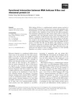
Dose distribution in the optic pathways
The maximum dose in the optic chiasma, ipsilateral and
controlateral optic nerves are represented in the Figure
1a,b,c. No difference was noted between NCF and CF
techniques.
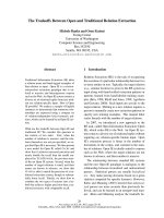
Likewise, the doses delivered in 1, 2 and 5% of these vol-
umes (D1, D2 and D5%) were similar for the two tech-
niques (Figure 2a,b,c). The differences did not exceed 2 Gy
except for patient n° 2, where the dose in the controlateral
optic nerve was more than 3 Gy lower with the CF tech-
nique.
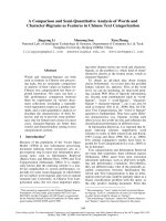
Dose distribution in organs at risk
In Figure 3, we show the most characteristic tolerance
doses for each organ at risk (OAR). Also represented are
the mean values for each organ with their standard devia-
tion. No difference was noted between CF and NCF for the
ipsilateral and controlateral lenses, brainstem, temporo-
mandibular joints, inner ears or parotids. Moreover, we
did not find a subgroup of patients who benefited from
either of the techniques.
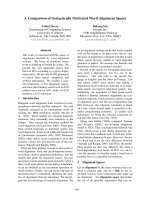
Dose distribution in the target volume
The mean dose volume histogram for the PTV1 is shown
in Figure 4. We can observe a slight difference in favour of
the NCF compared with the CF. The HI confirm this ten-
dency with a mean value of 0.14 for the NCF and 0.16 for
the CF; HIs are represented for each patient in Figure 5.
The conformity index for the two techniques was similar
as shown in Figure 6. In the subgroup of patients with
skull base involvement (T4), a significant benefit was
noticed with the CF compared to the NCF (Fig. 7). Ninety
percent and 95% of the volume received respectively 96%
and 94.5% of the prescribed dose with the CF technique
and 94.5% and 93.5% with the NCF technique.
In the other subgroups, we observed no dosimetric impact
of either of the techniques.
Discussion
The implementation of modern radiotherapy (3D confor-
mal and IMRT) has led to a reduction of the mean total
dose to organs at risk and particularly to the optic nerves
when compared to conventional radiotherapy [12]. Radi-
ation optic neuropathy is highly dependant on the radia-
tion dose [13-15]. Consequently, the high incidence of
radiotherapy-induced blindness, as much as 37%, with
conventional radiotherapy could be reduced by the use of
3D radiotherapy [16-18]. However in a single institution
study with 40 patients [19], conformal radiotherapy for
paranasal sinus carcinoma seems to be safer with only a 5
% incidence of cataract and only 2.5% unilateral blind-
ness.
The benefits, in dose reduction to the optic pathways, of
IMRT over 3D conformal radiotherapy have already been
investigated [20], with an average maximum dose of 56.4
Gy for IMRT and of 64.2 Gy for 3D conformal radiother-
apy. However, a complex treatment planning with 4-field
3D conformal radiotherapy and forward treatment plan-
ning will yield similar results to CF IMRT [21]. We have to
point out though, that since high doses were delivered to
both optic nerves, this diminishes the case for IMRT in
this study.
One other advantage of IMRT is the mode of administra-
tion of radiotherapy: as integrated concomitant boosts
with a daily dose fraction to OAR of less than 2 Gy which
can prevent late complications [14].
In a publication by the M. D. Anderson Cancer Center, the
maximum point dose for the controlateral optic nerve was
significantly reduced when using 5 non coplanar field
IMRT rather than the 9 coplanar field method [22]. On
Table 1: Dose-volume constraint set used for inverse planning
optimization
Volume Constraint
PTV V 95% > 95% prescribed dose
CTV V 99% > 95% prescribed dose
Spinal cord D max < 40 Gy
Ext cord D max < 45 Gy
Brainstem D max < 55 Gy
Frontal lob D max < 60 Gy
Parietal lob D max < 60 Gy
Hypophyse D max < 55 Gy
Temporomandibular joints D max < 60 Gy
Parotid D mean < 26 Gy
V 50% < 30 Gy
Lens D max < 12 Gy
Optic nerve D max < 55 Gy
Chiasma D max < 55 Gy
Retina D max < 55 Gy
Lachrymal glands No constraint
Radiation Oncology 2007, 2:35 />Page 4 of 9
(page number not for citation purposes)
Maximum doses in optic pathways, respectively optic chiasma (a), ipsilateral optic nerve (b) and controlateral optic nerve (c)Figure 1
Maximum doses in optic pathways, respectively optic chiasma (a), ipsilateral optic nerve (b) and controlateral optic nerve (c).
For each patient (Px), the maximum dose for coplanar field CF (grey) and for non coplanar field NCF (black) is represented.
Radiation Oncology 2007, 2:35 />Page 5 of 9
(page number not for citation purposes)
Dose distribution in optic pathways for each patient (Px); optic chiasma (a), ipsilateral optic nerve (b) and controlateral optic nerve (c) respectivelyFigure 2
Dose distribution in optic pathways for each patient (Px); optic chiasma (a), ipsilateral optic nerve (b) and controlateral optic
nerve (c) respectively. The dose difference in Gy between non coplanar field NCF and coplanar field CF (D NCF – D CF) is
represented in terms of D1%, D2%, and D5% corresponding to the doses in 1, 2 and 5 % of the volumes respectively. This
means that when the difference is negative, the dose to optic pathways is higher when using coplanar field technique.
Radiation Oncology 2007, 2:35 />Page 6 of 9
(page number not for citation purposes)
Homogeneity index (HI) for each patient in the coplanar field CF (grey) and non coplanar field NCF (black) techniqueFigure 5
Homogeneity index (HI) for each patient in the coplanar field
CF (grey) and non coplanar field NCF (black) technique. HI is
defined as the difference between D1 and D99% divided by
the prescribed dose. A perfect homogeneity would be
reached with a zero index.
Comparison of dose distribution in organs at risk for the coplanar field technique CF (grey) and non coplanar field technique NCF (black)Figure 3
Comparison of dose distribution in organs at risk for the coplanar field technique CF (grey) and non coplanar field technique
NCF (black). The most characteristic tolerance dose for each organ at risk is represented. D1%, Dmean and Dmax are the 1%
of organ volume doses, mean dose and maximum dose respectively. Mean dose values and standard deviation are shown.
Mean dose volume histogram in the planning target volume for the coplanar field CF (grey) and non coplanar field tech-nique NCF (black)Figure 4
Mean dose volume histogram in the planning target volume
for the coplanar field CF (grey) and non coplanar field tech-
nique NCF (black). Minimum and maximum doses of the
study group are represented by dotted lines.
Radiation Oncology 2007, 2:35 />Page 7 of 9
(page number not for citation purposes)
the other hand, there was no significant difference for the
ipsilateral nerve. In our experience, we did not observe
better D1, D2 and D5% to the optic pathways with the
non coplanar fields when using the same maximum dose
for both techniques.
The significance of maximum point dose to serial organs
such as chiasma or optic nerves in fractionated radiother-
apy is not well known, and many teams use D1 or D2 as
the maximum tolerated dose for the validation of dosim-
etry. A Belgian study by Claus et al. introduced a planning
organ at risk volume (PRV) made of a 2 mm isotropic
expansion around the optic pathway with the following
constraint: less than 5% of the PRV should receive more
than 60 Gy [23]. A median follow up of 31 months [24]
for the 39 patients did not show any radiotherapy-
induced blindness. Moreover, a recent publication [25]
with 36 patients treated by IMRT for paranasal carcinoma
did not report decreased vision.
For small organ volumes such as the lens, we used a PRV
with 2 mm isotropic margin. During optimization, con-
straints were modified in order to have the lowest dose
possible. Maximum dose to the lens was very similar for
both techniques, around 16 Gy. Probability of cataract
remained high at 27% at 5 years and 57% at 8 years for a
dose of 15 Gy in 15 fractions, as described by Henk et al.
[26].
This excess dose to the lens does not depend on the radi-
otherapy technique, and even with conformal 3D treat-
ment [12,21], the ipsilateral and controlateral lenses are
irradiated more than the tolerated dose given by TD 5/5
published by Emami [27]. Proton therapy could be used
to reduce the dose to the controlateral lens but 40% of the
controlateral lens will still be overdosed [12], without any
impact on the dose delivered to the ipsilateral lens.
It would be tempting to compare the two treatment
modalities for the entire ocular globe. We know that
coplanar IMRT and 3D conformal radiotherapy reduce the
mean total dose in a similar way when compared to con-
ventional irradiation [12], with a slight benefit with a 5-
beam conformal radiotherapy over coplanar IMRT [28]. A
significant dosimetric advantage represented by the mean
dose is noted for non coplanar over coplanar field IMRT
when considering the two ocular globes [22]. Neverthe-
less, the pertinence of these comparisons for clinical prac-
tice is doubtful because of the difference in
radiosensibility for the diverse components of the eye
[29].
The DVH and the conformity index for the PTV1 are sim-
ilar between the two techniques. No available data dem-
onstrating the superiority of the non coplanar over
coplanar fields on the target volume has been published.
This lack of difference can be explained by the ease with
which the inverse planning system can follow the con-
straints prescribed for sliding window IMRT. No superior-
ity of conformal 3D treatment over conventional
treatment planning was demonstrated in maxillary sinus
tumors [20]. In ethmoid carcinoma, conformal radiother-
apy with forward planning is better than conventional
planning [21,28].
PTV mean dose volume histogram in the T4 subgroup for the coplanar field CF (grey) and non coplanar field technique NCF (black)Figure 7
PTV mean dose volume histogram in the T4 subgroup for the
coplanar field CF (grey) and non coplanar field technique
NCF (black). Minimum and maximum doses of the study
group are represented by dotted lines.
Conformal index CI for each patient in the coplanar field CF (grey) and non coplanar field NCF (black) techniqueFigure 6
Conformal index CI for each patient in the coplanar field CF
(grey) and non coplanar field NCF (black) technique. CI is
defined as follows: CI = (TV/V
PTV
) × (TV/V
95%
) TV: Treated
Volume is the volume of PTV1 receiving the prescribed dose
(95%); V
PTV
is the volume of PTV; V
95%
is the volume
enclosed in the isodose 95%
Radiation Oncology 2007, 2:35 />Page 8 of 9
(page number not for citation purposes)
In the publication of Adams et al., the conformity and
homogeneity indices are better with IMRT than with con-
formal treatment [20]. The same observations were made
when comparing multifield dynamic IMRT and step-and-
shoot IMRT to 3D conformal treatment [30]. Interest-
ingly, no dosimetric benefits for the target volume were
noted with IMRT over conformal treatment planning in
the maxillary sinus [12], probably due to the distance
from the optic pathway and the lack of concave organs.
The MD Anderson Cancer Center experience did not
directly compare coplanar and non coplanar field IMRT;
however, they used a parallelized multi-resolution beam
angle optimization (PMBAO) which included non copla-
nar fields and obtained thus better dose homogeneity
without a real impact on the conformity index [22]. Their
results could be explained by the use of only two non
coplanar beams in a 5-beam configuration. The impact
noted on the homogeneity in the target volume is in
agreement with our results and seems to be due to non
coplanar fields rather than the PMBAO.
Conclusion
IMRT using the non coplanar field technique in ethmoid
carcinoma is an effective approach for treating this tumor.
A slight impact was shown on the PTV coverage for the
non coplanar set up compared with the coplanar tech-
nique. Using our beam configuration, T4 tumors with
skull base involvement were better treated with coplanar
fields. In all cases, inverse planning allows for a dosimet-
ric sparing of the optic pathways with good target volume
coverage whatever the set up employed. The clinical
impact on local control and on late effects is still not
known with IMRT and a retrospective analysis of this
cohort of patients is required.
Abbreviations
IMRT- Intensity modulated radiotherapy;
NCF- Non coplanar field;
CF- Coplanar field;
GTV- Gross tumor volume;
CTV- Clinical target volume;
PTV- Planning target volume;
Dmax- Maximum delivered dose;
DVH- Dose-volume histogram;
HI- Homogeneity index;
OAR- Organ at risk;
PMBAO- Parallelized multi-resolution beam angle opti-
mization.
Competing interests
The author(s) declare that they have no competing inter-
ests.
Authors' contributions
AS, KI, and PF conceived the study, collected data, and
drafted the manuscript.
AA, LS, and NA collected data.
JBD and DA participated in coordination and helped to
draft the manuscript.
CL, RG, and MM participated in the design of the study
and assisted in data collection.
DA provided mentorship and edited the manuscript.
All authors have read and approved the final manuscript.
Acknowledgements
The authors would like to thank B. Hawkins and F. Godson for the excel-
lent assistance in the preparation of this manuscript.
References
1. Dulguerov P, Jacobsen MS, Allal AS, Lehmann W, Calcaterra T: Nasal
and paranasal sinus carcinoma: are we making progress? A
series of 220 patients and a systematic review. Cancer 2001,
92:3012-3029.
2. Dulguerov P, Allal AS: Nasal and paranasal sinus carcinoma:
how can we continue to make progress? Current opinion in
otolaryngology & head and neck surgery 2006, 14:67-72.
3. Jansen EP, Keus RB, Hilgers FJ, Haas RL, Tan IB, Bartelink H: Does
the combination of radiotherapy and debulking surgery
favor survival in paranasal sinus carcinoma? Int J Radiat Oncol-
Biol Phys 2000, 48:27-35.
4. Amendola BE, Eisert D, Hazra TA, King ER: Carcinoma of the
maxillary antrum: surgery of radiation therapy? Int J Radiat
Oncol Biol Phys 1981, 7:743-746.
5. Hordijk GJ, Brons EN: Carcinomas of the maxillary sinus: a ret-
rospective study. Clinical otolaryngology and allied sciences 1985,
10:285-288.
6. Jiang GL, Ang KK, Peters LJ, Wendt CD, Oswald MJ, Goepfert H:
Maxillary sinus carcinomas: natural history and results of
postoperative radiotherapy. Radiother Oncol 1991, 21:193-200.
7. Jiang GL, Morrison WH, Garden AS, Geara F, Callender D, Goepfert
H, Ang KK: Ethmoid sinus carcinomas: natural history and
treatment results. Radiother Oncol 1998, 49:21-27.
8. Paulino AC, Fisher SG, Marks JE: Is prophylactic neck irradiation
indicated in patients with squamous cell carcinoma of the
maxillary sinus? Int J Radiat Oncol Biol Phys 1997, 39(2):283-289.
9. Tsujii H, Kamada T, Arimoto T, Mizoe J, Shirato H, Matsuoka Y, Irie
G: The role of radiotherapy in the management of maxillary
sinus carcinoma. Cancer 1986, 57:2261-2266.
10. Tsien C, Eisbruch A, McShan D, Kessler M, Marsh R, Fraass B: Inten-
sity-modulated radiation therapy (IMRT) for locally
advanced paranasal sinus tumors: incorporating clinical deci-
sions in the optimization process. Int J Radiat Oncol Biol Phys
2003, 55:776-784.
Publish with BioMed Central and every
scientist can read your work free of charge
"BioMed Central will be the most significant development for
disseminating the results of biomedical research in our lifetime."
Sir Paul Nurse, Cancer Research UK
Your research papers will be:
available free of charge to the entire biomedical community
peer reviewed and published immediately upon acceptance
cited in PubMed and archived on PubMed Central
yours — you keep the copyright
Submit your manuscript here:
/>BioMedcentral
Radiation Oncology 2007, 2:35 />Page 9 of 9
(page number not for citation purposes)
11. Feuvret L, Noel G, Mazeron JJ, Bey P: Conformity index: a review.
Int J Radiat Oncol Biol Phys 2006, 64:333-342.
12. Mock U, Georg D, Bogner J, Auberger T, Potter R: Treatment
planning comparison of conventional, 3D conformal, and
intensity-modulated photon (IMRT) and proton therapy for
paranasal sinus carcinoma. Int J Radiat Oncol Biol Phys 2004,
58:147-154.
13. Jiang GL, Tucker SL, Guttenberger R, Peters LJ, Morrison WH, Gar-
den AS, Ha CS, Ang KK: Radiation-induced injury to the visual
pathway. Radiother Oncol 1994, 30:17-25.
14. Parsons JT, Bova FJ, Fitzgerald CR, Mendenhall WM, Million RR: Radi-
ation optic neuropathy after megavoltage external-beam
irradiation: analysis of time-dose factors. Int J Radiat Oncol Biol
Phys 1994, 30:755-763.
15. Takeda A, Shigematsu N, Suzuki S, Fujii M, Kawata T, Kawaguchi O,
Uno T, Takano H, Kubo A, Ito H: Late retinal complications of
radiation therapy for nasal and paranasal malignancies: rela-
tionship between irradiated-dose area and severity. Int J
Radiat Oncol Biol Phys 1999, 44:599-605.
16. Ellingwood KE, Million RR: Cancer of the nasal cavity and eth-
moid/sphenoid sinuses. Cancer 1979, 43:1517-1526.
17. Katz TS, Mendenhall WM, Morris CG, Amdur RJ, Hinerman RW, Vil-
laret DB: Malignant tumors of the nasal cavity and paranasal
sinuses. Head Neck 2002, 24:821-829.
18. Shukovsky LJ, Fletcher GH: Retinal and optic nerve complica-
tions in a high dose irradiation technique of ethmoid sinus
and nasal cavity. Radiology 1972, 104:629-634.
19. Pommier P, Ginestet C, Sunyach M, Zrounba P, Poupart M, Ceruse P,
Ciupea C, Carrie C, Montbarbon X: Conformal radiotherapy for
paranasal sinus and nasal cavity tumors: three-dimensional
treatment planning and preliminary results in 40 patients. Int
J Radiat Oncol Biol Phys 2000, 48:485-493.
20. Adams EJ, Nutting CM, Convery DJ, Cosgrove VP, Henk JM, Dearna-
ley DP, Webb S: Potential role of intensity-modulated radio-
therapy in the treatment of tumors of the maxillary sinus. Int
J Radiat Oncol Biol Phys 2001, 51:579-588.
21. Pacholke HD, Amdur RJ, Louis DA, Yang H, Mendenhall WM: The
role of intensity modulated radiation therapy for favorable
stage tumor of the nasal cavity or ethmoid sinus. Am J Clin
Oncol 2005, 28:474-478.
22. Wang X, Zhang X, Dong L, Liu H, Gillin M, Ahamad A, Ang K, Mohan
R: Effectiveness of noncoplanar IMRT planning using a paral-
lelized multiresolution beam angle optimization method for
paranasal sinus carcinoma. Int J Radiat Oncol Biol Phys 2005,
63:594-601.
23. Claus F, De Gersem W, De Wagter C, Van Severen R, Vanhoutte I,
Duthoy W, Remouchamps V, Van Duyse B, Vakaet L, Lemmerling M,
Vermeersch H, De Neve W: An implementation strategy for
IMRT of ethmoid sinus cancer with bilateral sparing of the
optic pathways. Int J Radiat Oncol Biol Phys 2001, 51:318-331.
24. Duthoy W, Boterberg T, Claus F, Ost P, Vakaet L, Bral S, Duprez F,
Van Landuyt M, Vermeersch H, De Neve W: Postoperative inten-
sity-modulated radiotherapy in sinonasal carcinoma: clinical
results in 39 patients. Cancer 2005, 104:71-82.
25. Daly ME, Chen AM, Bucci MK, El-Sayed I, Xia P, Kaplan MJ, Eisele
DW: Intensity-modulated radiation therapy for malignancies
of the nasal cavity and paranasal sinuses. Int J Radiat Oncol Biol
Phys 2007, 67:151-157.
26. Henk JM, Whitelocke RA, Warrington AP, Bessell EM: Radiation
dose to the lens and cataract formation. Int J Radiat Oncol Biol
Phys 1993, 25:815-820.
27. Emami B, Lyman J, Brown A, Coia L, Goitein M, Munzenrider JE, Shank
B, Solin LJ, Wesson M: Tolerance of normal tissue to therapeu-
tic irradiation. Int J Radiat Oncol Biol Phys 1991, 21:109-122.
28. Huang D, Xia P, Akazawa P, Akazawa C, Quivey JM, Verhey LJ, Kaplan
M, Lee N: Comparison of treatment plans using intensity-
modulated radiotherapy and three-dimensional conformal
radiotherapy for paranasal sinus carcinoma. Int J Radiat Oncol
Biol Phys 2003, 56:158-168.
29. Nakissa N, Rubin P, Strohl R, Keys H: Ocular and orbital compli-
cations following radiation therapy of paranasal sinus malig-
nancies and review of literature. Cancer 1983, 51:980-986.
30. O'Daniel JC, Dong L, Kuban DA, Liu H, Schechter N, Tucker SL,
Rosen I: The delivery of IMRT with a single physical modula-
tor for multiple fields: a feasibility study for paranasal sinus
cancer. Int J Radiat Oncol Biol Phys 2004, 58:876-887.