Báo cáo y học: " Importance of spliceosomal RNP1 motif for intermolecular T-B cell spreading and tolerance restoration in lupus" pptx
Bạn đang xem bản rút gọn của tài liệu. Xem và tải ngay bản đầy đủ của tài liệu tại đây (653.32 KB, 10 trang )
Open Access
Available online />Page 1 of 10
(page number not for citation purposes)
Vol 9 No 5
Research article
Importance of spliceosomal RNP1 motif for intermolecular T-B cell
spreading and tolerance restoration in lupus
Fanny Monneaux, Véronique Parietti, Jean-Paul Briand and Sylviane Muller
Centre National de la Recherche Scientifique UPR9021, Institut de Biologie Moléculaire et Cellulaire, 15 rue René Descartes, 67000 Strasbourg,
France
Corresponding author: Sylviane Muller,
Received: 5 Jul 2007 Revisions requested: 31 Jul 2007 Revisions received: 7 Aug 2007 Accepted: 26 Oct 2007 Published: 26 Oct 2007
Arthritis Research & Therapy 2007, 9:R111 (doi:10.1186/ar2317)
This article is online at: />© 2007 Monneaux et al.; licensee BioMed Central Ltd.
This is an open access article distributed under the terms of the Creative Commons Attribution License ( />),
which permits unrestricted use, distribution, and reproduction in any medium, provided the original work is properly cited.
Abstract
We previously demonstrated the importance of the RNP1 motif-
bearing region 131–151 of the U1-70K spliceosomal protein in
the intramolecular T-B spreading that occurs in MRL/lpr lupus
mice. Here, we analyze the involvement of RNP1 motif in the
development and prevention of naturally-occurring
intermolecular T-B cell diversification. We found that MRL/lpr
peripheral blood lymphocytes proliferated in response to
peptides containing or corresponding exactly to the RNP1 motif
of spliceosomal U1-70K, U1-A and hnRNP-A2 proteins. We
also demonstrated that rabbit antibodies to peptide 131–151
cross-reacted with U1-70K, U1-A and hnRNP-A2 RNP1-
peptides. These antibodies recognized the U1-70K and U1-A
proteins, and also U1-C and SmD1 proteins, which are devoid
of RNP1 motif. Repeated administration of phosphorylated
peptide P140 into MRL/lpr mice abolished T-cell response to
several peptides from the U1-70K, U1-A and SmD1 proteins
without affecting antibody and T-cell responses to foreign (viral)
antigen in treated mice challenged with infectious virus. These
results emphasized the importance of the dominant RNP1
region, which seems to be central in the activation cascade of B
and T cells reacting with spliceosomal RNP1
+
and RNP1
-
spliceosomal proteins. The tolerogenic peptide P140, which is
recognized by lupus patients' CD4
+
T cells and known to protect
MRL/lpr mice, is able to thwart emergence of intermolecular T-
cell spreading in treated animals.
Introduction
Longitudinal studies of spontaneously lupus-prone inbred
mouse strains and patients with systemic lupus erythematosus
(SLE) consistently show an ordered appearance of typical
auto-antibodies in the serum of individuals [1-4]. With time the
fine specificity of the antibody response initially focused
against one or few autoepitopes diversifies to other epitopes
of the same protein (intramolecular spreading) and to other
components that are physically associated within the same
antigenic macromolecular particles, such as nucleosome, spli-
ceosome, and Ro particle (intermolecular spreading). Epitope
spreading is thus a process whereby epitopes distinct from
and non-cross-reactive with an inducing epitope become
major targets of an ongoing immune response. This phenome-
non is not limited to autoimmunity; it has also been described
in experimental and natural situations as a consequence of
acute or persistent infection. Although the concept of epitope
spreading was introduced more than 15 years ago [5], the cel-
lular components that catalyze the spreading hierarchy have
not been well defined, and certain aspects of this process
remain unexplained. Recent studies suggest that autoreactive
B cells are important cellular mediators contributing to autore-
active T-cell response diversification via their functions that
mediate antigen processing and presentation [6,7].
Previous work from our laboratory demonstrated that peptide
131–151 of the spliceosomal U1-70K protein as well as a
peptide analogue containing a phosphoserine residue at posi-
tion 140 (peptide P140) are recognized by CD4
+
T cells from
lupus mice. Both peptides were shown to behave as promis-
cuous epitopes and bind a large panel of murine and human
MHC class II molecules [8-11]. Administration into young
MRL/lpr lupus-prone mice of P140 peptide in saline, but not of
the non-phosphorylated peptide 131–151, led to a dramatic
amelioration of the clinical and biological manifestations of
treated animals and significantly prolonged their survival [9].
ELISA = enzyme-linked immunosorbent assay; FA = Freund's adjuvant; PBL = peripheral blood lymphocyte; SLE = systemic lupus erythematosus;
Arthritis Research & Therapy Vol 9 No 5 Monneaux et al.
Page 2 of 10
(page number not for citation purposes)
The peptide P140 administrated in Freund's adjuvant (FA)
accelerated the renal disease in MRL/lpr mice [9]. Our studies
revealed further that peripheral CD4
+
T cells from lupus
patients, but not from patients with other autoimmune dis-
eases (such as rheumatoid arthritis, primary Sjögren's syn-
drome, autoimmune deafness, polymyositis, primary billiary
cirrhosis and autoimmune hepatitis) or infectious diseases,
very specifically recognized the 131–151 and P140 peptides,
and that phosphorylation of Ser
140
prevented ex vivo prolifera-
tion of lupus patients' CD4
+
T cells but not secretion of high
levels of regulatory cytokines [11].
The 131–151 sequence of the spliceosomal U1-70K protein
is located within a 80–90 amino acid-long RNA-binding
domain. It encompasses a conserved sequence, called RNP1
motif, which is also present in other RNA-binding proteins,
such as small nuclear (sn)RNP (such as U1-A) and heteroge-
neous nuclear (hn)RNP (such as hnRNP-A2/B1) proteins.
Starting from the observation that sequences containing this
RNP1 motif are often targeted by antibodies from lupus
patients and mice, we hypothesized that the RNP1 motif could
be involved in the earliest stages of the T-B intramolecular
diversification process to other regions of one of the spliceo-
somal proteins that contain this unique motif, and might pro-
mote intermolecular spreading to epitopes of other proteins
present within the same spliceosomal particle and containing
or not an RNP1 motif [12,13]. We demonstrated that an
intramolecular T and B cell spreading effectively occurs in
MRL/lpr mice tested at different ages and emphasized the
importance of the RNP1 region in the cascade of events
observed in the murine lupus response [14]. In the present
study, we investigated the potential capacity of peptide 131–
151 to generate antibodies that react with spliceosomal pep-
tides and proteins encompassing, or not, the RNP1 motif. We
tested the reactivity of peripheral blood lymphocytes (PBLs)
from unprimed lupus mice to react with RNP1-containing pep-
tides from several spliceosomal proteins. We also examined
whether administration of protecting P140 peptide into MRL/
lpr mice could modulate intermolecular epitope spreading in
vivo by preventing T-cell response to peptides from heterolo-
gous spliceosomal proteins that do or do not contain an RNP1
motif. We provide data showing that P140 peptide treatment
efficiently suppresses T-cell responses to other, unrelated spli-
ceosomal epitopes, in addition to the tolerizing peptide.
Remarkably, however, the B and T cell immune response to
foreign antigens (such as viral particles) remains unaffected in
treated animals.
Materials and methods
Peptides and proteins
Eleven synthetic peptides of the U1-70K, U1-A, SmD1,
hnRNP-A2 and La ribonucleoproteins were used in this study
(Figure 1). Six of them were described previously, namely
131–151, 183–202 and P140 peptides of the U1-70K pro-
tein, peptide 35–55 of the hnRNP-A2 protein, and peptides
77–96 and 97–119 of SmD1 [3,8,15,16]. The synthesis of
new peptides, including four peptides, the sequence of which
corresponded exactly to the respective RNP1 motifs of the
four proteins U1-70K, U1-A, hnRNP-A2 and La, and peptide
35–54 of the U1-A protein was performed using the same
classical N-[9-fluorenyl] methoxycarbonyl (Fmoc) solid-phase
chemistry. The homogeneity of each purified peptide was
checked by analytical high-performance liquid chromatogra-
phy (HPLC), and their identity was assessed by matrix-
assisted laser desorption and ionization time-of-flight (TOF)
mass spectrometry (MS) using a Protein TOF apparatus
(Bruker Spectrospin, Bremen, Germany). A large fragment
encompassing residues 21–119 of the SmD1 protein (human
sequence [15]) was assembled using optimized Fmoc chem-
istry protocols with a multichannel peptide synthesizer [17].
Ser
35
, Ser
59
and Thr
78
residues were incorporated into the
growing peptide chain by pseudoproline-protected dipeptides
to increase solvation and coupling rates during peptide
assembly [18]. Side chain deprotection and cleavage of pep-
tides from the solid support was performed by trifluoroacetic
acid (TFA) treatment in the presence of appropriate scaven-
gers. The SmD1 fragment 21–119 was purified by reversed-
phase HPLC (RP-HPLC) using a Beckman preparative HPLC
system on a Nucleosil C18 (1 × 30 cm) column. The elution
was achieved with a linear gradient of aqueous 0.1% TFA (A)
and 0.08% TFA in 80% acetonitrile, 20% water (B) at a flow
rate of 6 ml/min with UV detection at 230 nm. The purity of the
SmD1 fragment was controlled by analytical RP-HPLC on a
Beckman instrument on two types of column, namely a Nucle-
osil C18 5 μm-column (150 × 4.6 mm) and a Nucleosil C4 5
μm-column (150 × 4.6 mm), using a linear gradient of 0.1%
TFA in water and acetonitrile containing 0.08% TFA at a flow
rate of 1.2 ml/min. The mass of the long SmD1 fragment 21–
119 was assessed by LC/MS using a ThermoFinnigan LCQ
advantage/LC surveyor. Recombinant U1-A, U1-C, U1-70K
and SmBB' proteins (human sequences) were purchased
from Diarect AG (Freiburg, Germany).
Rabbit immunization
On days 1, 21 and 42, outbred Fauves de Bourgogne female
rabbits (Dombes Romans, Chatillon, France) were injected
subcutaneously (sc) with peptide 131–151 of the U1-70K
protein (100 μg/rabbit/injection) emulsified in complete FA
(CFA) for the first injection and incomplete FA for the subse-
quent injections. Rabbits were bled before immunization and
then regularly throughout the protocol.
ELISA and Western immunoblotting
Standard ELISA procedures with peptide 131–151 (2 μM in
0.05 M carbonate buffer, pH 9.6) or dsDNA (100 ng/ml in 25
mM citrate buffer, pH 5.4) directly coated onto polyvinyl micro-
titer plates (Falcon, Oxnard, California, USA), and goat anti-
rabbit IgG-horseradish peroxidase (HRP) conjugate (Jackson
ImmunoResearch Laboratories, West Groves, Pennsylvania)
diluted 1:50,000 in phosphate-buffered saline containing
Available online />Page 3 of 10
(page number not for citation purposes)
0.05% Tween (PBS-T) were used to measure antibody reac-
tivity in rabbit antisera [8]. The final reaction was visualized
with H2O2 and 3,3',5,5'-tetramethyl benzidine used as chro-
mogen, and absorbance was measured at 450 nm. For com-
petitive ELISA, increasing amounts of inhibitor peptides were
first incubated for 1 h at 37°C and then overnight at 4°C with
constant dilutions of rabbit antisera in PBS-T. The mixtures
were subsequently transferred to polyvinyl Falcon plates pre-
coated with peptide 131–151, and post-coated with bovine
serum albumin in PBS-T (0.4% w/v). After a 1 h incubation at
37°C and washing, bound antibodies were detected as indi-
cated above. Preliminary experiments were conducted in a
direct format to define the working dilution of rabbit antiserum.
For Western immunoblotting, U1-A, U1-C, U1-70K, SmBB'
and SmD1 proteins were first subjected to electrophoresis in
12.5% SDS-polyacrylamide gels and then transferred to nitro-
cellulose. The blotted strips were saturated in Tris-buffered
saline, pH 7.5, containing 0.5% Tween (TBS-T) and 5% milk
for 1 h at room temperature (RT), and then incubated with rab-
bit antisera diluted 1:500 in TBS-T-milk for 1 h at RT. After
washing, strips were incubated with HRP-conjugated second
antibodies to rabbit IgG (1:5,000 in TBS-T). Enhanced chemi-
luminescent (ECL™) reagents (Amersham Pharmacia Biotech,
Buckinghamshire, UK) were used to reveal positive reactions.
Cellular assays
To study the natural T-cell reactivity occurring in unprimed
lupus-prone mice, MRL/lpr mice were bled at weeks 8, 10, 12
and 14, and lymphocytes were purified by density separation
(Lympholyte-M, d = 1.0875; Cedarlane, Hornby, Canada).
PBLs were collected, washed three times, and resuspended
at 3 × 10
6
cells/ml in L-alanyl-L-glutamine-enriched RPMI
1640 medium (Cambrex, Verviers, Belgium) containing 10%
fetal calf serum (FCS; Dutscher, Brumath, France), HEPES,
gentamycine, and β-mercaptoethanol. The proliferative
response to peptides was measured in duplicate using 3 ×
10
5
cells/well and a single peptide concentration (100 μM).
After 72 h, the cultures were pulsed for 18 h with [
3
H]-thymi-
dine (6.7 Ci/mmol; 1μCi/well) and DNA-incorporated radioac-
tivity was measured using a Matrix 9600 direct beta counter
(Packard, Meriden, Connecticut, USA; cpm range from 100 to
30,000). The SD of duplicate cultures was always below 20%
of the mean. Control tests were performed by adding Con-A
(100 μl/well; 5 μg/ml) to cells during the time of the culture
(90 h).
To analyze the effect of P140 peptide administration on spon-
taneous T cell spreading occurring in lupus-prone mice, MRL/
lpr mice received peptide P140 (100 μg in saline/mouse/
injection; 10 mice) or PBS alone (10 mice) intravenously (iv) at
weeks 5, 7 and 9 [9]. Treated mice were bled at week 10, and
peptides of interest (100 μM in the cultures) were tested for
their ability to induce proliferation of PBLs, as described
above.
To examine the possible effect of P140 treatment on the ability
of mice to mount a normal immune reaction after viral infection,
P140-treated MRL/lpr mice (18-week-old at the time of the
experiment) were challenged intranasally (in) with A/NT/60/68
influenza virus (H3N2 strain) in allantoic fluid using a dose pre-
determined to provoke ~30% of weight loss [19]. The
required dose was of the order of 40 μl of undiluted allantoic
fluid (HA titer 1260) per mouse. As a control, a group of
untreated MRL/lpr mice was challenged in parallel. Mice were
followed daily for their body weight. The capacity of PBLs from
challenged mice to proliferate and secrete INF-γ ex vivo in the
presence of increasing concentrations of the HA peptide
307–319 (PKYVKQNTLKLAT [20]) representing a promiscu-
ous T-helper epitope from influenza virus HA, was evaluated
13 days after challenge, as described above. The anti-virus
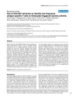
Figure 1
Synthetic peptides used in this studySynthetic peptides used in this study. (a) Peptides of U1-A, U1-70K,
hnRNP-A2 and La proteins containing or corresponding to RNP1
motifs. The RNP1 motif (complete or partial) is highlighted by an empty
box. In peptides corresponding exactly to the motif (gray boxes) amino
acid similarities are in bold face. The serine
140
residue, which is phos-
phorylated in the P140 peptide is underlined. (b) Sequence of other
peptides used in this study.
Arthritis Research & Therapy Vol 9 No 5 Monneaux et al.
Page 4 of 10
(page number not for citation purposes)
antibody production in the serum from challenged mice was
measured in ELISA [19] 28 days after challenge.
All animal experiments were performed with the approval of
the local Institutional Animal Care and Use Committee
(CREMEAS).
Statistics
Analysis for statistically significant differences was performed
with Student's t-test. P values < 0.05 were considered
significant.
Results
Recognition by T cells from unprimed MRL/lpr mice of
peptides containing or corresponding exactly to the
RNP1 motif
In a first set of experiments, 20 non-immunized MRL/lpr mice
were bled longitudinally at weeks 8, 10, 12 and 14, and in
order to have enough cells to test individually several peptides
in duplicate (only 1.5 to 2.10
6
PBLs can be collected from the
blood of a single living mouse), PBLs were pooled and tested
for their ability to proliferate ex vivo in response to 100 μM of
each peptide. Owing to the well-documented accumulation of
CD4
-
CD8
-
double negative (DN) T cells in PBLs of MRL/lpr
mice (80% of DN T cells at 17 weeks of age, data not shown),
we focused our measurement of specific CD4
+
T cells prolif-
eration in a window ending at week 14. Responses were con-
sidered to be positive when stimulation indices (SI) were
higher than 2 in the proliferation assay. In good agreement
with previous findings [14], a proliferative response of PBLs
purified from 8 to 14-week-old MRL/lpr mice was observed in
response to peptides 131–151 and P140 of the U1-70K pro-
tein (Figure 2a). In addition, U1-A peptide 35–54, which con-
tains a part of the RNP1 motif (Figure 1a), induced
proliferation of MRL/lpr PBLs that was similar in intensity to the
proliferative response to P140 peptide (Figure 2a), and was at
its maximum at week 12. A proliferative response was also
measured when PBLs were recalled with hnRNP-A2 peptide
35–55 (Figure 2a), which also contains only a part of the
RNP1 motif (Figure 1a).
In order to determine whether MRL/lpr T cells precisely recog-
nized the RNP1 motif present in the respective spliceosomal
proteins, we synthesized three additional peptides corre-
sponding exactly to the RNP1 motifs of the U1-70K, U1-A and
hnRNP-A2 proteins, called RNP1-70K, RNP1-U1A and
RNP1-A2, respectively (Figure 1a), and tested, as above, the
ability of these peptides to stimulate the proliferation of PBLs
from 8-14-wk-old MRL/lpr mice (Figure 2b). No or very weak
proliferation was observed in response to the RNP1-70K pep-
tide 139–151 at any age. A modest but significant proliferative
response with RNP1-A2 peptide 45–57 was measurable with
PBLs from 10, 12 and 14-wk-old MRL/lpr mice. In contrast, as
early as 8 weeks of age, and at least until 14 weeks, a strong
proliferative response was observed when MRL/lpr PBLs
were cultured in the presence of the 13-residue-long RNP1-
U1A peptide 47–59 (Figure 2b; maximum at week 12). This
response was inhibited in the presence of neutralizing anti-
CD4 monoclonal antibody GK1.5 (10 μg/ml; not shown) indi-
cating that the main cell population reacting with the RNP1-
U1A peptide 47–59 does correspond to CD4
+
T cells.
Reactivity of rabbit antibodies to peptide 131–151 of the
U1-70K protein with RNP1-peptide
In our diversification model [13], the RNP1 motif could be
involved at an early stage of the anti-spliceosomal autoimmune
response. We first used competitive ELISA to measure the
cross-capacity of antibodies generated against the RNP1
sequence of the U1-70K protein to recognize RNP1-peptides
from U1-A and hnRNP-A2 proteins (Figure 3a). The binding to
the plastic-bound 131–151 peptide of IgG antibodies from an
outbred rabbit immunized against the same peptide 131–151
was almost completely inhibited by the homologous peptide
131–151, and to a lower extent by the RNP1-70K peptide
139–151. The amount of peptide required to inhibit 50% of
the antibody reaction was 5 nM of the homologous peptide
131–151 and 80 nM of the RNP1-70K peptide. Although
much more poorly (IC
50
~3 μM), RNP1-U1A peptide was also
able to inhibit the IgG antibody binding to peptide 131–151 in
a dose-dependent manner. However, both the RNP1-A2 and
RNP1-La peptides, the latter significantly differing in its
sequence from the RNP1 sequence present in the U1-70K
protein (Figure 1a), were very poor inhibitors (Figure 3a).
These findings indicate that antibodies raised to peptide 131–
151 of the U1-70K protein recognize the RNP1 motif present
within U1-A, but also show that punctual mutations present in
the respective RNP1 motifs (Figure 1a) strongly affect their
conformation in solution and consequently their cross-reactiv-
ity with antibodies.
Antibodies generated in outbred rabbits to peptide 131–
151 of the U1-70K protein react with the cognate protein
and with other spliceosomal proteins
We then questioned whether antibodies raised to peptide
131–151 are able to recognize the whole cognate protein and
other proteins present in the same spliceosomal particle (Fig-
ure 3b). Ponceau red staining of membranes blotted with the
three recombinant proteins U1-70K, U1-A, and U1-C and with
the long synthetic fragment 21–119 of SmD1 revealed a sin-
gle band at 65 kD, 35 kD, 20 kD and 12 kD, respectively, as
determined with MW markers (Figure 3b, lanes 1–4). IgG anti-
bodies from the rabbit immunized against peptide 131–151 in
CFA (see above) strongly reacted with the U1-70K (lane 5)
and the U1-A (lane 6) proteins, as well as with the U1-C (lane
7) and SmD1 (lane 8) proteins, which do not contain any
RNP1 motif, suggesting the establishment of a heterologous
B-cell epitope spreading phenomenon, and not solely immune
cross-reactivity. No reactivity was found with SmBB' protein
(not shown) or with any of the five proteins when tested with
the serum from an outbred rabbit that received several
Available online />Page 5 of 10
(page number not for citation purposes)
injections of CFA alone (lanes 9–12). This rabbit did not
develop any IgG reactivity to dsDNA (as measured by ELISA),
and no clinical sign of autoimmunity was measurable 8 weeks
after the last of three peptide administrations (result confirmed
in several outbred rabbits). New Zealand White rabbits were
not used in this set of experiments because they have a natural
tendency to produce auto-antibodies [21]. A similar result was
obtained with two outbred mice (ICR, Harlan) immunized with
peptide 131–151, for which antisera react in Western immu-
noblot with the U1-70K protein and also with U1-A and U1-C
proteins (data not shown).
Effect of P140 therapy on spontaneous T cell spreading
We previously demonstrated that phosphorylated P140 pep-
tide administrated in saline into MRL/lpr mice transiently
abolished T cell spreading to other regions of the cognate pro-
tein U1-70K [14]. We hypothesized that through its RNP1
motif, the P140 peptide could originate a mechanism of
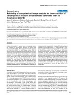
Figure 2
Spontaneous T-cell reactivity to peptides containing or corresponding to RNP1 motifs in unprimed MRL/lpr miceSpontaneous T-cell reactivity to peptides containing or corresponding to RNP1 motifs in unprimed MRL/lpr mice. The proliferative response of
peripheral blood lymphocytes (PBLs) from 8-, 10-, 12- and 14-week-old mice was measured in the presence of U1-70K, U1-A and hnRNP-A2 pep-
tides (100 μM) encompassing completely or partially the RNP1 motif of each protein (a) or corresponding exactly to the RNP1 motif of each protein
(b). The results are expressed as SI corresponding to the ratio cpm in the culture with peptide to cpm in the culture without peptide. A mean SI > 2
was considered to be positive (horizontal line). The average tritiated thymidine incorporation in the absence of peptide and in the presence of Con-A
was 50 and 3,000 cpm, respectively. This experiment is one of two individual experiments that showed similar results. Bars show the mean ± SEM.
Arthritis Research & Therapy Vol 9 No 5 Monneaux et al.
Page 6 of 10
(page number not for citation purposes)
'tolerance spreading' leading to the beneficial effect observed
in treated MRL/lpr mice. With the aim of further investigating
whether peptide P140 could also alter spontaneous intermo-
lecular T cell spreading in this pathway, we studied the prolif-
erative response of PBLs collected from P140-treated MRL/
lpr mice to a set of selected spliceosomal peptides. We con-
firmed our previous findings showing that the proliferative
response of PBLs from P140-treated mice to the homologous
peptide P140 and to peptide 183–202 of the same parent
U1-70K protein was significantly abolished (79 and 83% of
inhibition, respectively; P = 0.006 and P = 0.002, respec-
tively; Figure 4). In the same assay, a 56%-reduction of the
proliferative response to the RNP1-UA peptide was detecta-
ble (Figure 4) but was not statistically significant (P = 0.059).
However, we found a statistically significant drop of PBL pro-
liferative response in the presence of peptide 35–54 of U1-A
(47% decrease; P = 0.02), peptide 35–55 of hnRNP-A2
(67% decrease; P = 0.0004) and the RNP1 A2 peptide (74%
decrease; P = 0.002).
As U1-A and hnRNP-A2 peptides do contain an RNP1 motif,
we could not rule out the possibility that the observed drop of
responsiveness was simply due to the extinction of P140-spe-
cific T cells by a homologous-like, cross-reactive effect. To
ensure that P140 treatment was leading to a true (non-cross-
reactive) mechanism of 'intermolecular tolerance spreading',
the goal was thus to demonstrate that T cell response to spli-
ceosomal proteins that do not contain an RNP1 motif was also
abolished in P140-treated MRL/lpr mice. U1-C and SmD1
proteins, which are associated to the U1-snRNP particle and
are devoid of RNP1 motif, were appropriate candidates in this
context. Epitopes recognized by CD4
+
T cells are not known
in U1-C protein. However, an epitope recognized by T cells
from SLE patients and (NZBxNZW)F1 mice has been identi-
fied in the C-terminal end of SmD1 [22,23]. This epitope is
present in peptide 83–119, which encompasses the
sequence 97–119 shown earlier in our laboratory to contain a
B-cell epitope recognized by IgG antibodies from MRL/lpr and
(NZBxNZW)F1 lupus mice and from patients with lupus
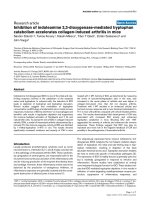
Figure 3
Reactivity of rabbit antibodies directed to peptide 131–151 of the U1-70K protein with RNP1-peptides and spliceosomal proteinsReactivity of rabbit antibodies directed to peptide 131–151 of the U1-70K protein with RNP1-peptides and spliceosomal proteins. (a) The antise-
rum was diluted 1/20,000 and incubated first for 1 h at 37°C and then overnight at 4°C with increasing amounts of different RNP1-peptides used as
competitors in the fluid-phase. The homologous peptide 131–151 was used as control. The mixtures were then added to microtiter plates pre-
coated with 2 μM of immunizing peptide 131–151. The results are expressed as the percentage of inhibition of the ELISA reaction measured with-
out competitor peptide. (b) U1-70K, U1-A, U1-C, and SmD1 proteins were subjected to electrophoresis, transferred to nitrocellulose, and colored
with Ponceau red (lanes 1–4). The blotted strips were then incubated with the serum (diluted 1/500) from rabbits that received either peptide 131–
151 (lanes 5–8) or CFA alone (lanes 9–12). IgG antibodies only were tested. ECL reagents were used to reveal positive reactions.
Available online />Page 7 of 10
(page number not for citation purposes)
[3,10,15]. In preliminary experiments with peptide dose-
response measurements, we found that SmD1 peptide 97–
119, but not SmD1 peptide 77–96, was efficiently recognized
by MRL/lpr PBLs (not shown). With this new target, as above,
we analyzed the reactivity of MRL/lpr PBLs (the peptide 77–
96 was used as control) after P140 treatment. As shown in
Figure 4, we observed that PBLs from P140-treated MRL/lpr
mice did no longer proliferate ex vivo in response to SmD1
peptide 97–119 used as recall antigen in the culture (81%
decrease; P = 0.0005).
Most importantly, we demonstrated that the tolerogenic effect
of P140 peptide on T- and B-cell reactivity to auto-antigens
was not generalized to the total T- and B-cell response. We
found that P140-treated mice display normal capacity to suc-
cessfully mount T- and B-cell responses after a viral challenge.
This was demonstrated by testing the ability of PBLs from
MRL/lpr mice treated with either peptide P140 or PBS alone,
and then challenged with an infectious dose of influenza virus,
to proliferate (Figure 5a) and secrete IFN-γ (not shown) ex
vivo, in response to a CD4
+
T cell promiscuous influenza
hemaglutinin epitope used as recall antigen. As shown, both
groups behave very similarly. Both groups also produced sim-
ilar levels of anti-virus IgG antibodies (not shown) and equally
recovered body weight loss resulting from infection (Figure
5b).
Discussion
A crucial question in our understanding of SLE is to identify the
early cellular and molecular events that predispose to the loss
of tolerance in individuals and those that trigger the transition
from a preclinical status to overt disease and perpetuate the
immune-mediated, inflammatory response. The concept of the
T-B cell spreading phenomenon based on an initial acute
autoimmune response restricted to a few epitopes using a lim-
ited number of T-cell clones for expansion is now widely
accepted. However, the possible initiator antigens and the
mechanism that render this(these) antigen(s) immunogenic in
a predisposed context is not clear [1,24-26]. It is also not clear
what the link is that drives T-B cell diversification from these
putative initial epitopes to a particular set of antigens. Our
hypothesis is that the RNP1 motif, which is present in a
number of RNA-binding proteins and represents a frequent
target for B- and T-cell response, might be central in this proc-
ess and partly explain autoimmunity to RNP proteins in lupus
[12,13].
In this model, the RNP1 motif initiates the spreading of the
immune response to the whole protein (intramolecular spread-
ing), then proceeds in an ordered manner to other proteins
containing the RNP1 motif, and finally, to proteins that do not
contain any RNP1 motif but are colocalized in the same parti-
cle (intermolecular spreading). In our previous work [14], we
clearly demonstrated that the first step of this process effec-
tively occurs in MRL/lpr lupus mice, in which the immune
response diversifies from the RNP1 motif to the whole U1-70K
protein (intramolecular spreading). In the present study, we
validated the second step of our proposed model [13] by dem-
onstrating that an immune response induced against the
RNP1 motif can drive the diversification to other spliceosomal
proteins that may or may not contain an RNP1 motif, such as
U1A, hnRNP-A2, U1C and SmD1 proteins (intermolecular
spreading).
Moreover, we not only confirmed that the sequence 131–151
of the U1-70K protein contains a recurrent epitope recognized
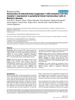
Figure 4
Effect of a brief peptide P140 therapy on the spontaneous T cell spreading in MRL/lpr miceEffect of a brief peptide P140 therapy on the spontaneous T cell spreading in MRL/lpr mice. Mice were either treated with peptide P140 (hatched
bars) administrated intravenously in saline at weeks 5, 7 and 9 or received saline alone (solid bars). The proliferative response of PBLs was meas-
ured ex vivo at 10 weeks in the presence of selected peptides (100 μM) containing (P140, 35–54 U1-A, RNP1-U1A, 35–55 hnRNP-A2, RNP1-A2)
or not (183–202 of the U1-70K protein, 97–119 SmD1) an RNP1 motif. A mean SI > 2 was considered to be positive (horizontal line). The average
tritiated thymidine incorporation in the absence of peptide and in the presence of Con-A was 50 and 2,000 cpm, respectively. Bars show the mean
± SEM. Significant differences are indicated. ns: non-significant reduction.
Arthritis Research & Therapy Vol 9 No 5 Monneaux et al.
Page 8 of 10
(page number not for citation purposes)
by CD4
+
T cells from MRL/lpr mice, but we also showed that
the RNP1 motif present in other spliceosomal proteins can be
recognized by MRL/lpr PBLs. Clearly, the RNP1 sequence
47
RSLKMRGQAFVIF
59
present within the U1-A protein
induced a stronger proliferation of MRL/lpr PBLs compared to
other RNP1 sequences, which were much less efficient and
for some, very poor inducers. It has to be noted that the T cell
epitopes of U1-A characterized previously by others [27-29] in
MRL/lpr and (NZBxNZW)F1 mice and patients with SLE or
mixed-connective tissue disease did not or only partially
encompass the RNP1 motif. Epitopes recognized by autore-
active CD4
+
T cells either in patients or in murine models of
lupus or rheumatoid arthritis are not known in the case of the
hnRNP-A2 protein.
The RNP1 motif is present in B-cell epitopes recognized in dif-
ferent proteins by antibodies from autoimmune mice and
patients [[13]; and references therein]. In the present study,
we show that antibodies elicited after immunizing a rabbit with
the peptide 131–151 of the U1-70K protein cross-react with
the short RNP1 motif contained within the immunizing
sequence but significantly less well with the RNP1 motifs of
U1-A and hnRNP-A2 proteins, indicating that in solution these
short peptides do adopt different conformations. In Western
immunoblotting, antibodies from this immunized outbred rab-
bit reacted strongly with the cognate protein U1-70K and with
U1-A, but also with U1-C and SmD1, which are both devoid of
RNP1 motif. These results fit well with the previous findings
from several groups, which showed similar intermolecular anti-
body diversification in longitudinal studies of lupus patients
and mice [1,30,31]. However, in our rabbits immunized with
the 131–151 peptide, we found no antibodies to dsDNA or
chromatin, no antibodies to recombinant SmBB', and no clini-
cal sign of autoimmunity. Immunization of experimental rabbits
and mouse models with the proline-rich peptide PPPGMRPP,
another key sequence, which is present in several spliceo-
somal proteins and was reported to trigger spreading in immu-
nized animals [32,33], gave contradictory results in
independent studies regarding the presence or not of anti-
DNA antibodies and signs of lupus-like autoimmunity [34-36].
To our knowledge, PPPGMRPP peptide administrated in
lupus mice was never shown to possess protective or tolero-
genic properties.
The reasons that underlie the successful treatment of MRL/lpr
mice with peptide P140, which significantly retards the emer-
gence of dsDNA IgG antibodies and the progression of
nephritis and prolongs the survival of treated mice, are not yet
fully elucidated. We showed here that P140 administration not
only abolishes T cell intramolecular spreading to other regions
of the cognate U1-70K protein, but also leads to an impressive
unresponsiveness of PBLs reacting with RNP1-peptides or
longer peptides containing the motif, and with a peptide from
the C terminus of SmD1 protein, newly characterized in this
study and originated from a spliceosomal protein that does not
contain any RNP1 motif. Thus, in treated animals, P140 pep-
tide elicits clearly a state of bystander suppression leading to
the downregulation of autoreactive T- and B-cell response to
other self-antigens (so-called tolerance spreading [37]).
Important questions remain at this stage as to whether P140
peptide induces initial tolerance by playing the role of
antagonist or partial agonist of the receptor of autoreactive T
Figure 5
Brief peptide P140 therapy does not affect immune responses to viral challenge in MRL/lpr miceBrief peptide P140 therapy does not affect immune responses to viral challenge in MRL/lpr mice. P140-treated (closed symbols) and untreated
MRL/lpr mice (open symbols) (10 mice/group) were challenged intra-nasally with infectious influenza virus. (a) Thirteen days after viral challenge, the
proliferative response of PBLs to increasing concentrations of HA peptide 307–319 was measured ex vivo. Results are expressed as SI. Bars show
the mean ± SEM. The average tritiated thymidine incorporation in the absence of peptide and in the presence of Con-A was 50 and 4,000 cpm,
respectively. (b) Weight loss pattern after intra-nasal viral challenge. The weight of MRL/lpr mice treated or untreated with P140 peptide was
compared.
Available online />Page 9 of 10
(page number not for citation purposes)
cells, or by using another mechanism, as previously discussed
[9]. The fact that P140-treated animals could mount a normal
immune response to foreign antigens is not an unexpected
result if we consider that P140 peptide primarily target the
TCR of specific autoreactive T cells.
Conclusion
Our results confirm the importance of the RNP1 motif in the
pathway of events leading to autoimmunity in lupus. This motif
is unique within the 131–151 sequence, which triggers both
intramolecular and intermolecular T and B cell diversification.
It is contained in peptides recognized by human autoreactive
T cell clones [38] and CD4
+
T cells from lupus patients [11].
Thus, our experimental data strongly reinforce the hypothetical
model we proposed some years ago [12,13] (Figure 6). How-
ever, to advance our understanding of this mechanism it will be
necessary to elucidate why, in lupus, T and B cells have not
been rendered tolerant to the RNP1 motif. Yet, a very positive
aspect of our findings is to provide clear evidence that target-
ing appropriate autoreactive T cells with a single peptide can
be sufficient to efficiently immunomodulate the complex
autoimmune response in lupus.
Competing interests
Several patents (holders CNRS and ImmuPharma) cover the
P140 project. The following authors declare that they have
financial competing interest, as holders of stocks and/or
options in ImmuPharma: SM and JPB. FM received a salary
from ImmuPharma and CNRS for 2 years. The CNRS research
lab received bench fees for part of this work from
ImmuPharma.
Authors' contributions
FM performed the experimental work, designed the study and
prepared the manuscript. VP performed the experimental
work. JP performed peptide synthesis, purification and analy-
sis. SM is the head of laboratory and supervisor of the work,
she designed the study and prepared the manuscript. All
authors read and approved the final manuscript.
Acknowledgements
This work was supported by CNRS and a grant from the Fondation pour
la Recherche Médicale. VP was supported by a fellowship from the
Association de Recherche sur la Polyarthrite (ARP). We thank M. Val-
ette (Faculté de Médecine RTH Laennec, Lyon, France) for providing
influenza virus (strain A/NT/60/68).
References
1. James JA, Harley JB: B-cell epitope spreading in autoimmunity.
Immunol Rev 1998, 164:185-200.
2. Hirata D, Iwamoto M, Yoshio T, Okazaki H, Masuyama J, Mimori A,
Minota S: Nucleolin as the earliest target molecule of autoan-
tibodies produced in MRL/lpr lupus-prone mice. Clin Immunol
2000, 97:50-58.
3. Dumortier H, Monneaux F, Jahn-Schmid B, Briand JP, Skriner K,
Cohen PL, Smolen JS, Steiner G, Muller S: B and T cell
responses to the spliceosomal heterogeneous nuclear ribo-
nucleoproteins A2 and B1 in normal and lupus mice. J
Immunol 2000, 165:2297-2305.
4. Arbuckle MR, McClain MT, Rubertone MV, Scofield RH, Dennis
GJ, James JA, Harley JB: Development of autoantibodies before
the clinical onset of systemic lupus erythematosus. N Engl J
Med 2003, 349:1526-1533.
5. Lehmann PV, Forsthuber T, Miller A, Sercarz EE: Spreading of T-
cell autoimmunity to cryptic determinants of an autoantigen.
Nature 1992, 358:155-157.
6. Dai YD, Carayanniotis G, Sercarz E: Antigen processing by auto-
reactive B cells promotes determinant spreading. Cell Mol
Immunol 2005, 2:169-175.
7. Tian J, Zekzer D, Lu Y, Dang H, Kaufman DL: B cells are crucial
for determinant spreading of T cell autoimmunity among beta
cell antigens in diabetes-prone nonobese diabetic mice. J
Immunol 2006, 176:2654-2661.
Figure 6
Model illustrating the possible role of RNP1 motif in the initiation of T-B cell spreading pathway (adapted from [13])Model illustrating the possible role of RNP1 motif in the initiation of T-B cell spreading pathway (adapted from [13]). Epitopes containing the RNP1
motif (in dark blue) are presented to specific T cells that in turn activate B cells to produce anti-RNP1 antibodies (Abs). These B cells then bind and
process the RNP1 epitope present within RNP1+ proteins, such as U1-70K (in blue) and U1-A (in green), but also the whole spliceosomal particle
that contains RNP1-proteins, such as U1-C (in yellow) and SmD1 (in pink) proteins. This leads to the activation of Th and B cells and results in the
production of diverse sets of auto-antibodies, which then deposit in tissues (IC, immune complexes) and trigger organ damage.
Arthritis Research & Therapy Vol 9 No 5 Monneaux et al.
Page 10 of 10
(page number not for citation purposes)
8. Monneaux F, Briand JP, Muller S: B and T cell immune response
to small nuclear ribonucleoprotein particles in lupus mice:
autoreactive CD4(+) T cells recognize a T cell epitope located
within the RNP80 motif of the 70 K protein. Eur J Immunol
2000, 30:2191-2200.
9. Monneaux F, Lozano JM, Patarroyo ME, Briand JP, Muller S: T cell
recognition and therapeutic effect of a phosphorylated syn-
thetic peptide of the 70 K snRNP protein administered in MR/
lpr mice. Eur J Immunol 2003, 33:287-296.
10. Monneaux F, Dumortier H, Steiner G, Briand JP, Muller S: Murine
models of systemic lupus erythematosus: B and T cell
responses to spliceosomal ribonucleoproteins in MRL/
Fas(lpr) and (NZB × NZW)F(1) lupus mice. Int Immunol 2001,
13:1155-1163.
11. Monneaux F, Hoebeke J, Sordet C, Nonn C, Briand JP, Maillère B,
Sibillia J, Muller S: Selective modulation of CD4+ T cells from
lupus patients by a promiscuous, protective peptide analog. J
Immunol 2005, 175:5839-5847.
12. Monneaux F, Muller S: Key sequences involved in the spreading
of the systemic autoimmune response to spliceosomal
proteins. Scand J Immunol 2001, 54:45-54.
13. Monneaux F, Muller S: Epitope spreading in systemic lupus ery-
thematosus: identification of triggering peptide sequences.
Arthritis Rheum 2002, 46:1430-1438.
14. Monneaux F, Parietti V, Briand JP, Muller S: Intramolecular T cell
spreading in unprimed MRL/lpr mice: importance of the U1-
70k protein sequence 131–151. Arthritis Rheum 2004,
50:3232-3238.
15. Barakat S, Briand JP, Weber JC, van Regenmortel MH, Muller S:
Recognition of synthetic peptides of Sm-D autoantigen by
lupus sera. Clin Exp Immunol 1990, 81:256-262.
16. Dumortier H, Abbal M, Fort M, Briand JP, Cantagrel A, Muller S:
MHC class II gene associations with autoantibodies to U1A
and SmD1 proteins. Int Immunol 1999, 11:249-257.
17. Neimark J, Briand JP: Development of a fully automated mul-
tichannel peptide synthesizer with integrated TFA cleavage
capability. Pept Res 1993, 6:219-228.
18. Mutter M, Nefzi A, Sato T, Sun X, Wahl F, Wöhr T: Pseudo-pro-
lines (psi Pro) for accessing "inaccessible" peptides. Pept Res
1995, 8:
145-153.
19. Muller S, Plaué S, Samama JP, Valette M, Briand JP, Van Regen-
mortel MH: Antigenic properties and protective capacity of a
cyclic peptide corresponding to site A of influenza virus
haemagglutinin. Vaccine 1990, 8:308-314.
20. Beignon AS, Briand JP, Muller S, Partidos CD: Immunization
onto bare skin with synthetic peptides: immunomodulation
with a CpG-containing oligodeoxynucleotide and effective
priming of influenza virus-specific CD4+ T cells. Immunology
2002, 105:204-212.
21. Furuichi K, Ezoe H, Obara T, Oka T: Evidence for a naturally
occurring anti-spermine antibody in normal rabbit serum. Proc
Natl Acad Sci USA 1980, 77:2904-2908.
22. Riemekasten G, Weiss C, Schneider S, Thiel A, Bruns A, Schu-
mann F, Bläss S, Burmester GR, Hiepe F: T cell reactivity against
the SmD1(83–119) C terminal peptide in patients with sys-
temic lupus erythematosus. Ann Rheum Dis 2002,
61:779-785.
23. Riemekasten G, Kawald A, Weiss C, Meine A, Marell J, Klein R,
Hocher B, Meisel C, Hausdorf G, Manz R, et al.: Strong acceler-
ation of murine lupus by injection of the SmD1(83–119)
peptide. Arthritis Rheum 2001, 44:2435-45.
24. Singh RR: Prevention and control of reciprocal T-B cell diversi-
fication: implications for lupus-like autoimmunity. Mol
Immunol 2004, 40:1137-45.
25. Sercarz EE: Immune focusing vs diversification and their con-
nection to immune regulation. Immunol Rev 1998, 164:5-10.
26. Deshmukh US, Bagavant H, Lewis J, Gaskin F, Fu SM: Epitope
spreading within lupus-associated ribonucleoprotein
antigens. Clin Immunol 2005, 117:112-120.
27. Okubo M, Kokubun M, Nishimaki T, Kasukawa R, Ohto H,
Yamamoto K, Muller S: T cell epitope mapping of U1-A RNP.
Arthritis Rheum 1995, 38:1170-1172.
28. Suen JL, Wu CH, Chen YY, Wu WM, Chiang BL: Characteriza-
tion of self-T-cell response and antigenic determinants of U1A
protein with bone marrow-derived dendritic cells in NZB ×
NZW F1 mice. Immunology 2001,
103:301-309.
29. Yang MH, Suen JL, Li SL, Chiang BL: Identification of T-cell
epitopes on U1A protein in MRL/lpr mice: double-negative T
cells are the major responsive cells. Immunology 2005,
115:279-286.
30. Hassan AB, Gunnarsson I, Karlsson G, Klareskog L, Forslid J, Lun-
dberg IE: Longitudinal study of interleukin-10, tumor necrosis
factor-alpha, anti-U1-snRNP antibody levels and disease activ-
ity in patients with mixed connective tissue disease. Scand J
Rheumatol 2001, 30:282-289.
31. McClain MT, Lutz CS, Kaufman KM, Faig OZ, Gross TF, James JA:
Structural availability influences the capacity of autoantigenic
epitopes to induce a widespread lupus-like autoimmune
response. Proc Natl Acad Sci USA 2004, 101:3551-3556.
32. James JA, Gross T, Scofield RH, Harley JB: Immunoglobulin
epitope spreading and autoimmune disease after peptide
immunization: Sm B/B'-derived PPPGMRPP and PPPGIRGP
induce spliceosome autoimmunity. J Exp Med 1995,
181:453-461.
33. James JA, Harley JB: A model of peptide-induced lupus autoim-
mune B cell epitope spreading is strain specific and is not H-
2 restricted in mice. J Immunol 1998, 160:502-508.
34. Mason LJ, Timothy LM, Isenberg DA, Kalsi JK: Immunization with
a peptide of Sm B/B' results in limited epitope spreading but
not autoimmune disease. J Immunol 1999, 162:5099-5105.
35. Vlachoyiannopoulos PG, Petrovas C, Tzioufas AG, Alexopoulos C,
Tsikaris V, Guialis A, Nakopoulou L, Sakarellos-Daitsiotis M,
Sakarellos C, Davaris P, Moutsopoulos HM: No evidence of
epitope spreading after immunization with the major Sm
epitope P-P-G-M-R-P-P anchored to sequential oligopeptide
carriers (SOCs). J Autoimmun 2000, 14:53-61.
36. Rai G, Ray S, Shaw RE, Degrange PF, Mage RG, Newman BA:
Models of systemic lupus erythematosus: development of
autoimmunity following peptide immunizations of noninbred
pedigreed rabbits. J Immunol 2006, 176:660-667.
37. Kaliyaperumal A, Michaels MA, Datta SK: Antigen-specific ther-
apy of murine lupus nephritis using nucleosomal peptides:
tolerance spreading impairs pathogenic function of autoim-
mune T and B cells. J Immunol 1999, 162:5775-5783.
38. Greidinger EL, Foecking MF, Schäfermeyer KR, Bailey CW, Primm
SL, Lee DR, Hoffman RW: T cell immunity in connective tissue
disease patients targets the RNA binding domain of the U1-70
kDa small nuclear ribonucleoprotein. J Immunol 2002,
169:3429-3437.