Báo cáo y học: "Inorganic pyrophosphate generation by transforming growth factor-beta-1 is mainly dependent on ANK induction by Ras/Raf-1/extracellular signal-regulated kinase pathways in chondrocytes" docx
Bạn đang xem bản rút gọn của tài liệu. Xem và tải ngay bản đầy đủ của tài liệu tại đây (883.24 KB, 13 trang )
Open Access
Available online />Page 1 of 13
(page number not for citation purposes)
Vol 9 No 6
Research article
Inorganic pyrophosphate generation by transforming growth
factor-beta-1 is mainly dependent on ANK induction by
Ras/Raf-1/extracellular signal-regulated kinase pathways in
chondrocytes
Frederic Cailotto, Arnaud Bianchi, Sylvie Sebillaud, Narayanan Venkatesan, David Moulin, Jean-
Yves Jouzeau and Patrick Netter
UMR 7561 CNRS-Nancy-Université, Laboratoire de Physiopathologie et Pharmacologie Articulaires (LPPA) and Faculté de Médecine, Avenue de la
forêt de Haye, BP184, 54505 Vandœuvre-Lès-Nancy, France
Corresponding author: Arnaud Bianchi,
Received: 21 Sep 2007 Revisions requested: 22 Oct 2007 Revisions received: 12 Nov 2007 Accepted: 22 Nov 2007 Published: 22 Nov 2007
Arthritis Research & Therapy 2007, 9:R122 (doi:10.1186/ar2330)
This article is online at: />© 2007 Cailotto et al.; licensee BioMed Central Ltd.
This is an open access article distributed under the terms of the Creative Commons Attribution License ( />),
which permits unrestricted use, distribution, and reproduction in any medium, provided the original work is properly cited.
Abstract
ANK is a multipass transmembrane protein transporter thought
to play a role in the export of intracellular inorganic
pyrophosphate and so to contribute to the pathophysiology of
chondrocalcinosis. As transforming growth factor-beta-1 (TGF-
β1) was shown to favor calcium pyrophosphate dihydrate
deposition, we investigated the contribution of ANK to the
production of extracellular inorganic pyrophosphate (ePPi) by
chondrocytes and the signaling pathways involved in the
regulation of Ank expression by TGF-β1. Chondrocytes were
exposed to 10 ng/mL of TGF-β1, and Ank expression was
measured by quantitative polymerase chain reaction and
Western blot. ePPi was quantified in cell supernatants. RNA
silencing was used to define the respective roles of Ank and
PC-1 in TGF-β1-induced ePPi generation. Finally, selective
kinase inhibitors and dominant-negative/overexpression plasmid
strategies were used to explore the contribution of several
signaling pathways to Ank induction by TGF-β1. TGF-β1
strongly increased Ank expression at the mRNA and protein
levels, as well as ePPi production. Using small interfering RNA
technology, we showed that Ank contributed approximately
60% and PC-1 nearly 20% to TGF-β1-induced ePPi generation.
Induction of Ank by TGF-β1 required activation of the
extracellular signal-regulated kinase (ERK) pathway but not of
p38-mitogen-activated protein kinase or of protein kinase A. In
line with the general protein kinase C (PKC) inhibitor calphostin
C, Gö6976 (a Ca
2+
-dependent PKC inhibitor) diminished TGF-
β1-induced Ank expression by 60%, whereas a 10% inhibition
was observed with rottlerin (a PKCδ inhibitor). These data
suggest a regulatory role for calcium in TGF-β1-induced Ank
expression. Finally, we demonstrated that the stimulatory effect
of TGF-β1 on Ank expression was inhibited by the suppression
of the Ras/Raf-1 pathway, while being enhanced by their
constitutive activation. Transient overexpression of Smad 7, an
inhibitory Smad, failed to affect the inducing effect of TGF-β1 on
Ank mRNA level. These data show that TGF-β1 increases ePPi
levels, mainly by the induction of the Ank gene, which requires
activation of Ras, Raf-1, ERK, and Ca
2+
-dependent PKC
pathways in chondrocytes.
Introduction
Chondrocalcinosis is a frequent human disease characterized
by the deposition of calcium-containing crystals, mostly cal-
cium pyrophosphate dihydrate (CPPD), within joints. CPPD
crystals contribute to cartilage destruction by stimulating
mitogenesis of synovial cells as well as synthesis and secre-
tion of proteases, prostanoids, and proinflammatory cytokines
that are implicated in cartilage matrix degradation [1]. Several
forms of chondrocalcinosis have been described, including
idiopathic ones, the frequency of which increases with aging,
APase = alkaline phosphatase; CPPD = calcium pyrophosphate dihydrate; Ct = threshold cycle; DMEM = Dulbecco's modified Eagle's medium; ePPi
= extracellular inorganic pyrophosphate; ERK = extracellular signal-regulated kinase; FCS = fetal calf serum; iPPi = intracellular inorganic pyrophos-
phate; MAPK = mitogen-activated protein kinase; MEK-1 = mitogen-activated protein kinase/extracellular signal-regulated kinase kinase 1; NPPase
= nucleotide pyrophosphatase phosphodiesterase; PBS = phosphate-buffered saline; PCR = polymerase chain reaction; PKA = protein kinase A;
PKC = protein kinase C; p-NP = para-nitrophenol; p-NPP = para-nitrophenyl phosphate; p-NPTMP = para-nitrophenylthymidine 5'-monophosphate;
PPi = inorganic pyrophosphate; siRNA = small interfering RNA; TBS = Tris-buffered saline; TGF-β1 = transforming growth factor-beta-1; TNAP =
tissue-nonspecific alkaline phosphatase.
Arthritis Research & Therapy Vol 9 No 6 Cailotto et al.
Page 2 of 13
(page number not for citation purposes)
and familial forms. Some forms of familial chondrocalcinosis,
typically inherited in an autosomal dominant manner, were
reported to be linked to human chromosomes 8q (CCAL1) or
5p (CCAL2) [2]. Complementary genetic studies demon-
strated the linkage between familial forms and the Ank gene,
located on the CCAL 2 locus. More recently, mutations in the
5' untranslated region of Ank mRNA were also correlated with
sporadic forms of chondrocalcinosis [3]. Mutations in the Ank
gene were reported additionally in autosomal dominant crani-
ometaphyseal dysplasia and ankylosing spondylitis [4,5], sup-
porting a key role for the Ank gene in the field of mineralizing
arthropathy.
It is generally recognized that a local buildup of excess extra-
cellular inorganic pyrophosphate (ePPi), the anionic compo-
nent of CPPD crystals, supports CPPD formation [6].
Intracellular inorganic pyrophosphate (iPPi) is a by-product of
many synthetic intracellular reactions [7], but there is evidence
that it is not able to diffuse across healthy cell membranes. As
a consequence, ePPi generation by chondrocytes results from
its de novo synthesis of ePPi by ecto-enzymes and/or from the
contribution of a transport system allowing iPPi to reach the
extracellular milieu where CPPD deposition takes place.
Among ecto-enzymes, the ecto-nucleoside triphosphate pyro-
phosphohydrolase, also known as PC-1 (or NPP1), which is
abundant in cell membrane [8], hydrolyzes extracellular nucle-
oside triphosphates into their monophosphate esters and ePPi
[9]. On the other hand, the ANK protein was recently postu-
lated to play a key role in the transport of iPPi across the cell
membrane. ANK is a multipass transmembrane protein
thought to serve either as an anion channel or as a regulator of
such a channel [10]. Progressive ankylosis in (ank/ank) mice
is an autosomal recessive form of joint destruction character-
ized by pathological mineralization in the articular surfaces and
synovium [11]. This 'loss of function' mutation in the Ank gene
increased iPPi concentration while reducing ePPi concentra-
tion in (ank/ank) mouse fibroblasts [10], and these alterations
were reversed by overexpression of wild-type Ank. This cor-
recting effect of Ank was blocked by probenecid, a general
inhibitor of organic anion transport [10], which was also
shown to inhibit transforming growth factor-beta-1 (TGF-β1)-
induced inorganic pyrophosphate (PPi) elaboration by
chondrocytes [12]. These data indicate an important role for
ANK in the regulation of PPi export.
Finally, accumulation of ePPi in the extracellular milieu could
also result from its reduced degradation in the pericellular
matrix. Therefore, one must keep in mind that alkaline phos-
phatase (APase), also known as tissue-nonspecific alkaline
phosphatase (TNAP), which is very abundant in chondrocytes
adjacent to subchondral bone [13], can hydrolize ePPi into
two molecules of extracellular inorganic phosphate. CPPD
deposition is therefore highly dependent on the interplay
among PC-1, ANK, and TNAP, which tightly regulate the bal-
ance between ePPi production and ePPi degradation.
TGF-β1 was shown to be the major growth factor that elevated
the production of ePPi by normal chondrocytes [6]. Moreover,
it was demonstrated that chondrocyte responsiveness to
TGF-β1 increased with aging [14] and that TGF-β1 stimulated
ePPi production by articular chondrocytes significantly more in
old patients than in younger subjects [15]. These effects were
closely related to the occurrence of sporadic chondrocalcino-
sis [16]. Previous studies showed that Ank mRNA level was
higher in human chondrocytes exposed to TGF-β1 than in con-
trols [17], as was also the case for murine cartilage and bone
[18]. TGF-β1 was shown to induce two major signaling path-
ways, referred to as TGF-β1 Smad-dependent [19] or Smad-
independent [20] signaling, in many cell types. However, intra-
cellular pathways involved in the regulation of the Ank gene
are not well documented in chondrocytes, and the contribu-
tion of ANK relative to other ePPi-regulating proteins remains
unclear. Therefore, the identification of the signaling pathway
implicated in Ank regulation and subsequent ePPi production
by TGF-β1 warrants interest and could lead to insights in the
therapy of sporadic chondrocalcinosis.
The present work aimed to investigate the molecular mecha-
nisms underlying the induction of the Ank gene by TGF-β1 and
to evaluate the relative contribution of ANK to ePPi production
in chondrocytes. To that end, we characterized the kinetics of
expression of Ank, PC-1, and TNAP in response to TGF-β1.
Then, using small interfering RNA (siRNA), we evaluated the
respective contributions of Ank and PC-1 in ePPi production.
Finally, using selective kinase inhibitors and a dominant-nega-
tive/overexpression plasmid strategy, we distinguished the
Smad from the non-Smad signaling events downstream of
TGF-β1.
Materials and methods
Chondrocyte isolation and culture
Normal articular cartilage was obtained from 6-week-old male
Wistar rats (130 to 150 g) killed under dissociative anesthesia
(ketamine [Rhône-Mérieux, Lyon, France] and acepromazine
[Sanofi Santé Animale, Libourne, France]) in accordance with
local ethics committee and national animal care guidelines.
Articular cartilage pieces were collected aseptically by joint
surgery and were dissected from femoral head caps, and
chondrocytes were obtained by sequential digestion with pro-
nase and collagenase B (Roche Diagnostics, Meylan, France)
as described previously [21]. Cells were washed twice in
phosphate-buffered saline (PBS) and cultured to confluence
in 75-cm
2
flasks at 37°C in a humidified atmosphere contain-
ing 5% CO
2
. Cells were maintained in Dulbecco's modified
Eagle's medium (DMEM)/F-12 supplemented with L-
glutamine (2 mM), gentamicin (50 μg/mL), amphotericin B
(0.5 μg/mL), and heat-inactivated fetal calf serum (FCS)
(10%) (Invitrogen Corporation, Cergy Pontoise, France). All
experiments reported here were performed with first-passage
chondrocytes plated at 4 × 10
5
cells per well in six-well plates.
Available online />Page 3 of 13
(page number not for citation purposes)
Chemicals
All chemical reagents, including para-nitrophenylthymidine 5'-
monophosphate (p-NPTMP), para-nitrophenyl phosphate (p-
NPP), and para-nitrophenol (p-NP), were obtained from
Sigma-Aldrich (Saint-Quentin Fallavier, France) unless other-
wise indicated. Chemical kinase inhibitors were purchased
from Calbiochem (now part of EMD Biosciences, Inc., San
Diego, CA, USA): RcAMP (a protein kinase A [PKA] inhibitor),
calphostin C (a general inhibitor of protein kinase C [PKC]),
rottlerin (an inhibitor of PKCδ) or Gö6976 (an inhibitor of
Ca
2+
-dependent PKCα and PKCβI isoenzymes), SB203580
(a p38-mitogen-activated protein kinase [MAPK] inhibitor), or
PD98059 (a MAPK extracellular signal-regulated kinase [ERK]
kinase 1 [MEK-1] inhibitor).
Study design
First, we controlled the cell phenotype by measuring the
expression level of specific chondrocyte markers (type I, II, and
X collagens as well as aggrecan) by real-time quantitative
polymerase chain reaction (PCR). Second, we studied the
kinetics of expression of Ank, PC-1, and TNAP in response to
TGF-β1 (PeproTech France, Neuilly-sur-Seine, France) at the
mRNA and protein levels. To that end, chondrocytes were
incubated for 1, 3, 6, 12, 24, or 48 hours in the presence or
absence of 10 ng/mL of TGF-β1 (prepared from a stock solu-
tion at 10 μg/mL in 2 mM citric acid containing 2 mg/mL
bovine serum albumin) in DMEM/F-12 containing a final con-
centration of 1% FCS. Third, to investigate the respective con-
tributions of Ank and PC-1 in ePPi production, we compared
the response with TGF-β1 in chondrocytes transfected with
50 nM of siRNA for each gene. Fourth, to investigate signaling
pathways implicated in Ank induction by TGF-β1, we per-
formed the kinetics of activation (10 to 180 minutes) of PKC,
MAPK, and Smad in cells stimulated with 10 ng/mL of TGF-
β1. The contribution of each signaling pathway was assessed
by pretreating chondrocytes for 1 hour with the following inhib-
itors before exposure to 10 ng/mL of TGF-β1 (12 hours): 10
μM of RcAMP (PKA), 1 μM of calphostin C, 5 μM of rottlerin
or 5 μM of Gö6976 (PKC), 10 μM of SB203580 (p38-
MAPK), or 10 μM of PD98059 (MEK-1). These final concen-
trations were chosen after preliminary experiments that dem-
onstrated that inhibitors were active (Western blot assessing
the phosphorylation of the concerned signaling pathway; data
not shown) and not cytotoxic in the MTT (3-[4,5-dimethylthia-
zol-2-yl]-2,5-diphenyltetrazolium bromide) assay (data not
shown). For these experiments, all inhibitors (MAPK, PKA, and
PKC) were dissolved in dimethyl sulfoxide (0.1% final concen-
tration). In the last set of experiments, Ank expression was
studied in chondrocytes electroporated (Nucleofector
®
;
amaxa AG, Cologne, Germany) with Ras, Raf-1 (wild-type,
constitutively active, or dominant-negative forms) (Clontech-
Takara Bio Europe, Saint-Germain-en-Laye, France) or wild-
type Smad 7 (Addgene plasmid 11733 [22]) overexpressing
plasmids before stimulation or not with 10 ng/mL of TGF-β1.
RNA extraction and reverse transcription-polymerase
chain reaction analysis
Total RNA from cultured chondrocytes was isolated by the
acidified guanidinium isothiocyanate method, using TRIZOL
®
reagent (Sigma-Aldrich). Two micrograms of total RNA was
reverse-transcribed for 90 minutes at 37°C in a 20-μL reaction
mixture containing 2.5 mM dNTPs, 5 μM random hexamer
primers, 1.5 mM MgCl
2
, and 200 U Moloney murine leukemia
virus reverse transcriptase (Sigma-Aldrich).
Real-time quantitative polymerase chain reaction
To quantify aggrecan, Ank, L27, PC-1, TNAP, type IA2, II, or X
collagens, and S29 mRNA expression, a real-time quantitative
PCR was performed using Lightcycler
®
(Roche Diagnostics)
technology. PCR was performed with SYBR green master mix
system (Qiagen S.A., Courtaboeuf, France). The gene-specific
primer pairs are described in Table 1. Melting curve was per-
formed to determine the melting temperature of each PCR
product, as a control of its specificity, and after amplification,
the product size was checked on a 2% agarose gel stained
with ethidium bromide (0.5 μg/mL). Analyses depended upon
a threshold cycle (Ct) that corresponded to the first clearly
detectable increase in fluorescence secondary to SYBR
green incorporation into double-stranded DNA. Briefly, the Ct
was converted into picograms of DNA using calibration curves
made of serial dilutions of known amounts of corresponding
purified PCR products. Each run included positive standards
and negative reaction controls. The mRNA levels of the gene
of interest and of the housekeeping gene S29 were deter-
mined in parallel for each sample, and results were expressed
as the ratio of mRNA level of each gene of interest over the
S29 gene.
Western blot analysis
Rat chondrocytes stimulated or not with TGF-β1 were har-
vested and lysed in 1× Laemmli buffer (2% SDS, 10% glyc-
erol, 5% 2-β mercaptoethanol, 0.002% bromophenol blue,
and 125 mM Tris HCl [pH 6.8]). Protein samples were run on
SDS-polyacrylamide gels (10%) and transferred onto a polyvi-
nylidene fluoride membrane (Immobilon; Sigma-Aldrich) as
previously described [23]. After 1 hour in blocking buffer
(Amersham Biosciences, now part of GE Healthcare, Little
Chalfont, Buckinghamshire, UK), membranes were incubated
overnight at 4°C with primary antibodies. ANK protein level
was determined using rabbit antiserum Ab3 (1:5,000), kindly
provided by David Kingsley, Stanford University School of
Medicine, Stanford, CA, USA. PC-1 protein level was evalu-
ated using antiserum (1:500) (designed by Eurogentec S.A.
[Seraing, Belgium] using a keyhole limpet hemocyanin-cou-
pled peptide of the following sequence: NH
2
-Glu-Arg-Asp-
Gly-Glu-Gln-Ala-Gly-Gln-Gly-Pro-Arg-His-Gly-Pro-Cys-
COOH) and a polyclonal antibody against β-actin (1:4,000)
(Sigma-Aldrich). In signaling pathway experiments, incubation
was carried out with anti-phospho-ERK 1/2, anti-phospho-
p38-MAPK, anti-phospho-pan-PKC, and anti-phospho-Smad
Arthritis Research & Therapy Vol 9 No 6 Cailotto et al.
Page 4 of 13
(page number not for citation purposes)
3/1 (each at 1:500) (Cell Signaling Technology, Inc., Danvers,
MA, USA) or β-actin (1:4,000). After three washings with Tris-
buffered saline (TBS)-Tween, the blot was incubated with an
anti-rabbit immunoglobulin G conjugated with horseradish
peroxidase (Cell Signaling Technology, Inc.) diluted at
1:2,000 in blocking buffer for 1 hour at room temperature.
After four washings with TBS-Tween, protein bands were
detected by chemiluminescence with the Phototope Detec-
tion system (Cell Signaling Technology, Inc.) according to the
manufacturer's recommendations. The band intensities were
quantified by densitometry with a computerized image
processing system (Geldoc 2000
®
; Bio-Rad Laboratories,
Inc., Hercules, CA, USA).
Nucleotide pyrophosphatase phosphodiesterase and
alkaline phosphatase activity
Rat chondrocytes stimulated or not with TGF-β1 were har-
vested and lysed in a buffer containing 1% triton X-100, 1.6
mM MgCl
2
, and 0.2 M Tris Base (pH 8.1). Total protein extract
(quantified by bicinchonic acid assay) was incubated for 15
minutes with 1 μmol of p-NPTMP for nucleotide pyrophos-
phatase phosphodiesterase (NPPase) activity or for 2.5 hours
with 5 μmol of p-NPP for APase activity; these enzymatic activ-
ities both generate p-NP. At the end of incubation, the reaction
was stopped by adding exactly 10 μmol of EDTA (ethylenedi-
aminetetraacetic acid) and 200 μmol of NaOH and the
absorbance was read at 410 nm. The standard concentra-
tions, ranging from 0 to 0.2 mM p-NP, were included in each
assay. Results were expressed as units per milligram of total
cell proteins, which were quantified by bicinchonic acid assay
[24].
Radiometric assay for extracellular inorganic
pyrophosphate
ePPi levels were measured using the differential adsorption of
UDP-(6-
3
H) glucose (GE Healthcare), and its reaction product
6-phospho-(6-
3
H) gluconate on activated charcoal, as previ-
ously described [25]. The standard concentrations, ranging
from 10 to 400 pmol of PPi, were included in each assay. After
adsorption of the reaction mixture on charcoal, and centrifuga-
tion at 14,000 rpm for 10 minutes, 100 μL of the supernatant
was removed carefully and counted for radioactivity in 5 mL of
Bio-Safe II (Research Products International Corp, Mt. Pros-
pect, IL, USA). Results were expressed as picomole of ePPi
per microgram of total cell proteins.
Silencing experiments with small interfering RNA
siRNA sequences (designed by Eurogentec S.A.) were Ank
sense 5'-CUGGCCAACACGAACAACA-3' and antisense 5'-
UGUUGUUCGUGUUGGCCAG-3' and PC-1 sense 5'-GAG-
GAUGUUUACUCUAUGA-3' and antisense 5'-UCAUAGA-
GUAAACAUCCUC-3' and were used at final concentrations
of 50 nM. Transfections were carried out with one or the other
siRNA using X-treme reagent (Roche Diagnostics). Briefly,
siRNA and X-treme reagent were diluted separately in serum-
free medium, and then diluted X-treme reagent was added to
siRNA. After a short incubation at room temperature, cells
were washed with PBS and incubated for 4 hours with siRNA-
X-treme mix. After this time, medium containing 2% FCS was
added to this mix. Stimulations with TGF-β1 (10 ng/mL) were
performed the following day. L27 mRNA expression was used
as a negative control to check for the specificity of siRNA
effects.
Table 1
Gene-specific primer pairs used in real-time quantitative polymerase chain reaction
Gene Sense Antisense Amplicon
length
(base pair)
GenBank
accession
number
Aggrecan 5'-ACA CCC CTA CCC TTG CTT CT-3' 5'-AAA GTG TCC AAG GCA TCC AC-3' 124 NM_022190
Ank 5'-CAA GAG AGA CAG GGC CAA AG-3' 5'-AAG GCA GCG AGA TAC AGG AA-3' 173 NM_053714
L27 5'-TCC TGG CTG GAC GCT ACT C-3' 5'-CCA CAG AGT ACC TTG TGG GC-3' 227 NM_022514
PC-1 5'-TAT GCC CAA GAA AGG AAT GG-3' 5'-GCA GCT GGT AAG CAC AAT GA-3' 165 NM_053535
S29 5'-AAG ATG GGT CAC CAG CAG CTC TAC TG-3' 5'-AGA CGC GGC AAG AGC GAG AA-3' 67 NM_012876
TNAP 5'-GAA CGT CAA TTA ACG GCT GA-3' 5'-CAG ATG GGT GGG AAG AGG T-3' 50 NM_013059
Type IA2
collagen
5'-TTG ACC CTA ACC AAG GAT GC-3' 5'-CAC CCC TTC TGC GTT GTA TT-3' 197 NM_053356
Type II
collagen
5'-TCC CTC TGG TTC TGA TGG TC-3' 5'-CTC TGT CTC CAG ATG CAC CA-3' 161 NM_012929
Type X
collagen
5'-ATA TCC TGG GGA TCC AGG TC-3' 5'-TGG GTC ACC CTT AGA TCC AG-3' 241 AJ131848
TNAP, tissue-nonspecific alkaline phosphatase.
Available online />Page 5 of 13
(page number not for citation purposes)
Plasmid electroporation
Chondrocytes were electroporated with plasmids encoding
for Ras, Raf-1 (wild-type, constitutively active, or dominant-
negative forms) or wild-type Smad 7 (overexpressing plasmid)
using Human Chondrocyte Nucleofector
®
Kit (amaxa AG)
according to the manufacturer's protocol. Briefly, trypsinized
chondrocytes (1 × 10
6
cells) were gently mixed with 6 μg of
either plasmid and then were electroporated using Nucleofec-
tor
®
program U-28. Immediately after transfection, cells were
split equally into three wells containing 20% FCS-DMEM/F-12
culture medium and were left to recover for 24 hours. Cells
were then stimulated or not with 10 ng/mL of TGF-β1 for 12
hours before mRNA (Ank, aggrecan) or 15 minutes before pro-
tein (ERK 1/2) extraction. Plasmid pmaxGFP™ (amaxa AG),
encoding a green fluorescent protein, was used to determine
the transfection efficiency.
Statistical analysis
Results were expressed as the mean ± standard deviation of
at least three independent assays. Comparisons were made
by analysis of variance, followed by Fisher t post hoc test using
Statview™ 5.0 software (SAS Institute Inc., Cary, NC, USA). A
p value of less than 0.05 was considered significant.
Results
Effect of TGF-β1 on the kinetics of proteins regulating
inorganic pyrophosphate metabolism in chondrocytes
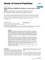
Our preliminary experiments confirmed that chondrocytes
strongly expressed the cartilage-specific markers aggrecan
and type II collagen, whereas the expression of type IA2 and X
collagens was not detected (Figure 1a). This confirmed the
mature phenotype of the articular chondrocytes used through-
out the study, all the more so considering that experiments
were carried out with first-passage cells.
Then, we examined the time course of Ank, PC-1, and TNAP
mRNA expression in TGF-β1-stimulated chondrocytes (Figure
1b). Ank was upregulated from 6 hours and reached a peak
value of 4.5-fold at 12 hours after TGF-β1 exposure. In these
conditions, PC-1 was induced approximately 2-fold after 12
hours and reached a maximal induction of approximately 3.5-
fold after 24 hours of stimulation with TGF-β1. In contrast, we
failed to detect any expression of TNAP in resting chondro-
cytes or after stimulation with TGF-β1.
Western blotting indicated that ANK protein was induced from
6 hours after TGF-β1 challenge, whereas PC-1 was upregu-
lated after 12 hours (Figure 1c). Taken together, these data
demonstrated that Ank was induced by TGF-β1 more rapidly
than PC-1 in chondrocytes.
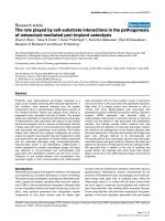
Effect of TGF-β1 on extracellular inorganic
pyrophosphate
As shown in Figure 2a, ePPi level increased by 3-fold after 6
hours of stimulation with TGF-β1 and reached a plateau from
24 hours (5-fold). In these experimental conditions, TGF-β1
stimulated NPPase activity by 5-fold at 24 hours (Figure 2b).
However, we failed to detect any APase activity in our experi-
mental conditions, which is consistent with the lack of expres-
sion of type X collagen. These data suggested that TGF-β1
Figure 1
Effect of transforming growth factor-beta-1 (TGF-β1) on proteins regu-lating inorganic pyrophosphate metabolismEffect of transforming growth factor-beta-1 (TGF-β1) on proteins regu-
lating inorganic pyrophosphate metabolism. (a) Phenotypic characteri-
zation of chondrocytes. Total RNA was extracted from untreated rat
chondrocytes and subjected to real-time polymerase chain reaction
(PCR) analysis. The relative abundance of gene mRNAs was normal-
ized to that of S29 mRNA. Results are presented in histograms as
mean percentages (± standard deviation [SD]) over S29 value. (b)
Effect of TGF-β1 on Ank, PC-1, and TNAP mRNA levels. Total RNA
was extracted from rat chondrocytes exposed to 10 ng/mL of TGF-β1
from 1 to 48 hours and subjected to real-time PCR analysis. The rela-
tive abundance of gene mRNAs was normalized to that of S29 mRNA.
Results are expressed as mean percentages (± SD) over control val-
ues. Statistically significant differences from the control are indicated
as *p < 0.05. (c) Effect of TGF-β1 on ANK or PC-1 protein levels. Total
proteins were extracted from rat chondrocytes exposed to 10 ng/mL of
TGF-β1 from 6 to 48 hours and subjected to Western blotting using
polyclonal anti-ANK and anti-PC-1 antibody. The protein band intensi-
ties were quantified by densitometry from enhanced chemilumines-
cence immunoblots. The relative abundance of these proteins was
normalized to that of β-actin protein and expressed as induction folds
over control value. N.D., not detected; TNAP, tissue-nonspecific alka-
line phosphatase.
Arthritis Research & Therapy Vol 9 No 6 Cailotto et al.
Page 6 of 13
(page number not for citation purposes)
increased ePPi levels by activating ecto-enzymes and not by
reducing TNAP activity, which remained undetectable.
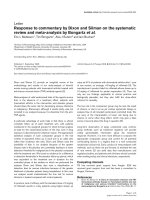
Respective contributions of Ank and PC-1 to TGF-β1-
induced increase in extracellular inorganic
pyrophosphate levels in chondrocytes
The siRNA technology was used to investigate the respective
contributions of Ank and PC-1 on TGF-β1-induced produc-
tion of ePPi. Control experiments showed that siRNAs were
efficient, as they reduced mRNA level by more than 80% in
basal conditions (Figure 3a,b) and diminished the stimulating
effect of TGF-β1 below control level (Figure 3a,b) at the time
of maximal gene expression. No effect was observed on L27
mRNA level in either condition (Figure 3a,b), confirming that
the concentration of 50 nM of siRNA was gene-specific for
chondrocytes. When ePPi levels were measured in culture
supernatant of chondrocytes transfected with siRNA, inhibi-
tion of PC-1 was ineffective on basal ePPi level and
accounted for only a 16% decrease in ePPi level in TGF-β1-
stimulated cells (Figure 3c). In contrast, transfection with Ank
siRNA reduced the basal ePPi level by 33% and reduced the
ePPi level by 60% in TGF-β1-stimulated cells (Figure 3c).
These data demonstrated that Ank played a major role com-
pared with PC-1 in the regulation of ePPi level in resting and
TGF-β1-stimulated chondrocytes.
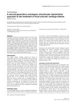
Identification of TGF-β1-induced signaling pathways
contributing to the regulation of Ank gene
As shown in Figure 4a, Western blotting revealed that TGF-β1
induced the phosphorylation of p38-MAPK and PKC as early
as 10 minutes after stimulation. A major activation of ERK 1/2
was also observed from 10 to 15 minutes after TGF-β1
challenge, whereas a strong phosphorylation of Smad 3 was
noted at 30 and 60 minutes. In our experimental conditions,
the corresponding non-phosphorylated proteins remained
unchanged (data not shown).
When chondrocytes were stimulated by TGF-β1 in the pres-
ence of SB203580, a selective p38-MAPK inhibitor, the
increase in Ank mRNA level was not affected (Figure 4b). In
contrast, PD98059, a selective MEK-1 inhibitor, reduced the
stimulatory effect of TGF-β1 by 60% (Figure 4b), supporting a
contribution of this MAPK to the induction of the Ank gene.
RcAMP, a selective PKA inhibitor, was also ineffective in these
experimental conditions (Figure 4b). These data showed that
TGF-β1 induced multiple signaling pathways in chondrocytes
but that neither p38-MAPK nor PKA contributed to its stimu-
lating effect on the Ank gene.
Contribution of protein kinase C pathway to TGF-β1-
induced Ank expression
As shown in Figure 5a, the stimulatory effect of TGF-β1 on
Ank expression was suppressed almost completely by cal-
phostin, a general PKC inhibitor. In these conditions, rottlerin,
a specific inhibitor of PKCδ, decreased Ank induction by 10%
whereas Gö6976, a specific inhibitor of Ca
2+
-dependent
PKCs (PKCα and PKCβI), had a 60% inhibitory effect (Figure
5a). A subsequent dose-ranging study showed that inhibition
of TGF-β1-induced expression of Ank by Gö6976 was clearly
dose-dependent and reached the control level at 10 μM (Fig-
ure 5b). Taken together, these data supported a major role for
PKCα and PKCβI isoenzymes in TGF-β1-stimulated expres-
sion of Ank.
Contribution of Ras and Raf-1 pathways to TGF-β1-
induced Ank expression
The experiments evaluating transfection efficiency did not
show any significant difference among the wild-type, the con-
stitutively active, or the dominant-negative forms of Ras and
Raf-1 (data not shown). When chondrocytes were transfected
to overexpress constitutively active Ras (Figure 6a) or Raf-1
(Figure 6b), the basal expression of Ank was increased by 2-
fold and 1.5-fold, respectively. In these conditions, the induc-
tion of Ank by TGF-β1 was increased by 3-fold (Figure 6a) and
4.3-fold, respectively (Figure 6b), which remained similar to
cells transfected either with an empty vector or with wild-type
Ras or Raf-1 (Figure 6a,b). Transfection with dominant-nega-
Figure 2
Effect of transforming growth factor-beta-1 (TGF-β1) on extracellular inorganic pyrophosphate (ePPi) and nucleotide pyrophosphatase phos-phodiesterase (NPPase) activityEffect of transforming growth factor-beta-1 (TGF-β1) on extracellular
inorganic pyrophosphate (ePPi) and nucleotide pyrophosphatase phos-
phodiesterase (NPPase) activity. (a) Kinetics of ePPi levels in culture
supernatant of rat chondrocytes stimulated with TGF-β1 (10 ng/mL).
ePPi was assayed radiometrically and normalized to the amount of total
cell proteins (n = 6). Data are expressed as mean (± standard deviation
[SD]) in picomoles per microgram of protein. (b) NPPase activity in cul-
tured rat chondrocytes stimulated with 10 ng/mL of TGF-β1. Enzyme
activity was normalized to the amount of total cell proteins (n = 3).
Results are expressed as mean (± SD) in micromoles of paranitrophe-
nol per minute per milligram of protein. Statistically significant differ-
ences from the control are indicated as *p < 0.05.
Available online />Page 7 of 13
(page number not for citation purposes)
tive forms of Ras (Figure 6a) or Raf-1 (Figure 6b) reduced both
basal (by 75% and 45%, respectively) and TGF-β1-induced
(by 3-fold and 2-fold, respectively) Ank expression. These data
supported a major role for the Ras/Raf-1 pathway in TGF-β1-
induced expression of Ank.
Western blot analysis of ERK 1/2 phosphoproteins showed an
activation of ERK 1/2 pathway by TGF-β1 in cells transfected
with wild-type Ras (Figure 6c). More importantly, the phospho-
rylation of ERK 1/2 was suppressed almost completely in cells
electroporated with a dominant-negative form of Ras, whereas
Figure 3
Respective contributions of Ank and PC-1 to transforming growth factor-beta-1 (TGF-β1)-induced increase in extracellular inorganic pyrophosphate (ePPi) productionRespective contributions of Ank and PC-1 to transforming growth factor-beta-1 (TGF-β1)-induced increase in extracellular inorganic pyrophosphate
(ePPi) production. Effect of small interfering RNA (siRNA) on Ank (a) and PC-1 (b) mRNA levels. Rat chondrocytes were transfected with siRNA 24
hours before TGF-β1 stimulation. Total RNA was extracted from rat chondrocytes exposed to 10 ng/mL of TGF-β1 for 12 hours (Ank) (a) or 24
hours (PC-1) (b) and then subjected to real-time polymerase chain reaction analysis. The level of Ank, PC-1, and L27 mRNAs was normalized to that
of S29 mRNA and expressed as mean percentages (± SD) over control values. (c) Effect of Ank or PC-1 siRNA on ePPi levels. Shown are levels of
ePPi in culture supernatant of rat chondrocytes transfected with siRNA and then stimulated for 12 or 24 hours with 10 ng/mL of TGF-β1. ePPi levels
were normalized to the amount of total cell proteins (n = 6) and are expressed as mean (± SD) in picomoles per microgram of protein. Statistically
significant differences from the control are indicated as *p < 0.05 and from TGF-β1-treated cells as #p < 0.05.
Arthritis Research & Therapy Vol 9 No 6 Cailotto et al.
Page 8 of 13
(page number not for citation purposes)
overexpression of constitutively active Ras raised both basal
and TGF-β1-induced levels of ERK 1/2 phosphorylation (Fig-
ure 6c). These results, similar to those obtained with
equivalent Raf-1 constructs, demonstrated that activation of
ERK 1/2 by TGF-β1 involved the Ras/Raf-1 pathway in
chondrocytes.
Induction of Ank expression by TGF-β1 is a Smad-
independent event
In chondrocytes transfected to overexpress wild-type Smad 7,
an inhibitory Smad, control experiments showed that the basal
expression of aggrecan, chosen as a specific Smad-depend-
ent gene in chondrocytes, was reduced marginally (Figure 7a).
In contrast, the stimulating effect of TGF-β1 on aggrecan was
abolished, thus demonstrating the efficiency of the construct
(Figure 7a). However, neither the basal expression of Ank nor
its induction by TGF-β1 was affected by overexpression of
Smad 7 in chondrocytes (Figure 7b). These data demon-
strated that the Smad pathway did not play a major role in the
induction of the Ank gene by TGF-β1.
Discussion
Previous studies demonstrated a major contribution of ANK in
the regulation of ePPi levels. ANK is a transporter able to
export iPPi from the cells and is known to be upregulated in
osteoarthritis [17,26]. Moreover, chondrocytes and cartilage
extracts from patients with CPPD disease express high levels
of Ank mRNA [17]. Besides, chondrocytes treated with TGF-
β1 generated more ePPi than normal chondrocytes [15], and
chondrocyte sensitivity to TGF-β1 increased with aging [14].
These data led us to suppose that Ank could be a major target
of TGF-β1 in chondrocytes, likely contributing to its patho-
physiological relevance to CPPD deposition.
In our experimental conditions, TGF-β1 increased both Ank
mRNA and ANK protein levels, this induction beginning as
early as 6 hours. PC-1 mRNA was also induced by TGF-β1 but
in a more delayed fashion, whereas expression of TNAP could
not be detected. Our data confirmed that TGF-β1 stimulated
ePPi production [15] and demonstrated that this was concom-
itant with the increased expression of PC-1 and Ank. Moreo-
Figure 4
Identification of several signaling pathways in transforming growth factor-beta-1 (TGF-β1)-induced expression of the Ank geneIdentification of several signaling pathways in transforming growth factor-beta-1 (TGF-β1)-induced expression of the Ank gene. (a) Kinetics of sign-
aling events induced by TGF-β1. Total proteins were extracted from rat chondrocytes exposed to 10 ng/mL of TGF-β1 for 10 to 180 minutes and
subjected to Western blotting using anti-phospho-ERK 1/2, anti-phospho-p38-MAPK, anti-phospho-pan-PKC, or anti-phospho-Smad 3/1. The rela-
tive abundance of these proteins was normalized to that of β-actin protein. (b) Effect of specific signaling inhibitors on TGF-β1-induced expression
of Ank mRNA. Total RNA was extracted from rat chondrocytes stimulated with 10 ng/mL of TGF-β1 in the presence of 10 μM RcAMP (PKA inhibi-
tor) or 10 μM SB203580 (a selective p38-MAPK inhibitor) or 10 μM PD98059 (a MEK-1 inhibitor) added 1 hour before TGF-β1. The mRNA level of
Ank obtained from real-time polymerase chain reaction analysis was normalized to that of S29 mRNA and is expressed as mean percentages (±
standard deviation) over control values from three independent experiments. Statistically significant differences from the control are indicated as *p
< 0.05 and from TGF-β1-treated cells as #p < 0.05. ERK, extracellular signal-regulated kinase; MEK-1, mitogen-activated protein kinase/extracellu-
lar signal-regulated kinase kinase 1; p38-MAPK, p38-mitogen-activated protein kinase; PKA, protein kinase A; PKC, protein kinase C.
Available online />Page 9 of 13
(page number not for citation purposes)
ver, similar experiments performed on cartilage explants
showed an identical pattern of response, thus validating our
monolayer culture system of chondrocytes (F. Cailotto, A.
Bianchi, S. Sebillaud, N. Venkatesan, D. Moulin, J-Y. Jouzeau,
P. Netter, unpublished data). Based on the higher levels of
TGF-β1 found in the synovial fluid of osteoarthritis patients
having developed CPPD deposition [27], our findings are in
favor of a pathophysiological contribution of TGF-β1-induced
dysregulation of Ank and PC-1 in sporadic chondrocalcinosis.
To date, no data are available on the relative contributions of
ANK and PC-1 to TGF-β1-induced changes in ePPi produc-
tion. Therefore, we developed an siRNA technology to clarify
the respective roles of ANK and PC-1 since both genes dif-
fered rather in their kinetics of expression than in their extent
of induction in response to TGF-β1. We demonstrated that, for
a comparable knockdown of target genes by siRNA, Ank con-
tributed 4-fold more than PC-1 to TGF-β1-induced increase in
ePPi level. The minor effect of PC-1 siRNA on ePPi level could
possibly be explained by a stronger basal expression of PC-1
(and, consequently, a residual enzymatic activity even after
siRNA transfection) compared with ANK in our cell culture sys-
tem. However, our data were not biased by the kinetics of
induction of each gene since the consequence of their knock-
down was studied at the time of their maximal expression in
response to TGF-β1. Moreover, we also observed that Ank
siRNA diminished TGF-β1-induced ePPi production by 65%
at 24 hours (data not shown), confirming that the contribution
of Ank was still the most significant at the time of maximal
expression of PC-1 mRNA. Our results could be partly
explained by the fact that ANK is a multipass transmembrane
protein thought to serve either as an anion channel or as a
regulator of such a channel [10]. As a consequence, an
increase or a decrease of protein level could have a profound
and rapid impact on PPi transport across the membrane. PC-
1 has been shown to be strongly induced by TGF-β1, as well
as NPPase activity in chondrocytes [28], and our data con-
firmed these observations. However, the contribution of PC-1
to ePPi generation could be estimated to be between 35% to
50% in osteoblasts [29], suggesting that PC-1 is an important
contributor but not the major contributor of ePPi generation,
which is consistent with our findings in chondrocytes. One
important consideration is that, even though knockdown with
siRNA was very efficient at the mRNA level, neither the repres-
sion of Ank nor that of PC-1 was sufficient to diminish ePPi
levels below control level in TGF-β1-stimulated cells. This sug-
Figure 5
Contribution of protein kinase C (PKC) pathway to transforming growth factor-beta-1 (TGF-β1)-induced expression of the Ank geneContribution of protein kinase C (PKC) pathway to transforming growth factor-beta-1 (TGF-β1)-induced expression of the Ank gene. (a) Effect of
PKC inhibitors on Ank expression. Total RNA was extracted from rat chondrocytes stimulated with 10 ng/mL of TGF-β1 in the presence of 10 μM of
calphostin C (general PKC inhibitor) or 5 μM of rottlerin (PKCδ inhibitor) or 5 μM of Gö6976 (PKC α/β1 inhibitor) added 1 hour before TGF-β1. (b)
Dose-response study of PKC-dependent induction of Ank by TGF-β1. Total RNA was extracted from rat chondrocytes stimulated with 10 ng/mL of
TGF-β1 in the presence of 0, 1, 2.5, 5, or 10 μM of Gö6976 added 1 hour before TGF-β1. The mRNA level of Ank obtained from real-time polymer-
ase chain reaction analysis was normalized to that of S29 mRNA and is expressed as mean percentages (± standard deviation) over control values
from three independent experiments. Statistically significant differences from the control are indicated as *p < 0.05 and from TGF-β1-treated cells as
#p < 0.05.
Arthritis Research & Therapy Vol 9 No 6 Cailotto et al.
Page 10 of 13
(page number not for citation purposes)
gests that, although Ank seemed to play a major role, the gen-
eration of ePPi could require a coordinated contribution of Ank
and PC-1, as suggested by others [26].
When we studied the regulation of Ank expression by TGF-β1,
we demonstrated firstly that ERK 1/2 and p38-MAPKs were
activated in our cell culture system. These results agree well
with their contribution to the inducing effect of TGF-β1 on
aggrecan [30] or TIMP-3 [31] expression in chondrogenic
cells. Complementary experiments with specific kinase inhibi-
tors showed that inhibition of MEK-1 by PD98059 reduced
the stimulating effect of TGF-β1 by 2-fold. In contrast, the lack
of efficiency of SB203580 demonstrated that p38-MAPK was
not involved in TGF-β1-induced expression of Ank. Our results
Figure 6
Effect of Ras/Raf-1 modulation on transforming growth factor-beta-1 (TGF-β1)-induced expression of the Ank geneEffect of Ras/Raf-1 modulation on transforming growth factor-beta-1 (TGF-β1)-induced expression of the Ank gene. Rat chondrocytes were electro-
porated with empty vector, wild-type, constitutively active, or dominant-negative plasmids for Ras (a) or Raf-1 (b) (2 μg/well of six-well plate) before
stimulation with 10 ng/mL of TGF-β1 for 12 hours. Total RNA was extracted and subjected to real-time polymerase chain reaction analysis. The
mRNA level of Ank was normalized to that of S29 mRNA and is expressed as mean percentages (± standard deviation) over control values from
three independent experiments. Statistically significant differences from the control are indicated as *p < 0.05 and from TGF-β1-treated cells as #p
< 0.05. (c) Effect of TGF-β1 on extracellular signal-regulated kinase (ERK) 1/2 phosphorylation in electroporated cells with wild-type, constitutively
active, or dominant-negative plasmids for Ras (2 μg/well of six-well plate). Total proteins were extracted from rat chondrocytes exposed to 10 ng/mL
of TGF-β1 for 15 minutes and subjected to Western blotting using anti-phospho- and anti-total-ERK 1/2 antibodies. The relative abundance of these
proteins was normalized to that of β-actin protein.
Available online />Page 11 of 13
(page number not for citation purposes)
are in accordance with other studies reporting that the contri-
bution of the p38-MAPK pathway to the TGF-β1 effect in car-
tilage was influenced greatly by the species [32] and cell
culture system [33].
To investigate further the contribution of the MEK-1 pathway,
we modulated the expression of Ras and Raf-1 proteins, which
are known to trigger MEK-1 activation in chondrocytes [34].
Our results showed that Ras and, to a lesser extent, Raf-1
were implicated in TGF-β1-induced expression of Ank in
chondrocytes, a finding consistent with data demonstrating
the activation of Ras and Raf-1 by TGF-β1 in several cell sys-
tems [35,36].
Furthermore, we demonstrated that the activation of ERK 1/2
by TGF-β1 was strongly dependent on the activation of Ras
and Raf-1 since transfection with dominant-negative forms of
these proteins completely inhibited the stimulating effect of
TGF-β1. Moreover, constitutively active forms raised both
basal and TGF-β1-induced ERK 1/2 phosphorylation. Taken
together, these findings confirmed the linkage between activa-
tion of Ras/Raf-1, MEK-1, and ERK pathways [34-36] and
highlighted their contribution to the induction of Ank by TGF-
β1 in chondrocytes.
Ryan and colleagues [37] demonstrated that, in addition to
MAPKs, adenylate cyclase activation and generation of cAMP
reduced ePPi production in chondrocytes. The PKA pathway
was also described to be induced by TGF-β1 in chondrocytes
[38,39]. However, using RcAMP as a PKA inhibitor, we failed
to demonstrate any contribution of PKA to the induction of Ank
by TGF-β1.
In chondrocytes, the production of ePPi was also reported to
be stimulated by PKC-dependent pathways [37], which are
known to be activated by TGF-β1 [40]. We showed that the
stimulation of Ank expression depended on PKC activation
but with a variable effect of the inhibitors depending on their
specificity for PKC isoenzymes. Thus, when cells were pre-
treated with calphostin C (a broad PKC inhibitor), a strong
effect on Ank mRNA expression was observed whereas rott-
lerin (a PKCδ inhibitor) was weakly effective. These findings
suggested that PKCδ, alongside the other members of the
novel PKC family (PKCε, PKCη, and PKCθ [41]), was not
implicated in the regulation of Ank expression by TGF-β1. In
contrast, we demonstrated that Gö6976 (a Ca
2+
-dependent
PKCα and PKCβI isoenzyme inhibitor) was strongly inhibitory.
Since the only difference between novel PKC and conven-
tional PKC (for example, Ca
2+
-dependent PKCα and PKCβI)
activation is the dependence on calcium [41], our findings
support a possible regulatory role for calcium in the induction
of Ank expression by TGF-β1.
Some genes induced by TGF-β1 are partly or totally regulated
by Smad proteins. We demonstrated that, in our experimental
conditions, TGF-β1 was able to induce the phosphorylation of
Smad 3, known to modulate the expression of chondrocyte-
specific genes, such as aggrecan [42]. Smad 7 is a natural
inhibitor of the Smad pathway, preventing Smad 3 phosphor-
ylation in the cytosol and therefore suppressing Smad
signaling [19]. Scharstuhl and colleagues [42] demonstrated
that Smad 7 overexpression was able to counteract TGF-β1-
induced expression of aggrecan in chondrocytes, as was the
case in our experiments. However, we demonstrated that over-
expression of wild-type Smad 7 failed to reduce TGF-β1-
induced expression of Ank in chondrocytes, thus demonstrat-
ing that it was a non-Smad signaling event.
Taken as a whole, these observations suggest that, contrary to
degenerative joint pathologies [43], targeting the non-Smad
TGF-β1 signaling events may lead to insights in the field of
Figure 7
Effect of Smad 7 overexpression on transforming growth factor-beta-1 (TGF-β1)-induced responses in rat chondrocytesEffect of Smad 7 overexpression on transforming growth factor-beta-1
(TGF-β1)-induced responses in rat chondrocytes. Rat chondrocytes
were electroporated with either empty vector or wild-type Smad 7 over-
expressing plasmid (2 μg/well of six-well plate) and then treated for 12
hours with 10 ng/mL of TGF-β1. Total RNA was extracted and sub-
jected to real-time polymerase chain reaction analysis. The mRNA level
of aggrecan (a) and Ank (b) was normalized to that of S29 mRNA and
is expressed as mean percentages (± standard deviation) over control
values from three independent experiments. Statistically significant dif-
ferences from the control are indicated as *p < 0.05 and from TGF-β1-
treated cells as #p < 0.05.
Arthritis Research & Therapy Vol 9 No 6 Cailotto et al.
Page 12 of 13
(page number not for citation purposes)
sporadic chondrocalcinosis since the repression of Ank would
translate into reduced ePPi levels in synovial fluid. Thus, selec-
tive conventional PKC inhibitors should be powerful agents,
considering that some of them, such as Gö6976 [44,45], are
currently in clinical development for cardiovascular diseases
and cancer. Moreover, probenecid was shown to inhibit Ank
function [10] and TGF-β1-induced PPi elaboration by
chondrocytes [12]. Therefore, the effect of the combination of
probenecid and Gö6976 on CPPD formation could be worthy
of study.
Conclusion
To summarize, the present study shows that TGF-β1
increases ePPi levels, with a major contribution of Ank, despite
its similar inducing effect on Ank and PC-1. These results are
in favor of a main role of Ank as an effector of TGF-β1 in CPPD
formation. Induction of Ank is mediated by the dual activation
of Ras, Raf-1, and MEK-1/ERK cascade and Ca
2+
-dependent
PKC but is independent of the Smad signaling pathway. Our
data support the strong contribution of Ank to the pathogene-
sis of sporadic chondrocalcinosis, in addition to familial chon-
drocalcinosis, and identify signaling pathways involved in TGF-
β1-induced Ank expression which could lead to new therapies
for CPPD disease.
Competing interests
The authors declare that they have no competing interests.
Authors' contributions
FC performed cell culture, RNA extraction, real-time quantita-
tive PCR, ePPi assays, and siRNA assays and was involved in
drafting the manuscript. AB carried out the inhibitor
experiments, drafted the manuscript, and contributed to the
study design. SS performed chondrocyte isolation, cell cul-
ture, Western blot analysis, and real-time quantitative PCR. NV
participated in revising the manuscript and in the interpretation
of data. DM carried out RNA extraction and participated in
revising the manuscript. J-YJ and PN contributed to the study
design and revised the manuscript for intellectual content. All
authors read and approved the final manuscript.
Acknowledgements
This work was supported by grants from the CPRC/PHRC, the CHU
Nancy, and the Communauté Urbaine du Grand Nancy.
References
1. Liu-Bryan R, Liote F: Monosodium urate and calcium pyrophos-
phate dihydrate (CPPD) crystals, inflammation, and cellular
signaling. Joint Bone Spine 2005, 72:295-302.
2. Pendleton A, Johnson MD, Hughes A, Gurley KA, Ho AM, Doherty
M, Dixey J, Gillet P, Loeuille D, McGrath R, et al.: Mutations in
ANKH cause chondrocalcinosis. Am J Hum Genet 2002,
71:933-940.
3. Zhang Y, Johnson K, Russell RG, Wordsworth BP, Carr AJ, Terkel-
taub RA, Brown MA: Association of sporadic chondrocalcinosis
with a – 4-basepair G-to-A transition in the 5'-untranslated
region of ANKH that promotes enhanced expression of ANKH
protein and excess generation of extracellular inorganic
pyrophosphate. Arthritis Rheum 2005, 52:1110-1117.
4. Timms AE, Zhang Y, Bradbury L, Wordsworth BP, Brown MA:
Investigation of the role of ANKH in ankylosing spondylitis.
Arthritis Rheum 2003, 48:2898-2902.
5. Reichenberger E, Tiziani V, Watanabe S, Park L, Ueki Y, Santanna
C, Baur ST, Shiang R, Grange DK, Beighton P, et al.: Autosomal
dominant craniometaphyseal dysplasia is caused by muta-
tions in the transmembrane protein ANK. Am J Hum Genet
2001, 68:1321-1326.
6. Rosenthal AK, McCarty BA, Cheung HS, Ryan LM: A comparison
of the effect of transforming growth factor beta 1 on pyrophos-
phate elaboration from various articular tissues. Arthritis
Rheum 1993, 36:539-542.
7. Rachow JW, Ryan LM: Inorganic pyrophosphate metabolism in
arthritis. Rheum Dis Clin North Am 1988, 14:289-302.
8. Howell DS, Martel-Pelletier J, Pelletier JP, Morales S, Muniz O:
NTP pyrophosphohydrolase in human chondrocalcinotic and
osteoarthritic cartilage. II. Further studies on histologic and
subcellular distribution. Arthritis Rheum 1984, 27:193-199.
9. Ryan LM, Wortmann RL, Karas B, Lynch MP, McCarty DJ: Pyro-
phosphohydrolase activity and inorganic pyrophosphate con-
tent of cultured human skin fibroblasts. Elevated levels in
some patients with calcium pyrophosphate dihydrate deposi-
tion disease. J Clin Invest 1986, 77:1689-1693.
10. Ho AM, Johnson MD, Kingsley DM: Role of the mouse ank gene
in control of tissue calcification and arthritis. Science 2000,
289:265-270.
11. Sweet HO, Green MC: Progressive ankylosis, a new skeletal
mutation in the mouse. J Hered 1981, 72:87-93.
12. Rosenthal AK, Ryan LM: Probenecid inhibits transforming
growth factor-beta 1 induced pyrophosphate elaboration by
chondrocytes. J Rheumatol 1994, 21:896-900.
13. Xu Y, Pritzker KP, Cruz TF: Characterization of chondrocyte
alkaline phosphatase as a potential mediator in the dissolu-
tion of calcium pyrophosphate dihydrate crystals. J Rheumatol
1994, 21:912-919.
14. Guerne PA, Blanco F, Kaelin A, Desgeorges A, Lotz M: Growth
factor responsiveness of human articular chondrocytes in
aging and development. Arthritis Rheum 1995, 38:960-968.
15. Rosen F, McCabe G, Quach J, Solan J, Terkeltaub R, Seegmiller
JE, Lotz M: Differential effects of aging on human chondrocyte
responses to transforming growth factor beta: increased pyro-
phosphate production and decreased cell proliferation. Arthri-
tis Rheum 1997, 40:1275-1281.
16. Ryan LM, McCarty DJ: Calcium pyrophosphate crystal deposi-
tion disease, pseudogout, and articular chondrocalcinosis. In
Arthritis and Allied Conditions: A Textbook of Rheumatology 13th
edition. Edited by: Koopman W. Baltimore: Williams and Wilkins;
1997:2103-2126.
17. Hirose J, Ryan LM, Masuda I: Up-regulated expression of carti-
lage intermediate-layer protein and ANK in articular hyaline
cartilage from patients with calcium pyrophosphate dihydrate
crystal deposition disease. Arthritis Rheum 2002,
46:3218-3229.
18. Sohn P, Crowley M, Slattery E, Serra R: Developmental and TGF-
beta-mediated regulation of Ank mRNA expression in carti-
lage and bone. Osteoarthritis Cartilage 2002, 10:482-490.
19. Moustakas A, Souchelnytskyi S, Heldin CH: Smad regulation in
TGF-beta signal transduction. J Cell Sci 2001, 114:4359-4369.
20. Moustakas A, Heldin CH: Non-Smad TGF-beta signals. J Cell
Sci 2005, 118:3573-3584.
21. Kuettner KE, Pauli BU, Gall G, Memoli VA, Schenk RK: Synthesis
of cartilage matrix by mammalian chondrocytes in vitro. I. Iso-
lation, culture characteristics, and morphology. J Cell Biol
1982, 93:
743-750.
22. Hayashi H, Abdollah S, Qiu Y, Cai J, Xu YY, Grinnell BW, Richard-
son MA, Topper JN, Gimbrone MA Jr, Wrana JL, et al.: The MAD-
related protein Smad7 associates with the TGFbeta receptor
and functions as an antagonist of TGFbeta signaling. Cell
1997, 89:1165-1173.
23. Towbin H, Staehelin T, Gordon J: Electrophoretic transfer of pro-
teins from polyacrylamide gels to nitrocellulose sheets: proce-
dure and some applications. Proc Natl Acad Sci USA 1979,
76:4350-4354.
24. Smith PK, Krohn RI, Hermanson GT, Mallia AK, Gartner FH,
Provenzano MD, Fujimoto EK, Goeke NM, Olson BJ, Klenk DC:
Measurement of protein using bicinchoninic acid. Anal
Biochem 1985, 150:76-85.
Available online />Page 13 of 13
(page number not for citation purposes)
25. Terkeltaub R, Rosenbach M, Fong F, Goding J: Causal link
between nucleotide pyrophosphohydrolase overactivity and
increased intracellular inorganic pyrophosphate generation
demonstrated by transfection of cultured fibroblasts and oste-
oblasts with plasma cell membrane glycoprotein-1. Relevance
to calcium pyrophosphate dihydrate deposition disease.
Arthritis Rheum 1994, 37:934-941.
26. Johnson K, Terkeltaub R: Upregulated ank expression in oste-
oarthritis can promote both chondrocyte MMP-13 expression
and calcification via chondrocyte extracellular PPi excess.
Osteoarthritis Cartilage 2004, 12:321-335.
27. Punzi L, Oliviero F, Ramonda R: Transforming growth factor-
beta levels in synovial fluid of osteoarthritis with or without
calcium pyrophosphate dihydrate crystals. J Rheumatol 2003,
30:420. author reply 420–421
28. Lotz M, Rosen F, McCabe G, Quach J, Blanco F, Dudler J, Solan J,
Goding J, Seegmiller JE, Terkeltaub R: Interleukin 1 beta sup-
presses transforming growth factor-induced inorganic pyro-
phosphate (PPi) production and expression of the PPi-
generating enzyme PC-1 in human chondrocytes. Proc Natl
Acad Sci USA 1995, 92:10364-10368.
29. Hessle L, Johnson KA, Anderson HC, Narisawa S, Sali A, Goding
JW, Terkeltaub R, Millan JL: Tissue-nonspecific alkaline phos-
phatase and plasma cell membrane glycoprotein-1 are central
antagonistic regulators of bone mineralization. Proc Natl Acad
Sci USA 2002, 99:9445-9449.
30. Watanabe H, de Caestecker MP, Yamada Y: Transcriptional
cross-talk between Smad, ERK1/2, and p38 mitogen-activated
protein kinase pathways regulates transforming growth fac-
tor-beta-induced aggrecan gene expression in chondrogenic
ATDC5 cells. J Biol Chem 2001, 276:14466-14473.
31. Qureshi HY, Sylvester J, El Mabrouk M, Zafarullah M: TGF-beta-
induced expression of tissue inhibitor of metalloproteinases-
3 gene in chondrocytes is mediated by extracellular signal-
regulated kinase pathway and Sp1 transcription factor. J Cell
Physiol 2005, 203:345-352.
32. Badger AM, Roshak AK, Cook MN, Newman-Tarr TM, Swift BA,
Carlson K, Connor JR, Lee JC, Gowen M, Lark MW, et al.: Differ-
ential effects of SB 24 a selective p38 mitogen-activated pro-
tein kinase inhibitor, on IL-1 treated bovine and human
cartilage/chondrocyte cultures. Osteoarthritis Cartilage 2002,
8:434-443.
33. Poleni PE, Bianchi A, Etienne S, Koufany M, Sebillaud S, Netter P,
Terlain B, Jouzeau JY: Agonists of peroxisome proliferators-
activated receptors (PPAR) alpha, beta/delta or gamma
reduce transforming growth factor (TGF)-beta-induced prote-
oglycans' production in chondrocytes. Osteoarthritis Cartilage
2007, 15:493-505.
34. Krejci P, Masri B, Fontaine V, Mekikian PB, Weis M, Prats H, Wil-
cox WR: Interaction of fibroblast growth factor and C-natriu-
retic peptide signaling in regulation of chondrocyte
proliferation and extracellular matrix homeostasis. J Cell Sci
2005, 118:5089-5100.
35. Mulder KM, Morris SL: Activation of p21ras by transforming
growth factor beta in epithelial cells. J Biol Chem 1992,
267:5029-5031.
36. Reimann T, Hempel U, Krautwald S, Axmann A, Scheibe R, Seidel
D, Wenzel KW: Transforming growth factor-beta1 induces acti-
vation of Ras, Raf-1, MEK and MAPK in rat hepatic stellate
cells. FEBS Lett 1997, 403:57-60.
37. Ryan LM, Kurup IV, Cheung HS: Transduction mechanisms of
porcine chondrocyte inorganic pyrophosphate elaboration.
Arthritis Rheum 1999, 42:555-560.
38. Rosado E, Schwartz Z, Sylvia VL, Dean DD, Boyan BD: Trans-
forming growth factor-beta1 regulation of growth zone
chondrocytes is mediated by multiple interacting pathways.
Biochim Biophys Acta 2002, 1590:1-15.
39. Li TF, O'Keefe RJ, Chen D: TGF-beta signaling in chondrocytes.
Front Biosci 2005, 10:681-688.
40. Su S, DiBattista JA, Sun Y, Li WQ, Zafarullah M: Up-regulation of
tissue inhibitor of metalloproteinases-3 gene expression by
TGF-beta in articular chondrocytes is mediated by serine/
threonine and tyrosine kinases. J Cell Biochem 1998,
70:517-527.
41. Jaken S: Protein kinase C isozymes and substrates. Curr Opin
Cell Biol 1996, 8:168-173.
42. Scharstuhl A, Diepens R, Lensen J, Vitters E, van Beuningen H, van
der Kraan P, van den Berg W: Adenoviral overexpression of
Smad-7 and Smad-6 differentially regulates TGF-beta-medi-
ated chondrocyte proliferation and proteoglycan synthesis.
Osteoarthritis Cartilage 2003, 11:773-782.
43. Blom AB, van der Kraan PM, van den Berg WB: Cytokine target-
ing in osteoarthritis. Curr Drug Targets 2007, 8:283-292.
44. Goekjian PG, Jirousek MR: Protein kinase C in the treatment of
disease: signal transduction pathways, inhibitors, and agents
in development. Curr Med Chem 1999, 6:877-903.
45. Hanauske AR, Oberschmidt O, Hanauske-Abel H, Lahn MM, Eis-
mann U: Antitumor activity of enzastaurin (LY317615.HCl)
against human cancer cell lines and freshly explanted tumors
investigated in vitro soft-agar cloning experiments. Invest New
Drugs 2007, 25:205-210.